Nano-scale Impedance Spectroscopy Probe... · Nano-scale Impedance Spectroscopy ... Topic: Scanning...
2
Nano-scale Impedance Spectroscopy Laura Fumagalli 1 , Ignacio Casuso 2 , Giorgio Ferrari 1 , Gabriel Gomila 2 , Marco Sampietro 1 , Josep Samitier 2 1 Politecnico di Milano, Dipartimento di Elettronica e Informazione, P.za L. da Vinci 32, 20133 Milano (Italy) 2 Laboratori de Nanobioenginyeria-CREBEC, Parc Científic de Barcelona and Departament d’Electrónica, Universitat de Barcelona, C/ Martí i Franquès 1, 08028 Barcelona, Spain. E-mail: [email protected] Topic: Scanning Probe Microscopy (SPM). Impedance spectroscopy is a well-established method for characterization of frequency-dependent electronic transport and for extraction of equivalent electrical circuit in a wide variety of systems, both solids and liquids. In addition, impedance characterization has recently become of major importance in the field of biosensors as a powerful technique in order to transduce bio-recognition events [1]. In particular, an interesting possibility is to apply the impedance spectroscopy to nanobiosensor based on single proteins, such as receptors or ion channels, in natural lipid membrane or immobilized on chemically functionalized substrates [2]. To the aim of electrical characterization at single-molecule scale, the Atomic Force Microscope (AFM) has proved to be a very powerful tool, especially for biological samples allowing measurements under physiological conditions. AFM impedance spectroscopy has already been reported [3, 4] in recent years, referred to as nano-impedance microscopy (NIM) or scanning impedance microscopy (SIM). The measurement system is commonly made up of a standard AFM coupled with a commercial impedance analyzer. The conductive AFM tip across the sample acts as a probe for impedance measurement, both fixed in a point and in imaging mode. However, this setup shows limited capabilities in terms of capacitance resolution and time requirements. In this presentation, we describe a new implementation of AFM impedance spectroscopy, based on specific amplifier circuit for impedance measurement mounted with the AFM. The circuit is a wide- band low-noise transimpedance amplifier designed to convert alternating current from the sample into amplified output voltage. The bandwidth extends from below 1Hz up to 1MHz, with a very low input noise, flat down at 10fA/√Hz up to 10kHz. Custom developed software automates impedance data acquisition and processing, by using a PC board with an A/D converter (see Fig1). Real part and imaginary part of impedance are extracted and they can be measured simultaneously with topography and dc current. The proposed setup shows capacitance resolution of few AttoFarad and a resistance resolution greater than GOhms with a measurement time of 10ms, that is few minutes required for an impedance image. Better performance can be obtained increasing the measuring time. Tests measurements (see Fig.2) as well as measurements on biological samples will be presented and discussed. Scanning Probe Microscopies (SPM) Poster 2 nd NanoSpain Worshop March 14-17, 2005 Barcelona-Spain
Transcript of Nano-scale Impedance Spectroscopy Probe... · Nano-scale Impedance Spectroscopy ... Topic: Scanning...
Nano-scale Impedance Spectroscopy
Laura Fumagalli1, Ignacio Casuso2, Giorgio Ferrari1, Gabriel Gomila2, Marco Sampietro1, Josep
Samitier2 1 Politecnico di Milano, Dipartimento di Elettronica e Informazione, P.za L. da Vinci 32, 20133 Milano (Italy)
2 Laboratori de Nanobioenginyeria-CREBEC, Parc Científic de Barcelona and Departament d’Electrónica, Universitat de Barcelona, C/ Martí i Franquès 1, 08028 Barcelona, Spain.
E-mail: [email protected] Topic: Scanning Probe Microscopy (SPM).
Impedance spectroscopy is a well-established method for characterization of frequency-dependent electronic transport and for extraction of equivalent electrical circuit in a wide variety of systems, both solids and liquids. In addition, impedance characterization has recently become of major importance in the field of biosensors as a powerful technique in order to transduce bio-recognition events [1]. In particular, an interesting possibility is to apply the impedance spectroscopy to nanobiosensor based on single proteins, such as receptors or ion channels, in natural lipid membrane or immobilized on chemically functionalized substrates [2].
To the aim of electrical characterization at single-molecule scale, the Atomic Force Microscope
(AFM) has proved to be a very powerful tool, especially for biological samples allowing measurements under physiological conditions. AFM impedance spectroscopy has already been reported [3, 4] in recent years, referred to as nano-impedance microscopy (NIM) or scanning impedance microscopy (SIM). The measurement system is commonly made up of a standard AFM coupled with a commercial impedance analyzer. The conductive AFM tip across the sample acts as a probe for impedance measurement, both fixed in a point and in imaging mode. However, this setup shows limited capabilities in terms of capacitance resolution and time requirements.
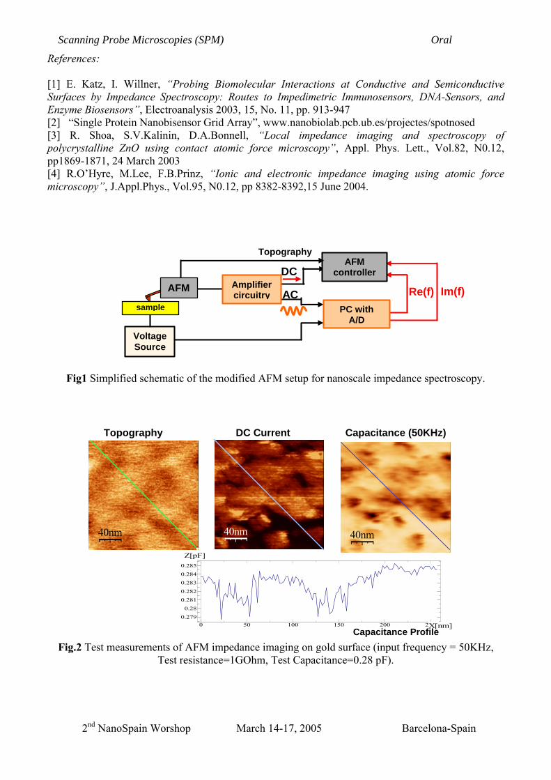
In this presentation, we describe a new implementation of AFM impedance spectroscopy, based on
specific amplifier circuit for impedance measurement mounted with the AFM. The circuit is a wide-band low-noise transimpedance amplifier designed to convert alternating current from the sample into amplified output voltage. The bandwidth extends from below 1Hz up to 1MHz, with a very low input noise, flat down at 10fA/√Hz up to 10kHz. Custom developed software automates impedance data acquisition and processing, by using a PC board with an A/D converter (see Fig1). Real part and imaginary part of impedance are extracted and they can be measured simultaneously with topography and dc current. The proposed setup shows capacitance resolution of few AttoFarad and a resistance resolution greater than GOhms with a measurement time of 10ms, that is few minutes required for an impedance image. Better performance can be obtained increasing the measuring time.
Tests measurements (see Fig.2) as well as measurements on biological samples will be presented
and discussed.
Scanning Probe Microscopies (SPM) Poster
2nd NanoSpain Worshop March 14-17, 2005 Barcelona-Spain
References: [1] E. Katz, I. Willner, “Probing Biomolecular Interactions at Conductive and Semiconductive Surfaces by Impedance Spectroscopy: Routes to Impedimetric Immunosensors, DNA-Sensors, and Enzyme Biosensors”, Electroanalysis 2003, 15, No. 11, pp. 913-947 [2] “Single Protein Nanobisensor Grid Array”, www.nanobiolab.pcb.ub.es/projectes/spotnosed [3] R. Shoa, S.V.Kalinin, D.A.Bonnell, “Local impedance imaging and spectroscopy of polycrystalline ZnO using contact atomic force microscopy”, Appl. Phys. Lett., Vol.82, N0.12, pp1869-1871, 24 March 2003 [4] R.O’Hyre, M.Lee, F.B.Prinz, “Ionic and electronic impedance imaging using atomic force microscopy”, J.Appl.Phys., Vol.95, N0.12, pp 8382-8392,15 June 2004.
Fig1 Simplified schematic of the modified AFM setup for nanoscale impedance spectroscopy.
Fig.2 Test measurements of AFM impedance imaging on gold surface (input frequency = 50KHz,
Test resistance=1GOhm, Test Capacitance=0.28 pF).
0 50 100 150 200 250
0.279
0.28
0.281
0.282
0.283
0.284
0.285
X[nm]
Z[pF]
Capacitance Profile
40nm
Topography
40nm
DC Current
40nm
Capacitance (50KHz)
Im(f) Re(f)
PC with A/D
Voltage Source
sample
AFM
AFM controller
Amplifier circuitry
DC
AC
Topography
Scanning Probe Microscopies (SPM) Oral
2nd NanoSpain Worshop March 14-17, 2005 Barcelona-Spain