Nano-hydroxyapatite preparation from biogenic raw · PDF fileNano-hydroxyapatite preparation...
7
Central European Journal of Chemistry Nano-hydroxyapatite preparation from biogenic raw materials * E-mail: [email protected] Received 26 August 2009; Accepted 10 November 2009 Abstract: Hydroxyapatite (HAp) was successfully produced from recycled eggshell, seashell and phosphoric acid. The phases obtained depended on the ratio of calcined eggshell/ seashell to phosphoric acid, the calcination temperature and the mechanochemical activation method (ball milling or attrition milling). The HAp structures were characterized by X-ray diffraction, scanning electron microsopy and infrared spectroscopy. Attrition milling was more effective than ball milling, yielding nanosize, homogenous and pure Hap. © Versita Sp. z o.o. Keywords: HAp • Raw material • Ball milling • Attrition • Characterization 1 Ceramics and Nanocomposites Department, Research Institute for Technical Physics and Materials Science, Hungarian Academy of Sciences, 1121 Budapest, Hungary 2 Nanostructures Department, Research Institute for Technical Physics and Materials Science, Hungarian Academy of Sciences, 1121 Budapest, Hungary 3 IR and Raman Spectroscopy Laboratory, Chemical Research Center, Hungarian Academy of Sciences, 1025 Budapest, Hungary Gréta Gergely 1* , Ferenc Wéber 1 , István Lukács 1 , Levente Illés 1 , Attila L. Tóth 1 , Zsolt E. Horváth 2 , Judit Mihály 3 , Csaba Balázsi 1 Research Article 1. Introduction Hydroxyapatite, (Ca 10 (PO 4 ) 6 (OH) 2 is chemically similar to the mineral component of bones and teeth [1-3]. HAp is among the few bioactive materials, meaning that it will support bone ingrowth and osseointegration when used in orthopaedic, dental and maxillofacial applications. Hydroxyapatite coatings are often applied to metallic implants, especially stainless steels and titanium alloys, to improve the surface properties. It may be employed in powders, porous blocks and hybrid composites to fill bone defects or voids when large sections of bone have had to be removed, or when bone augmentations are required (e.g. dental applications). HAp can be produced from biogenic materials like coral [4], seashell, [5], eggshell [3,6,7], body fluids [8] and by some chemical synthetic methods. These are based on solid state reactions [9], chemical precipitation [10], hydrothermal reactions [11], sol-gel methods [12] and mechanochemical methods [13,14] using different calcium and phosphorus-containing starting materials. Hydroxyapatite nanopowders have been produced from eggshell and seashell-derived raw materials and phosphoric acid using a 1.67 Ca/P ratio [15]. The structures were characterized by X-ray diffraction (XRD) and scanning electron microsopy (SEM). The phases observed during the powder synthesis depended on the ratio of calcined eggshell/seashell to phosphoric acid, the calcination temperature, and the duration of the mechanochemical synthesis [7]. The present work aimed to synthesize HAp via mechanochemical activation with three objectives: (1) to form HAp from two different raw materials: eggshell and seashell; (2) to compare two mechanochemical activation process: ball milling and attrition milling; and (3) to study the phase stability and powder characteristics. Cent. Eur. J. Chem. • 8(2) • 2010 • 375–381 DOI: 10.2478/s11532-010-0004-4 375
Transcript of Nano-hydroxyapatite preparation from biogenic raw · PDF fileNano-hydroxyapatite preparation...

Central European Journal of Chemistry
Nano-hydroxyapatite preparation from biogenic raw materials
* E-mail: [email protected]
Received 26 August 2009; Accepted 10 November 2009
Abstract: Hydroxyapatite (HAp) was successfully produced from recycled eggshell, seashell and phosphoric acid. The phases obtained depended on the ratio of calcined eggshell/ seashell to phosphoric acid, the calcination temperature and the mechanochemical activation method (ball milling or attrition milling). The HAp structures were characterized by X-ray diffraction, scanning electron microsopy and infrared spectroscopy. Attrition milling was more effective than ball milling, yielding nanosize, homogenous and pure Hap.
© Versita Sp. z o.o.
Keywords: HAp • Raw material • Ball milling • Attrition • Characterization
1 Ceramics and Nanocomposites Department, Research Institute for Technical Physics and Materials Science, Hungarian Academy of Sciences, 1121 Budapest, Hungary
2 Nanostructures Department, Research Institute for Technical Physics and Materials Science, Hungarian Academy of Sciences, 1121 Budapest, Hungary
3 IR and Raman Spectroscopy Laboratory, Chemical Research Center, Hungarian Academy of Sciences, 1025 Budapest, Hungary
Gréta Gergely1*, Ferenc Wéber1, István Lukács1, Levente Illés1, Attila L. Tóth1,Zsolt E. Horváth2, Judit Mihály3, Csaba Balázsi1
Research Article
1. Introduction Hydroxyapatite, (Ca10(PO4)6(OH)2 is chemically similar to the mineral component of bones and teeth [1-3]. HAp is among the few bioactive materials, meaning that it will support bone ingrowth and osseointegration when used in orthopaedic, dental and maxillofacial applications. Hydroxyapatite coatings are often applied to metallic implants, especially stainless steels and titanium alloys, to improve the surface properties. It may be employed in powders, porous blocks and hybrid composites to fill bone defects or voids when large sections of bone have had to be removed, or when bone augmentations are required (e.g. dental applications).
HAp can be produced from biogenic materials like coral [4], seashell, [5], eggshell [3,6,7], body fluids [8] and by some chemical synthetic methods. These are based on solid state reactions [9], chemical precipitation [10], hydrothermal reactions [11], sol-gel methods [12]
and mechanochemical methods [13,14] using different calcium and phosphorus-containing starting materials.
Hydroxyapatite nanopowders have been produced from eggshell and seashell-derived raw materials and phosphoric acid using a 1.67 Ca/P ratio [15]. The structures were characterized by X-ray diffraction (XRD) and scanning electron microsopy (SEM). The phases observed during the powder synthesis depended on the ratio of calcined eggshell/seashell to phosphoric acid, the calcination temperature, and the duration of the mechanochemical synthesis [7].
The present work aimed to synthesize HAp via mechanochemical activation with three objectives: (1) to form HAp from two different raw materials: eggshell and seashell; (2) to compare two mechanochemical activation process: ball milling and attrition milling; and (3) to study the phase stability and powder characteristics.
Cent. Eur. J. Chem. • 8(2) • 2010 • 375–381DOI: 10.2478/s11532-010-0004-4
375
Nano-hydroxyapatite preparation from biogenic raw materials
2. Experimental Procedure
Seashell and eggshell were collected and washed with detergent, then calcined in air at 900oC for 3 h. Every 10 minutes samples were removed for analysis. During the first 30 minutes most of the organic materials were burnt out, then the eggshell and seashell were converted to calcium oxide.
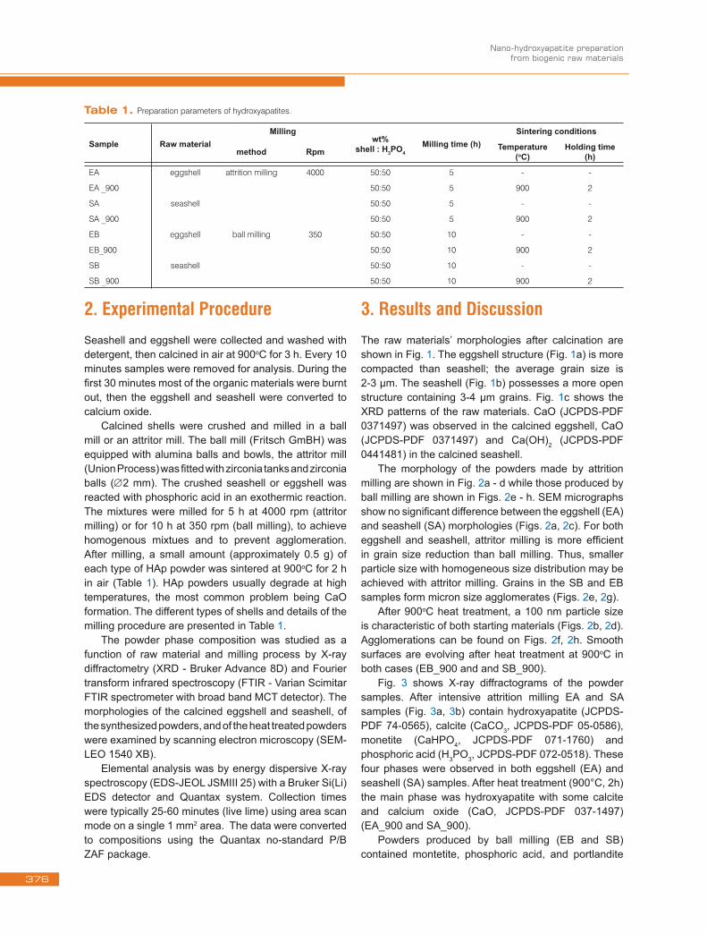
Calcined shells were crushed and milled in a ball mill or an attritor mill. The ball mill (Fritsch GmBH) was equipped with alumina balls and bowls, the attritor mill (Union Process) was fitted with zirconia tanks and zirconia balls (∅2 mm). The crushed seashell or eggshell was reacted with phosphoric acid in an exothermic reaction. The mixtures were milled for 5 h at 4000 rpm (attritor milling) or for 10 h at 350 rpm (ball milling), to achieve homogenous mixtues and to prevent agglomeration. After milling, a small amount (approximately 0.5 g) of each type of HAp powder was sintered at 900oC for 2 h in air (Table 1). HAp powders usually degrade at high temperatures, the most common problem being CaO formation. The different types of shells and details of the milling procedure are presented in Table 1.
The powder phase composition was studied as a function of raw material and milling process by X-ray diffractometry (XRD - Bruker Advance 8D) and Fourier transform infrared spectroscopy (FTIR - Varian Scimitar FTIR spectrometer with broad band MCT detector). The morphologies of the calcined eggshell and seashell, of the synthesized powders, and of the heat treated powders were examined by scanning electron microscopy (SEM-LEO 1540 XB).
Elemental analysis was by energy dispersive X-ray spectroscopy (EDS-JEOL JSMIII 25) with a Bruker Si(Li) EDS detector and Quantax system. Collection times were typically 25-60 minutes (live lime) using area scan mode on a single 1 mm2 area. The data were converted to compositions using the Quantax no-standard P/B ZAF package.
3. Results and DiscussionThe raw materials’ morphologies after calcination are shown in Fig. 1. The eggshell structure (Fig. 1a) is more compacted than seashell; the average grain size is 2-3 μm. The seashell (Fig. 1b) possesses a more open structure containing 3-4 μm grains. Fig. 1c shows the XRD patterns of the raw materials. CaO (JCPDS-PDF 0371497) was observed in the calcined eggshell, CaO (JCPDS-PDF 0371497) and Ca(OH)2 (JCPDS-PDF 0441481) in the calcined seashell.
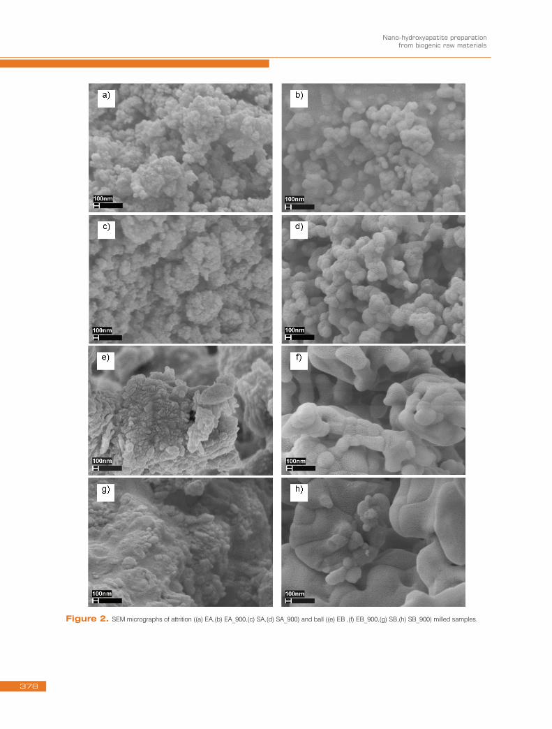
The morphology of the powders made by attrition milling are shown in Fig. 2a - d while those produced by ball milling are shown in Figs. 2e - h. SEM micrographs show no significant difference between the eggshell (EA) and seashell (SA) morphologies (Figs. 2a, 2c). For both eggshell and seashell, attritor milling is more efficient in grain size reduction than ball milling. Thus, smaller particle size with homogeneous size distribution may be achieved with attritor milling. Grains in the SB and EB samples form micron size agglomerates (Figs. 2e, 2g).
After 900oC heat treatment, a 100 nm particle size is characteristic of both starting materials (Figs. 2b, 2d). Agglomerations can be found on Figs. 2f, 2h. Smooth surfaces are evolving after heat treatment at 900oC in both cases (EB_900 and and SB_900).
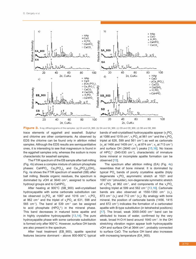
Fig. 3 shows X-ray diffractograms of the powder samples. After intensive attrition milling EA and SA samples (Fig. 3a, 3b) contain hydroxyapatite (JCPDS-PDF 74-0565), calcite (CaCO3, JCPDS-PDF 05-0586), monetite (CaHPO4, JCPDS-PDF 071-1760) and phosphoric acid (H3PO3, JCPDS-PDF 072-0518). These four phases were observed in both eggshell (EA) and seashell (SA) samples. After heat treatment (900°C, 2h) the main phase was hydroxyapatite with some calcite and calcium oxide (CaO, JCPDS-PDF 037-1497) (EA_900 and SA_900).
Powders produced by ball milling (EB and SB) contained montetite, phosphoric acid, and portlandite
Sample Raw materialMilling
wt%shell : H3PO4
Milling time (h)Sintering conditions
method Rpm Temperature (oC)
Holding time (h)
EA eggshell attrition milling 4000 50:50 5 - -
EA _900 50:50 5 900 2
SA seashell 50:50 5 - -
SA _900 50:50 5 900 2
EB eggshell ball milling 350 50:50 10 - -
EB_900 50:50 10 900 2
SB seashell 50:50 10 - -
SB _900 50:50 10 900 2
Table 1. Preparation parameters of hydroxyapatites.
376

G. Gergely et al.
(Ca(OH)2, JCPDS-PDF 044-1481) (Figs. 3c, 3d). In contrast to attrition milled samples the HAp phase was not detected before heat treatment.
After heating at 900oC for 2 h hydroxyapatite, CaO, and calcite appeared in both EB_900 and SB_900 samples, independent of the raw material. In the EB_900 sample, calcium phosphate (β-Ca3(PO4)2, JCPDS-PDF 70-2065) and whitlockite (Ca3(PO4)2, JCPDS-PDF 002-0786) were observed. Calcium phosphate (β-Ca3(PO4)2, JCPDS-PDF 005-0586) was found in the SB_900 powder.
In order to check the presence of elements in addition to the diffractograms energy dispersive X-ray microanalysis (EDS) measurements were performed on the milled powder samples. As the hydrogen content can not be analyzed by X-ray methods, furthermore
the irregular and porous samples do not allow rigorous correction of the EDS spectra, the results can be considered as semiquantitative ones. The spectra were collected in a JSM-25 S-III SEM using Bruker Si(Li) EDS detector and Quantax system using area scan mode for averaging. The collection times were typically 25-60 minutes (live lime) on a single 1 mm2 area of each sample, then the data were turned into numerical composition values using the no-standard P/B ZAF package of the Quantax system. The normalized mass percent output values of the P/B ZAF program are summarized in Table 2. Zirconia may be found in the samples prepared by attrition milling only, because of wearing of zirconia milling parts (balls, tanks). Other elements found by EDS are sodium and silicon (as shown in Table 2). These elements are characteristic
Sample O Na Mg Si P S Cl Ca Zn Zr Ca/P
wt% at.ratio
EA 42.05 0.08 0.45 0.06 16.01 0.1 0.03 41.01 0.06 0.16 1.98
EA_900 31.76 0.05 0.47 0.1 18.34 0.1 0.01 48.98 0.06 0.14 2.06
SA 41.83 0.1 0 0.16 15.72 0.06 0.03 41.93 0.04 0.11 2.06
SA_900 27.89 0.11 0 0.15 18.95 0.03 0.01 52.52 0.05 0.29 2.14
EB 49.67 0.01 0.4 0.03 14.81 0.09 0.01 34.87 0.06 0.05 1.82
EB_900 41.25 0.06 0.47 0.07 16.85 0.08 0 40.99 0.04 0.17 1.88
SB 37.63 0.2 0 0.09 17.08 0.02 0 44.77 0.07 0.16 2.03
SB_900 35.73 0.28 0 0.13 17.63 0.03 0 46.16 0.04 0.01 2.02
Table 2. Elemental composition of hydroxyapatites.
Figure 1. SEM images (a) eggshell and (b) seashell and XRD pattern of the calcinated material (c) eggshell and seashell.
377
Nano-hydroxyapatite preparation from biogenic raw materials
Figure 2. SEM micrographs of attrition ((a) EA,(b) EA_900,(c) SA,(d) SA_900) and ball ((e) EB ,(f) EB_900,(g) SB,(h) SB_900) milled samples.
378
G. Gergely et al.
Figure 3. X-ray diffractograms of the samples: (a) EA and EA_900, (b) SA and SA_900, (c) EB and EB_900, (d) SB and SB_900.
trace elements of eggshell and seashell. Sulphur and chlorine are other contaminants. As observed by EDS the chlorine can be found only in attrition milled samples. Although the EDS results are semiquantitative ones, it is interesting to see that magnesium is found in the eggshell samples only, whereas the sodium is more characteristic for seashell samples.
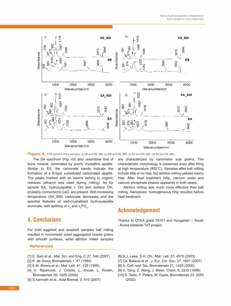
The FTIR spectrum of the EB sample after ball milling (Fig. 4b) shows a complex mixture of calcium phosphate phases: CaHPO4, Ca3(PO4)2 and Ca10(PO4)6(OH)2. Fig. 4a shows the FTIR spectrum of seashell (SB) after ball milling. Beside organic residues, the spectrum is dominated by νOH at 3640 cm-1, assigned to surface hydroxyl groups and to CaHPO4.
After heating at 900°C (SB_900) well-crystallised hydroxyapatite with some carbonate substitution can be observed (ν3PO4 at 1087 and 1016 cm-1, ν1PO4 at 962 cm-1 and the triplet of ν4PO4 at 631, 598 and 565 cm-1). The band at 539 cm-1 can be assigned to acid phosphate (HPO4
2-) in the mineral phase. This band decreases in matured bone apatite and in highly crystalline hydroxyapatite [13,14]. The pure hydroxyapatite phase with some carbonate substitution is formed only after 900°C. However, surface OH bands are also present in the spectrum.
After heat treatment (EB_900), apatite spectral features become dominant - above 800-900°C typical
bands of well-crystallised hydroxyapatite appear (ν3PO4 at 1086 and 1018 cm-1, ν1PO4 at 961 cm-1 and the ν4PO4 triplet at 626, 599 and 561 cm-1) as well as carbonate (ν3 at 1466 and 1409 cm-1, ν2 at 874 cm-1, ν4 at 713 cm-1) and surface OH (3640 cm-1) peaks [15,16]. No traces of HPO4
2- (540-530 cm-1), characteristic of immature bone mineral or incomplete apatite formation can be observed [15].
The spectrum after attrition milling (EA) (Fig. 4c) resembles that of bone mineral. It is dominated by typical PO4 bands of poorly crystalline apatite (triply degenerate ν3PO4 asymmetric stretch at 1021 and 1087 cm-1 (shoulder), non-degenerate symmetric stretch of ν1PO4 at 962 cm-1, and components of the ν4PO4 bending triplet at 599 and 562 cm-1 [13,16]. Carbonate bands are also observed at 1550-1350 cm-1 (ν3), 873 cm-1 (ν2) and 712 cm-1 (ν4). By analogy with bone mineral, the position of carbonate bands (1456, 1415 and 872 cm-1) indicates the formation of a carbonated apatite with B-type substitution (in tetrahedral positions) [17]. The broad, weak 3000-3400 cm-1 band can be attributed to traces of water, confirmed by the very weak, broad H-O-H bend around 1640 cm-1. In the OH stretching vibration region appear both hydroxyapatite νOH and surface OH at 3644 cm-1, probably connected to surface CaO. The surface OH band also increases with increasing temperature (EA_900).
379
Nano-hydroxyapatite preparation from biogenic raw materials
The SA spectrum (Fig. 4d) also resembles that of bone mineral, dominated by poorly crystalline apatite. Similar to EA, the carbonate bands indicate the formation of a B-type substituted carbonated apatite. The peaks marked with an asterix belong to organic residues (ethanol was used during milling). As for sample EA, hydroxyapatite ν OH and surface OH, probably connected to CaO, are present. With increasing temperature (SA_900) carbonate decreases and the spectral features of well-crystallised hydroxyapatite dominate, with splitting of ν3 and ν4PO4.
4. ConclusionsFor both eggshell and seashell samples ball milling resulted in micrometer sized aggregated coarse grains with smooth surfaces, while attrition milled samples
are characterized by nanometer size grains. The characteristic morphology is preserved even after firing at high temperature (900°C). Samples after ball milling include little or no Hap, but attrition milling yielded mainly Hap. After heat treatment HAp, calcium oxide and calcium phosphate phases appeared in both cases.
Attrition milling was much more effective than ball milling. Nanosized, homogeneous HAp resulted before heat treatment.
AcknowledgementThanks to OTKA grant 76181 and Hungarian – South –Korea bilaterial TéT project.
Figure 4. FTIR spectra of the samples: a) SB and SB_900, (b) EB and EB_900, (c) EA and EA_900, (d) SA and SA_900.
References
[1] E. Saiz et al., Mat. Sci. and Eng. C 27, 546 (2007)[2] K. de Groot, Biomaterials 1, 47 (1980)[3] E.M. Rivera et al., Mat. Lett. 41, 128 (1999)[4] U. Ripamonti, J. Crooks, L. Khoali, L. Roden,
Biomaterials 30, 1428 (2009)[5] S.Kenneth et al., Acta Biomat. 3, 910 (2007)
[6] S.J. Leea, S.H. Oh., Mat. Lett. 57, 4570 (2003)[7] Cs. Balázsi et al., J. Eur. Cer. Soc. 27, 1601 (2007)[8] A. CuK neyt Tas, Biomaterials 21, 1429 (2000)[9] X. Yang, Z. Wang, J. Mater. Chem. 8, 2233 (1998)[10] D. Tadic, F. Peters, M. Epple, Biomaterials 23, 2555
(2002)
380
G. Gergely et al.
[11] S. Jinawath, D. Polchai, M. Yoshimura, Mater. Sci. and Eng. C 22, 35 (2002)
[12] C.S. Chai, K.A. Gross, B.B. Nissan, Biomaterials 19, 2291 (1998)
[13] B. Nasari-Tabrizi et al., Mat. Lett. 63, 543 (2009) [14] K.C.B. Yeong, J. Wang, S.C. Ng, Biomaterials 22,
2705 (2001)[15] Cs. Balázsi et al., Mat. Sci. For. 537-538, 105
(2007) [16] K.C.B. Yeong, J. Wang, S.C. Ng, Biomaterials 22,
271 (2001) [17] A. Slosarczyk, E. Stobicrska, Z. Paszkicwicz,
M. Gawlicki, J. Am. Ceram. Soc. 79, 2539 (1996)
[18] N.O. Engin, A.C. Tas, J. Am. Ceram. Soc. 83, 1581-1584 (2000)
[19] L. M. Miller et al, Biochim. et Biophys. Acta 1527, 11 (2001)
[20] F.Z. Zakaria et al., J. Raman Spectrosc. 39, 1204 (2008)
[21] V.C. Farmer, The Infrared Spectra of Minerals (Bartholomew Press, Dorkung, Surrey, 1974) 390
[22] A. Slosarczyk et al, Cer. Inter. 23, 297 (1997)[23] C. Rey, V. Renugopalakrishnan, B. Collins,
M.J. Glimcher, Calcif. Tissue Int. 49, 251 (1991)
381


![Isao KIMURA, Ph.D.gsweb/gs/teacher/pdf/29.pdf · and emulsion Eng., vol. 18, hydroxyapatite vol.28, no.6, [3] "Preparation of hollow hydroxyapatite microspheres containing magnesium](https://static.fdocuments.us/doc/165x107/5f0c5a867e708231d434fba8/isao-kimura-phd-gswebgsteacherpdf29pdf-and-emulsion-eng-vol-18-hydroxyapatite.jpg)


![Synthesis of Hydroxyapatite from egg shell and preparation ... · Ahmed M. Saeed [6] have worked on synthesis of calcium hydroxyapatite powder from hen’s eggshell and orthophosphoric](https://static.fdocuments.us/doc/165x107/603a19cdd92f8913c757fb39/synthesis-of-hydroxyapatite-from-egg-shell-and-preparation-ahmed-m-saeed-6.jpg)