Advances in Collagen/Hydroxyapatite Composite …cdn.intechweb.org/pdfs/14592.pdfAdvances in...
31
1 Advances in Collagen/Hydroxyapatite Composite Materials Anton Ficai, Ecaterina Andronescu, Georgeta Voicu and Denisa Ficai Politehnica University of Bucharest, Faculty of Applied Chemistry and Materials Science Romania 1. Introduction The annually necessary human bone grafts are in continuous grown due to the increasing of fractals, congenital and non-congenital diseases. Based on statistical reports (Murugan and Ramakrishna 2005), only in USA about 6.3 million fractures occur every year and about 550.000 of these require bone grafting. The most frequently fractures occur at the level of hip, ankle, tibia and fibula. Due to the higher physical effort, the men are more exposed to fracture than women (2.8% in the case of men comparing to 2.0% in the case of women). The number of fractures increases year by year and consequently many researchers from different research fields co-operate in order to develop new bone graft materials. Also, it is important to mention that, in the present, the bone diseases are overlapped only by hearth diseases. The history of bone grafting is starting in 1913 when Dr. D.E. Robertson assays a piece of cat’s bone and a piece of human bone for bone grafting into dogs (Gallie and Toronto 1914). The microscopic analysis of implanted graft after 20 days shows that the space between graft and living bone is filled with new cancellous bone. These early works made the premises for the development of the bone grafts. Due to the increasing of the necessarily bone grafts, autografts and allografts can not cover the overall need of bone grafts. For compensate this gap, artificial (synthetic) grafts are necessary and, consequently used. The use of synthetic grafts has some advantages versus allografts, autografts and xenografts: the possibility to obtain unlimited number/quantity of synthetic grafts, more safety use of artificial bone grafts without disease transmission risk, pain limitation by elimination of some secondary surgical intervention. The need of bone grafts materials lead to the synthesis of many kind of materials with different properties. Function of the nature of these materials and the relation between these grafts and the host tissue, these materials can be divided into 4 generations (Fig. 1). The components of the first generation of bone grafting biomaterials have remarkable mechanical properties but they are neither bioresorbable nor bioactive. More than, the use of these kind of bone grafts have limited lifetime (usually less than 10-15 years) and need to be extracted and replaced surgically. Some of the most representative biomaterials from the first generation of bone grafting biomaterials are: the iron, cobalt, chromium, titan or their alloys: steel (especially 316 L), cobalt or titan based alloys (Corces 2002; Corces and Garcia 2007) etc. www.intechopen.com
Transcript of Advances in Collagen/Hydroxyapatite Composite …cdn.intechweb.org/pdfs/14592.pdfAdvances in...

1
Advances in Collagen/Hydroxyapatite Composite Materials
Anton Ficai, Ecaterina Andronescu, Georgeta Voicu and Denisa Ficai Politehnica University of Bucharest, Faculty of Applied Chemistry and Materials Science
Romania
1. Introduction
The annually necessary human bone grafts are in continuous grown due to the increasing of
fractals, congenital and non-congenital diseases. Based on statistical reports (Murugan and
Ramakrishna 2005), only in USA about 6.3 million fractures occur every year and about
550.000 of these require bone grafting. The most frequently fractures occur at the level of
hip, ankle, tibia and fibula. Due to the higher physical effort, the men are more exposed to
fracture than women (2.8% in the case of men comparing to 2.0% in the case of women). The
number of fractures increases year by year and consequently many researchers from
different research fields co-operate in order to develop new bone graft materials. Also, it is
important to mention that, in the present, the bone diseases are overlapped only by hearth
diseases.
The history of bone grafting is starting in 1913 when Dr. D.E. Robertson assays a piece of
cat’s bone and a piece of human bone for bone grafting into dogs (Gallie and Toronto 1914).
The microscopic analysis of implanted graft after 20 days shows that the space between graft
and living bone is filled with new cancellous bone. These early works made the premises for
the development of the bone grafts.
Due to the increasing of the necessarily bone grafts, autografts and allografts can not cover
the overall need of bone grafts. For compensate this gap, artificial (synthetic) grafts are
necessary and, consequently used. The use of synthetic grafts has some advantages versus
allografts, autografts and xenografts: the possibility to obtain unlimited number/quantity of
synthetic grafts, more safety use of artificial bone grafts without disease transmission risk,
pain limitation by elimination of some secondary surgical intervention.
The need of bone grafts materials lead to the synthesis of many kind of materials with
different properties. Function of the nature of these materials and the relation between these
grafts and the host tissue, these materials can be divided into 4 generations (Fig. 1).
The components of the first generation of bone grafting biomaterials have remarkable
mechanical properties but they are neither bioresorbable nor bioactive. More than, the use of
these kind of bone grafts have limited lifetime (usually less than 10-15 years) and need to be
extracted and replaced surgically. Some of the most representative biomaterials from the
first generation of bone grafting biomaterials are: the iron, cobalt, chromium, titan or their
alloys: steel (especially 316 L), cobalt or titan based alloys (Corces 2002; Corces and Garcia
2007) etc.
www.intechopen.com

Advances in Composite Materials for Medicine and Nanotechnology
4
Fig. 1. Biomaterials evolution in the field of bone grafting
The components of the second generation of bone grafting biomaterials are at least
bioresorbable or bioactive and they do not require to be replaced in time. The most
representative biomaterials from the second generation of bone grafting biomaterials are:
calcium phosphates (especially hydroxyapatite and tricalcium phosphate), the bioglasses
(Hench et al. 2004), alumina (Sedel et al. 1994); zirconia (Clarke et al. 2003) and the following
polymers: poly ε-caprolactone (Oláh et al. 2007), polyurethanes (Bonzani et al. 2007 ;
Guelcher et al. 2004) etc.
The components of the third generation of bone grafting biomaterials are both bioresorbable
and bioactive and have superior properties. It has to mention that these biomaterials present
higher specific properties than the first two generations of bone graft materials and short
time after implantation these materials are resorbed and in time, in the place of the bone
graft the new bone is formed. The properties of these (nano)composite materials is strongly
influenced by the nature of components, the composition and the morphology. That is why
many researchers tried to obtain not only compositional similitude with natural bones from
mineralogical and morphological point of view. The most representative biomaterials of the
third generation of bone grafting biomaterials are: (nano)hydroxyapatite/collagen (Wahl
and Czernuszka 2006), (nano)hydroxyapatite/collagen/hyaluronic acid (Bakos et al. 1999),
hydroxyapatite/poly-L-lactic acid (Kesenci et al. 2000), hydroxyapatite/chitosan (Wang and
Li 2007).
The fourth generation of bone grafting biomaterials is similar with the third generation
materials but improved by the presence of bonny cells, growth factors, bone morphogenetic
proteins etc.
One of the most important characteristic of bone grafts materials is the osteointegration. The
osteointegration (and also osteoconductivity) of the grafts is related to the degree of porosity
and pore size (applicable especially to the last three generation of bone grafting
biomaterials) (Develioglu et al. 2005). Based on the literature data and also based on the size
of osteoblasts (which vary up to 20-25 μm) (Chang et al. 2000; Develioglu et al. 2005;
Gauthier et al. 1998) the optimum pore size was found to be 50-550μm.
The natural bones contain mainly collagen and hydroxyapatite. That is the reason because
many researchers try to understand and obtain (nano)hydroxyapatite/collagen composite
for hard tissue repairing. The composition of bones varies function of many factors such as:
specie, sex, age, bone type, location etc.. The relative composition of bone is presented in the
Table 1, while the most important biomedical properties of the bones are presented in
Table2. The in vivo bone biosynthesis is controlled by many factors such as: BMP (bone
morphogenetic proteins) (Abe et al. 2000), transforming growth factors (Tashjian et al. 1985),
cytokines (de Vernejoul et al. 1993 ), hormones (Bollerslev et al. 1991 ; De Vernejoul et al.
www.intechopen.com

Advances in Collagen/Hydroxyapatite Composite Materials
5
1990; Hock and Gera 1992), transcription factors (Cui et al. 2003; Ogawa et al. 2000),
adhesion molecules (Miyake et al. 1991) and so on.
Components wt %
Mineral phase
Hydroxyapatite Carbonate (mostly as carbonated hydroxyapatite) Citrate Na+ Mg2+
Others
60–66 ~ 4 ~ 0,9 ~ 0.7 ~ 0.5 Traces
Organic phase
Collagen Non-collagenous proteins: (osteocalcin, osteonectin, osteopontin, sialoprotein, BMP) Others: (polysaccharides, lipids, cytokines)
20–25 2 – 3 Traces
Water 8 – 9
Table 1. The composition of healthy bone
Property Cortical
Bone
Cancellous
Bone
Young’s (Tensile) Modulus (GPa) 7–30 0.05–0.5
Compresive strength (MPa) 100–230 2–12
Flexural Strength (MPa) 50–150 10–20
Fracture toughness (MPa m1/2) 2–12 0.1
Strain to failure 1–3 5–7
Apparent density (g/cm3) 1.8–2.0 0.1–1.0
Surface area/volume ratio (mm2/mm3) 2.5 20
Table 2. Biomechanical properties of healthy bone
Bone formation and remodelation is controlled by many factors: physical, chemical, hormonal, growth factors, and anti-mineralization agents (Karsenty 2000; Karsenty et al. 2009; Wallach et al. 1989). The bone tissue piezoelectricity is the key element which assists the bone formation, remodelation and growth, especially in the early stages of the bone formation and. The piezoelectricity of the bone is due, among the rests, due to the bone
www.intechopen.com

Advances in Composite Materials for Medicine and Nanotechnology
6
tissue anisotropy. The main elements which induce the anisotropy of the collagen based structures are: the anisotropy of the collagen molecules themselves and the anisotropy of collagen fibrils and fibres as well as the oriented morphology of the fibrils and fibres disposed in the isotropic extrafibrillar space (Hellmich et al. 2004). Trying to obtain such composite with same composition and structure with the natural
bone, the researchers elaborated many synthesis methods (Wahl and Czernuszka 2006) such
as: in vitro collagen mineralization (Lawson and Czernuszka 1998), thermally – triggered
assembly of hydroxyapatite/collagen gels (Pederson et al. 2003), vacuum infiltration of
collagen into a ceramic matrix (Werner et al. 2002), enzymatic mineralization of collagen
sheets (Yamauchi et al. 2004), freeze drying and supercritical point drying (Pompe et al.
2001), biomimetic synthesis (Li and Chang 2008).
One of the most promising bone graft material seems to be the
collagen/nanohydroxyapatite composite (Li et al. 2006) due to its very good compositional
and structural similarity with natural bone (Yunoki et al. 2007; Yunoki et al. 2006). The role
of each component is not very well known. It is generally accepted that mineral phase,
mainly containing hydroxyapatite, provide toughness and rigidity while the organic matrix
provide tensile strength and flexibility of bone. In composite materials, collagen and
hydroxyapatite play the same roles as they play in natural bone.
The mineral phase is deposited on the organic phase through electrostatic interactions
between the carboxyl groups from collagen and Ca2+ from hydroxyapatite, but there is no
unison about the mineralization sites. Many researchers assert that hydroxyapatite is
deposited only onto the non-collagenous proteins and citrate anions (Rhee and Tanaka 1998)
while others assert that mineralization occurs also on the pure collagen (Lin et al. 2004;
Zhang et al. 2003). It is well known that materials properties are influenced not only by
composition but also by morphology. This is the reason because even for similar
composition of many bones their properties differ very much. Starting from these
hypothesis, scientists tried to improve or induce new properties of the COLL/HA composite
materials by addition of third component (or even more components) or by inducing
different morphology. The influence of the morphology can be easily marked out by
comparing the mechanical properties of compact and cancellous bone (Table 2) (Hench and
Wilson 1993).
Collagen mineralization occurs due to the interactions which appear between collagenous
structures and hydroxyapatite nanocrystals. In fact, these interactions occur between
carboxylate groups and Ca2+ cations and can be illustrated as presented in Fig. 2. This
hypothesis is supported by FTIR data and was explained based on the spectral shifts of C-O
and C=O bands in pure collagen and COLL/HA composites (Ficai et al. 2009a).
I II
Fig. 2. Mezomeric form stabilization of carboxylate group due to mineralization
www.intechopen.com

Advances in Collagen/Hydroxyapatite Composite Materials
7
As a result of these interactions, hydroxyapatite particles are preferentially deposited onto the collagenous support forming so-called nucleation centers. Once formed, the nucleation centers increase and can lead even to the formation of a thin film. If the amount of deposited HA is low enough, the mineralization centers can be visualized by scanning electron microscopy (Fig. 3).
Fig. 3. The SEM micrograph of mineralized collagen fibres
In order to obtain good bone substitutes, researchers have to understand the biosynthesis of
natural bone and to control some parameters such as composition, hydroxyapatite shape
and size, collagen fibrils and hydroxyapatite blade orientation.
The synthesis of COLL/HA composite materials with different porosity/density is very important in order to obtain materials with tailored properties. COLL/HA composite materials with tailored ceramic properties can be easily obtained by combining the controlled air drying with freeze drying. The controlled air drying followed by freeze drying can be easily used for the synthesis of COLL/HA composite materials with any composition. In this scope, the wet composite materials are let, under controlled atmosphere for certain time (controlled temperature and humidity) followed by a final drying, realized by freeze drying. The use of mixed drying method (controlled air drying combined with freeze drying) is technically easy to control and economically sustainable. The ceramic properties of the COLL/HA composite materials can be easily controlled by the air drying step conditions (especially drying time); the longer controlled air drying time lead to composite materials with lower porosity and increasing density. Scanning electron microscopy can be used for qualitative analysis of these materials obtained by controlled air drying followed by freeze drying (Fig. 4). Analysing the SEM images of the materials obtained by air drying (increasing drying time) followed by freeze drying it is stand out a mile that the increasing air drying lead to denser materials.
www.intechopen.com

Advances in Composite Materials for Medicine and Nanotechnology
8
Fig. 4. SEM images of COLL/HA composite materials obtained by controlled air drying (0, 48 and 199h) followed by freeze drying
0.00
0.20
0.40
0.60
0.80
1.00
1.20
1.40
1.60
0 50 100 150 200 250 300
air drying time, h
den
sity
, g/c
m3
COLL/HA 1:4
COLL/HA 1:3
COLL/HA 1:1
Fig. 5. Ceramic properties of the three series of COLL/HA composite materials versus air drying time
It has to mention that during the air drying process, simultaneous with the water evaporation the morpho-structural restructuring of the composite happened which leads to more compact materials, the density of the COLL/HA composite materials being 0.15-1.6g/cm3 while the open porosity 25-95% (Fig. 5). As a general rule, the density of the
www.intechopen.com

Advances in Collagen/Hydroxyapatite Composite Materials
9
composite materials increases with the increase of the air drying time. This method is very useful because allow to tailor the ceramic properties of the materials without modify the composition. The orientation of collagen/hydroxyapatite composite is induced by the mutual interaction
between collagen and hydroxyapatite in aqueous solutions. For better orientation, it is
possible to use electric and / or magnetic field. Cunyou Wu and co-workers (Wu et al. 2007)
report unidirectional oriented hydroxyapatite/ collagen composite using 10 T magnetic field
starting from calcium containing collagen solution and phosphate solution at 37 0C. If
magnetic field is imposed, perpendicularly to the rotation axis, unidirectional oriented
collagen fibrils and hydroxyapatite crystals have obtained due to their magnetic
susceptibility anisotropy (Hellmich et al. 2004).
Due to the importance of these composite materials thousands of papers and many
comprehensive reviews are published yearly. Some of the most comprehensive reviews
about bones and bone grafts materials based on collagen and/or hydroxyapatite were
published in the last years by Cui at al (Cui et al. 2007), Dorozhkin (Dorozhkin 2009),
Murugan and Ramakrishna (Murugan and Ramakrishna 2005) and by Wahl and
Czernuszka (Wahl and Czernuszka 2006).
Some of the most recent advances in the field of COLL/HA composite materials are
systematically presented below.
2. The influence of organic and inorganic species on the COLL/HA composite materials
Recent studies made by Ficai et al. (Ficai et al. 2010b; Ficai et al. 2010e) reveal the influences
of the PVA, denaturated collagen (hydrolysate collagen and ionic species on the
collagen/hydroxyapatite composite materials. These works are essential because neither in
vitro nor in vivo mineralization is readily understood, and the synthesis of materials that
mimic the characteristics of bone has not been previously accomplished.
The properties of COLL/HA composite materials can be tailored by addition of different
organic or inorganic components; well studied in the literature being that ions which
naturally occur in natural bone: citrate, fluoride, chloride, carbonate, magnesium or some
ions which accidentally occur in natural bones and, usual is responsible for different
diseases (Pb2+, Sr2+ etc.).
The synthesis of complex COLL/HA composite materials which also contain PHA, collagen
hydrolysate or other ionic species than calcium and phosphate were synthesized by co-
precipitation of hydroxyapatite on collagenous matrices (Ficai et al. 2009b). The
mineralization process consisted of two successive stages. In the first stage, a Ca(OH)2
suspension was added drop-wise and allowed to interact for 24 h. In the second stage, a
stoichiometric quantity of NaH2PO4 solution was added drop-wise. The quantities of
collagen matrix and HA precursors were used at a ratio of 1:4. After the hydroxyapatite
precursors were added, the pH was adjusted to 9 using a solution of NaOH, in order to
assure brushite-free HA precipitation. During the synthesis and drying of the composite
material, the temperature was maintained at approximately 37 0C.
To study the effect of the citrate ions on the mineralization process, the collagen matrices
were dipped into a dilute citrate solution (5 ‰) for 30 minutes and then mineralized as
presented above. Upon completion of this process, the resulting materials were analyzed.
www.intechopen.com

Advances in Composite Materials for Medicine and Nanotechnology
10
In this chapter, two collagenous matrices that contained 5% and 70% of the hydrolysate
were mineralized following the aforementioned procedure. The COLL/PVA and COLL-
PVA/HA hybrid materials were obtained starting from collagen gel and PVA solution, the
mixing ration being COLL:PVA = 1:2. the as obtained gel was than mineralized as presented
above.
In order to obtain fluoride-substituted hydroxyapatite, a modified mineralization method
was used. For this purpose, in the second stage of mineralization, a corresponding amount
of NaF was added to the phosphate solution to assure the transformation of
5%hydroxyapatite into fluoroapatite.
In order to design COLL/HA composite materials and to obtain a bone-like morphology, it
is important to understand the influence of each component on the morphology of the
material.
The influence of the collagen hydrolysate, PVA and ionic species were analyzed by XRD,
FTIR and SEM (-EDS) from the point of view of composition and morphology. The synthetic
conditions and characteristics of the resulting hydroxyapatite were strongly influenced by
the organic support.
Fig. 6. XRD patterns of different collagen/hydroxyapatite-based composite materials: A. mineralized cross-linked matrix (glutaraldehyde, 1%); B. mineralized cross-linked matrix (glutaraldehyde, 1%), the mineralization occurs in the presence of F- (5 % (molar) reported to HA); C. mineralized hydrolysate-enriched collagen matrix; and D. crystalline HA (ASTM 74-5966)
www.intechopen.com
Advances in Collagen/Hydroxyapatite Composite Materials
11
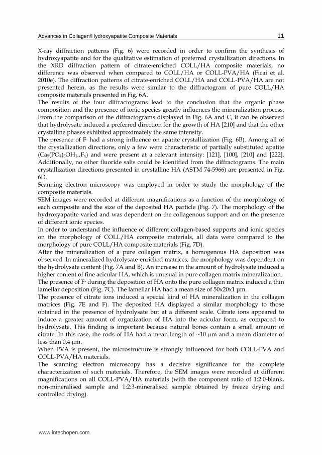
X-ray diffraction patterns (Fig. 6) were recorded in order to confirm the synthesis of hydroxyapatite and for the qualitative estimation of preferred crystallization directions. In the XRD diffraction pattern of citrate-enriched COLL/HA composite materials, no difference was observed when compared to COLL/HA or COLL-PVA/HA (Ficai et al. 2010e). The diffraction patterns of citrate-enriched COLL/HA and COLL-PVA/HA are not presented herein, as the results were similar to the diffractogram of pure COLL/HA composite materials presented in Fig. 6A. The results of the four diffractograms lead to the conclusion that the organic phase composition and the presence of ionic species greatly influences the mineralization process. From the comparison of the diffractograms displayed in Fig. 6A and C, it can be observed that hydrolysate induced a preferred direction for the growth of HA [210] and that the other crystalline phases exhibited approximately the same intensity. The presence of F- had a strong influence on apatite crystallization (Fig. 6B). Among all of the crystallization directions, only a few were characteristic of partially substituted apatite (Ca5(PO4)3OH1-xFx) and were present at a relevant intensity: [121], [100], [210] and [222]. Additionally, no other fluoride salts could be identified from the diffractograms. The main crystallization directions presented in crystalline HA (ASTM 74-5966) are presented in Fig. 6D. Scanning electron microscopy was employed in order to study the morphology of the composite materials. SEM images were recorded at different magnifications as a function of the morphology of each composite and the size of the deposited HA particle (Fig. 7). The morphology of the hydroxyapatite varied and was dependent on the collagenous support and on the presence of different ionic species. In order to understand the influence of different collagen-based supports and ionic species on the morphology of COLL/HA composite materials, all data were compared to the morphology of pure COLL/HA composite materials (Fig. 7D). After the mineralization of a pure collagen matrix, a homogenous HA deposition was observed. In mineralized hydrolysate-enriched matrices, the morphology was dependent on the hydrolysate content (Fig. 7A and B). An increase in the amount of hydrolysate induced a higher content of fine acicular HA, which is unusual in pure collagen matrix mineralization. The presence of F- during the deposition of HA onto the pure collagen matrix induced a thin lamellar deposition (Fig. 7C). The lamellar HA had a mean size of 50x20x1 μm. The presence of citrate ions induced a special kind of HA mineralization in the collagen matrices (Fig. 7E and F). The deposited HA displayed a similar morphology to those obtained in the presence of hydrolysate but at a different scale. Citrate ions appeared to induce a greater amount of organization of HA into the acicular form, as compared to hydrolysate. This finding is important because natural bones contain a small amount of citrate. In this case, the rods of HA had a mean length of ~10 µm and a mean diameter of less than 0.4 µm. When PVA is present, the microstructure is strongly influenced for both COLL-PVA and COLL-PVA/HA materials. The scanning electron microscopy has a decisive significance for the complete characterization of such materials. Therefore, the SEM images were recorded at different magnifications on all COLL-PVA/HA materials (with the component ratio of 1:2:0-blank, non-mineralised sample and 1:2:3-mineralised sample obtained by freeze drying and controlled drying).
www.intechopen.com
Advances in Composite Materials for Medicine and Nanotechnology
12
Fig. 7. SEM images of different collagen and hydroxyapatite composite materials: A. composite obtained by mineralization of a collagen-hydrolysate matrix (5% of hydrolysate); B. composite obtained by mineralization of a collagen-hydrolysate matrix (70% of hydrolysate); C. composite obtained by mineralization of a pure collagen matrix (the mineralization was achieved in the presence of F- (5%-molar reported to HA); D. mineralized (pure) collagen matrix; E, F. composite obtained by mineralization of a collagen-citrate matrix (citrate less than 2%)
www.intechopen.com

Advances in Collagen/Hydroxyapatite Composite Materials
13
The SEM images have given some morphological information about the four materials. Fig.
8a, b and Fig. 9a, b show the morphology of the two materials obtained by controlled drying
in air at 30oC; they are stratified but compact materials. Fig. 8c, f and Fig. 9c, f show the
morphology of the freeze dried composite materials; such materials exhibiting stratified but
porous morphologies. Based on the SEM images, the freeze dried COLL-PVA/HA 1:2:0
hybrid material has the mean distance of 60-120μm between the sheets while for the freeze
dried COLL-PVA/HA 1:2:3 composite, the hybrid material has the mean distance of 40-
70μm between the sheets.
Fig. 8. SEM images of the COLL–PVA hybrid materials (weight ratio of 1:2) obtained by (a and b) controlled drying and (c–f) freeze drying
In fact, the two morphologies are similar, the compact materials resulted from the
continuous remodelling and restructuring until it becomes compact; if the freeze drying is
used, once the materials are frozen the restructuring is blocked and the materials retain their
initial high porosity.
For both compositions, the controlled air drying at 30oC has led to parallel layers bounded
each other by fibres of different diameters (from less than 1µm up to 10µm).
www.intechopen.com

Advances in Composite Materials for Medicine and Nanotechnology
14
A higher magnification (Fig. 9f, for instance) has revealed the very good homogeneity and compatibility of the three components practically, no free HA agglomerates being visible.
Fig. 9. SEM images of the COLL–PVA/HA hybrid composite materials (weight ratio of 1:2:3) obtained by (a and b) controlled drying and (c–f) freeze drying
The osteointegration of the bone graft materials is induced, among the rests, by the porosity.
The density, porosity and absorption (Table 1) were measured by the Arthur method. In all
cases three replicates were done, the experimental error being less than 1-2%. The results are
in very good agreement with the SEM observation. The highest porosities correspond to the
freeze dried materials and have reached about 80 and 90%. For the materials obtained by
controlled drying at 30oC in air, the porosity was less than 20%. The xylene absorption is
proportional with the porosity while the density is in inverse proportion. The presence of
HA has induced the density increase and a decrease in the porosity and xylene absorption.
Hybrid materials with intermediary properties were obtained by a mixed drying method which involves controlled drying followed by freeze drying (not presented in this paper).
www.intechopen.com

Advances in Collagen/Hydroxyapatite Composite Materials
15
Sample Density,
g/cm3
Porosity,
% Absorption,
%
COLL-PVA 1:2, freeze drying 0.10 92.7 90.0
COLL-PVA 1:2, controlled drying 0.75 19.3 19.0
COLL-PVA/HA 1:2:3, freeze drying 0.44 79.2 78.9
COLL-PVA/HA 1:2:3, controlled drying 0.88 13.7 13.0
Table 3. Ceramic properties of the COLL-PVA 1:2 (wt) hybrid materials and COLL-PVA/HA 1:2:3 (wt) composite materials
In order to understand the influence of citrate ions on the morphology of acicular and non-acicular zones, the EDS spectra of these two zones were recorded (Fig. 10). The results indicated that the ratio of Ca to P differed between these two zones, where the Ca:P ratio was 1.6 (similar to stoichiometric HA) in the non-acicular zone (Fig. 10A) and 1.1 in the acicular zone (Fig. 10B). The acicular zone occurs due to a higher content of Na+ and citrate.
Fig. 10. EDS spectra of a) a non-acicular and b) an acicular reach zone
Infrared spectroscopy (Fig. 11) is an efficient tool for the investigation of organic-inorganic interactions. The differences in morphology may be explained by these strong interactions and are evidenced in the shift of peaks or in peaks duplications. In order to understand organic-inorganic interactions, the IR spectrum of a pure collagen matrix was recorded (Fig. 11A). The most important peaks in the pure matrix were the amide peaks (Chang and Tanaka 2002; Ficai et al. 2009b). After collagen matrix mineralization with HA precursors, the main amide peak of pure collagen (1630 cm-1) was shifted to a higher wave number for the COLL/HA composite (1650 cm-1), while the phosphate peaks appeared at 1030, 609 and 564 cm-1 (Fig. 11B). The infrared spectrum of the mineralized hydrolysate-enriched collagen matrix (Fig. 11C) was characterized by the duplication of each main collagen peak and led to the differentiation of the phosphate peak and the appearance of a shoulder at about 1110 cm-1. These results can be explained by the degree of condensation between collagen and collagen hydrolysate, which induces interactions of varying strength between mineral and organic components. As reported in the literature (Silva et al. 2001), the degradation of collagen
www.intechopen.com

Advances in Composite Materials for Medicine and Nanotechnology
16
leads to a decrease in the absorption band at 1240 cm-1. In composites obtained with hydrolysate, this band being less intense than that in pure collagen.
Fig. 11. FTIR spectra of: A. a collagen matrix (cross-linked with glutaraldehyde, 1%); B. a mineralized cross-linked collagen matrix (glutaraldehyde, 1%); C. a mineralized hydrolysate-enriched collagen matrix; and D. a mineralized cross-linked collagen matrix achieved in the presence of F- (5 % (molar) reported to HA)
When the mineralization of the collagen matrix occurs in the presence of fluoride, the collagen peaks were not shifted (compared to the COLL\HA composite), but the phosphate peaks of HA were split due to the partial substitution of hydroxyl groups with fluoride (Fig. 11D).
3. The synthesis of COLL/HA composite materials with oriented structure
In this field of orientation some works were published in the last 5 years. The synthesis of COLL/HA composite materials with oriented structure can be induced by self-assembly, electric field orientation or by using a high magnetic field (Wu et al. 2007), best results being reported by self-assembly (Ficai et al. 2010c) and pulsed electric field orientation (Ficai et al. 2010a). The magnetic field orientation is possible due to the magnetic anisotropy of the hydroxyapatite (Wu et al. 2007).
3.1 Self-assembling of COLL/HA composite materials The self-assembly is one of the most easy to realize orientation which not imply any external influences but require slow drying at pH=6.9-9 which can easily induce denaturation of the collagen. The synthesis of COLL/HA composite materials by self-assembling consists by two successive stages but have some particularities. In the first stage, the collagen gel is treated with the desired amount of Ca(OH)2 suspension, drop-wise and magnetically stirred for 24 h and let to interact. In the second stage, the stoichiometric quantity of H3PO4 solution was added also dropwise. Ca(OH)2 was used in order to assure the necessary basic pH. During the first stage of mineralization the pH was maintained at pH 0 9 by addition of HCl.
www.intechopen.com

Advances in Collagen/Hydroxyapatite Composite Materials
17
The ratio of collagen, Ca(OH)2 and H3PO4 was so chosen that the final ratio of COLL:HA to be 20:80 (wt) and at the end of synthesis the concentration of collagen to became 1.66%. After the H3PO4 solution is added, the pH was adjusted at about 9 using NaOH solution, in order to assure pure HA precipitation. During the synthesis and drying of the composite
material, the temperature was kept at ~37 OC. pH 9 was choose based on two characteristics of the collagen molecules:
a. at pH= 6.9–8 the collagen molecules are in an extended conformation (length of collagen molecule 180–200 nm) and,
b. the fibrillogenesis is increasing in the range of 6.6–9.2. The main processes which occur during the synthesis are represented in Fig. 12. The collagen molecules have different conformation, function of the pH can be linear (at pH≥9) or crimpy (for pH< 7); at intermediate pH the collagen molecules being crimpy but with a more pronounced linear aspect. In order to be sure that collagen molecules are in elongated form (linear) the working pH was set at 9.
Fig. 12. Specific processes occurred in solution during the self-assembly of COLL/HA composite materials
Function of the mineralization pH, we can obtain different morphologies (Fig. 12b and d). If the pH increases, the crimpy structure of collagen molecules from gel and also from
www.intechopen.com

Advances in Composite Materials for Medicine and Nanotechnology
18
composite can be modified into the elongated one. If the drying time is slowly enough, these linear (elongated) collagen molecules can self-assembly and form cylindrical fibrils and fibres (Fig. 12e), otherwise they will form fibrils and fibres, growth tri-dimensional, with crimpy collagen molecules (for pH< 7) or with linear collagen molecules (for pH ≥ 9). The structure presented in Fig. 12b can be transformed into the structure presented in Fig. 12d by increasing of pH at an adequate value. In this case, the pH must be higher than in case of transformation of the structure illustrated in Fig. 12a into the structure presented in Fig. 12c, probably due to the interaction between HA and collagen. The self-assembling process was characterized especially by XRD and SEM. XRD was used to point out the mineralization process. The XRD spectrum, in Fig. 13, shows the formation of HA.
Fig. 13. XRD pattern of COLL/HA composite material (a) before sodium chloride removal and (b) after sodium chloride removal
In the case of COLL/HA composite obtained by self-assembly comparing with the HA
obtained by precipitation, the X-ray diffraction pattern exhibit a much higher intensity for
the 2 1 1 peak reported to the intensity of the other peaks characteristic to HA. This result
can be attributed to a preferential growth of the HA crystals in the 2 1 1 direction due to the
collagen influence. As it can see, the composite material also contains NaCl (Fig. 13a). The
www.intechopen.com

Advances in Collagen/Hydroxyapatite Composite Materials
19
removal of NaCl can be easily made by washing the composite materials with distilled
water, following the next procedure: after drying, the COLL/HA composite material with
uniaxial orientation of the constitutive fibres is crosslinked with glutaraldehyde, washed
with plenty of water and dried. Following this procedure, the chloride was completely
removed (Fig. 13b) without altering the mineral phase, especially from the point of view of
crystallinity and preferential crystallization direction.
The self-assembling structure of collagen molecules and hydroxyapatite particles can be
proved by SEM (Fig. 14). The samples were analyzed in perpendicular and parallel section
reported to mineralized collagen fibres, in order to study the formation and the orientation
of collagen fibrils and fibres. The sodium chloride removal not alters the composite
morphology; the SEM images recorded before and after sodium chloride removal being
similar. Whatever the analysis section, the recorded SEM images show the formation of
collagen fibrils and fibres, which are mineralized with HA.
Fig. 14. The SEM images of collagen/hydroxyapatite composite materials, recorded at different magnification; (a) parallel view with the fibres, (b–c) perpendicular view with the fibres, (d) high resolution SEM at 80,000× magnification
The SEM image presented in Fig. 14a is recorded in a fibres perpendicular section. This
image shows a homogenous arrangement of the fibres. Fig. 14b and c are recorded in a
fibres parallel profile and shows, at different magnification the stratified structure of the
composite materials, the fibrils and fibres being organized in layers. It can also observe
highly oriented fibres and fibres bundle. The homogenous arrangement can be evidentiated
www.intechopen.com

Advances in Composite Materials for Medicine and Nanotechnology
20
also in fibres parallel profile. Analyzing the rupture profile, it can be observed a shift
between the rupture point, comparing different neighbor adjacent layers. In Fig. 14c,
recorded at higher magnification, it can better observe the mineral deposition. It can
conclude, that at the end of collagen fibres, the HA density is higher. At broken, the rupture
seems to follow the highly mineralized zone of interfibrilar gap which exist at the end of
two successive fibres. Finally, at a much higher magnification (Fig. 14d) the SEM images of
fibres’ end allow the estimation of the dimensions of HA, these particles being in the
nanometric range. At high magnification from SEM image, it can be seen that the
dimensions of the HA crystals are in the nanometric range (4–40 nm), with an elongated
morphology. These results are in good agreement with the observation reporting to natural
HA from the natural bone.
The SEM images obtained at different magnification, in the rupture, on parallel and
perpendicular direction of collagen fibres, are showing a homogenous microstructure, with
fibres organized in layers, with high orientation. Taking also in consideration the way in
which the rupture took place, and also the fact that we have a high concentration of HA
grains in the rupture points we might conclude that the COLL/HA composite material
synthesized has a very similar structure with that of the natural long bone, that have been
obtained through auto-assembling process.
Fig. 15. Straight line projection onto the three dimensions and visualization of the three angles formed between the projections and the three dimensions
The orientation degree can be quantified function of the deviation angle of each collagen
fibers from the direction of the applied electric field. The orientation degree of mineralized
collagen fibers is highly influenced by magnification; with the increasing magnification the
orientation degree increase. At a magnification of 1000× the average of deviation degree is
less than 5% while the orientation degree is more than 95%. The average fiber deviation and
also the orientation degree of collagen fibers can be quantified based on the following
equations:
Ni
i=1
100 DAD(%) = and the OD(%) = 100 D
N 90⋅ −∑
www.intechopen.com

Advances in Collagen/Hydroxyapatite Composite Materials
21
where, N is the number of fibers; DAi — correspond to deviation angle of each fiber; D (%) correspond to the average deviation of the fibers toward the applied electric field and OD(%) correspond to the orientation degree. Obviously the orientation is difficult to quantify and only a few methods permit this kind of measurements (and usually these are indirect methods); the orientation will be quantify based on the SEM images. Hydroxyapatite was obtained by co-precipitation in the presence of collagen gel. In fact, starting from collagen gel and hydroxyapatite precursors, in certain conditions, due to the interactions between collagen, hydroxyapatite and water it was obtained self-assembled, highly oriented composite materials. It can note that, many authors published a lot of papers dealing with COLL/HA composite materials which start from collagen gel and calcium hydroxide and ortophosphoric acid as precursors, but due to the inadequate processing conditions they do not obtained the uniaxial orientation of the constitutive fibres. In aqueous solution the collagen molecules and hydroxyapatite precursors have the capacity to induce synthetic bone formation by self-assembling. The obtained composite material is more similar with compact bones morphology; the recorded analysis being very similar with these bones. Very important is that we also propose a new way to estimate the average deviation of fibres and we determined it, assuming a 2D model. The average deviation is 2.54±0.2% which means that the degree of orientation is 97.46±0.2%.
3.2 Electric field orientation of COLL/HA composite materials Also, orientation can occur due to different external factors (magnetic or electric fields), most efficient and easy to realize being the electric field orientation. The electric field orientation occur really fast (less than 1h) at low electric field (<1V/cm), the best electric field being the pulsatory electric filed. The magnetic orientation can be realized but require very high magnetic field (10T). First time in the literature, the degree of orientation was mathematically quantified using a very simple equation, based on the SEM images. The synthesis of COLL/HA composite materials via electric field orientation is realized in similar conditions with the mention that, before drying different kinds of electric fields are applied. By short, the collagen mineralization was conducted in two stages, in order to mimic the in vivo osteosynthesis. First stage is the calcium deposition onto the collagen that takes 24 h. The second stage consists in phosphate addition leading to hydroxyapatite precipitation (Ficai et al. 2009a). After 24 h, at 370C and pH=9–10, the mineralization can be considered completed and the orientation process can be initiated. For orientation purpose of the mineralized collagen gel, there were considered two types of electric field: pulsatory field (0.93 V/cm) and superposed field (direct field -0.67 V/cm and pulsatory field -0.93 V/cm). In order to be able to compare the obtained results, all the samples were obtained in similar hollow mold with the dimensions of W×L×H=2×3×2 cm3; if electric field is applied, the electrodes are fixed in the hollow mold at 3 cm distance. The mineralized collagen gel is maintained for 1 h in the desired electric field. After that, the samples were introduced in the freeze drier and frozen at -350C. The freezing of the material is compulsory, in order to preserve the obtained structure. After freezing, the electrodes can be removed and the freeze drying process is started. When no electric field is applied, the collagen molecules dipoles are randomly disposed, without any orientation. If an electric field is imposed, the collagen dipoles are becoming oriented, due to the interaction between collagen and the electric field (Fig. 16).
www.intechopen.com

Advances in Composite Materials for Medicine and Nanotechnology
22
Fig. 16. Electric field orientation of collagen dipoles
The electric field is obtained with a “home-made” device, consisting in two different electric generators, each with its own role. One is responsible for the direct current, while the other for the pulsatory current generation. These two generators can work separately, at a maximum potential of 30 V. The SEM micrographs (Fig. 17) show relevant difference between the samples obtained in the presence and respectively in the absence of electric field. If different types of electric field are applied, different degrees of orientation are achieved. Fig. 17a, d show the microstructure of collagen/hydroxyapatite composite material obtained without electric field, Fig. 17b, e show the microstructure of collagen/ hydroxyapatite composite material, obtained by orientation in a pulsatory electric field and Fig. 17c, f show the microstructure of the composite material obtained in a combined (superposed) electric field.
Fig. 17. SEM images of coll/HA composite: a, d) control – no electric field is applied; b, e) pulsatory electric field is applied, 0,93 V/cm, frequency = 1 Hz; c, f) superposed electric field: 0,67 V/cm direct electric field and 0,93 V/cm and 1Hz pulsatory electric field
The mineralization process of collagen gels leads to composite materials with a structure similar to that of spongy bone (Fig. 17a, d), without any orientation of the collagen fibers. If
www.intechopen.com

Advances in Collagen/Hydroxyapatite Composite Materials
23
a pulsatory electric field is applied the collagen molecules and fibril are becoming oriented and the structure became more compact (Fig. 17b, e), like compact bone. The superposed electric field induces an intermediary degree of orientation of the composite material (Fig. 17c, f). The presence of the direct electric field induces not only orientation but also migration processes of the collagen molecules and due to the collision with the collagen molecules or hydroxyapatite particle the orientation degree will be worse comparing with pure pulsatory electric field. If a combined electric field is applied, the influence of the electric field on the microstructure of samples is not that evident, the structure exhibiting a very low degree of orientation (Fig. 17c, f). It is well known that the natural osteogenesis is assisted by the collagen piezoelectricity. Briefly, the piezoelectricity can be defined as the translation of the mechanical stress into electric field (Ferreira et al. 2008; Noris-Suarez et al. 2007). The osteogenesis is a very complex process which consists of continuous collagen deposition and mineralization. Piezoelectricity influence not only the mineralization process but, due to the electrical surface charge induced by the mechanical stimuli, also the collagen deposition is assisted. The electric field produced by piezoelectricity is similar with the pulsatory electric field (the frequency of the pulsatory electric field being of less than 5–10 Hz) and due to this similarity the results are the same: in the cortical bone the microstructure is orientated (these bones are exposed to mechanical stress) as well as in the case of in vitro, pulsatory electric field assisted COLL/HA composite materials synthesis.
4. The synthesis of COLL/HA composite materials by “layer by layer” method
The LbL deposition of hydroxyapatite (HA) onto a collagen matrix involves HA synthesis on the collagen matrix starting from electrically charged support. (Ficai et al. 2009c). The mineralization process was performed directly on the collagen matrix starting from Ca(OH)2 suspension and NaH2PO4 solution. In all cases, the collagen matrices weighed between 0.06 and 0.07 g in order to obtain reproducible depositions. The precursors were prepared in order to obtain a 0.026 M Ca(OH)2 suspension and a 0.015 M NaH2PO4 solution. Nine pieces of collagen matrix of similar mass were used for the mineralization process. The layer by layer method is schematically represented in Fig. 18. The mineralization process was performed as follows. In the first stage, the collagen matrices were bound on a stainless steel net and immersed into the mechanically stirred Ca(OH)2 suspension for 20 minutes. In the second step, the matrices were immersed in the phosphate solution for 10 minutes. When the matrices were immersed in the phosphate solution, hydroxyapatite precipitation occurred due to the retained Ca2+ ions. Between the two immersions, the matrices were allowed to stay exposed to air for 5 minutes to allow for water draining. The mineralization process can be analyzed by SEM, FTIR and ATD-TG. Scanning electron microscopy images were recorded in order to study the mineralization,
the morphology and the porosity of the obtained deposition products. The recorded SEM (Fig. 19) images revealed that better mineralization occurred when a larger number of hydroxyapatite layers were deposited. The increased number of HA layers led to an increased amount of deposited HA. When studying different composites with an increasing number of deposited HA layers, we can conclude that the composite materials have become denser, due to the filling of the matrix holes with HA.
www.intechopen.com

Advances in Composite Materials for Medicine and Nanotechnology
24
Fig. 18. Schematically representation of layer by layer deposition method
LbL1; 1000x LbL3; 1000x
LbL5; 1000x LbL5; 3500x
Fig. 19. SEM images of layer by layer mineralized collagen matrix: 1, 3, 5 layers of HA
www.intechopen.com

Advances in Collagen/Hydroxyapatite Composite Materials
25
The amount of deposited HA is dependent on the mineralization process, and particularly on the citrate ions present within the collagenous materials. The amount of HA is seen to be directly proportional to the number of deposited HA layers. At higher SEM resolutions, the size of HA agglomerates was determined. The typical size of the HA agglomerates increases with the number of layers from less than 100 nm for the composite obtained by LbL1 up to several micrometers in the case of composite obtained by LbL5. Infrared spectroscopy (Fig. 20) confirmed the composite structure of the obtained materials. IR analysis also confirmed the increasing amount of deposited HA by showing increasing
3-4PO to carbonyl group peak intensity ratios with increasing numbers of HA layers. For
visual quantification of the mineralization process, the IR spectra of collagen matrix and mineralised matrices were worked up to obtain the same intensity of the carbonyl peaks (1637 cm−1) without modifying the peak ratios. In this case, it can be assumed that the ratio between the intensity of HA peaks for each multilayered composites is proportional to the ratio of the deposited HA amounts.
Fig. 20. FT-IR spectra of pure and mineralized collagen matrix with 1 and 3 layers of HA
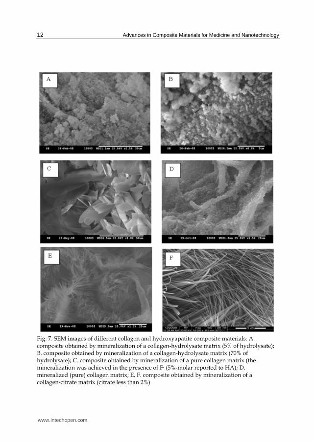
When the collagenous matrix is immersed for 24 h into Ca(OH)2 suspension and than into NaH2PO4 solution, about 32% of HA is deposited onto the matrix (Fig. 21, dotted line). The same results were obtained when alternately immersing the matrix three or four times into a Ca(OH)2 suspension (for 20 min) and a NaH2PO4 solution (for 10 min). The amount of deposited HA was determined by gravimetric analysis by measuring the initial mass of collagen matrices and the mass of the dried mineralised matrices. The amount of deposited HA is quite linear with respect to the number of deposited layers. This relationship was quantified and the root-mean-square deviation was found to be 0.9857. The layer by layer deposition method was successfully applied in order to increase the amount of mineral phase that is deposited on the collagen matrix. The root-mean-square-deviation is very close to 1, meaning highly linear layer growth was observed for even six HA layers.
www.intechopen.com
Advances in Composite Materials for Medicine and Nanotechnology
26
y = 4.6529x + 15.458
R2 = 0.9857
20
30
40
50
1 2 3 4 5 6 7 8number of layers
HA
co
nte
nt
(%)
■ value obtained by TG analysis
♦ value obtained by gravimetric measurements
Fig. 21. Gravimetric variation of deposited HA with the number of deposited layers
The classical mineralization method conducted by dipping a collagen matrix in Ca(OH)2 suspension for 24 h followed by dipping the collagen matrix in NaH2PO4 solution for a further 24 h can be improved by using the LbL deposition method. Deposition of six HA layers obtained by alternately soaking in a Ca(OH)2 suspension for 20 min and then in a NaH2PO4 solution for 10 min resulted in a 12% increase in HA deposition. Extrapolating from the obtained results, it can be estimated that for a collagen matrix, such as the ones used in these experiments, one would need to deposit about 14 layers of HA to obtain a composite material similar to whale bone, while antler bone composition can be obtained if seven HA layers are deposited. The obtained composite materials can be assumed to be hydroxyapatite matrices reinforced with mineralized collagen fibers as they have a structure very similar to the bone structure suggested by Hellmich et al. (Hellmich et al. 2004). The good linearity of the quantity of deposited HA with respect to the number of layers is due to interactions occurring not only between collagen and HA, but also between deposited HA and Ca2+ ions. From the parameters that influence the deposition of HA, we can distinguish three main categories: support dependent, solution/suspension dependent and processing parameters. While the first two categories have been extensively studied and can be quantified, the third category is very difficult to be quantified. Under similar mineralization conditions, the amount of deposited HA is greater in the presence of citrate ions, but the LbL method applied to a pure collagen matrix can increase the amount of deposited HA even more than in the case of one layer deposition (24+24 h) onto citrate enriched collagen matrices.
5. The synthesis of complex COLL/HA+Fe3O4 composite materials
Magnetite is a mineral with multiple roles in both medical (Ito et al. 2005; Mornet et al. 2006; Zhang and Misra 2007) and non-medical applications (Ficai et al. 2010d; Ju and Bian 2006). The addition of magnetic nanoparticles (especially magnetite nanoparticles) induces new
properties to the COLL/HA composite materials (Andronescu et al. 2010). The bone regenerative effects are due to the presence of COLL/HA while the anti-tumoral effects are due to the presence of magnetite which can produce hyperthermia when an electromagnetic
www.intechopen.com

Advances in Collagen/Hydroxyapatite Composite Materials
27
field is applied. These systems, even at low magnetite concentration (5%) can be used for curative purpose because can generate the necessary hyperthermia and consequently induce tumoral cell apoptosis. It is important to mention that the presence of magnetite even at low
concentration (1-2%) may induce hyperthermia but, in order to be useful for medical applications (hyperthermia – cancer treatment) at least 5% of magnetite is required. One of the most important advantage of the use of magnetite based composite materials is that hyperthermia can be activated only when is necessary and consequently the side effects is
limited comparing with chemotherapy, for instance. As a matter of course these materials will be improved by the addition of other antitumoral agents such as silver or gold nanoparticles, cytostatics or other drugs for pain managements.
6. Conclusions
Collagen/hydroxyapatite composite materials are the most similar synthetic grafts with
bone from many points of view, bone being composed from collagen and hydroxyapatite as main components and few percent of other components. The morphology and subsequent the properties of the composite materials is strongly
influenced by the presence of different components, even when they are present in small proportions. The reason that perfect bone graft materials have not been successfully synthesized is due to the limited number of components used in the synthesis of bone graft materials, which typically include only collagen and hydroxyapatite or carbonated apatite.
It is worth to mention that all commercially available collagen forms can be converted into COLL/HA composite materials with dense or spongious microstructure. If collagen gel can be easily converted in dense or spongious materials by a proper choice of the drying method, collagen
Fig. 22. The influence of collagen form on the composite materials microstructure
Based on the presented results, it can be concluded that the presence of additional components (which are usually found in natural bone in small concentrations) is of
significant importance. For instance, the presence of fluoride induces a higher crystallinity in the deposited mineral phase. The morphology of the apatite phase was found to be lamellar by SEM, where visible pores were not observed, even at relatively high
magnification.
Collagen gel Collagen fibres Collagen matrix
Dense structrures Spongious structrures
Similar with compact bone Similar with spongy bone
≡ ≡
www.intechopen.com

Advances in Composite Materials for Medicine and Nanotechnology
28
The shape of the mineral phase of the composite material obtained by in vitro co-precipitation in the presence of fluoride is biomimetic and similar to the mineral phase of natural bone. The main difference between the mineral phases of bone and composite materials obtained in the presence of F- is the size of the crystal. In order to obtain natural-sized crystals in the mineral phase, crystallization inhibitors may be used. Under these conditions, we expect to reduce the size of the crystals. Not only the presence of different components can induce morpho-structural modifications but also the synthesis route. For instance, the proper, applied electric or magnetic field or the drying method corroborated with ionic strength and pH lead to the formation of highly oriented COLL/HA composite materials. The synthesis of COLL/HA composite materials with oriented morphology of the mineralized collagen fibrils and fibres is an essential step to obtain bone grafts of the long bones. The orientation degree, based on SEM images, was of great importance and allows the quantification of the orientation. Based on the existing data, best orientation can achieve with self-assembling, the mean orientation degree being of ~ 97%.
7. Aknowledgements
Authors recognize financial support from the European Social Fund through POSDRU/89/1.5/S/54785 project: "Postdoctoral Program for Advanced Research in the field of nanomaterials and from Romanian Authority for Scientific Research through the project 72-198. We also thank to Elsevier for the amability to reuse some parts of text or figures.
8. References
Abe, E., Yamamoto, M., Taguchi, Y., Lecka- Czernik, B., O'Brien, C. A., Economides, A. N., Stahl, N., Jilka, R. L. and Manolagas, S. C. 2000. Essential requirement of BMPs-2/4 for both osteoblast and osteoclast formation in murine bone marrow cultures from adult mice: antagonism by noggin. J Bone Miner Res 15:663-673.
Andronescu, E., Ficai, M., Voicu, G., Ficai, D., Maganu, M. and Ficai, A. 2010. Synthesis and characterization of collagen/hydroxyapatite:magnetite composite material for bone cancer treatment. Journal of Materials Science - Materials in Medicine 21(7):2237-2242.
Bakos, D., Soldan, M. and Hernandez-Fuentes, I. 1999. Hydroxyapatite-collagen-hyaluronic acid composite. Biomaterials 20(2):191-195.
Bollerslev, J., Gram, J., Nielsen, H., Brixen, K., Storm, T., Larsen, H. and Mosekilde, L. 1991 Effect of a short course of 1,25-dihydroxyvitamin D3 on biochemical markers of bone remodeling in adult male volunteers. Bone 12 339-343.
Bonzani, I. C., Adhikari, R., Houshyar, S., Mayadunne, R., Gunatillake, P. and Stevens, M. M. 2007 Synthesis of two-component injectable polyurethanes for bone tissue engineering. Biomaterials 28(3):423-433.
Chang, M. C. and Tanaka, J. 2002. FT-IR study for hydroxyapatite/collagen nanocomposite cross-linked by glutaraldehyde. Biomaterials 23:4811–4818.
Clarke, I. C., Manaka, M., Green, D. D., Williams, P., Pezzotti, G., Kim, Y.-H., Ries, M., Sugano, N., Sedel, L., Delauney, C. and others. 2003. Current Status of Zirconia Used in Total Hip Implants. Journal of Bone and Joint Surgery 85(Supplement 4):73-84.
www.intechopen.com

Advances in Collagen/Hydroxyapatite Composite Materials
29
Corces, A. 2002. Metal Alloys. Proc. III Miami Symposium for Total Joint Replacement, South Miami Hospital, Miami, Florida.
Corces, A. and Garcia, M. 2007. Metallic Alloys. [Online] Available: http://www.emedicine.com/orthoped/TOPIC610.HTM [2008].
Cui, C. B., Cooper, L. F., Yang, X., Karsenty, G. and Aukhil, I. 2003. Transcriptional Coactivation of Bone-Specific Transcription Factor Cbfa1 by TAZ. Mol Cell Biol 23(3):1004-1013.
Cui, F.-Z., Li, Y. and Ge, J. 2007. Self-assembly of mineralized collagen composites. Materials Science and Engineering R 57:1-27.
de Vernejoul, M., Cohen-Solal, M. and Orcel, P. 1993 Bone cytokines. Curr Opin Rheumatol 5:332-338.
De Vernejoul, M., Pointillart, A., Bourdeau, A., Morieux, C., Modrowski, D., Miravet, L. and Caulin, F. 1990. Effect of calcitonin administration on young pig trabecular bone remodeling. Bone 11:29-33.
Develioglu, H., Koptagel, E., Gedik, R. and Dupoirieux, L. 2005. The effect of a biphasic ceramic on calvarial bone regeneration in rats. Journal of Oral Implantology 31(6):309-312.
Dorozhkin, S. V. 2009. Calcium orthophosphate-based biocomposites and hybrid biomaterials. J Mater Sci 44:2343-2387.
Ferreira, A. M., Noris-Suarez, K., Bello, A., Marquez, A. H., Feijoo, J. L. and Lira-Olivares, J. 2008. Effect of type I collagen piezoelectricity on cellular adhesion. Iv Latin American Congress on Biomedical Engineering 2007, Bioengineering Solutions for Latin America Health, Vols 1 and 2 18(1,2):659-662.
Ficai, A., Andronescu, E., Ghitulica, C., Voicu, G., Trandafir, V., Manzu, D., Ficai, M. and Pall, S. 2009a. Colagen/Hydroxyapatite Interactions in Composite Biomaterials. Materiale Plastice 46(1):11-15.
Ficai, A., Andronescu, E., Trandafir, V., Ghitulica, C. and Voicu, G. 2010a. Collagen/hydroxyapatite composite obtained by electric field orientation. Materials Letters 64(4):541-544.
Ficai, A., Andronescu, E., Voicu, G., Ghitulica, C. and Ficai, D. 2010b. The influence of collagen support and ionic species on the morphology of collagen/hydroxyapatite composite materials. Materials Characterization 61(4):402-407.
Ficai, A., Andronescu, E., Voicu, G., Ghitulica, C., Vasile, B. S., Ficai, D. and Trandafir, V. 2010c. Self assembled collagen/ hydroxyapatite composite materials. Chemical Engineering Journal 160(2):794-800.
Ficai, A., Andronescu, E., Voicu, G., Manzu, D. and Ficai, M. 2009b. Layer by layer deposition of hydroxyapatite onto the collagen matrix. Materials Science and Engineering: C 29(7):2217-2220.
Ficai, A., Andronescu, E., Voicu, G., Manzu, D. and Ficai, M. 2009c. Layer by layer deposition of hydroxyapatite onto the collagen matrix. Materials Science & Engineering C-Materials for Biological Applications 29(7):2217-2220.
Ficai, D., Ficai, A., Voicu, G., Vasile, B. S., Guran, C. and Andronescu, E. 2010d. Polysulfone based Membranes with Desired Pores Characteristics. Materiale Plastice 47(1):24-27.
Maria Ficai, Ecaterina Andronescu, Denisa Ficai, Georgeta Voicu, Anton Ficai; Synthesis and characterization of COLL-PVA/HA hybrid materials with stratified morphology; Colloids and Surfaces B: Biointerfaces; 2010:81(2): 614-619
www.intechopen.com

Advances in Composite Materials for Medicine and Nanotechnology
30
Gallie, W. E. and Toronto, M. B. 1914. The histrory of a bone graft. The Journal of Bone and Joint Surgery s2-12:201-212.
Guelcher, S., Patel, V., Gallagher, K., Connolly, S., Didier, J., Doctor, J. and Hollinger, J. 2004. Synthesis of Polyurethane Foam Scaffolds for Bone Tissue Engineering. Proc. AIChE Anual meeting Austin, Texas.
Hellmich, C., Barthelemy, J. F. and Dormieux, L. 2004. Mineral-collagen interactions in elasticity of bone ultrastructure - a continuum micromechanics approach. European Journal of Mechanics A/Solids 23(5):783-810.
Hench, L. L., Hench, J. W. and Greenspan, D. C. 2004. BIOGLASS®: A SHORT HISTORY AND BIBLIOGRAPHY. Journal of Australian Ceramic Society 40(1):1-42.
Hench, L. L. and Wilson, J., (eds.) 1993. An Introduction to Bioceramics. World Scientific Publishing Co., Singapore.
Hock, J. and Gera, I. 1992. Effects of continuous and intermittent administration and and inhibition of resorption on the anabolic response of bone to PTH. J Bone Miner Res 7:65-72.
Ito, A., Shinkai, M., Honda, H. and Kobayashi, T. 2005. Medical application of functionalized magnetic nanoparticles. Journal of Bioscience and Bioengineering 100(1):1-11.
Ju, D. Y. and Bian, P. 2006. Development of ferrite magnetic materials with high strength by a low-temperature sintering method. Science of Engineering Ceramics Iii 317-318:893-898.
Karsenty, G. 2000. How many factors are required to remodel bone? Nature Medicine 6:970 - 971.
Karsenty, G., Kronenberg, H. M. and Settembre, C. 2009. Genetic Control of Bone Formation. Annual Review of Cell and Developmental Biology 25:629-648.
Kesenci, K., Fambri, L., Migliaresi, C. and Pişkin, E. 2000. Preparation and properties of poly(L-lactide)/hydroxyapatite composites. Journal of Biomaterials Science-Polymer Edition 11(6):617-632.
Lawson, A. and Czernuszka, J. 1998. Collagen-calcium phosphate composites. Proc Inst Mech Eng [H] 212:413-425.
Li, X. K. and Chang, J. 2008. Preparation of bone-like apatite-collagen nanocomposites by a biomimetic process with phosphorylated collagen. Journal of Biomedical Materials Research Part A 85A(2):293-300.
Li, X. M., Feng, Q. L., Liu, X. H., Dong, W. and Cui, F. H. 2006. Collagen-based implants reinforced by chitin fibres in a goat shank bone defect model. Biomaterials 27(9):1917-1923.
Lin, X. Y., Li, X. D., Fan, H. S., Wen, X. T., Lu, J. and Zhang, X. D. 2004. In situ synthesis of bone-like apatite/collagen nano-composite at low temperature. Materials Letters 58(27-28):3569-3572.
Miyake, K., Medina, K., Ishihara, K., Kimoto, M., Auerbach, R. and Kincade, P. W. 1991. A VCAM-like Adhesion Molecule on Murine Bone Marrow Stromal Cells Mediates Binding of Lymphocyte Precursors in Culture. The Journal of Cell Biology 114:557-565.
Mornet, S., Vasseur, S., Grasset, F., Veverka, P., Goglio, G., Demourgues, A., Portier, J., Pollert, E. and Duguet, E. 2006. Magnetic nanoparticle design for medical applications. Progress in Solid State Chemistry 34(2-4):237-247.
www.intechopen.com

Advances in Collagen/Hydroxyapatite Composite Materials
31
Murugan, R. and Ramakrishna, S. 2005. Development of nanocomposites for bone grafting. Composites Science and Technology 65:2385–2406.
Noris-Suarez, K., Lira-Olivares, J., Ferreira, A. M., Feijoo, J. L., Suarez, N., Hernandez, M. C. and Barrios, E. 2007. In Vitro Deposition of Hydroxyapatite on Cortical Bone Collagen Stimulated by Deformation-Induced Piezoelectricity. Biomacromolecules 8:941-948.
Ogawa, S., Harada, H., Fujiwara, M., Tagashira, S., Katsumata, T. and Takada, H. 2000 Cbfa1, an essential transcription factor for bone formation, is expressed in testis from the same promoter used in bone. DNA Research 7(3):181-185.
Oláh, L., Filipczak, K., Czvikovszky, T., Czigánya, T. and Borbás, L. 2007. Changes of porous poly(ε-caprolactone) bone grafts resulted from e-beam sterilization process. Radiation Physics and Chemistry 76(8-9):1430-1434.
Pederson, A. W., Ruberti, J. W. and Messersmith, P. B. 2003. Thermal assembly of a biomimetic mineral/collagen composite. Biomaterials 24(26):4881-4890.
Pompe, W., Gelinsky, M., Hofinger, I. and Knepper-Nicolai, B. 2001. Functionally graded collagen-hydroxyapatite materials for bone replacement. Functionally Graded Materials 2000 114:65-72.
Rhee, S. H. and Tanaka, J. 1998. Hydroxyapatite coating on a collagen membrane by a biomimetic method. Journal of the American Ceramic Society 81(11):3029-3031.
Sedel, L., Nizard, R. S., Kerboull, L. and Witvoet, J. 1994. Alumina-Alumina Hip Replacement in Patients Younger Than 50 Years Old. Clinical Orthopaedics & Related Research 298:175-183.
Silva, C. C., Thomazini, D., Pinheiro, A. G., Aranha, N., Figueiro, S. D., Goes, J. C. and Sombra, A. S. B. 2001. Collagen-hydroxyapatite films: piezoelectric properties. Materials Science and Engineering B-Solid State Materials for Advanced Technology 86(3):210-218.
Tashjian, A. H., Voelkel, E. F., Lazzaro, M., Singers, F. R., Roberts, A. B., Derynck, R., Winklerii, M. E. and Levine, L. 1985. alpha and beta human transforming growth factors stimulate prostaglandin production and bone resorption in cultured mouse calvaria. Proc Natl Acad Sci 82:4535-4538.
Wahl, D. A. and Czernuszka, J. T. 2006. Collagen-hydroxyapatite composites for hard tissue repair. European Cells & Materials 11:43-56.
Wallach, S., Avioli, L. V. and Carstens, J. H. J. 1989. Factors in bone formation. Calcified Tissue International 45(1):4-6.
Wang, L. and Li, C. Z. 2007. Preparation and physicochemical properties of a novel hydroxyapatite/chitosan-silk fibroin composite. Carbohydrate Polymers 68(4):740-745.
Werner, J., Linner-Krcmar, B., Friess, W. and Greil, P. 2002. Mechanical properties and in vitro cell compatibility of hydroxyapatite ceramics with graded pore structure. Biomaterials 23(21):4285-4294.
Wu, C. Y., Sassa, K., Iwai, K. and Asai, S. 2007. Unidirectionally oriented hydroxyapatite/collagen composite fabricated by using a high magnetic field. Materials Letters 61(7):1567-1571.
Yamauchi, K., Goda, T., Takeuchi, N., Einaga, H. and Tanabe, T. 2004. Preparation of collagen/calcium phosphate multilayer sheet using enzymatic mineralization. Biomaterials 25(24):5481-5489.
www.intechopen.com
Advances in Composite Materials for Medicine and Nanotechnology
32
Yunoki, S., Ikoma, T., Monkawa, A., Ohta, K., Kikuchi, M., Marukawa, E., Sotome, S., Shinomiya, K. and Tanaka, J. 2007. Fabrication of three-dimensional porous hydroxyapatite/collagen composite with rubber-like elasticity. Journal of Biomaterial Science, Polymer Edition 18(4):393-409.
Yunoki, S., Ikoma, T., Monkawa, A., Ohta, K., Kikuchi, M., Sotome, S., Shinomiya, K. and Tanaka, J. 2006. Control of pore structure and mechanical property in hydroxyapatite/collagen composite using unidirectional ice growth. Materials Letters 60(8):999-1002.
Zhang, J. and Misra, R. D. K. 2007. Magnetic drug-targeting carrier encapsulated with thermosensitive smart polymer: Core-shell nanoparticle carrier and drug release response. Acta Biomaterialia 3(6):838-850.
Zhang, W., Liao, S. S. and Cui, F. Z. 2003. Hierarchical self-assembly of nano-fibrils in mineralized collagen. Chemistry of Materials 15(16):3221-3226.
www.intechopen.com

Advances in Composite Materials for Medicine andNanotechnologyEdited by Dr. Brahim Attaf
ISBN 978-953-307-235-7Hard cover, 648 pagesPublisher InTechPublished online 01, April, 2011Published in print edition April, 2011
InTech EuropeUniversity Campus STeP Ri Slavka Krautzeka 83/A 51000 Rijeka, Croatia Phone: +385 (51) 770 447 Fax: +385 (51) 686 166www.intechopen.com
InTech ChinaUnit 405, Office Block, Hotel Equatorial Shanghai No.65, Yan An Road (West), Shanghai, 200040, China
Phone: +86-21-62489820 Fax: +86-21-62489821
Due to their good mechanical characteristics in terms of stiffness and strength coupled with mass-savingadvantage and other attractive physico-chemical properties, composite materials are successfully used inmedicine and nanotechnology fields. To this end, the chapters composing the book have been divided into thefollowing sections: medicine, dental and pharmaceutical applications; nanocomposites for energy efficiency;characterization and fabrication, all of which provide an invaluable overview of this fascinating subject area.The book presents, in addition, some studies carried out in orthopedic and stomatological applications andothers aiming to design and produce new devices using the latest advances in nanotechnology. This widevariety of theoretical, numerical and experimental results can help specialists involved in these disciplines toenhance competitiveness and innovation.
How to referenceIn order to correctly reference this scholarly work, feel free to copy and paste the following:
Anton Ficai, Ecaterina Andronescu, Georgeta Voicu and Denisa Ficai (2011). Advances inCollagen/Hydroxyapatite Composite Materials, Advances in Composite Materials for Medicine andNanotechnology, Dr. Brahim Attaf (Ed.), ISBN: 978-953-307-235-7, InTech, Available from:http://www.intechopen.com/books/advances-in-composite-materials-for-medicine-and-nanotechnology/advances-in-collagen-hydroxyapatite-composite-materials