Nadler commonorthoproblems.ppt - UCSF CME commonorthopr… · Flexor tenosynovitis (trigger finger)...
38
10/21/2013 1 Common and Commonly Missed Orthopedic Problems UCSF Primary Care Medicine: Principles and Practice 2013 Paul L. Nadler, MD Professor of Medicine Director- UCSF Screening and Acute Care Clinic Disclosures I have no commercial involvement or financial interest in any medications or tests or procedures or durable medical equipment described in this lecture Graphics included under use license or attributed I wish to thank Carlin Senter, MD, UCSF Dept. of Orthopedics and Division of General Internal Medicine for reviewing major parts of this lecture to ensure accuracy and practice standards Objectives Discuss the initial assessment and management of orthopedic problems that commonly present to primary care physicians We will focus on acute, orthopedic problems Review physical exam techniques commonly used in orthopedic assessment, as well as specialized orthopedic examination techniques necessary to make the diagnosis Review significant conditions that are easily missed on standard x-rays Wrist Pain
Transcript of Nadler commonorthoproblems.ppt - UCSF CME commonorthopr… · Flexor tenosynovitis (trigger finger)...

10/21/2013
1
Common and Commonly Missed Orthopedic Problems
UCSF Primary Care Medicine: Principles and Practice 2013
Paul L. Nadler, MDProfessor of Medicine
Director- UCSF Screening and Acute Care Clinic
Disclosures
I have no commercial involvement or financial interest in any medications or tests or procedures or durable medical equipment described in this lecture
Graphics included under use license or attributed
I wish to thank Carlin Senter, MD, UCSF Dept. of Orthopedics and Division of General Internal Medicine for reviewing major parts of this lecture to ensure accuracy and practice standards
Objectives Discuss the initial assessment and management
of orthopedic problems that commonly present to primary care physicians
We will focus on acute, orthopedic problems
Review physical exam techniques commonly used in orthopedic assessment, as well as specialized orthopedic examination techniques necessary to make the diagnosis
Review significant conditions that are easily missed on standard x-rays
Wrist Pain
10/21/2013
2
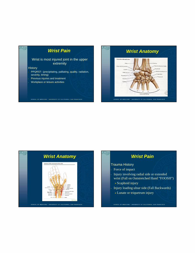
Wrist Pain
Wrist is most injured joint in the upper extremity
History• PPQRST- (precipitating, palliating, quality, radiation,
severity, timing)
• Previous injuries and treatment
• Workplace or leisure activities
Wrist Anatomy
Wrist Anatomy Wrist Pain
Trauma History
• Force of impact
• Injury involving radial side or extended wrist (Fall on Outstretched Hand “FOOSH”)
Scaphoid injury
• Injury loading ulnar side (Fall Backwards)
Lunate or triquetrum injury
10/21/2013
3
Wrist Pain
“Pain Pattern”1) Pain over the Dorsum of the Wrist with flexion and extension
Ligament injury
Fracture if post-traumatic
2) “Stiffness”
Rheumatoid arthritis
Carpal Tunnel
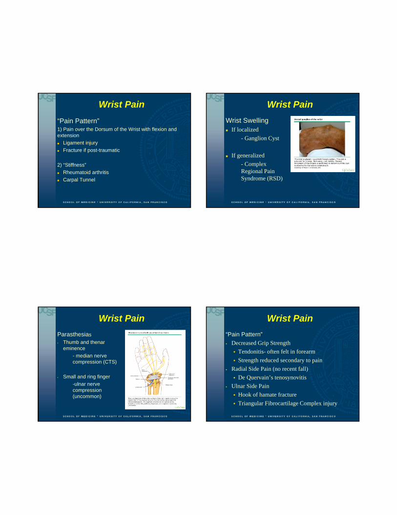
Wrist Pain
Wrist Swelling If localized
- Ganglion Cyst
If generalized
- Complex Regional Pain Syndrome (RSD)
Wrist Pain
Parasthesias
• Thumb and thenar eminence
- median nerve compression (CTS)
• Small and ring finger
-ulnar nerve compression (uncommon)
Wrist Pain
“Pain Pattern”
Decreased Grip Strength
Tendonitis- often felt in forearm
Strength reduced secondary to pain
Radial Side Pain (no recent fall)
De Quervain’s tenosynovitis
Ulnar Side Pain
Hook of hamate fracture
Triangular Fibrocartilage Complex injury
10/21/2013
4
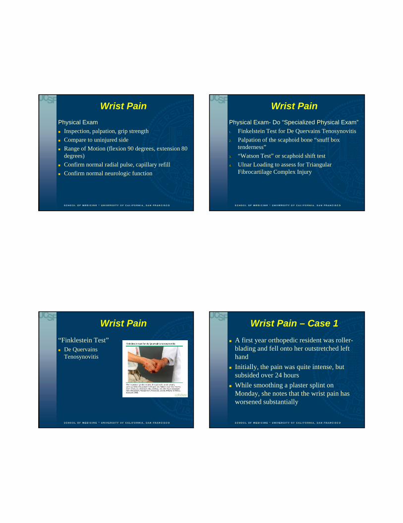
Wrist Pain
Physical Exam
Inspection, palpation, grip strength
Compare to uninjured side
Range of Motion (flexion 90 degrees, extension 80 degrees)
Confirm normal radial pulse, capillary refill
Confirm normal neurologic function
Wrist Pain
Physical Exam- Do “Specialized Physical Exam”
1. Finkelstein Test for De Quervains Tenosynovitis
2. Palpation of the scaphoid bone “snuff box tenderness”
3. “Watson Test” or scaphoid shift test
4. Ulnar Loading to assess for Triangular Fibrocartilage Complex Injury
Wrist Pain
“Finklestein Test” De Quervains
Tenosynovitis
Wrist Pain – Case 1
A first year orthopedic resident was roller-blading and fell onto her outstretched left hand
Initially, the pain was quite intense, but subsided over 24 hours
While smoothing a plaster splint on Monday, she notes that the wrist pain has worsened substantially
10/21/2013
5
Wrist Pain – Case 1
There is mild swelling over the wrist with point tenderness distal to the radius and proximal to the first MCP joint
There is full range of motion of the wrist
X-ray is negative for fracture
Wrist Pain – Case 1
Based on this history and physical presented, what do you suspect?
1) Wrist sprain
2) Occult Radial Head Fracture
3) Scaphoid Fracture
4) Scapholunate Dissociation
Wrist Pain – Case 1
Based on this history and physical presented, what do you suspect?
1) Wrist sprain
2) Occult Radial Head Fracture
3) Scaphoid Fracture
4) Scapholunate Dissociation
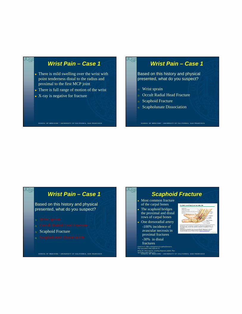
Scaphoid Fracture Most common fracture
of the carpal bones The scaphoid bridges
the proximal and distal rows of carpal bones
One dorsoradial artery-100% incidence of avascular necrosis in proximal fractures-30% in distal fractures
Gutierrez G. Office management of scaphoid fractures. Phys SportsMed 1996;24(8):60-70.
Rettig AC. Wrist injuries: avoiding diagnostic pitfalls. Phys SportsMed 1994;22(8):33-9
10/21/2013
6
Scaphoid Fractures
• Seventy percent are through the waist
• Twenty percent are proximal
• Ten percent are distal– delay in diagnosis
of one to two weeks increases risk of non-union and subsequent arthrosis
Richard JR. Office orthopedics: thumb spica casting for scaphoid fractures. Am Family
Physician 1995;52: 1113-9.
Scaphoid Fracture
• Non-union of scaphoid fracture (occurs in 5% of fractures)
• Wrist arthrosis and pain
• Long term occupational disability
Tiel-van Buul MM, van Beek EJ, Borm JJ, Gubler FM, Broekhuizen AH, van Royen EA. The value of radiographs and bone scintigraphy in suspected scaphoid fracture. A statistical analysis. J Hand Surg [Br] 1993;18:403-6.
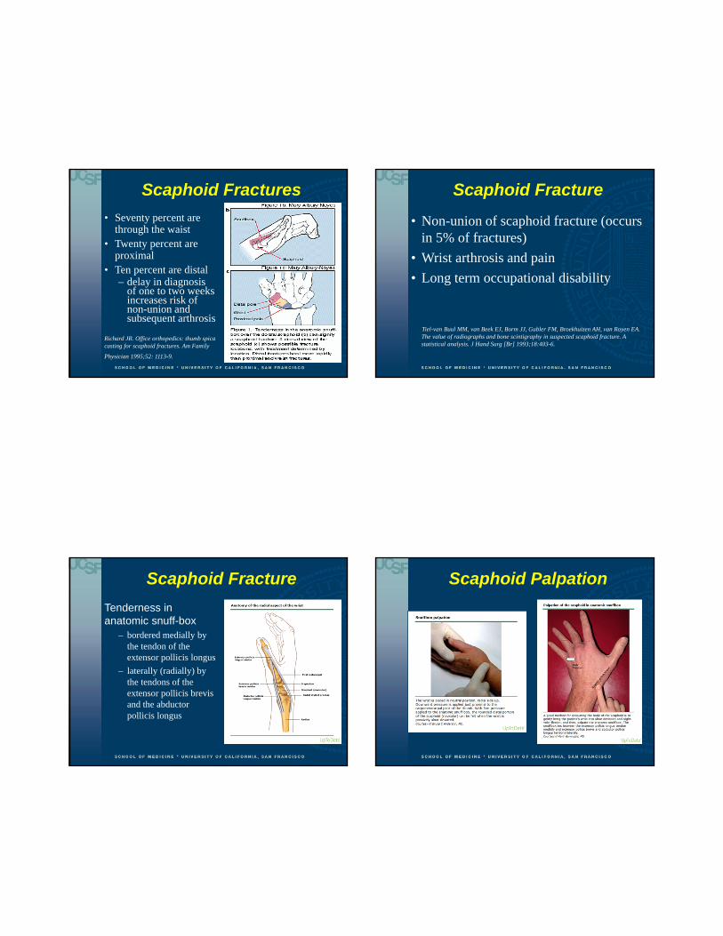
Scaphoid Fracture
Tenderness in anatomic snuff-box
– bordered medially by the tendon of the extensor pollicis longus
– laterally (radially) by the tendons of the extensor pollicis brevis and the abductor pollicis longus
Scaphoid Palpation

10/21/2013
7
Scaphoid Fracture
• X-rays should include a scaphoid view– antero-posterior
with 30 degree supination and ulnar deviation
Scaphoid Fracture
TreatmentI) Snuff box pain and x-ray is POSITIVE for fracture
Urgent Ortho Consultation
II) Snuff box pain and x-ray is NEGATIVE for fracture
Urgent Ortho Consultation
Discharge patient with Thumb Spica Splint
Richard JR. Office orthopedics: thumb spica casting for scaphoid fractures. Am Fam Physician 1995;52: 1113-9.
Gultierrez G. Office management of scaphoid fractures. Phys SportsMed 1996;24(8):60-70
Thumb Spica Splint Wrist Pain – Case 2Fortunately for this ortho resident, careful follow-up showed no scaphoid fracture
• But the wrist pain persists
• The pain is worse with dorsiflexion
• There is point tenderness over the dorsal mid-wrist, on the ulnar side of the scaphoid snuff box
10/21/2013
8
Wrist Pain – Case 2Based on the history and physical presented, what do you suspect?
1) Injury to the distal radial ulnar joint (DRUJ)
2) Occult Scaphoid Fracture
3) Scapholunate dissociation
4) Injury to the Triangular Fibrocartilage Complex
Wrist Pain – Case 2Based on the history and physical presented, what do you suspect
1) Injury to the distal radial ulnar joint (DRUJ)
2) Occult Scaphoid Fracture
3) Scapholunate dissociation
4) Injury to the Triangular Fibrocartilage Complex
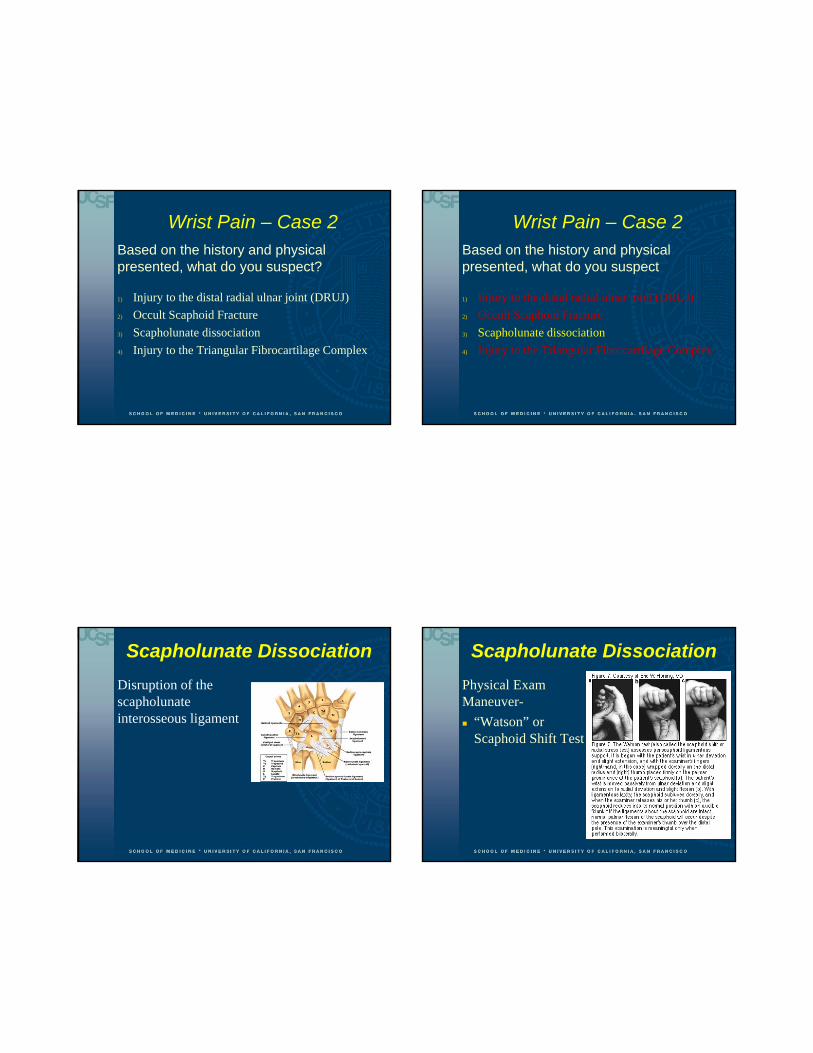
Scapholunate Dissociation
Disruption of the scapholunate interosseous ligament
Scapholunate Dissociation
Physical Exam Maneuver-
“Watson” or Scaphoid Shift Test
10/21/2013
9
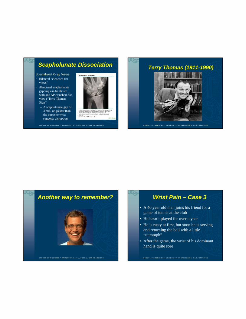
Scapholunate Dissociation
Specialized X-ray Views
• Bilateral “clenched fist views”
• Abnormal scapholunate gapping can be shown with and AP clenched-fist view (“Terry Thomas Sign”)
– A scapholunate gap of 3 mm, or greater than the opposite wrist suggests disruption
Terry Thomas (1911-1990)
Another way to remember? Wrist Pain – Case 3
• A 40 year old man joins his friend for a game of tennis at the club
• He hasn’t played for over a year
• He is rusty at first, but soon he is serving and returning the ball with a little “uummph”
• After the game, the wrist of his dominant hand is quite sore
10/21/2013
10
Wrist Pain – Case 3
• At first, a little RICE (rest, ice, compression, elevation) seems to help
• But later, at work, he has trouble writing because of wrist pain
• He consults you for evaluation
Wrist Pain – Case 3
• X-ray is negative for fracture
• Wrist pain is primarily on the lateral wrist (ulnar side)
• There is some localized swelling and loss of grip strength
• With active ulnar deviation, he (and you) feels a “click”
• There is point tenderness distal to the ulnar styloid
• There is significant pain with ulnar deviation of the wrist and axial loading
Wrist Pain – Case 3
Based on this history and physical exam, what do you suspect?
1. Ulnar Styloid Fracture
2. Hook of Hamate Fracture
3. Acute Ulnar Nerve Neuropathy
4. Triangular Fibrocartilage Complex Injury (TFCC)
Wrist Pain – Case 3
Based on this history and physical exam, what do you suspect?
1. Ulnar Styloid Fracture
2. Hook of Hamate Fracture
3. Acute Ulnar Nerve Neuropathy
4. Triangular Fibrocartilage Complex Injury (TFCC)
10/21/2013
11
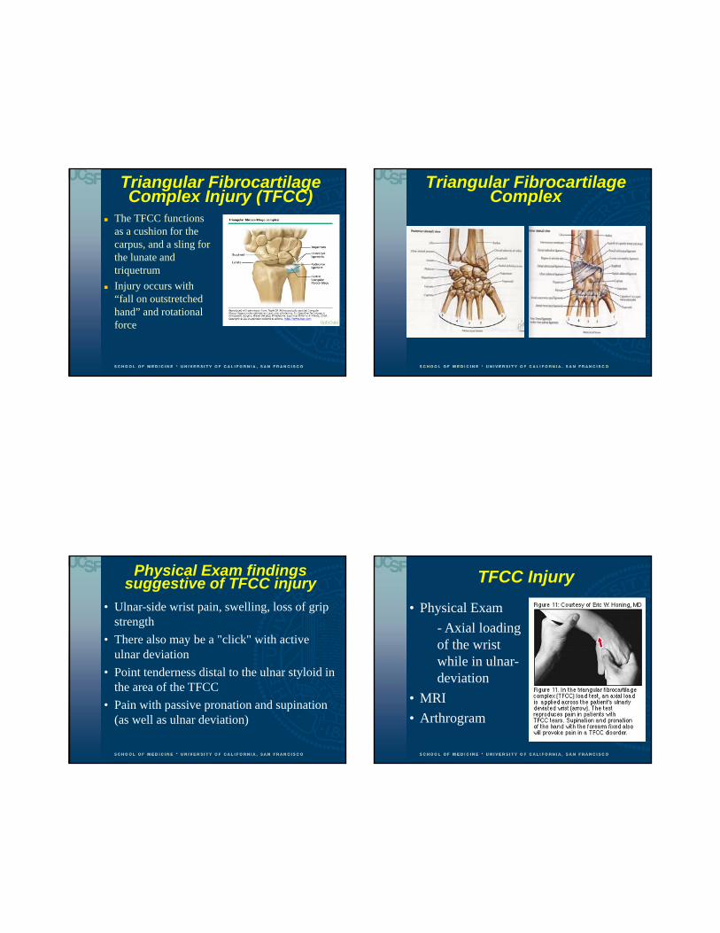
Triangular Fibrocartilage Complex Injury (TFCC)
The TFCC functions as a cushion for the carpus, and a sling for the lunate and triquetrum
Injury occurs with “fall on outstretched hand” and rotational force
Triangular Fibrocartilage Complex
Physical Exam findings suggestive of TFCC injury
• Ulnar-side wrist pain, swelling, loss of grip strength
• There also may be a "click" with active ulnar deviation
• Point tenderness distal to the ulnar styloid in the area of the TFCC
• Pain with passive pronation and supination (as well as ulnar deviation)
TFCC Injury
• Physical Exam
- Axial loading of the wrist while in ulnar-deviation
• MRI
• Arthrogram

10/21/2013
12
TFCC Injury
• Injection of the ulnar carpal space with a corticosteroid and lidocaine (mild cases)
• Arthroscopy• Prompt surgery if Distal Radial Ulnar
Joint (DRUJ) is also disrupted
Loftus JB, Palmer AK: Disorders of the distal radioulnar joint and triangularfibrocartilage complex: an overview, in Lichtman DM, Alexander AH (eds): The Wristand Its Disorders, ed 2. Philadelphia, WB Saunders Co, 1997, pp 385-414 Halikis MN, Taleisnik J: Soft-tissue injuries of the wrist. Clin Sports Med1996;15(2):235-259
Hand and Finger Pain
Hand and Finger Pain Five metacarpals
Two phalanges in thumb/three phalanges in the other fingers
Joints Metacarpophalangeal
(MCP) Proximal
interphlanageal (PIP) Distal Interphalangeal
(DIP) Interphalangeal Joint
(IP) in thumb
Hand and Finger Pain
Finger Flexor Tendons
Travel in a fibro-osseous tunnel between the metacarpal and the DIP joint
Superificialis Tendon attaches to the middle phalanges
Profundus Tendon attaches to the distal phalanges
10/21/2013
13
Hand and Finger Pain
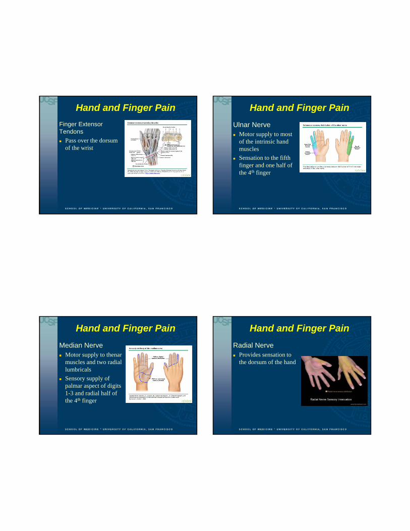
Finger Extensor Tendons
Pass over the dorsum of the wrist
Hand and Finger Pain
Ulnar Nerve Motor supply to most
of the intrinsic hand muscles
Sensation to the fifth finger and one half of the 4th finger
Hand and Finger Pain
Median Nerve Motor supply to thenar
muscles and two radial lumbricals
Sensory supply of palmar aspect of digits 1-3 and radial half of the 4th finger
Hand and Finger Pain
Radial Nerve Provides sensation to
the dorsum of the hand

10/21/2013
14
Hand and Finger Pain
Examination Pain, stiffness, enlargement of DIP joints
Osteoarthritis
Pain, swelling and fusiform enlargement of multiple hand joints
Rheumatoid arthritis
Tenderness over a single MP joint with loss of smooth digit function
Flexor tenosynovitis (trigger finger)
Assessment of ulnar ligament laxity if a patient experiences forceful abduction of thumb
Hand and Finger Pain Case 1
A 35 year old man consults you for thumb pain
He just returned from a ski trip to Tahoe
He reports pushing hard on his pole on a sharp turn and loosing his grip
He states that the pole “pushed out” this thumb
He says that he was initially evaluated at a local urgent care, and x-rays were taken (and were negative for fracture)
He would like you to treat him for his “sprained thumb”
Hand and Finger Pain Case 1
Based on this history, what do you suspect?
1) Occult 1st MCP Joint Fracture
2) Ulnar Collateral Ligament Tear
3) Acute Median Nerve Palsy
4) Occult fracture of the 1st metacarpal
Hand and Finger Pain Case 1
Based on this history, what do you suspect?
1) Occult 1st MCP Joint Fracture
2) Ulnar Collateral Ligament Tear
3) Acute Median Nerve Palsy
4) Occult fracture of the 1st metacarpal

10/21/2013
15
Ulnar Collateral Ligament Tear
“Gamekeeper’s Thumb” or “Skier’s” thumb
Rupture of the 1st MCP joint of the hand
Ulnar Collateral Ligament Tear
Ulnar collateral ligament tear represents 60% of upper limb problems in skiers
Frequently overlooked and under-diagnosed
Untreated tears can cause disabling instability of the hand, as the ulnar collateral ligament stabilizes the first metacarpalphalangeal (MCP) joint
Early surgical repair (within two or three weeks of the injury) are superior to results of late repair
Reid DC. Forearm, wrist and hand. In: Sports injury assessment and rehabilitation. New York: Churchill Livingstone,1992:1089-92. Richard JR. Gamekeeper's thumb: ulnar collateral ligament injury. Am Fam
Physician 1996;53:1775-81.
Ulnar Collateral Ligament Tear
Stress testing is performed by stabilizing the metacarpophalangeal joint in flexion and radially deviating the thumb
More than 30 degrees deviation (or 20 degrees more than opposite side) suggests significant damage to ulnar collateral ligament
Ulnar Collateral Ligament Tear
X-rays should be taken BEFORE stress testing as it might further displace the tear and lead to soft-tissue trapping
Consider stress testing under fluoroscopy
10/21/2013
16
Ulnar Collateral Ligament Tear
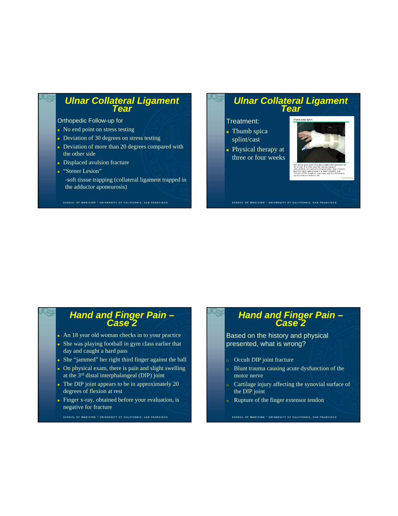
Orthopedic Follow-up for
No end point on stress testing
Deviation of 30 degrees on stress testing
Deviation of more than 20 degrees compared with the other side
Displaced avulsion fracture
“Stener Lesion”
-soft tissue trapping (collateral ligament trapped in the adductor aponeurosis)
Ulnar Collateral Ligament Tear
Treatment:
Thumb spica splint/cast
Physical therapy at three or four weeks
Hand and Finger Pain –Case 2
An 18 year old woman checks in to your practice
She was playing football in gym class earlier that day and caught a hard pass
She “jammed” her right third finger against the ball
On physical exam, there is pain and slight swelling at the 3rd distal interphalangeal (DIP) joint
The DIP joint appears to be in approximately 20 degrees of flexion at rest
Finger x-ray, obtained before your evaluation, is negative for fracture
Hand and Finger Pain –Case 2
Based on the history and physical presented, what is wrong?
1) Occult DIP joint fracture
2) Blunt trauma causing acute dysfunction of the motor nerve
3) Cartilage injury affecting the synovial surface of the DIP joint
4) Rupture of the finger extensor tendon

10/21/2013
17
Hand and Finger Pain –Case 2
Based on the history and physical presented, what is wrong?
1) Occult DIP joint fracture
2) Blunt trauma causing acute dysfunction of the motor nerve
3) Cartilage injury affecting the synovial surface of the DIP joint
4) Rupture of the finger extensor tendon
Mallet Finger
Mallet Finger Injury Mallet Finger Injury

10/21/2013
18
Mallet Finger Injury
• The distal interphalangeal (DIP) joint is extended by the– medial and lateral bands of the extensor tendon,
which inserts into the dorsal base of the distal phalanx
– injury results from loss of bony (avulsion fracture) or ligamentous attachment of the extensor mechanism into the distal phalanx
Mallet Finger Injury
History Hyperflexion
Hyperextension
Axial Loading
Physical Exam Extensor “lag” in the DIP joint
Mallet Finger Injury
X-Rays:• Lateral and PA radiographs should be obtained
initially to assess injury (and also after splinting) to determine joint congruity
• However, in many cases the x-rays do not show a fracture as the tendon has ruptured without bony avulsion
– The distal phalanx may appear to have subluxed in a volar direction from the unopposed pull of the flexor tendon
Mallet Finger Injury
Treatment:• Dorsal splinting or the use
of a “stack” splint
• The joint must be splinted in full extension (or 10 degrees of hyperextension)
• Failure to properly splint can lead to long-term flexion deformity and weakness
J Orthop Trauma 1991;1(2):105-111

10/21/2013
19
Mallet Finger InjuryTime in Splint-
• 6 weeks for a fracture
• 8 weeks for tendon failure
• 24 HOURS A DAY
• If the finger droops, the clock starts overLange RH, Engber WD: Hyperextension mallet finger. Orthop 1983;6(11):1426-1431
Follow up:
• Regular follow-up with patients help ensure good outcome (every 2 weeks)
• Evaluate for skin maceration
Mallet Finger
Surgical Pinning is required if:• Full extension not
achieved by second visit
• Subluxation of the distal phalanx volarly
• Avulsed bony fragment involves more than one-third of the joint
Ankle and Foot Pain
Ankle and Foot Pain
Approximately 20 percent of all musculoskeletal complaints are related to the foot and ankle
One to 10 million ankle injuries in the US per year
85% are sprains
Functions• Stable base for body weight support
• Rigid lever to propel body forward during walking
• Shock absorption for walking and running (two to six times an individual’s body weight

10/21/2013
20
Ankle and Foot Pain
Plantarflexion and dorsiflexion are the primary actions of the ankle
Inversion (supination) and eversion (pronation) are secondary
Ankle and Foot Pain
Ankle Anatomy
Bones Distal tibia and fibula
(superior)
Dome of the talus (inferior)
Ankle and Foot Pain
Ankle Anatomy
Ligaments Lateral Ligaments
Ankle and Foot Pain
Ankle Anatomy Ligaments Medial Ligaments

10/21/2013
21
Ankle and Foot Pain
The tibia and the fibula are bound together by
the anterior inferior tibio-fibular ligament
the posterior inferior tibio-fibular ligament
the interosseous membrane, which runs between the two long bones
Ankle and Foot Pain
Ankle Anatomy
Tendons Posterior Tendon
- Achilles Tendon
Ankle and Foot Pain
Ankle Anatomy Tendons Medial Tendons
-Posterior Tibialis Tendon
Ankle and Foot Pain
History Position of the ankle
during plantar flexion the anterior talofibular ligament is at greatest tension making it prone to injury
Can the patient bear weight after the injury?
Pop or snap may mean partial or full tendon injury
Previous strains? (More susceptible to injury, slower recovery)
10/21/2013
22
Ankle and Foot Pain
Observation
Can the patient bear weight?
Swelling? (lateral or medial or posterior)
Ecchymosis?• More likely in Grade 2 and Grade 3 ankle
sprains
Ankle and Foot Pain
Physical Exam:
Careful palpation over the lateral and medial malleolus and Achilles tendon
At this point, I STOP and ask-
Does this patient need an x-ray?
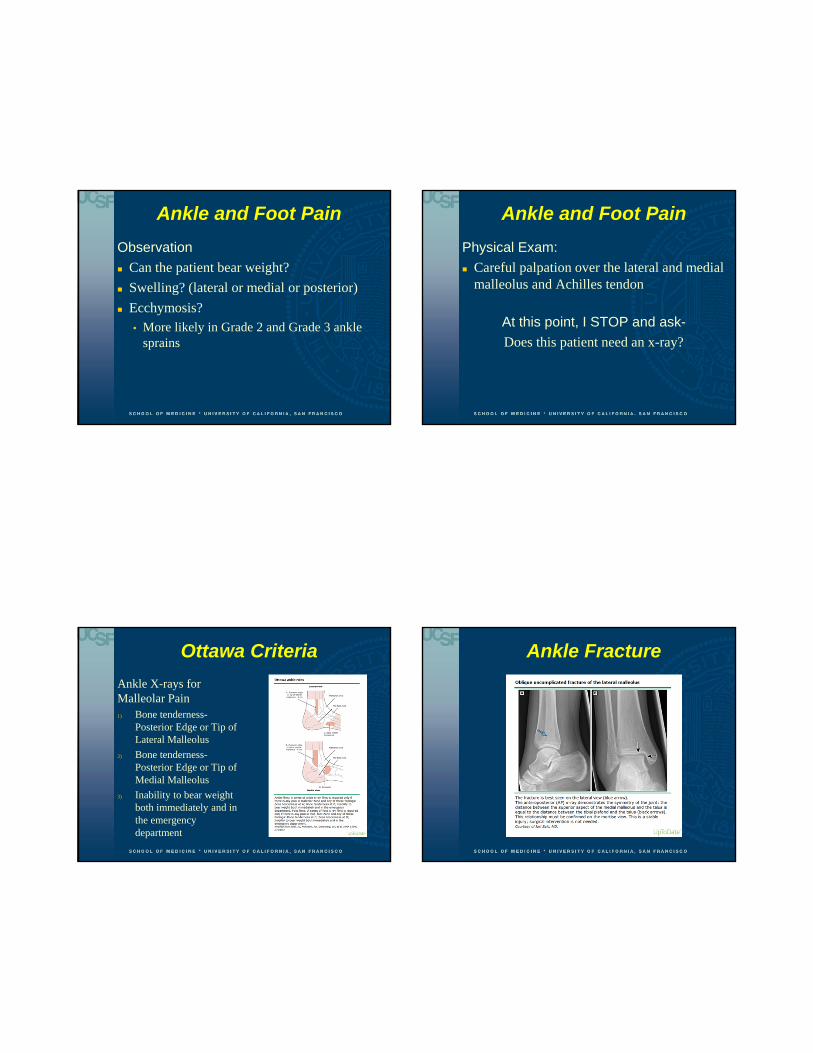
Ottawa Criteria
Ankle X-rays for Malleolar Pain1) Bone tenderness-
Posterior Edge or Tip of Lateral Malleolus
2) Bone tenderness-Posterior Edge or Tip of Medial Malleolus
3) Inability to bear weight both immediately and in the emergency department
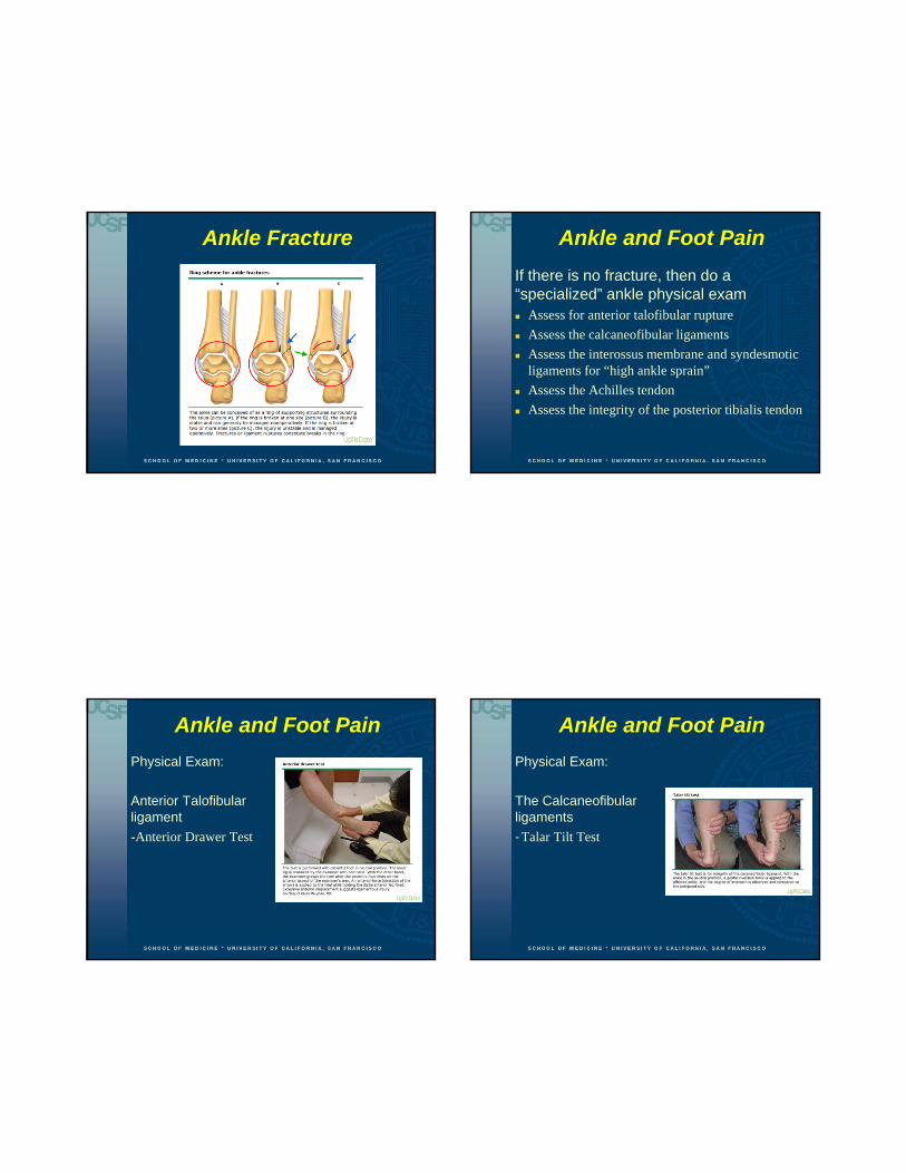
Ankle Fracture
10/21/2013
23
Ankle Fracture Ankle and Foot Pain
If there is no fracture, then do a “specialized” ankle physical exam Assess for anterior talofibular rupture
Assess the calcaneofibular ligaments
Assess the interossus membrane and syndesmotic ligaments for “high ankle sprain”
Assess the Achilles tendon
Assess the integrity of the posterior tibialis tendon
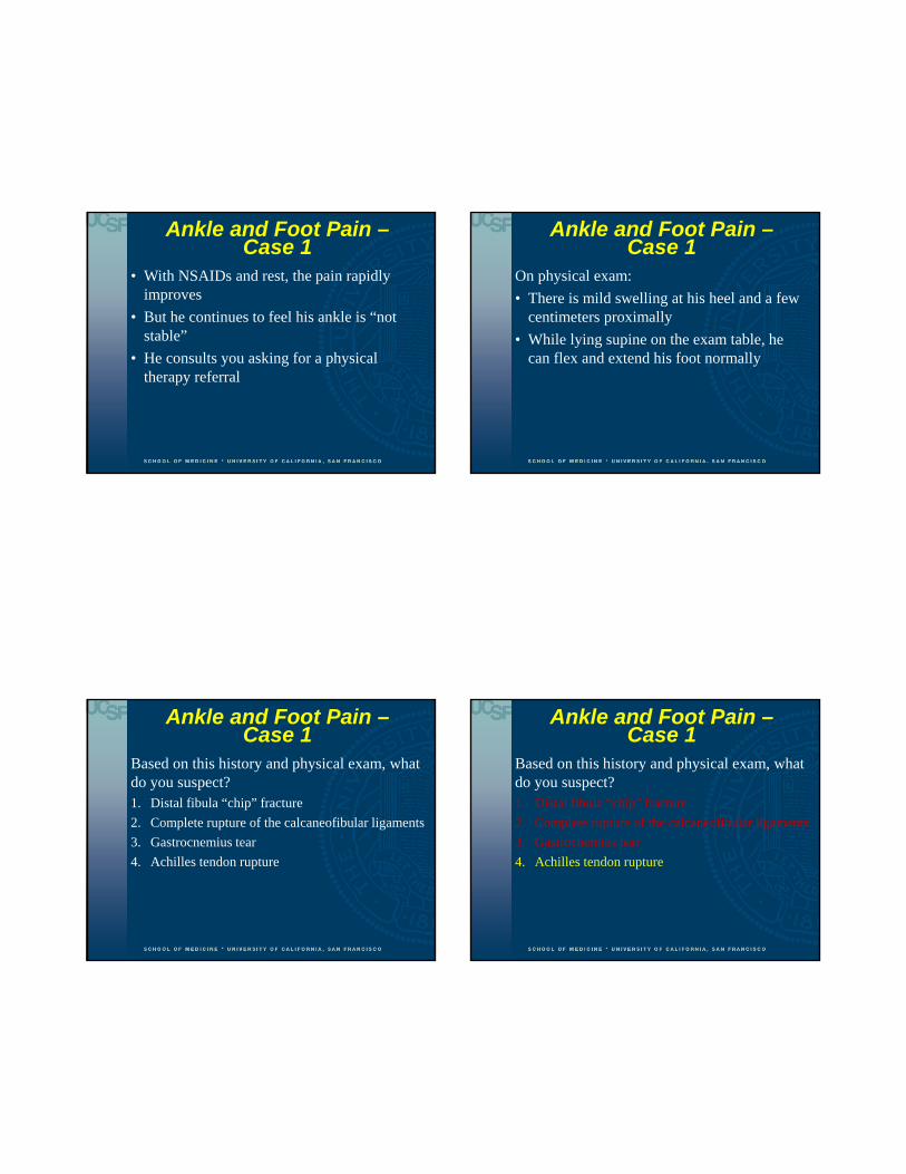
Ankle and Foot Pain
Physical Exam:
Anterior Talofibular ligament
-Anterior Drawer Test
Ankle and Foot Pain
Physical Exam:
The Calcaneofibular ligaments
-Talar Tilt Test

10/21/2013
24
Ankle and Foot Pain
Physical Exam:
Assess the interosseus membrane
-“Squeeze Test”
Ankle and Foot Pain
Physical Exam
Test syndesmotic ligaments (Distal Tibiofibular Syndesmosis Complex DTFSC)
“External Rotation
Stress Test”
Ankle and Foot Pain
Physical Exam:
Another test for the Distal Tibiofibular Syndesmosis Complex (DTFSC)
Ankle and Foot Pain –Case 1
• A 42 year old man is competing in a tennis tournament at his health club
• While starting forward to meet his opponent’s serve, he feels as if someone came up behind him and kicked him in the heel
10/21/2013
25
Ankle and Foot Pain –Case 1
• With NSAIDs and rest, the pain rapidly improves
• But he continues to feel his ankle is “not stable”
• He consults you asking for a physical therapy referral
Ankle and Foot Pain –Case 1
On physical exam:
• There is mild swelling at his heel and a few centimeters proximally
• While lying supine on the exam table, he can flex and extend his foot normally
Ankle and Foot Pain –Case 1
Based on this history and physical exam, what do you suspect?1. Distal fibula “chip” fracture
2. Complete rupture of the calcaneofibular ligaments
3. Gastrocnemius tear
4. Achilles tendon rupture
Ankle and Foot Pain –Case 1
Based on this history and physical exam, what do you suspect?1. Distal fibula “chip” fracture
2. Complete rupture of the calcaneofibular ligaments
3. Gastrocnemius tear
4. Achilles tendon rupture
10/21/2013
26
Achilles Tendon Rupture
• Second most frequently ruptured tendon
• Diagnosis is missed 25% of the time on initial exam
Hang DW, Bach BR, Bojchuk J. Partial Achilles tendon rupture following corticosteroid injection. A caveat to practitioners. Phys SportsMed 1995:23 (2):57-58; 63-66.
Kvist M. Achilles tendon injuries in athletes. Sports Med 1994;18:173-201
Achilles Tendon Rupture
History
• Patients often report a “pop” at the back of the heel
• Other terms used by patients include “kick” and “being shot in the heel”
• Symptoms of pain may rapidly improve
• Reasonably good plantar flexion of the foot may be preserved (especially in partial tears)
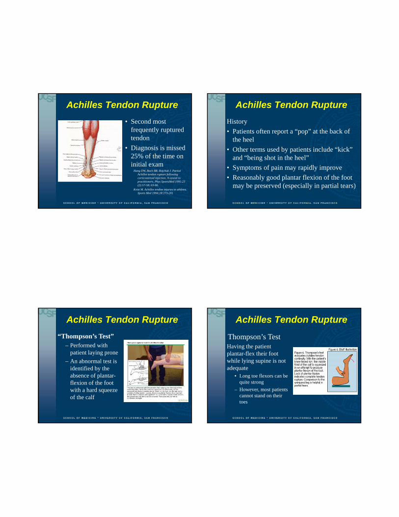
Achilles Tendon Rupture
“Thompson’s Test”– Performed with
patient laying prone
– An abnormal test is identified by the absence of plantar-flexion of the foot with a hard squeeze of the calf
Achilles Tendon Rupture
Thompson’s TestHaving the patient plantar-flex their foot while lying supine is not adequate
• Long toe flexors can be quite strong
– However, most patients cannot stand on their toes
10/21/2013
27
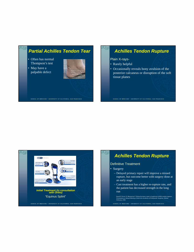
Partial Achilles Tendon Tear
• Often has normal Thompson’s test
• May have a palpable defect
Achilles Tendon Rupture
Plain X-rays-
• Rarely helpful
• Occasionally reveals bony avulsion of the posterior calcaneus or disruption of the soft tissue planes
Initial Treatment (in consultation with Ortho)
“Equinus Splint”
Achilles Tendon Rupture
Definitive Treatment
• Surgery– Delayed primary repair will improve a missed
rupture, but outcome better with surgery done at an early stage
– Cast treatment has a higher re-rupture rate, and the patient has decreased strength in the long run
Snead P, Porter D, Mannarino F. Delayed primary repair of neglected achilles tendon ruptures. Presentation at Annual Meeting of American Academy of Orthopaedic Surgeons, Atlanta: February, 1996
10/21/2013
28
Ankle and Foot Pain –Case 2
• A 68 year old woman steps into a shallow, uncovered open hole in the sidewalk on the way to your office for routine medical follow-up
• She reports twisting her ankle and falling to the ground
Ankle and Foot Pain –Case 2
• She is unhurt except for her left ankle
• She is having some pain with weight bearing
• On exam, she has pain and some ecchymosis over the medial malleolus
• She is neurovascularly intact
• Her left foot seems to have a flattened arch, but she denies having “flat feet”
• X-ray of the foot and ankle is negative for fracture
Ankle and Foot Pain –Case 2
What do you suspect is the problem?1) Occult calcaneal compression fracture
2) Charcot joint from chronic denervation
3) Acute S1 motor neuropathy in the foot secondary to traumatic compression from fall
4) Posterior tibialis tendon rupture
Ankle and Foot Pain –Case 2
What do you suspect is the problem?1) Occult calcaneal compression fracture
2) Charcot joint from chronic denervation
3) Acute S1 motor neuropathy in the foot secondary to traumatic compression from fall
4) Posterior tibialis tendon rupture
10/21/2013
29
Posterior Tibialis Tendon Rupture
• Not a common problem, but associated with ankle sprains that are commonly evaluated by primary care physicians
• Delayed diagnosis can cause fixed bony planus and need for hindfoot fusion
Marcus RE, Goodfellow DB, Pfister ME. The difficult diagnosis of posterior tibialis tendon rupture in sports injuries. Orthopedics 1995;18: 715-21.
Janis LR, Wagner JT, Kravitz RD, Greenberg JJ. Posterior tibial tendon rupture: classification, modified surgical repair, and retrospective study. J Foot Ankle Surg 1993;32:2-13.
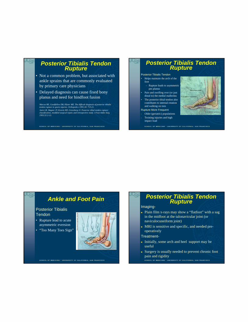
Posterior Tibialis Tendon Rupture
Posterior Tibialis Tendon
• Helps maintain the arch of the foot
– Rupture leads to asymmetric pes planus
• Pain and swelling over (or just distal to) the medial malleolus
• The posterior tibial tendon also contributes to internal rotation and walking on toes
Rupture More Frequent
Older (geriatric) populations
Twisting injuries and high impact load
Ankle and Foot Pain
Posterior Tibialis Tendon• Rupture lead to acute
asymmetric eversion
• “Too Many Toes Sign”
Posterior Tibialis Tendon Rupture
Imaging-
Plain film x-rays may show a “flatfoot” with a sag in the midfoot at the talonavicular joint (or naviculocuneiform joint)
MRI is sensitive and specific, and needed pre-operatively
Treatment-
Initially, some arch and heel support may be useful
Surgery is usually needed to prevent chronic foot pain and rigidity

10/21/2013
30
Foot Pain
Foot Pain
Anatomy 28 Bones
• 14 phalanges
• 7 tarsal bones
• 5 Metatarsals
• 2 Sesamoids
Hindfoot connects to the midfoot at the Chopart joint
Midfoot connects to the forefoot at the Lisfranc joint
Foot Pain
Physical Exam
Perform a neurovascular exam (pulses, sensation, capillary refill)
Inspect for wounds
Careful palpation for point tenderness which may indicate a fracture
Foot Pain
Following trauma, does this patient require a foot x-ray?
10/21/2013
31
Ottawa Foot Rules
X-ray is required if there is any pain in the mid-foot zone and ANY of these findings:
Bone tenderness at base of fifth metatarsal
Bone tenderness over the navicular
Inability to weight bear both immediately and in the casualty department
Foot Pain
Common soft-tissue causes
Plantar fasciitis
Morton’s neuroma
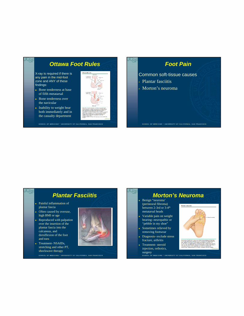
Plantar Fasciitis Painful inflammation of
plantar fascia
Often caused by overuse, high BMI or age
Reproduced with palpation over the insertion of the plantar fascia into the calcaneus, and dorsiflexion of the foot and toes
Treatment- NSAIDs, stretching and other PT, shockwave therapy
Morton’s Neuroma Benign “neuroma’
(perineural fibroma) between 2-3rd or 3-4th
metatarsal heads
Variable pain on weight bearing- neuropathic or “pebble in my shoe”
Sometimes relieved by removing footwear
Diagnosis- exclude stress fracture, arthritis
Treatment- steroid injection, orthotics, surgery

10/21/2013
32
Foot Pain
Common Acute Fractures
Fracture of the proximal 5th metatarsal
Fractures of the metatarsal shaft
Stress fractures
Foot Pain – Common Fractures
Three distinct fractures occur in the proximal fifth metatarsal
Each is treated differently
The joint between the bases of the fourth and fifth metatarsals is a key landmark for classifying proximal fifth metatarsal fractures
Foot Pain – Common Fractures
Minimally displaced avulsion fracture of the fifth metatarsal tubercle (styloid)
Ankle inversion while the foot is in plantar flexion
Can present like a lateral ankle sprain
Heals well- post-op shoe• Weight bear as tolerated
• Heals in three to six weeks
• Radiographic union 8 wks
Foot Pain – Common Fractures
Acute fifth metatarsal diaphysis (Jones) fracture
Occurs from a medial or mediolateral force on the base of the fifth metatarsal while weight is over the lateral aspect of the plantar flexed foot (heel off the ground injury)
Heal poorly- cast and surgery
10/21/2013
33
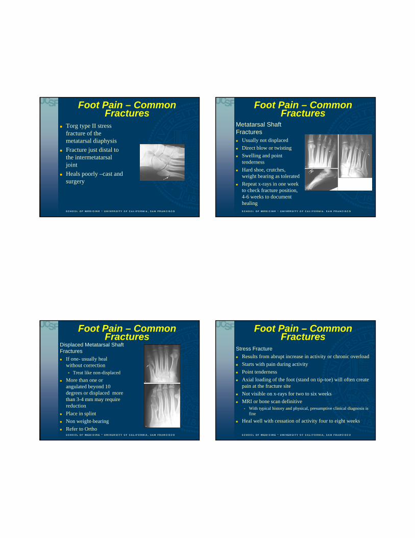
Foot Pain – Common Fractures
Torg type II stress fracture of the metatarsal diaphysis
Fracture just distal to the intermetatarsal joint
Heals poorly –cast and surgery
Foot Pain – Common Fractures
Metatarsal Shaft Fractures Usually not displaced
Direct blow or twisting
Swelling and point tenderness
Hard shoe, crutches, weight bearing as tolerated
Repeat x-rays in one week to check fracture position, 4-6 weeks to document healing
Foot Pain – Common Fractures
Displaced Metatarsal Shaft Fractures
If one- usually heal without correction Treat like non-displaced
More than one or angulated beyond 10 degrees or displaced more than 3-4 mm may require reduction
Place in splint
Non weight-bearing
Refer to Ortho
Foot Pain – Common Fractures
Stress Fracture
Results from abrupt increase in activity or chronic overload
Starts with pain during activity
Point tenderness
Axial loading of the foot (stand on tip-toe) will often create pain at the fracture site
Not visible on x-rays for two to six weeks
MRI or bone scan definitive• With typical history and physical, presumptive clinical diagnosis is
fine
Heal well with cessation of activity four to eight weeks

10/21/2013
34
Foot Pain – Common Fractures
Stress Fracture
If very painful, crutches and partial weight-bearing
Short leg cast for 1-3 weeks if severe pain
Consider custom orthotics, but no evidence for injury prevention
Foot Pain – Uncommon Fractures
Fractures of the proximal first through fourth metatarsal
- Endanger the Lisfranc ligament complex
Chopart's fracture–dislocation• Dislocation of the mid-tarsal (talonavicular and
calcaneocuboid joints of the foot
• Often with associated fractures of the calcaneus, cuboid and navicular
Foot Pain – Case 1 Your next patient is a 35 year old healthy woman
Last evening, she was sitting in the front seat of a car driven by a friend
The car in front of them suddenly slammed on their brakes to avoid hitting a deer in the road
The car your patient was in tried to stop, but couldn’t avoid a rear-end collision
Although your patient was wearing a seat belt, it was a very small car and she instinctively pushed both feet against the forward floorboard to brace for impact
Foot Pain – Case 1 Fortunately, she didn’t suffer any head or thorax or
upper extremity injury, or hip or knee or ankle trauma
But she injured her right foot, and she couldn’t bear weight without a lot of pain
EMS took her to the local ED, who X-rayed her foot
The x-ray was read as negative, on exam she was neurovascularly intact, and she was discharged with high dose NSAIDs and placed in post-operative shoe

10/21/2013
35
Foot Pain – Case 1 She consults you three days later, reporting
that she feels “no better”
On exam:• She has edema from the mid-tarsal area distally
into the toes
• Normal DP pulse and capillary refill
• Marked dorsal tenderness was noted over the second through fourth tarso-metatarsal joints
• You obtain the x-ray report from the ED and confirm that no fracture was found
Foot Pain – Case 1
Based on this history and physical exam, and x-ray report, what are you concerned about?
1. Traumatic stress fracture not yet evident on x-ray
2. Dorsalis pedis artery rupture endangering the navicular bone
3. Comminuted navicular fracture not visible on x-ray
4. Lisfranc fracture
Lisfranc Injury of the Foot
• “Lisfranc joint" - medial articulation involving the first and second metatarsals with the first and second cuneiforms
• Named for Jacques Lisfranc (1790-1847) a field surgeon in Napoleon’s army
– after an amputation he performed for gangrene

10/21/2013
36
Lisfranc Injury of the Foot
• This entire tarso-metatarsal complex is supported by the "keystone" wedging of the second metatarsal into the cuneiform
• There is a “weak link” – the first and second
metatarsal bases lack a transverse ligament
Vuori JP, Aro HT. Lisfranc joint injuries: trauma mechanisms and associated injuries. J Trauma 1993;35:40-5. Englanoff G, Anglin D, Hutson HR. Lisfranc fracture-dislocation: a frequently missed diagnosis in the emergency department. Ann Emerg Med 1995;26:229-33.
Lisfranc Injury of the Foot
The Lisfranc Ligament Complex• Hold metatarsal bases rigidly in place
• Maintain arch of foot
• Even subtle injuries can cause long-term disability
Lisfranc Injury of the Foot
Most common mechanism of injury is an axial load placed on a plantar-flexed foot
Lisfranc Injury of the Foot
One of the most commonly missed fractures by physicians (one of the top 5 missed diagnosis in the ER)
– Nearly 50 percent of Lisfranc joint injuries are missed on initial antero-posterior and oblique radiographs (even with significant injury)
– Most important initial diagnostic test is a clinician’s high index of suspicion
– Refer for unexplained tenderness near the tarsometatarsal joint
10/21/2013
37
Lisfranc Injury of the FootA Few Exam Pearls
Patient is unlikely to bear weight while standing on tip-toe
It may seem like a Grade III ankle sprain, • But no ankle ligamentous laxity
• If you stabilize the hindfoot (calcaneus) with one hand, and “twist” the forefoot, it will cause severe pain with a Lisfranc injury (and not with an ankle sprain)
Check and document neurovascular status• Dorsalis pedis artery passes over the proximal head
of the second metatarsal
Lisfranc Injury of the Foot
Plain X-rays
• Weight-bearing antero-posterior and lateral views, as well as a 30-degree oblique view
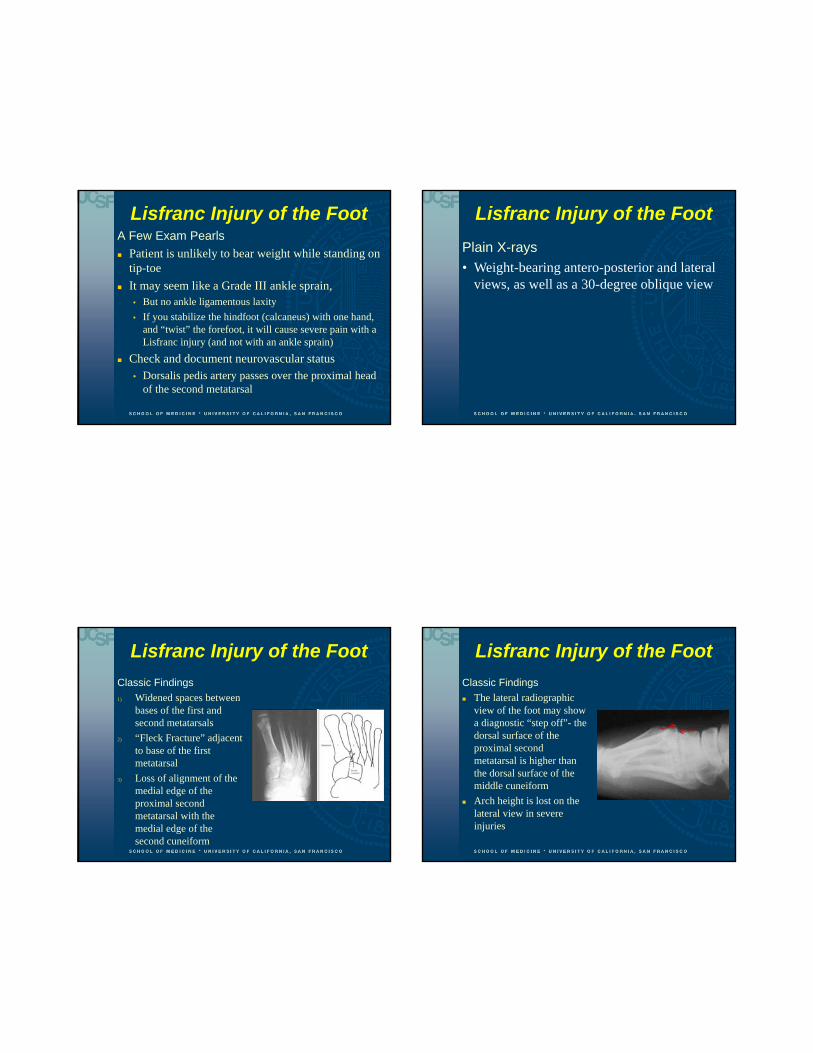
Lisfranc Injury of the Foot
Classic Findings
1) Widened spaces between bases of the first and second metatarsals
2) “Fleck Fracture” adjacent to base of the first metatarsal
3) Loss of alignment of the medial edge of the proximal second metatarsal with the medial edge of the second cuneiform
Lisfranc Injury of the Foot
Classic Findings
The lateral radiographic view of the foot may show a diagnostic “step off”- the dorsal surface of the proximal second metatarsal is higher than the dorsal surface of the middle cuneiform
Arch height is lost on the lateral view in severe injuries

10/21/2013
38
Lisfranc Injury of the Foot
Treatment Stage 1 injury – non-weight bearing cast Displacement of more than 2mm may need
open reduction and internal fixation (perhaps within 12 to 24 hours)
There is some evidence for closed reductionTrevino SG, Kodros S. Controversies in tarsometatarsal injuries. Orthop Clin North Am 1995;26:229-38. Myerson M. The diagnosis and treatment of injuries to the Lisfranc joint complex. Orthop Clin North Am 1989;20:655-64..Heckman JD. Fractures and dislocations of the foot. In: Rockwood CA, Green DP, Bucholz RD, eds. Rockwood and Green's Fractures in adults. Vol 2. 3d ed. Philadelphia: Lippincott, 1991:2140-51
Lisfranc Injury of the Foot
Failure to diagnose promptly• Compartment syndrome
• Ischemic contractures of the muscles (“claw toes”)
• Arthritis
• Permanent antalgic gait
• Chronic foot pain
Thank you!