My Method for Approaching Skin Biopsies - aad.org C017 - Haun...My Method for Approaching Skin...
34
PAUL HAUN, MD, MS, FAAD ASSISTANT PROFESSOR DERMATOLOGY AND DERMATOPATHOLOGY DEPARTMENT OF DERMATOLOGY UNIVERSITY OF PENNSYLVANIA CMC VAMC, PHILADELPHIA My Method for Approaching Skin Biopsies
Transcript of My Method for Approaching Skin Biopsies - aad.org C017 - Haun...My Method for Approaching Skin...

P A U L H A U N , M D , M S , F A A D
A S S I S T A N T P R O F E S S O R
D E R M A T O L O G Y A N D D E R M A T O P A T H O L O G Y
D E P A R T M E N T O F D E R M A T O L O G Y
U N I V E R S I T Y O F P E N N S Y L V A N I A
C M C V A M C , P H I L A D E L P H I A
My Method for Approaching Skin Biopsies

Disclosure
Royalties from Health Press, Inc
Fast Facts: Diagnosing Cutaneous T-Cell Lymphoma (P Haun and J Scarisbrick)
Q1:
Quick Audience Survey:
A) Medical Student
B) Dermatology Resident
C) Pathology Resident
D) Dermpath Fellow
E) NP/PA in Dermatology
F) Practicing Dermatologist/Dermatopathologist

Remember…
Don’t make it too difficult
Pattern recognition You are becoming a master of this skill
All of these conditions exist clinically
Utilize your knowledge from clinic while studying path and vice versa What will the biopsy look like of this eruption?
Learn the vocabulary of dermatopathology

Inflammatory Dermatopathology
Pick an approach/algorithm/process
Stick to It!
We all do it—some of us are just quicker than others
Low Power Magnification=High Power Thinking
Read the slide like a book—tell the biopsy’s story
Stratum Corneum>Epidermis>Dermis>Subcutis
Architecture/Pattern
Cell Types

My Approach
1st Question: Method of Biopsy
Punch=Inflammatory
Shave/Excision=Neoplastic

My Approach
2nd Question: Inflammatory vs. Neoplastic
Can be one of the most difficult questions
Overlap can occur
3rd Question: Anatomic Site
Can give a great deal of information—again, go back to your clinical training
Assists in ruling in/ruling out certain conditions
Can tie together a difficult histopathology case

Patterns
Superficial Perivascular
Superficial and Deep Perivascular
Nodular
Diffuse
Vasculitis
Intraepidermal Vesicular
Subepidermal Vesicular
Folliculitis/Perifolliculitis
Fibrosing
Panniculitis
Alopecia
“Others”
Depositions
Infestations
DDx Patterns

Superficial Perivascular
• No Epidermal Change
• Interface
• Vacuolar
• Lichenoid
• Spongiotic
• Spongiotic
• Psorasiform
• Lichenoid
• Psoriasiform
• +/- Lichenoid
Ackerman, et al. Histologic Diagnosis of Inflammatory Skin Diseases. 2nd Ed. 1997

Superficial and Deep Perivascular
Similar Patterns to SPV
• No Epidermal Involvement
• Interface
• Vacuolar
• Lichenoid
• Ballooning
• Spongiotic
• Psoriasiform

Superficial Perivascular Superficial and Deep Perivascular
Normal Skin Differential Urticaria Dermatophyte Atopic Dermatitis Allergic/Irritant Contact Lichen Planus PLEVA Erythema Multiforme Dermatomyositis/Lupus Psoriasis Syphillis Prurigo/LSC
Polymorphous Light Eruption
Pernio/chilblains
Arthropod assult
Fixed Drug Eruption
DLE
Mycosis Fungoides/LyP
Lichen Striatus
B-cell lymphoproliferative d/o
Necrobiosis lipoidica
Prototypes and DDx (some)

NodularLook for the cell type
• Lymphocytes
• Neutrophils
• Mixed Neuts/Eos
• Histiocytes
• Sarcoidal
• Tuberculoid
• Palisaded
• Suppurative

Diffuse
Much overlap with nodular patterns
Will discuss together

Lymphocytic Neutrophilic and Mixed
Lymphoma
Pseudolymphoma/CLH
Sweets Syndrome
Leukocytoclastic vasculitis
GPA/GPA with Eos
Acne keloidalis/dissecting cellulitis
Granuloma faciale/erythema elevatum diutinum
Nodular/Diffuse

Sarcoidal Tuberculoid
Foreign body reaction
Sarcoidosis
Rosacea
Leprosy
Lichen striatus
TB and its many flavors
Leishmaniasis
Syphillis
Nodular/Diffuse--Histiocytic

Palisaded Suppurative
Granuloma annulare
Necrobiosis lipoidica
Necrobiotic xanthogranuloma
Rheumatoid nodule
Gout
Ruptured cyst
Foreign body reaction
Infection
Infection
Infection
Nodular/Diffuse—Histiocytic

Vasculitis
• Small-Medium vs. Large Vessel
• Vessel Type
• +/- Neutrophils
• +/- Fibrin
• +/- Organisms
• Vasculitis vs. Vasculopathy

Vasculitis Vasculopathy/Occlusion
Small Vessel Drug
Malignancy
Infection
Literally hundreds of other causes
Degos
Medium-Large GPA
PAN
Nodular Vasculitis
DIC
TTP/ITP
Coumadin necrosis
PNH
Anti-phospholipid Ab
Cholesterol emboli
Catheter sheathing
Vasculitis/Vasculopathy

IntraepidermalVesicular• Balooning
• Spongiosis
• Acantholysis
• Upper spinous
• Mid spinous
• Suprabasal
• Pustular
• Intracorneal
• Subcorneal
• Spongiotic
Look for the deepest level involved
**Know you DIF Patterns**

Balooning Spongiosis
HSV/VZV
Orf
Vaccinia
Nutritional Deficiency
Hand-foot-mouth
EM
PLEVA
ACD/ICD
Dyshidrosis
Dermatophyte
EM
BP
PV
Fixed Drug
Intraepidermal Vesicular

Acantholysis Suprabasal
Upper Spinous
Bullous impetigo
Pemphigus foliaceous
Staph scalded skin
Mid Spinous
HSV/VZV
Dariers
Grovers
Pemphigus vulgaris
Hailey-Hailey
Pustular
Dermatophyte, Candida, Psoriasis, Impetigo, PF, SSSS, ACD, Dyshidrosis
Intraepidermal Vesicular

SubepidermalVesicular
Look for the Cell Type
• Paucicellular
• Lymphocytes
• Eosinophils
• Neutrophils
• Mixed
• Mast Cells
**Know you DIF Patterns**

Paucicellular Lymphocytes
Epidermolysis bullosa
Burn
EM
GVHD
PCT
Amylodosis
Cautery
EM
LSetA
PLEVA
Lichen Planus
Subepidermal Vesicular

Neutrophils Mixed
Dermatitis herpetiformis
Linear IgA
Bullous lupus
LCV
Infection
DH
BP
Linear IgA
EBA
Subepidermal Vesicular

Folliculitis
Infectious
Acne
Rosacea
Eosinophilic
Mycosis fungoides
Toxic erythema

Perifolliculitis
Discoid Lupus
Lichen planopilaris
Rosacea
Keratosis pilaris
Scurvy
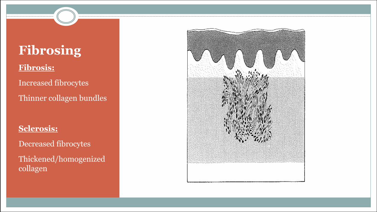
Fibrosing
Fibrosis:
Increased fibrocytes
Thinner collagen bundles
Sclerosis:
Decreased fibrocytes
Thickened/homogenized collagen

Panniculitis
• Septal (mostly)
• +/- Vasculitis
• Lobular (mostly)
• +/- Vasculitis

Septal Lobular
Erythema nodosum*
LCV
PAN
GA
Rheumatoid nodule
Necrobiotic xanthogranuloma
Nodular vasculitis Sclerema neonatorum Subcutaneous fat necrosis Cold panniculitis Lymphoma Pancreatic panniculitis Factitial Infection Sarcoidosis Trauma
Panniculitis

Alopecia
• Scarring or not
• Normal=~6 follicles per 4mm punch
• Inflammatory or not
• Cell Types
• Hair counts
• Catagen
• Telogen
• Anagen
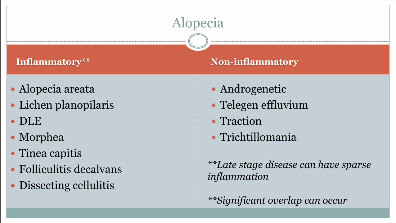
Inflammatory** Non-inflammatory
Alopecia areata
Lichen planopilaris
DLE
Morphea
Tinea capitis
Folliculitis decalvans
Dissecting cellulitis
Androgenetic
Telegen effluvium
Traction
Trichtillomania
Alopecia
**Late stage disease can have sparse inflammation
**Significant overlap can occur

Patterns
Superficial Perivascular
Superficial and Deep Perivascular
Nodular
Diffuse
Vasculitis
Intraepidermal Vesicular
Subepidermal Vesicular
Folliculitis/Perifolliculitis
Fibrosing
Panniculitis
Alopecia
“Others”
Depositions
Infestations
DDx Patterns

Others
Normal skin
Massive papillary dermal edema
Eosinophilic spongiosis
Deposition/Metabolic