Comparison of sample types and location of skin biopsies for BVDV detection using antigen capture...
18
Comparison of sample types and location of skin biopsies for BVDV detection using antigen capture ELISA Testing Dr. Brian Vander Ley BVD Symposium January 26, 2009 Phoenix, Arizona
-
Upload
juliet-wheeler -
Category
Documents
-
view
215 -
download
0
Transcript of Comparison of sample types and location of skin biopsies for BVDV detection using antigen capture...
Comparison of sample types and location of skin biopsies for BVDV detection using antigen capture
ELISA Testing
Dr. Brian Vander LeyBVD Symposium January 26, 2009
Phoenix, Arizona
Introduction
• We are looking for ways to screen for PI animals more effectively– Sample choices– Assay evaluation– Assay development
Overview
• 2 Projects– Sample project
• 40 PIs• Several different samples evaluated
– Hide project• 3 PIs• Involved systematic evaluation of whole hides for BVDV
antigen
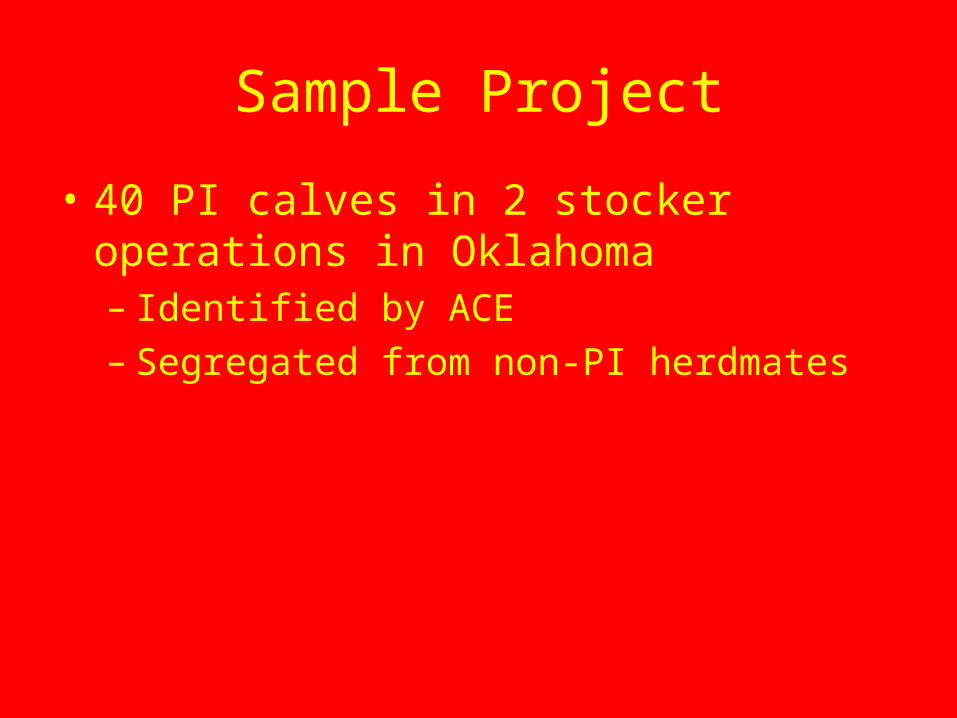
Sample Project
• 40 PI calves in 2 stocker operations in Oklahoma– Identified by ACE– Segregated from non-PI herdmates
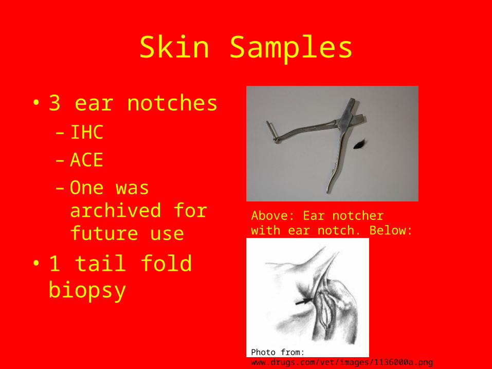
Skin Samples
• 3 ear notches– IHC– ACE– One was archived for
future use
• 1 tail fold biopsyAbove: Ear notcher with ear notch. Below: tail fold
Photo from: www.drugs.com/vet/images/1136000a.png

Swab Samples
• Nasal• Oral• Conjuctival• Vaginal/Preputial• Rectal
Photo from: www.puritanmedproducts.com
Collecting an oral swab

Sample Processing
• Skin Samples– Placed in dry tubes and frozen until testing – Incubated in 1 mL PBS for 24 hours at 4°C prior to
using ACE
• Swab Samples– Collected and place immediately in 1 mL PBS then
frozen – Thawed and allowed to incubate for 24 hours 4°C
prior to ACE
Results
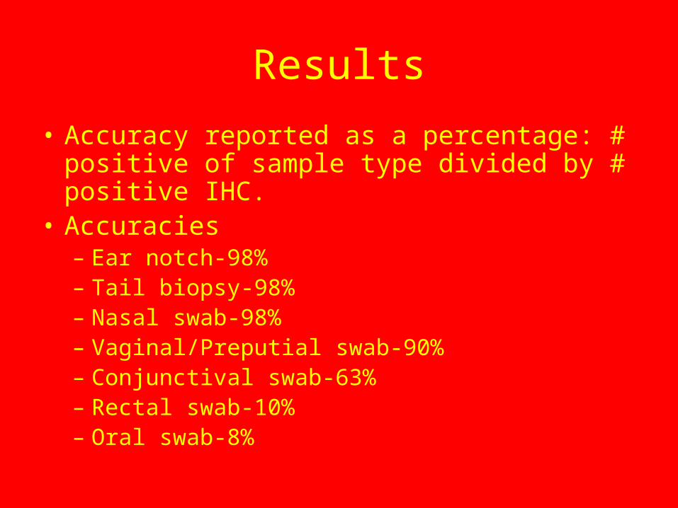
• Accuracy reported as a percentage: # positive of sample type divided by # positive IHC.
• Accuracies– Ear notch-98%– Tail biopsy-98%– Nasal swab-98%– Vaginal/Preputial swab-90%– Conjunctival swab-63%– Rectal swab-10%– Oral swab-8%

Sample Conclusions
• Nasal swabs could be a good sample for detecting PI cattle.– Simple– Non-invasive
• Other samples could also be good, but would pose some challenges
• More work would need to be done to validate any of these samples

Hide Project
• 3 PIs selected based on previous diagnosis as PI by ACE
• Each animal was euthanized and the skin was removed in its entirety
A complete hide

Hide Project
• Each hide was quartered in the field and packed on ice
• The hides were frozen until processing
Quarter division of hides
Hide Processing
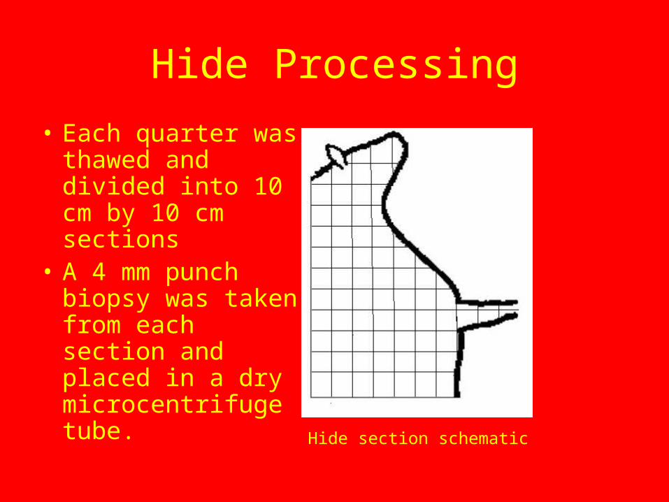
• Each quarter was thawed and divided into 10 cm by 10 cm sections
• A 4 mm punch biopsy was taken from each section and placed in a dry microcentrifuge tube.
Hide section schematic

Sample Evaluation
• All samples were incubated in 1 mL PBS at 4°C for the same amount of time.
• All steps were performed the same way on each plate and in sequential order from plate 1 to plate 7.
• Multiple controls were used in each plate– Kit positive control– Kit negative control– Titered BVDV infected cell culture– Titered BVDV infected cell culture diluted 1:10 in PBS
Results
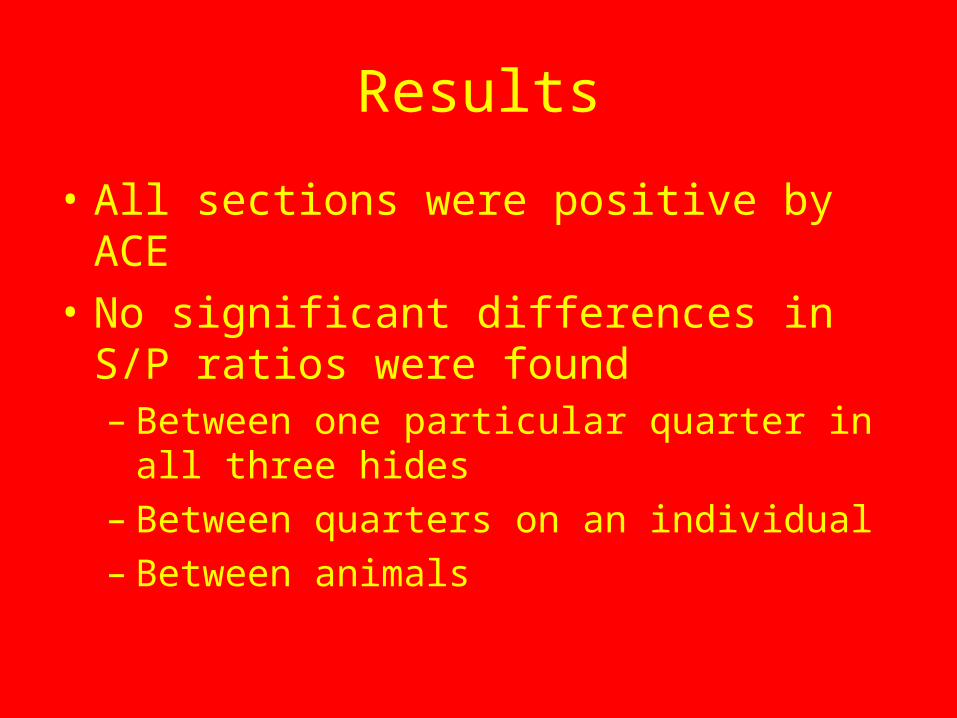
• All sections were positive by ACE• No significant differences in S/P ratios were
found– Between one particular quarter in all three hides– Between quarters on an individual– Between animals
Conclusions
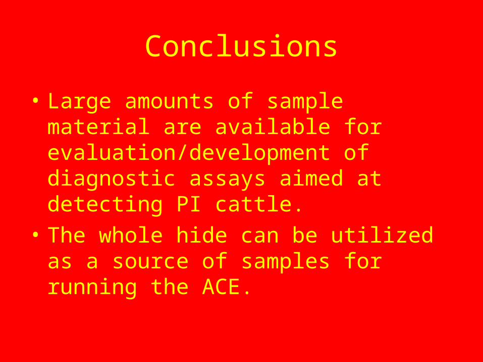
• Large amounts of sample material are available for evaluation/development of diagnostic assays aimed at detecting PI cattle.
• The whole hide can be utilized as a source of samples for running the ACE.
Overall Conclusions
• BVDV Antigen can be recovered from a variety of places.– Many swabs– Essentially any haired skin sample
• There are large amounts of material that can be harvested from an individual animal for evaluation or development of diagnostic assays, especially those that use skin.
• More research needs to be done to validate any of the samples.
References
• Idexx Herdchek® Bovine Virus Diarrhea Antigen Test Kit. U.S. Vet. License No. 313. 2007.

Questions?
Thank You