MUMMIFICATION OF CAT CADAVERS USING AN IMPROVED METHOD...
4
131 MUMMIFICATION OF CAT CADAVERS USING AN IMPROVED METHOD Ioana DUMITRU 1 , Cristian DEZDROBITU 1 , Irina IRIMESCU 1 , Cristian MARTONOS 1 , Bianca MATOSZ 1 , Florin SILAGHI 1 , Aurel DAMIAN 1 1 University of Agronomic Sciences and Veterinary Medicine of Cluj-Napoca, nr. 3-5, str. Calea Mănăştur, postal code 400372, Cluj-Napoca, Romania, 0741196318, Corresponding author email: [email protected] Abstract Some of the preservation techniques of human and animal cadavers have been known since antiquity, starting with the simplest ones, such as preservation by freezing, or with simple mummification, up to mummification by arterial injecting or by immersion. Their execution necessitates the usage of several combinations of substances with a single active component, such as: preservatives, to maintain tissue structure; disinfectants, to halt decay; moistening agents and coloring agents. The aim of this research was to obtain a cat mummy by means of an improved technique. The materials used were: one cat cadaver, dissection instruments, 7% and 15% formaldehyde solutions, technical grade glycerin and coloring agents. The mummification process consisted of injecting the cat body with the 7% formaldehyde solution, fitting it up on a mount and letting it fixate for 5 days, injecting it with the 15% formaldehyde solution, followed by another fixation interval of 10 days. Subsequently, we have removed the skin and the subcutaneous conjunctive tissue, underlining the musculature. The body was once again mounted and frozen for 14 days to dehydrate. It was then maintained at room temperature, followed by the application of the technical grade glycerin and of the coloring agents. The piece was then kept on the mount to dry until the completion of the mummification process. We conclude that this technique is successful at maintaining the anatomical characteristics of the body. Key words: preserving, cadaver, mummification, cat. INTRODUCTION Along with the recognition of anatomy as a scientific discipline, in Alexandria arose the interest to prevent the decomposing of biological tissues, which lead in time to the development of multiple preserving techniques (Singer C., 1957). Mummification can be defined as a method of preserving organic material, known since ancient times in many cultures, especially that of ancient Egypt. If certain environmental conditions are met, it is possible for the mummification to occur accidentally, without human intervention, even in the case of certain extremely sensitive and perishable tissues, like the nervous tissue (Radanov et al., 1992). The occurrence of natural mummification requires the meeting of certain special conditions such as: dry air, appropriate ventilation and high temperatures. The mummification speed depends very much on the state of the cadaver and on the external factors and varies between one month and twelve months. Armstrong K., (1993) states that “the methodology behind mummification is encompassed by the life of the King "Osiris" who brought civilization to Egypt. Ancient Egyptians believed that an intact preserved body was necessary for the soul to live forever”. The Greek historian Herodotus (450 BC) was the first to recount this technique (Putnam J., 1993). In the beginning, it was done as a simple dissection, but in time it became a complex process, performed only by embalmers (Yardley M, Rutka J., 1997). MATERIALS AND METHODS The experiment was carried out in the Comparative Anatomy Laboratory of the Faculty of Veterinary Medicine of Cluj- Napoca. The following materials were used to accomplish the mummification process: one cat cadaver (Fig. 1), dissection instruments, 7% and 15% solutions of formaldehyde, technical grade glycerin, and acrylic coloring agent. The first step of the mummification process consisted of injecting the cat cadaver with a 7% formaldehyde solution, then fitting it up on a mount (Fig. 2) and letting it fixate for 5 days. Scientific Works. Series C. Veterinary Medicine. Vol. LXI (2) ISSN 2065-1295; ISSN 2343-9394 (CD-ROM); ISSN 2067-3663 (Online); ISSN-L 2065-1295
Transcript of MUMMIFICATION OF CAT CADAVERS USING AN IMPROVED METHOD...
131
MUMMIFICATION OF CAT CADAVERS USING AN IMPROVED METHOD
Ioana DUMITRU1, Cristian DEZDROBITU1, Irina IRIMESCU1,
Cristian MARTONOS1, Bianca MATOSZ1, Florin SILAGHI1, Aurel DAMIAN1
1University of Agronomic Sciences and Veterinary Medicine of Cluj-Napoca,
nr. 3-5, str. Calea Mănăştur, postal code 400372, Cluj-Napoca, Romania, 0741196318,
Corresponding author email: [email protected] Abstract
Some of the preservation techniques of human and animal cadavers have been known since antiquity, starting with the simplest ones, such as preservation by freezing, or with simple mummification, up to mummification by arterial injecting or by immersion. Their execution necessitates the usage of several combinations of substances with a single active component, such as: preservatives, to maintain tissue structure; disinfectants, to halt decay; moistening agents and coloring agents. The aim of this research was to obtain a cat mummy by means of an improved technique. The materials used were: one cat cadaver, dissection instruments, 7% and 15% formaldehyde solutions, technical grade glycerin and coloring agents. The mummification process consisted of injecting the cat body with the 7% formaldehyde solution, fitting it up on a mount and letting it fixate for 5 days, injecting it with the 15% formaldehyde solution, followed by another fixation interval of 10 days. Subsequently, we have removed the skin and the subcutaneous conjunctive tissue, underlining the musculature. The body was once again mounted and frozen for 14 days to dehydrate. It was then maintained at room temperature, followed by the application of the technical grade glycerin and of the coloring agents. The piece was then kept on the mount to dry until the completion of the mummification process. We conclude that this technique is successful at maintaining the anatomical characteristics of the body. Key words: preserving, cadaver, mummification, cat. INTRODUCTION Along with the recognition of anatomy as a scientific discipline, in Alexandria arose the interest to prevent the decomposing of biological tissues, which lead in time to the development of multiple preserving techniques (Singer C., 1957). Mummification can be defined as a method of preserving organic material, known since ancient times in many cultures, especially that of ancient Egypt. If certain environmental conditions are met, it is possible for the mummification to occur accidentally, without human intervention, even in the case of certain extremely sensitive and perishable tissues, like the nervous tissue (Radanov et al., 1992). The occurrence of natural mummification requires the meeting of certain special conditions such as: dry air, appropriate ventilation and high temperatures. The mummification speed depends very much on the state of the cadaver and on the external factors and varies between one month and twelve months. Armstrong K., (1993) states that “the methodology behind mummification is
encompassed by the life of the King "Osiris" who brought civilization to Egypt. Ancient Egyptians believed that an intact preserved body was necessary for the soul to live forever”. The Greek historian Herodotus (450 BC) was the first to recount this technique (Putnam J., 1993). In the beginning, it was done as a simple dissection, but in time it became a complex process, performed only by embalmers (Yardley M, Rutka J., 1997). MATERIALS AND METHODS The experiment was carried out in the Comparative Anatomy Laboratory of the Faculty of Veterinary Medicine of Cluj-Napoca. The following materials were used to accomplish the mummification process: one cat cadaver (Fig. 1), dissection instruments, 7% and 15% solutions of formaldehyde, technical grade glycerin, and acrylic coloring agent. The first step of the mummification process consisted of injecting the cat cadaver with a 7% formaldehyde solution, then fitting it up on a mount (Fig. 2) and letting it fixate for 5 days.
Scientific Works. Series C. Veterinary Medicine. Vol. LXI (2)ISSN 2065-1295; ISSN 2343-9394 (CD-ROM); ISSN 2067-3663 (Online); ISSN-L 2065-1295
132
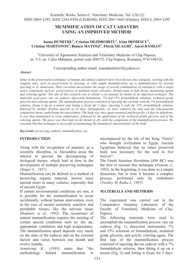
Next, it was injected with a 15% formaldehyde solution, followed by another fixation interval of 10 days. The injections were performed without the removal of the skin.
Fig. 1 Cat cadaver before injecting
Fig.2 Cadaver injected and mounted on a support
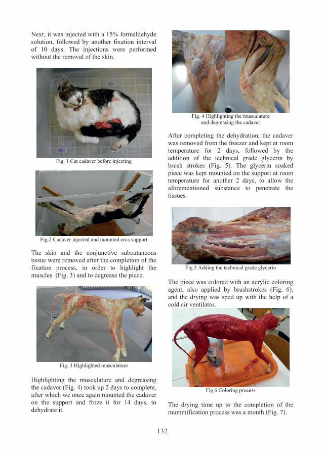
The skin and the conjunctive subcutaneous tissue were removed after the completion of the fixation process, in order to highlight the muscles (Fig. 3) and to degrease the piece.
Fig. 3 Highlighted musculature
Highlighting the musculature and degreasing the cadaver (Fig. 4) took up 2 days to complete, after which we once again mounted the cadaver on the support and froze it for 14 days, to dehydrate it.
Fig. 4 Highlighting the musculature
and degreasing the cadaver
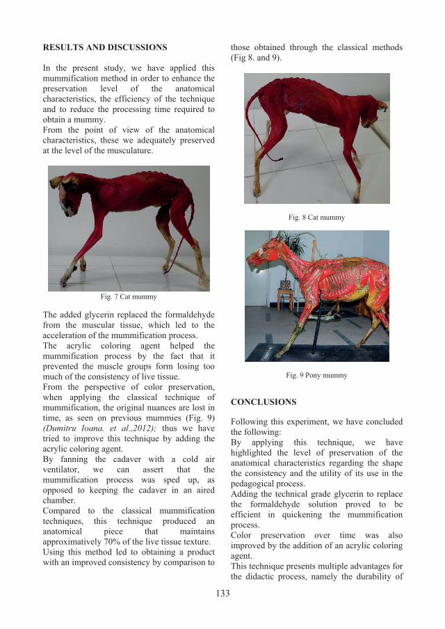
After completing the dehydration, the cadaver was removed from the freezer and kept at room temperature for 2 days, followed by the addition of the technical grade glycerin by brush strokes (Fig. 5). The glycerin soaked piece was kept mounted on the support at room temperature for another 2 days, to allow the aforementioned substance to penetrate the tissues.
Fig.5 Adding the technical grade glycerin
The piece was colored with an acrylic coloring agent, also applied by brushstrokes (Fig. 6), and the drying was sped up with the help of a cold air ventilator.
Fig.6 Coloring process
The drying time up to the completion of the mummification process was a month (Fig. 7).
133
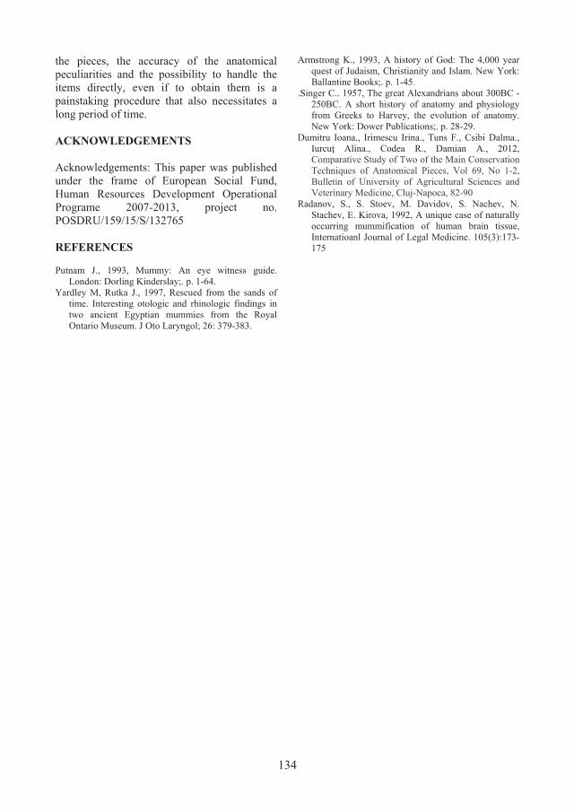
RESULTS AND DISCUSSIONS In the present study, we have applied this mummification method in order to enhance the preservation level of the anatomical characteristics, the efficiency of the technique and to reduce the processing time required to obtain a mummy. From the point of view of the anatomical characteristics, these we adequately preserved at the level of the musculature.
Fig. 7 Cat mummy
The added glycerin replaced the formaldehyde from the muscular tissue, which led to the acceleration of the mummification process. The acrylic coloring agent helped the mummification process by the fact that it prevented the muscle groups form losing too much of the consistency of live tissue. From the perspective of color preservation, when applying the classical technique of mummification, the original nuances are lost in time, as seen on previous mummies (Fig. 9) (Dumitru Ioana. et al.,2012); thus we have tried to improve this technique by adding the acrylic coloring agent. By fanning the cadaver with a cold air ventilator, we can assert that the mummification process was sped up, as opposed to keeping the cadaver in an aired chamber. Compared to the classical mummification techniques, this technique produced an anatomical piece that maintains approximatively 70% of the live tissue texture. Using this method led to obtaining a product with an improved consistency by comparison to
those obtained through the classical methods (Fig 8. and 9).
Fig. 8 Cat mummy
Fig. 9 Pony mummy
CONCLUSIONS
Following this experiment, we have concluded the following: By applying this technique, we have highlighted the level of preservation of the anatomical characteristics regarding the shape the consistency and the utility of its use in the pedagogical process. Adding the technical grade glycerin to replace the formaldehyde solution proved to be efficient in quickening the mummification process. Color preservation over time was also improved by the addition of an acrylic coloring agent. This technique presents multiple advantages for the didactic process, namely the durability of
134
the pieces, the accuracy of the anatomical peculiarities and the possibility to handle the items directly, even if to obtain them is a painstaking procedure that also necessitates a long period of time. ACKNOWLEDGEMENTS Acknowledgements: This paper was published under the frame of European Social Fund, Human Resources Development Operational Programe 2007-2013, project no. POSDRU/159/15/S/132765 REFERENCES Putnam J., 1993, Mummy: An eye witness guide.
London: Dorling Kinderslay;. p. 1-64. Yardley M, Rutka J., 1997, Rescued from the sands of
time. Interesting otologic and rhinologic findings in two ancient Egyptian mummies from the Royal Ontario Museum. J Oto Laryngol; 26: 379-383.
Armstrong K., 1993, A history of God: The 4,000 year quest of Judaism, Christianity and Islam. New York: Ballantine Books;. p. 1-45.
.Singer C.. 1957, The great Alexandrians about 300BC - 250BC. A short history of anatomy and physiology from Greeks to Harvey, the evolution of anatomy. New York: Dower Publications;. p. 28-29.
Dumitru Ioana., Irimescu Irina., Tuns F., Csibi Dalma., Iurcuţ Alina., Codea R., Damian A., 2012, Comparative Study of Two of the Main Conservation Techniques of Anatomical Pieces, Vol 69, No 1-2, Bulletin of University of Agricultural Sciences and Veterinary Medicine, Cluj-Napoca, 82-90
Radanov, S., S. Stoev, M. Davidov, S. Nachev, N. Stachev, E. Kirova, 1992, A unique case of naturally occurring mummification of human brain tissue, Internatioanl Journal of Legal Medicine. 105(3):173-175