Multiplex energy metabolism
1
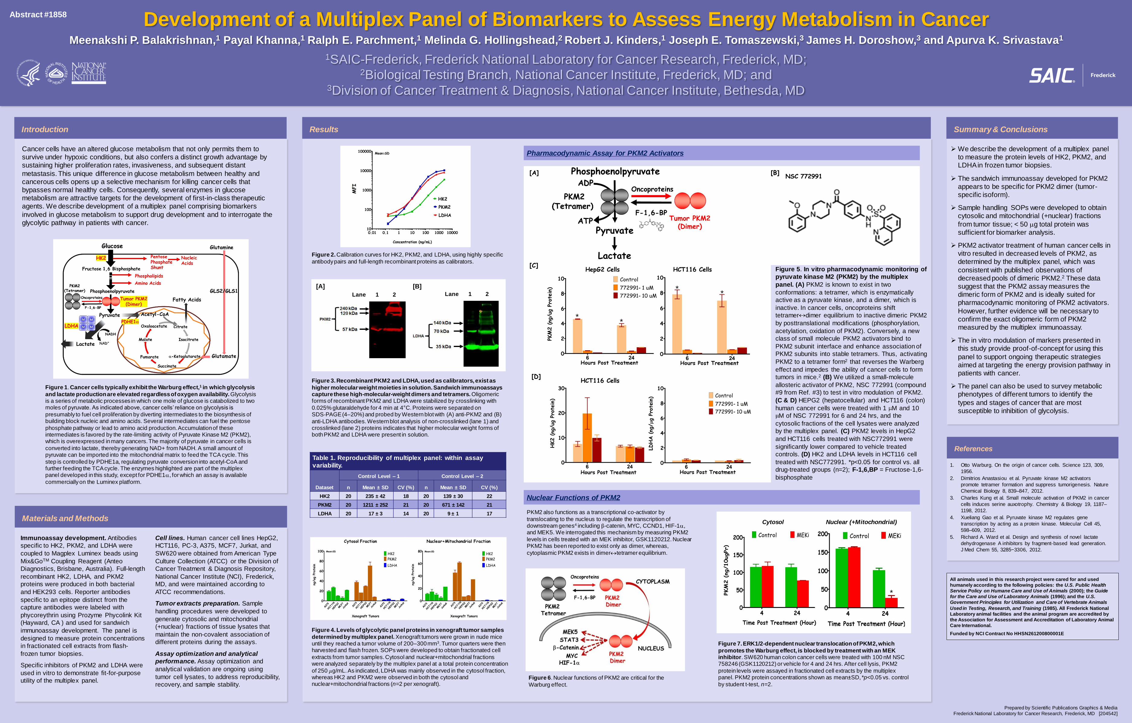
Immunoassay development. Antibodies specific to HK2, PKM2, and LDHA were coupled to Magplex Luminex beads using Mix&Go TM Coupling Reagent (Anteo Diagnostics, Brisbane, Australia). Full-length recombinant HK2, LDHA, and PKM2 proteins were produced in both bacterial and HEK293 cells. Reporter antibodies specific to an epitope distinct from the capture antibodies were labeled with phycoreythrin using Prozyme Phycolink Kit (Hayward, CA ) and used for sandwich immunoassay development. The panel is designed to measure protein concentrations in fractionated cell extracts from flash- frozen tumor biopsies. Specific inhibitors of PKM2 and LDHA were used in vitro to demonstrate fit-for-purpose utility of the multiplex panel. Cell lines. Human cancer cell lines HepG2, HCT116, PC-3, A375, MCF7, Jurkat, and SW620 were obtained from American Type Culture Collection (ATCC) or the Division of Cancer Treatment & Diagnosis Repository, National Cancer Institute (NCI), Frederick, MD, and were maintained according to ATCC recommendations. Tumor extracts preparation. Sample handling procedures were developed to generate cytosolic and mitochondrial (+nuclear) fractions of tissue lysates that maintain the non-covalent association of different proteins during the assays. Assay optimization and analytical performance. Assay optimization and analytical validation are ongoing using tumor cell lysates, to address reproducibility, recovery, and sample stability. Cancer cells have an altered glucose metabolism that not only permits them to survive under hypoxic conditions, but also confers a distinct growth advantage by sustaining higher proliferation rates, invasiveness, and subsequent distant metastasis. This unique difference in glucose metabolism between healthy and cancerous cells opens up a selective mechanism for killing cancer cells that bypasses normal healthy cells. Consequently, several enzymes in glucose metabolism are attractive targets for the development of first-in-class therapeutic agents. We describe development of a multiplex panel comprising biomarkers involved in glucose metabolism to support drug development and to interrogate the glycolytic pathway in patients with cancer. Figure 2. Calibration curves for HK2, PKM2, and LDHA, using highly specific antibody pairs and full-length recombinant proteins as calibrators. Development of a Multiplex Panel of Biomarkers to Assess Energy Metabolism in Cancer Meenakshi P. Balakrishnan, 1 Payal Khanna, 1 Ralph E. Parchment, 1 Melinda G. Hollingshead, 2 Robert J. Kinders, 1 Joseph E. Tomaszewski, 3 James H. Doroshow, 3 and Apurva K. Srivastava 1 1 SAIC-Frederick, Frederick National Laboratory for Cancer Research, Frederick, MD; 2 Biological Testing Branch, National Cancer Institute, Frederick, MD; and 3 Division of Cancer Treatment & Diagnosis, National Cancer Institute, Bethesda, MD We describe the development of a multiplex panel to measure the protein levels of HK2, PKM2, and LDHA in frozen tumor biopsies. The sandwich immunoassay developed for PKM2 appears to be specific for PKM2 dimer (tumor- specific isoform). Sample handling SOPs were developed to obtain cytosolic and mitochondrial (+nuclear) fractions from tumor tissue; < 50 g total protein was sufficient for biomarker analysis. PKM2 activator treatment of human cancer cells in vitro resulted in decreased levels of PKM2, as determined by the multiplex panel, which was consistent with published observations of decreased pools of dimeric PKM2. 2 These data suggest that the PKM2 assay measures the dimeric form of PKM2 and is ideally suited for pharmacodynamic monitoring of PKM2 activators. However, further evidence will be necessary to confirm the exact oligomeric form of PKM2 measured by the multiplex immunoassay. The in vitro modulation of markers presented in this study provide proof-of-concept for using this panel to support ongoing therapeutic strategies aimed at targeting the energy provision pathway in patients with cancer. The panel can also be used to survey metabolic phenotypes of different tumors to identify the types and stages of cancer that are most susceptible to inhibition of glycolysis. All animals used in this research project were cared for and used humanely according to the following policies: the U.S. Public Health Service Policy on Humane Care and Use of Animals (2000); the Guide for the Care and Use of Laboratory Animals (1996); and the U.S. Government Principles for Utilization and Care of Vertebrate Animals Used in Testing, Research, and Training (1985). All Frederick National Laboratory animal facilities and the animal program are accredited by the Association for Assessment and Accreditation of Laboratory Animal Care International. Funded by NCI Contract No HHSN261200800001E Introduction Results Summary & Conclusions Table 1. Reproducibility of multiplex panel: within assay variability. Dataset Control Level – 1 Control Level – 2 n Mean ± SD CV (%) n Mean ± SD CV (%) HK2 20 235 ± 42 18 20 139 ± 30 22 PKM2 20 1211 ± 252 21 20 671 ± 142 21 LDHA 20 17 ± 3 14 20 9 ± 1 17 Figure 7. ERK1/2-dependent nuclear translocation of PKM2, which promotes the Warburg effect, is blocked by treatment with an MEK inhibitor . SW620 human colon cancer cells were treated with 100 nM NSC 758246 (GSK1120212) or vehicle for 4 and 24 hrs. After cell lysis, PKM2 protein levels were assayed in fractionated cell extracts by the multiplex panel. PKM2 protein concentrations shown as mean±SD, *p<0.05 vs. control by student t-test, n=2. PKM2 also functions as a transcriptional co-activator by translocating to the nucleus to regulate the transcription of downstream genes 4 including -catenin, MYC, CCND1, HIF-1 , and MEK5. We interrogated this mechanism by measuring PKM2 levels in cells treated with an MEK inhibitor, GSK1120212. Nuclear PKM2 has been reported to exist only as dimer, whereas, cytoplasmic PKM2 exists in dimer tetramer equilibrium. Figure 3. Recombinant PKM2 and LDHA, used as calibrators, exist as higher molecular weight moieties in solution. Sandwich immunoassays capture these high-molecular-weight dimers and tetramers. Oligomeric forms of recombinant PKM2 and LDHA were stabilized by crosslinking with 0.025% glutaraldehyde for 4 min at 4°C. Proteins were separated on SDS‐PAGE (4–20%) and probed by Western blot with (A) anti‐PKM2 and (B) anti-LDHA antibodies. Western blot analysis of non-crosslinked (lane 1) and crosslinked (lane 2) proteins indicates that higher molecular weight forms of both PKM2 and LDHA were present in solution. Figure 1. Cancer cells typically exhibit the Warburg effect, 1 in which glycolysis and lactate production are elevated regardless of oxygen availability. Glycolysis is a series of metabolic processes in which one mole of glucose is catabolized to two moles of pyruvate. As indicated above, cancer cells’ reliance on glycolysis is presumably to fuel cell proliferation by diverting intermediates to the biosynthesis of building block nucleic and amino acids. Several intermediates can fuel the pentose phosphate pathway or lead to amino acid production. Accumulation of these intermediates is favored by the rate-limiting activity of Pyruvate Kinase M2 (PKM2), which is overexpressed in many cancers. The majority of pyruvate in cancer cells is converted into lactate, thereby generating NAD+ from NADH. A small amount of pyruvate can be imported into the mitochondrial matrix to feed the TCA cycle. This step is controlled by PDHE1a, regulating pyruvate conversion into acetyl-CoA and further feeding the TCA cycle. The enzymes highlighted are part of the multiplex panel developed in this study, except for PDHE1 , for which an assay is available commercially on the Luminex platform. Figure 5. In vitro pharmacodynamic monitoring of pyruvate kinase M2 (PKM2) by the multiplex panel. (A) PKM2 is known to exist in two conformations: a tetramer, which is enzymatically active as a pyruvate kinase, and a dimer, which is inactive. In cancer cells, oncoproteins shift tetramer dimer equilibrium to inactive dimeric PKM2 by posttranslational modifications (phosphorylation, acetylation, oxidation of PKM2). Conversely, a new class of small molecule PKM2 activators bind to PKM2 subunit interface and enhance association of PKM2 subunits into stable tetramers. Thus, activating PKM2 to a tetramer form 2 that reverses the Warberg effect and impedes the ability of cancer cells to form tumors in mice. 2 (B) We utilized a small-molecule allosteric activator of PKM2, NSC 772991 (compound #9 from Ref. #3) to test in vitro modulation of PKM2. (C & D) HEPG2 (hepatocellular) and HCT116 (colon) human cancer cells were treated with 1 M and 10 M of NSC 772991 for 6 and 24 hrs, and the cytosolic fractions of the cell lysates were analyzed by the multiplex panel. (C) PKM2 levels in HepG2 and HCT116 cells treated with NSC772991 were significantly lower compared to vehicle treated controls. (D) HK2 and LDHA levels in HCT116 cell treated with NSC772991. *p<0.05 for control vs. all drug-treated groups (n=2); F-1,6,BP = Fructose-1,6- bisphosphate References 1. Otto Warburg. On the origin of cancer cells. Science 123, 309, 1956. 2. Dimitrios Anastasiou et al. Pyruvate kinase M2 activators promote tetramer formation and suppress tumorigenesis. Nature Chemical Biology 8, 839–847, 2012. 3. Charles Kung et al. Small molecule activation of PKM2 in cancer cells induces serine auxotrophy. Chemistry & Biology 19, 1187– 1198, 2012. 4. Xueliang Gao et al. Pyruvate kinase M2 regulates gene transcription by acting as a protein kinase. Molecular Cell 45, 598–609, 2012. 5. Richard A. Ward et al. Design and synthesis of novel lactate dehydrogenase A inhibitors by fragment-based lead generation. J Med Chem 55, 3285−3306, 2012. Figure 4. Levels of glycolytic panel proteins in xenograft tumor samples determined by multiplex panel. Xenograft tumors were grown in nude mice until they reached a tumor volume of 200 –300 mm 3 . Tumor quarters were then harvested and flash frozen. SOPs were developed to obtain fractionated cell extracts from tumor samples. Cytosol and nuclear+mitochondrial fractions were analyzed separately by the multiplex panel at a total protein concentration of 250 g/mL. As indicated, LDHA was mainly observed in the cytosol fraction, whereas HK2 and PKM2 were observed in both the cytosol and nuclear+mitochondrial fractions (n=2 per xenograft). NSC 772991 [B] [D] HCT116 Cells HepG2 Cells HCT116 Cells [C] * * * * Figure 6. Nuclear functions of PKM2 are critical for the Warburg effect. [A] Lane 1 2 [A] [B] Lane 1 2 Abstract #1858 Nuclear+Mitochondrial Fraction Materials and Methods Prepared by Scientific Publications Graphics & Media Frederick National Laboratory for Cancer Research, Frederick, MD [204542] Cytosol Fraction Pharmacodynamic Assay for PKM2 Activators Nuclear Functions of PKM2 Cytosol Nuclear (+Mitochondrial) * * *
-
Upload
matt-sanderson -
Category
Health & Medicine
-
view
797 -
download
3
Transcript of Multiplex energy metabolism
Immunoassay development. Antibodies
specific to HK2, PKM2, and LDHA were
coupled to Magplex Luminex beads using
Mix&GoTM Coupling Reagent (Anteo
Diagnostics, Brisbane, Australia). Full-length
recombinant HK2, LDHA, and PKM2
proteins were produced in both bacterial
and HEK293 cells. Reporter antibodies
specific to an epitope distinct from the
capture antibodies were labeled with
phycoreythrin using Prozyme Phycolink Kit
(Hayward, CA ) and used for sandwich
immunoassay development. The panel is
designed to measure protein concentrations
in fractionated cell extracts from flash-
frozen tumor biopsies.
Specific inhibitors of PKM2 and LDHA were
used in vitro to demonstrate fit-for-purpose
utility of the multiplex panel.
Cell lines. Human cancer cell lines HepG2,
HCT116, PC-3, A375, MCF7, Jurkat, and
SW620 were obtained from American Type
Culture Collection (ATCC) or the Division of
Cancer Treatment & Diagnosis Repository,
National Cancer Institute (NCI), Frederick,
MD, and were maintained according to
ATCC recommendations.
Tumor extracts preparation. Sample
handling procedures were developed to
generate cytosolic and mitochondrial
(+nuclear) fractions of tissue lysates that
maintain the non-covalent association of
different proteins during the assays.
Assay optimization and analytical
performance. Assay optimization and
analytical validation are ongoing using
tumor cell lysates, to address reproducibility,
recovery, and sample stability.
Cancer cells have an altered glucose metabolism that not only permits them to
survive under hypoxic conditions, but also confers a distinct growth advantage by sustaining higher proliferation rates, invasiveness, and subsequent distant
metastasis. This unique difference in glucose metabolism between healthy and
cancerous cells opens up a selective mechanism for killing cancer cells that bypasses normal healthy cells. Consequently, several enzymes in glucose
metabolism are attractive targets for the development of first-in-class therapeutic
agents. We describe development of a multiplex panel comprising biomarkers
involved in glucose metabolism to support drug development and to interrogate the
glycolytic pathway in patients with cancer.
Figure 2. Calibration curves for HK2, PKM2, and LDHA, using highly specific
antibody pairs and full-length recombinant proteins as calibrators.
Development of a Multiplex Panel of Biomarkers to Assess Energy Metabolism in Cancer Meenakshi P. Balakrishnan,1 Payal Khanna,1 Ralph E. Parchment,1 Melinda G. Hollingshead,2 Robert J. Kinders,1 Joseph E. Tomaszewski,3 James H. Doroshow,3 and Apurva K. Srivastava1
1SAIC-Frederick, Frederick National Laboratory for Cancer Research, Frederick, MD; 2Biological Testing Branch, National Cancer Institute, Frederick, MD; and
3Division of Cancer Treatment & Diagnosis, National Cancer Institute, Bethesda, MD
We describe the development of a multiplex panel
to measure the protein levels of HK2, PKM2, and LDHA in frozen tumor biopsies.
The sandwich immunoassay developed for PKM2
appears to be specific for PKM2 dimer (tumor-specific isoform).
Sample handling SOPs were developed to obtain cytosolic and mitochondrial (+nuclear) fractions
from tumor tissue; < 50 g total protein was
sufficient for biomarker analysis.
PKM2 activator treatment of human cancer cells in
vitro resulted in decreased levels of PKM2, as
determined by the multiplex panel, which was
consistent with published observations of
decreased pools of dimeric PKM2.2 These data
suggest that the PKM2 assay measures the
dimeric form of PKM2 and is ideally suited for
pharmacodynamic monitoring of PKM2 activators.
However, further evidence will be necessary to
confirm the exact oligomeric form of PKM2
measured by the multiplex immunoassay.
The in vitro modulation of markers presented in
this study provide proof-of-concept for using this
panel to support ongoing therapeutic strategies aimed at targeting the energy provision pathway in
patients with cancer.
The panel can also be used to survey metabolic
phenotypes of different tumors to identify the
types and stages of cancer that are most susceptible to inhibition of glycolysis.
All animals used in this research project were cared for and used
humanely according to the following policies: the U.S. Public Health
Service Policy on Humane Care and Use of Animals (2000); the Guide
for the Care and Use of Laboratory Animals (1996); and the U.S.
Government Principles for Utilization and Care of Vertebrate Animals
Used in Testing, Research, and Training (1985). All Frederick National
Laboratory animal facilities and the animal program are accredited by
the Association for Assessment and Accreditation of Laboratory Animal
Care International.
Funded by NCI Contract No HHSN261200800001E
Introduction Results Summary & Conclusions
Table 1. Reproducibility of multiplex panel: within assay
variability.
Dataset
Control Level – 1 Control Level – 2
n Mean ± SD CV (%) n Mean ± SD CV (%)
HK2 20 235 ± 42 18 20 139 ± 30 22
PKM2 20 1211 ± 252 21 20 671 ± 142 21
LDHA 20 17 ± 3 14 20 9 ± 1 17
Figure 7. ERK1/2-dependent nuclear translocation of PKM2, which
promotes the Warburg effect, is blocked by treatment with an MEK inhibitor. SW620 human colon cancer cells were treated with 100 nM NSC 758246 (GSK1120212) or vehicle for 4 and 24 hrs. After cell lysis, PKM2
protein levels were assayed in fractionated cell extracts by the multiplex panel. PKM2 protein concentrations shown as mean±SD, *p<0.05 vs. control
by student t-test, n=2.
PKM2 also functions as a transcriptional co-activator by
translocating to the nucleus to regulate the transcription of downstream genes4 including -catenin, MYC, CCND1, HIF-1 , and MEK5. We interrogated this mechanism by measuring PKM2
levels in cells treated with an MEK inhibitor, GSK1120212. Nuclear PKM2 has been reported to exist only as dimer, whereas,
cytoplasmic PKM2 exists in dimer tetramer equilibrium.
Figure 3. Recombinant PKM2 and LDHA, used as calibrators, exist as
higher molecular weight moieties in solution. Sandwich immunoassays capture these high-molecular-weight dimers and tetramers. Oligomeric forms of recombinant PKM2 and LDHA were stabilized by crosslinking with
0.025% glutaraldehyde for 4 min at 4°C. Proteins were separated on SDS‐PAGE (4–20%) and probed by Western blot with (A) anti‐PKM2 and (B)
anti-LDHA antibodies. Western blot analysis of non-crosslinked (lane 1) and crosslinked (lane 2) proteins indicates that higher molecular weight forms of both PKM2 and LDHA were present in solution.
Figure 1. Cancer cells typically exhibit the Warburg effect,1 in which glycolysis
and lactate production are elevated regardless of oxygen availability. Glycolysis is a series of metabolic processes in which one mole of glucose is catabolized to two moles of pyruvate. As indicated above, cancer cells’ reliance on glycolysis is
presumably to fuel cell proliferation by diverting intermediates to the biosynthesis of building block nucleic and amino acids. Several intermediates can fuel the pentose
phosphate pathway or lead to amino acid production. Accumulation of these intermediates is favored by the rate-limiting activity of Pyruvate Kinase M2 (PKM2), which is overexpressed in many cancers. The majority of pyruvate in cancer cells is
converted into lactate, thereby generating NAD+ from NADH. A small amount of pyruvate can be imported into the mitochondrial matrix to feed the TCA cycle. This
step is controlled by PDHE1a, regulating pyruvate conversion into acetyl-CoA and further feeding the TCA cycle. The enzymes highlighted are part of the multiplex panel developed in this study, except for PDHE1 , for which an assay is available
commercially on the Luminex platform.
Figure 5. In vitro pharmacodynamic monitoring of
pyruvate kinase M2 (PKM2) by the multiplex
panel. (A) PKM2 is known to exist in two
conformations: a tetramer, which is enzymatically
active as a pyruvate kinase, and a dimer, which is
inactive. In cancer cells, oncoproteins shift
tetramer dimer equilibrium to inactive dimeric PKM2
by posttranslational modifications (phosphorylation,
acetylation, oxidation of PKM2). Conversely, a new
class of small molecule PKM2 activators bind to
PKM2 subunit interface and enhance association of
PKM2 subunits into stable tetramers. Thus, activating
PKM2 to a tetramer form2 that reverses the Warberg
effect and impedes the ability of cancer cells to form
tumors in mice.2 (B) We utilized a small-molecule
allosteric activator of PKM2, NSC 772991 (compound
#9 from Ref. #3) to test in vitro modulation of PKM2.
(C & D) HEPG2 (hepatocellular) and HCT116 (colon)
human cancer cells were treated with 1 M and 10
M of NSC 772991 for 6 and 24 hrs, and the
cytosolic fractions of the cell lysates were analyzed
by the multiplex panel. (C) PKM2 levels in HepG2
and HCT116 cells treated with NSC772991 were
significantly lower compared to vehicle treated
controls. (D) HK2 and LDHA levels in HCT116 cell
treated with NSC772991. *p<0.05 for control vs. all
drug-treated groups (n=2); F-1,6,BP = Fructose-1,6-
bisphosphate
References
1. Otto Warburg. On the origin of cancer cells. Science 123, 309,
1956.
2. Dimitrios Anastasiou et al. Pyruvate kinase M2 activators
promote tetramer formation and suppress tumorigenesis. Nature
Chemical Biology 8, 839–847, 2012.
3. Charles Kung et al. Small molecule activation of PKM2 in cancer
cells induces serine auxotrophy. Chemistry & Biology 19, 1187–
1198, 2012.
4. Xueliang Gao et al. Pyruvate kinase M2 regulates gene
transcription by acting as a protein kinase. Molecular Cell 45,
598–609, 2012.
5. Richard A. Ward et al. Design and synthesis of novel lactate
dehydrogenase A inhibitors by fragment-based lead generation.
J Med Chem 55, 3285−3306, 2012.
Figure 4. Levels of glycolytic panel proteins in xenograft tumor samples
determined by multiplex panel. Xenograft tumors were grown in nude mice until they reached a tumor volume of 200–300 mm3. Tumor quarters were then harvested and flash frozen. SOPs were developed to obtain fractionated cell
extracts from tumor samples. Cytosol and nuclear+mitochondrial fractions were analyzed separately by the multiplex panel at a total protein concentration
of 250 g/mL. As indicated, LDHA was mainly observed in the cytosol fraction, whereas HK2 and PKM2 were observed in both the cytosol and nuclear+mitochondrial fractions (n=2 per xenograft).
NSC 772991 [B]
[D] HCT116 Cells
HepG2 Cells HCT116 Cells [C]
* *
* *
Figure 6. Nuclear functions of PKM2 are critical for the
Warburg effect.
[A]
Lane 1 2
[A] [B]
Lane 1 2
Abstract #1858
Nuclear+Mitochondrial Fraction
Materials and Methods
Prepared by Scientific Publications Graphics & Media
Frederick National Laboratory for Cancer Research, Frederick, MD [204542]
Cytosol Fraction
Pharmacodynamic Assay for PKM2 Activators
Nuclear Functions of PKM2
Cytosol Nuclear (+Mitochondrial)
*
* *