Multimodal MRI quantification of the common neurostructural … · Accepted Manuscript Multimodal...
35
Accepted Manuscript Multimodal MRI quantification of the common neurostructural bases within the FTD- ALS continuum Chiara Crespi, Alessandra Dodich, Stefano F. Cappa, Nicola Canessa, Sandro Iannaccone, Massimo Corbo, Christian Lunetta, Andrea Falini, Chiara Cerami PII: S0197-4580(17)30315-9 DOI: 10.1016/j.neurobiolaging.2017.09.019 Reference: NBA 10039 To appear in: Neurobiology of Aging Received Date: 17 May 2017 Revised Date: 15 September 2017 Accepted Date: 19 September 2017 Please cite this article as: Crespi, C., Dodich, A., Cappa, S.F., Canessa, N., Iannaccone, S., Corbo, M., Lunetta, C., Falini, A., Cerami, C., Multimodal MRI quantification of the common neurostructural bases within the FTD-ALS continuum, Neurobiology of Aging (2017), doi: 10.1016/ j.neurobiolaging.2017.09.019. This is a PDF file of an unedited manuscript that has been accepted for publication. As a service to our customers we are providing this early version of the manuscript. The manuscript will undergo copyediting, typesetting, and review of the resulting proof before it is published in its final form. Please note that during the production process errors may be discovered which could affect the content, and all legal disclaimers that apply to the journal pertain.
Transcript of Multimodal MRI quantification of the common neurostructural … · Accepted Manuscript Multimodal...
Accepted Manuscript
Multimodal MRI quantification of the common neurostructural bases within the FTD-ALS continuum
Chiara Crespi, Alessandra Dodich, Stefano F. Cappa, Nicola Canessa, SandroIannaccone, Massimo Corbo, Christian Lunetta, Andrea Falini, Chiara Cerami
PII: S0197-4580(17)30315-9
DOI: 10.1016/j.neurobiolaging.2017.09.019
Reference: NBA 10039
To appear in: Neurobiology of Aging
Received Date: 17 May 2017
Revised Date: 15 September 2017
Accepted Date: 19 September 2017
Please cite this article as: Crespi, C., Dodich, A., Cappa, S.F., Canessa, N., Iannaccone, S.,Corbo, M., Lunetta, C., Falini, A., Cerami, C., Multimodal MRI quantification of the commonneurostructural bases within the FTD-ALS continuum, Neurobiology of Aging (2017), doi: 10.1016/j.neurobiolaging.2017.09.019.
This is a PDF file of an unedited manuscript that has been accepted for publication. As a service toour customers we are providing this early version of the manuscript. The manuscript will undergocopyediting, typesetting, and review of the resulting proof before it is published in its final form. Pleasenote that during the production process errors may be discovered which could affect the content, and alllegal disclaimers that apply to the journal pertain.
MANUSCRIP
T
ACCEPTED
ACCEPTED MANUSCRIPT
1
Multimodal MRI quantification of the common neurostructural bases within the FTD-
ALS continuum
Chiara Crespi 1*, Alessandra Dodich2,3, Stefano F. Cappa4,5, Nicola Canessa2,4, Sandro
Iannaccone2,3, Massimo Corbo6, Christian Lunetta7, Andrea Falini2,8,9, and Chiara Cerami2,3,9.
Affiliations
[1] Institute of Biomedical Technologies, National Research Council, Segrate, Italy
[2] Division of Neuroscience, San Raffaele Scientific Institute, Milano, Italy
[3] Department of Clinical Neurosciences, IRCCS San Raffaele Turro, Milano, Italy
[4] NeTS Center, Scuola Universitaria Superiore IUSS Pavia, Pavia, Italy
[5] IRCCS Fatebenefratelli di Brescia, Brescia, Italy
[6] Department of Neurorehabilitation Sciences, Casa Cura Policlinico, Milano, Italy
[7] NEuroMuscolar Omnicentre, Fondazione Serena Onlus, Niguarda Ca’ Granda Hospital,
Milano, Italy
[8] Neuroradiology Unit, San Raffaele Scientific Institute, Milano, Italy
[9] Vita-Salute San Raffaele University, Milano, Italy
*Corresponding author: Chiara Crespi
Institute of Biomedical Technologies, National Research Council (CNR)
E-mail: [email protected]
Street address: Via Fratelli Cervi 93, 20090, Segrate (MI), Italy
Phone: +39.02.26422629

MANUSCRIP
T
ACCEPTED
ACCEPTED MANUSCRIPT
2
Abstract
The continuum hypothesis linking the behavioral variant of Frontotemporal Dementia
(bvFTD) and Amyotrophic Lateral Sclerosis (ALS) is supported by clinical, pathological,
genetic and neuroimaging evidence. In the present multimodal magnetic resonance study, we
characterized the site and extent of shared neurostructural changes in gray and white matter in
20 bvFTD and 19 ALS patients without dementia.
We found an overlap of macrostructural and microstructural damage in both patient
groups compared to healthy controls, involving the right orbital and the bilateral anterior
cingulate cortices, the corticospinal tract and corpus callosum. The quantification of grey and
white matter damage within the areas of shared alterations highlighted a higher degree of
atrophy in orbitofrontal and frontomedial regions in patients with more severe executive
and/or behavioral symptoms, and a higher degree of degeneration in the motor pathway in
patients with more severe motor neuron disorders.
Our finding provides additional evidence confirming the FTD-ALS continuum
hypothesis, and supports the notion of a bimodal but convergent pattern of neurostructural
changes characterizing bvFTD and ALS.
Keywords
1. Behavioral variant of Frontotemporal Dementia
2. Amyotrophic Lateral Sclerosis
3. ALS-FTD Continuum Hypothesis
4. Tract-Based Spatial Statistics
5. Voxel-Based Morphometry
6. Corticospinal tract

MANUSCRIP
T
ACCEPTED
ACCEPTED MANUSCRIPT
3
1. Introduction
The Frontotemporal Dementia (FTD) – Amyotrophic Lateral Sclerosis (ALS) continuum
hypothesis, considering FTD and ALS as extreme points of a disease spectrum, is now
supported by a robust genetic and neuropathological evidence (Ferrari et al., 2011; Ling et al.,
2013). Above all, the TAR DNA binding protein (TPD-43) inclusions within the central
nervous system first, and then mutations in the hexanucleotide (GGGGCC) repeat expansion
of the chromosome 9 open reading frame 72 (C9orf72) gene have been consistently identified
in both diseases and, thus, proposed as the common neuropathological and genetic hallmarks,
respectively (Lattante et al., 2015; Ling et al., 2013; Weishaupt et al., 2016).
While the clinical presentation of these neurodegenerative diseases is extremely
different, a partial symptom overlap is not uncommon (Devenney et al., 2015; Lillo and
Hodges, 2009). Indeed, despite the different onset sites of neurodegenerative changes in ALS
and bvFTD – i.e., premotor and primary motor cortices in ALS (Turner and Verstraete, 2015),
fronto-limbic and temporal regions in bvFTD (Seeley et al., 2008) – both an early extra-motor
involvement in ALS (Goldstein and Abrahams, 2013), and the presence of damage of
corticospinal pathway in bvFTD (Lillo et al., 2012), have been consistently documented.
These findings can explain the overlap of signs and symptoms observed in clinical practice
(e.g., Cerami et al., 2015b; Consonni et al., 2013), as well as the co-occurrence of the two
diseases (i.e., ALS-FTD). It has been estimated that the 10-15% of patients diagnosed as FTD
– and in particular those individuals receiving a diagnosis of behavioral variant of FTD
(bvFTD) – can develop a motor neuron impairment, and are then diagnosed with ALS
(Burrell et al., 2011; Lomen-Hoerth, 2011). Conversely, about 13-15% of ALS patients may
display behavioral and/or cognitive changes typically observed in FTD, and particularly in
bvFTD, satisfying the clinical criteria for FTD diagnosis (Consonni et al., 2013; Lomen-
Hoerth, 2011; Phukan et al., 2012; Ringholz et al., 2005).
The continuum hypothesis is further supported by cases in which the presence of
subtle motor dysfunctions in FTD, as well as non-motor alterations in ALS, do not reach the
clinical significance required to make the diagnosis of ALS-FTD. For example,
MANUSCRIP
T
ACCEPTED
ACCEPTED MANUSCRIPT
4
neurophysiological investigations highlight about 25-30% of FTD patients present mild
clinical and EMG signs not fulfilling the clinical criteria for the diagnosis of ALS (Burrell et
al., 2011; Cerami et al., 2015a; Lomen-Hoerth et al., 2002). Conversely, it is recognized that
some ALS patients may show behavioral alterations or cognitive impairments (i.e., ALSbi
and ALSci) not fulfilling the criteria for dementia (Strong et al., 2009). Considerable
advances in the understanding of the neuropsychological profile of ALS patients triggered the
need for consensus criteria revision of the diagnosis of frontotemporal dysfunction in ALS
(Strong et al., 2017), then resulting in the concept of the frontotemporal spectrum disorder of
ALS (ALS-FTSD).
Finally, neuroimaging investigations provide further crucial evidence of the
involvement of frontal and temporo-limbic regions in ALS (Abrahams et al., 2005; Lillo et
al., 2012; Tsermentseli et al., 2012), suggesting a neurostructural overlap with bvFTD. This
pattern involves anterior cingulate, supramarginal gyrus, premotor and motor cortices,
alongside the alteration of the anterior portion of corpus callosum, the corticospinal tract and
the inferior longitudinal fasciculus (Lillo et al., 2012).
A precise quantification of the conjunction pattern between bvFTD and ALS, as well
as a detailed characterization of white-matter microstructural properties of commonly
degenerated pathways, have not been provided yet. The aim of the present study is to measure
both commonalities and differences across bvFTD and ALS compared to healthy controls
(HC). This was done by means of a conjunction analysis, considering grey matter (GM)
density values and white matter (WM) microstructural indices, i.e. fractional anisotropy (FA),
mode of anisotropy (MO), and mean diffusivity (MD).
2. Materials and methods
2.1 Participants
Twenty patients meeting the criteria for probable bvFTD (Rascovsky et al., 2011)
were enrolled in a multimodal Magnetic Resonance Imaging (MRI) study (see Table 1 for
demographics and clinical information). In-depth neuropsychological, behavioral and
MANUSCRIP
T
ACCEPTED
ACCEPTED MANUSCRIPT
5
instrumental assessment (i.e., needle electromyography (EMG), conventional MRI and
[18F]FDG-PET imaging) supported the clinical classification.
The neuropsychological exam included the evaluation of language abilities (picture
naming and single word comprehension), short-term (digit span forward) and long-term
memory (Rey auditory verbal learning; Rey-Osterrieth complex figure recall), visuo-spatial
abilities (Rey-Osterrieth complex figure copy), social cognition (Ekman 60-faces test, story-
based empathy task) and executive functions (Raven colored progressive matrices; digit span
backward; letter (P-F-L) and category (animals-fruits-cars) fluency; cognitive estimation task;
Stroop interference test and either Wisconsin card sorting test or Weigl’s sorting test). The
presence of behavioral alterations was assessed with the Frontal Behavioral Inventory and the
Neuropsychiatric Inventory questionnaires.
A detailed neurological examination recorded possible signs of upper (UMN) and
lower (LMN) motor neuron dysfunctions (MNDs). Needle EMG evaluated
neurophysiological evidence of acute and chronic neurogenic changes. Patients were thus
classified as pure bvFTD or bvFTD-MNDs according to the absence/presence of MNDs,
either on clinical or electromyographic evidence, as described in Cerami et al. (2015a). All
bvFTD patients were tested for the presence of C9ORF72 or GRN gene mutations. None of
them showed pathological mutations. Exclusion criteria for bvFTD were: left-handedness, a
positive medical history for other neuropsychiatric disorders, the presence of other
pathological findings on MRI scans, a Mini-Mental State Examination (MMSE) raw
score<21/30 and a Clinical Dementia Rating (CDR) scale global score>1.
Nineteen individuals without dementia (CDR range=0-0.5) with either probable or
definite ALS diagnosis (Brooks et al., 2000) were also enrolled (Table 1). Three out of 19
patients presented bulbar onset (i.e., dysarthria and/or dysphagia), while the rest had a spinal
onset. Clinical disability was assessed with the revised version of the ALS-Functional Rating
Scale (ALSFRS-r) (Cedarbaum et al., 1999). All ALS patients completed a
neuropsychological and behavioral assessment to evaluate the presence of cognitive and/or
behavioral impairments. The testing battery was the same used for bvFTD patients (see
MANUSCRIP
T
ACCEPTED
ACCEPTED MANUSCRIPT
6
above) except for the Rey-Osterrieth complex figure, which was not administered to ALS
patients. In order to control for individual differences in motor speed, we considered both the
mean fluency indexes and the reading time-difference condition of the Stroop test. Patients
with arm weakness were assisted by the examiner to move the cards during the task. Patients
were thus classified as pure ALS or ALS with frontotemporal spectrum disorders (i.e., ALS-
FTSD) according to Strong’s criteria (Strong et al., 2017).
ALS patients were tested for the presence of C9ORF72 or GRN gene mutations also,
showing no pathological mutation. Exclusion criteria for ALS were: left-handedness, a
positive medical history for other neuropsychiatric disorders as well as the presence of other
pathological findings on MRI scans, mild respiratory disorders (forced vital capacity <70% of
predicted capacity), severe dysarthria, and communication difficulties potentially invalidating
the interpretation of neuropsychological performance.
Twenty healthy controls (HC) matched with patients with respect to age and gender,
also participated in the study (Table 1). They were recruited from local senior community
centers on a voluntary basis. None of them was taking any medication potentially interfering
with neurobehavioral functioning. A next of kin (e.g., spouse) of each control subject was
interviewed to corroborate his/her normal daily functioning. Exclusion criteria for HC were
the presence of neuropsychiatric disorders, a positive neurologic examination, a global
Clinical Dementia Rating score>0, a Mini-Mental State Examination score ≤28/30, a verbal
and visuo-spatial delayed memory performance (Rey Auditory Verbal Learning test and Rey
Figure Recall task) ≤25th percentile.
All participants, or relative informants, gave their written informed consent to the
experimental procedure, which was approved by the Ethics Committee of the San Raffaele
Hospital.

MANUSCRIP
T
ACCEPTED
ACCEPTED MANUSCRIPT
7
Table 1. Demographic features and disease parameters of the enrolled sample
bvFTD (n=20) ALS (n=19) HC (n=20) Stats
Age [years; mean ± sd] 66.14 ± 6.81 60.97 ± 11 60.83 ± 8.03 F(2,56)=2.61,
p=0.08
Gender [m : f] 12 : 8 14 : 5 13 : 7 χ2=0.83,
p=0.66
Education [years; mean ± sd] 9.69 ± 4.53 10.3 ± 4.63 12.95 ± 4.17 F(2,56)=3.26,
p=0.046
Disease duration [months;
median (interquartile range)]
57 (24-84) 18 (7.5-
29.75)
- t(37)=2.3,
p=0.03
ALS-FSR-r global score
[mean ± sd]
46.5 ± 2.5 38.17 ±
7.54
- t(37)=4.68,
p<0.0001
CDR global score [range] 0-1 0-0.5 - t(37)=7.3,
p<0.0001
2.2 MRI data acquisition
All participants underwent a multimodal MRI scanning session, including T1-
weighted and Diffusion Tensor Imaging (DTI) sequences. Fluid-Attenuated-Inversion
Recovery (FLAIR) and T2-weighted images were also collected for diagnostic purposes. All
MRI scans were performed using a 3T Philips Achieva scanner (Philips Medical Systems,
Best, NL) with an 8-channels head coil. T1-weighted images were collected with a gradient-
echo sequence (220 slices, TR = 600 ms, TE = 20 ms, in-plane resolution 0.9 x 0.9 x 0.8
mm3). Whole-brain DTI images were acquired using a single-shot echo planar sequence
(TR/TE=8986/80 msec; FOV=240 mm2; 56 sections; 2.5 mm isotropic resolution) with
parallel imaging (SENSE factor=2.5) and diffusion gradients applied along 32 non-collinear
directions (b-value=1000 sec/mm2). One non-diffusion weighted volume was also collected.
2.3 VBM data preprocessing and analysis
T1-weighted structural brain images were pre-processed and quantitatively analyzed
in SPM8 (http://www.fil.ion.ucl.ac.uk/spm/software/spm8/) on Matlab v7.4 (Mathworks-Inc.,

MANUSCRIP
T
ACCEPTED
ACCEPTED MANUSCRIPT
8
Sherborn, MA), using the VBM8 (http://dbm.neuro.uni-jena.de/vbm/download/) and
Diffeomorphic Anatomical Registration Through Exponentiated Lie algebra (DARTEL)
(Ashburner, 2009, 2007) toolboxes. After a bias-correction for field-intensity
inhomogeneities, the T1-weighted images were registered using linear (12-parameter affine)
and non-linear (warping) transformations, and tissue-classified in GM, WM and cerebrospinal
fluid (CSF) components. The segmented tissue maps were then registered to the stereotactic
space of the Montreal Neurological Institute (MNI) using the iterative high-dimensional
normalization approach provided by DARTEL toolbox. Finally, the non-linear DARTEL
normalized gray matter images were smoothed with a 8-mm Full-Width-Half-Maximum
(FWHM) Gaussian-kernel and entered in the subsequent voxel-based statistical analyses.
Whole-brain analyses based on two-sample t-tests highlighted GM density
differences between healthy controls and ALS and bvFTD patients separately. Then, to
explore the overlap between the atrophic processes in ALS and bvFTD, we computed a
conjunction analysis between the statistical maps representing the common reduction of GM
in the two patient groups compared to healthy controls (Nichols et al., 2005). Total
intracranial volume was used as nuisance variables in order to correct for variation in
individual brain sizes. We excluded all voxels with a GM-value<0.15 (maximum=1) to avoid
edge effects at the border between GM and WM. The statistical thresholds were set at p<0.05
Family-Wise-Error (FWE) corrected for multiple comparisons at the cluster-level. Those
cerebral regions for which maps are provided were localized with reference to
cytoarchitectonical probabilistic maps of the human brain, using the SPM-Anatomy toolbox
v1.8 (Eickhoff et al., 2005).
2.4 DTI data preprocessing and analysis
We performed DTI data pre-processing and analysis with the FMRIB Software
Library tools (FSL: http://fsl.fmrib.ox.ac.uk/fsl/fslwiki/). Single-subject datasets were first
corrected for eddy current distortions and motion artifacts, skull-stripped and finally, as a
result of the fitting of the diffusion tensor model at each voxel, maps of diffusion scalar
MANUSCRIP
T
ACCEPTED
ACCEPTED MANUSCRIPT
9
indices were generated. We then carried out DTI group analyses with Tract-Based Spatial
Statistics (TBSS) (Smith et al., 2006). Briefly, the TBSS method includes a voxelwise non-
linear registration of all subjects’ Fractional Anisotropy (FA) maps that, once aligned, are
affine-transformed on a standard space (1x1x1 mm3 MNI152). After co-registration, FA maps
are averaged to create a mean FA image, and then used to generate a mean FA tract skeleton,
representing all common tracts across subjects. In order to exclude from further analysis those
parts of the skeleton that could not ensure a good correspondence across subjects, we applied
a threshold of 0.20 to the mean FA skeleton image. Finally, to account for residual
misalignments after the initial nonlinear registration, all subjects’ FA data were projected
onto the thresholded mean FA skeleton, creating a 4D dataset of all subjects’ FA skeletonized
data, which was fed into whole-brain voxel-wise statistical analysis. In addition, we ran the
non-FA TBSS script on maps of mean diffusivity (MD) and mode of anisotropy (MO).
Whole-brain voxelwise statistics were performed with randomise, software
implemented in FSL performing a permutation-based nonparametric approach within the
framework of the GLM. We employed the Threshold-Free Cluster Enhancement option
(Smith and Nichols, 2009) and set the significance threshold for group differences at p<0.05
corrected for multiple comparisons. We tested the presence of WM microstructural changes
in the two patient groups compared to controls separately – i.e., ALS versus HC and bvFTD
versus HC – setting a number of 10,000 random permutations per contrast. We then used the
easythresh_conj script to calculate the conjunction pattern between white-matter alterations
highlighted in ALS and bvFTD patients. The anatomical localization of significant clusters
was performed using the JHU White-Matter Tractography Atlas (Hua et al., 2008). Finally,
for graphical purposes, result maps were smoothed applying a Gaussian Kernel of 3 mm via
the tbss_fill script.
2.5 Statistical comparisons and quantification of neurostructural damage

MANUSCRIP
T
ACCEPTED
ACCEPTED MANUSCRIPT
10
In order to explore voxelwise results from conjunction analyses, we used an analysis
of variance (ANOVA) to compare mean GM density and WM microstructural indices
extracted from conjunction patterns between the two patient groups and HC.
Additionally, we used Chi-square tests to separately assess the association between
GM atrophy and WM microstructural changes within the bvFTD-ALS conjunction pattern
and, respectively, the presence of cognitive/behavioral symptoms in ALS patients (i.e, ALS-
FTSD) and of motor symptoms in bvFTD patients (i.e., bvFTD-MNDs). Preliminarily, we
classified all patients as impaired/non impaired based on the distribution of GM density and
diffusion indices (i.e., FA and MO) in HC (i.e., patients whose values fell below the 5th
percentile of HC were considered as impaired).
Finally, in order to evaluate the percent variation of GM density and FA/MO mean
indices we performed exploratory comparisons among bvFTD (i.e., pure bvFTD and bvFTD-
MNDs) and ALS (i.e., pure ALS and ALS-FTSD) subgroups with HC.
Statistical analyses were carried out with Statistica software
(http://www.statsoft.com).
3. RESULTS
3.1 BvFTD-ALS clinical continuum
Six out of 20 bvFTD patients showed clinical and/or EMG evidence of MNDs (i.e.,
bvFTD-MNDs), not satisfying criteria for ALS diagnosis (Brooks et al., 2000). BvFTD-
MNDs group of patients did not differ in terms of disease duration from pure bvFTD patients
(Mann-Whitney U=39, p=0.803). Similarly, 5 out of 19 ALS patients showed cognitive
and/or behavioral disorders (i.e., ALS-FTSD), which however did not satisfy criteria for
bvFTD diagnosis (Rascovsky et al., 2011). In detail, three out of these five ALS patients
presented with dysexecutive deficits (i.e., ALSci), the other two with apathy, irritability and
disinhibition (i.e., ALSbi). Pure ALS and ALS-FTSD did not differ in disease duration
(Mann-Whitney U=27, p=0.458) (Table 2).
MANUSCRIP
T
ACCEPTED
ACCEPTED MANUSCRIPT
11
Table 2. Neuropsychological performances of patients. Scores are corrected according to
Italian normative data. Mean and standard deviation (in brackets) for every variable
are reported in each group.
pure ALS
(14 patients)
ALS-FTDS
(5 patients)
pure bvFTD
(14 patients)
bvFTD-MND
(6 patients)
Mini Mental Status Examination 27.7 ± 1.2 28.4 ± 1.5 24.5 ± 4.1 23.9 ± 3.4
Verbal Fluency on phonemic cue 26.4 ± 6 28.4 ± 12.5 17.7 ± 9.8 15.8 ± 6
Verbal Fluency on semantic cue 44.2 ± 8.8 43 ± 15.6 29.1 ± 10.7 23.5 ± 10.7
Rey Auditory Verbal Learning test – immediate recall 42.2 ± 7.5 39.5 ± 13.6 31.8 ± 9.6 24.6 ± 14.8
Rey Auditory Verbal Learning test – delayed recall 8.6 ± 2.8 8.5 ± 2.4 5.9 ± 3.8 5.7 ± 4
Rey-Osterrieth Complex Figure – recall - - 16.5 ± 8.5 10.2 ± 1.7
Raven Progressive Colored Matrices 32.9 ± 3 29.2 ± 1.7 24.3 ± 6.9 20.9 ± 5.4
Digit Span Forward 5.8 ± 0.4 4.9 ± 1 4.5 ± 1 4.3 ± 1
Wisconsin Card Sorting Test 54.7 ± 28.7 67.7 ± 18 106.1 ± 16.7 104.9 ± 2.1
Cognitive Estimation Task 11.6 ± 2.7 14.4 ± 4.3 14.9 ± 5.3 15.7 ± 8
Stroop test 26.7 ± 8.6 55.1 ± 25.9 48.8 ± 6.9 45 ± 9.3
Rey-Osterrieth Complex Figure – copy - - 26.4 ± 4.8 32 ± 4.6
Naming 46.7 ± 1.6 44 ± 1.6 44.2 ± 3 44.7 ± 4.6
Ekman 60-faces task 48 ± 9.4 41.7 ± 7.5 31 ± 8.3 29.75 ± 5.3
Story-based empathy test 15.2 ± 2.3 14.25 ± 3.6 11.3 ± 5.3 10 ± 1.6
3.2 Neurostructural profiles of bvFTD and ALS groups
3.2.1 BvFTD patients versus healthy controls
As expected, VBM whole-brain analyses on GM density revealed a specific pattern
of atrophy in bvFTD (whole group, n=20) compared with HC, involving bilateral fronto-
temporal and limbic structures. In detail, the pattern of GM density reduction in bvFTD
involved the anterior cingulate cortex (ACC), the orbitofrontal cortex and the superior medial
frontal gyrus. A significant GM loss was also present in the anterior temporal lobe, including
the temporal pole and subcortical regions, such as the amygdala and the hippocampus (Figure
1, panel A).

MANUSCRIP
T
ACCEPTED
ACCEPTED MANUSCRIPT
12
We found a widespread FA reduction, mainly involving the limbic-prefrontal
circuitry, and including the uncinate (UF), inferior fronto-occipital (IFOF), inferior
longitudinal (ILF) fasciculi, and the anterior portion of the cingulum bundle (Figure 1, panel
B). This pattern also encompassed a consistent FA alteration along the forceps minor, the
corpus callosum (CC), the superior longitudinal fasciculus (SLF), the corticospinal tract
(CST) and the fornix (cluster size: 30558 voxels, cluster maximum: x=6, y=23, z=14). A
corresponding pattern of alteration emerged for MD, which was significantly increased in
bvFTD compared to HC (cluster size: 39832 voxels, cluster maximum: x=19, y=40, z=7).
Finally, bvFTD showed a relevant change in the mode of anisotropy (MO) in the anterior
commissural fibers and in the right CST, the left SLF, the ILF bilaterally and the IFOF.
3.2.2 ALS patients versus healthy controls
Compared to HC, ALS (whole group, n=19) showed GM reduction in limbic and
orbitofrontal regions, i.e., the ACC bilaterally and the right medial orbital gyrus (Figure 2,
panel A).
Whole-brain comparison on microstructural properties between ALS and HC
revealed a pattern of FA reduction involving the CC body and the CST bilaterally (cluster
size: 1151 voxels, cluster maximum: x=21, y=-21, z=46) (Figure 2, panel B). Additionally, a
significant MO decrease along the right CST (cluster size: 45 voxels, cluster maximum: x=21,
y=-26, z=41) emerged. ALS did not present significant changes in MD compared to HC.
3.3 Neurostructural conjunction patterns in bvFTD and ALS groups
Voxelwise conjunction analysis between patterns of GM atrophy in bvFTD and ALS
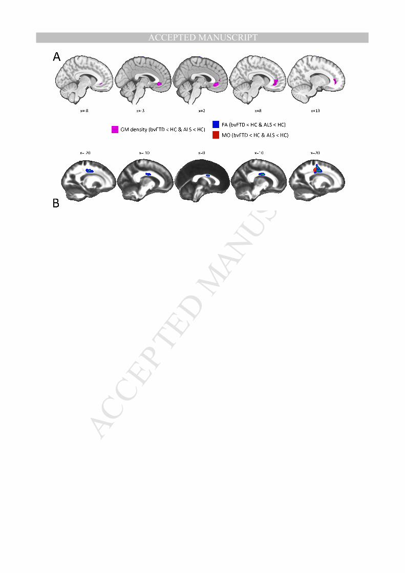
(i.e., the overlap between bvFTD<HC and ALS<HC statistical maps) showed a common
pattern of GM density reduction involving the ACC bilaterally and the right orbitofrontal
cortex (Figure 3, panel A). A one-way ANOVA on mean GM density values extracted from
the conjunction cluster highlighted a group effect (F(2,56)=18.541, p<0.00001). In particular,
post-hoc comparisons (Bonferroni correction) revealed significant differences in GM density

MANUSCRIP
T
ACCEPTED
ACCEPTED MANUSCRIPT
13
among all groups, with lower GM density values in bvFTD compared to both ALS
(p=0.0028) and HC (p<0.000001), and slightly lower GM density values in ALS compared to
HC (p=0.046).
Voxelwise conjunction analysis between clusters of WM microstructural changes in
bvFTD and ALS highlighted a common pattern of alterations, involving FA and MO indices
along the CC body and the right CST (Figure 3, panel B) including fibers connecting
homologous cortices of supplementary motor, premotor and primary motor areas. A one-way
ANOVA (performed separately on mean FA and MO indices extracted from conjunction
cluster among the three groups) resulted in a significant group effect (FA: F(2,56)=15.16,
p<0.00001; MO: F(2,56)=17.19, p<0.00001), showing a consistent pattern. Overall, we found
a significant decrease in FA and MO indices within the conjunction cluster in both ALS and
bvFTD compared to HC (ALS vs. HC: pFA<0.00001; pMO<0.000001; bvFTD vs. HC:
pFA=0.0023; pMO=0.008). Notably, post-hoc comparisons (Bonferroni correction) revealed a
significant difference between bvFTD and ALS with respect to mean MO values (p=0.025),
with ALS showing a greater decrease compared to bvFTD, but not in FA (p=0.189).
3.4 Neurostructural changes in bvFTD and ALS subgroups
The association analyses aiming to evaluate the strength of the relationship between
a) concomitant presence of MNDs and microstructural changes (FA/MO) within the
conjunction cluster involving the CST and the CC in bvFTD, and b) concomitant presence of
cognitive and/or behavioral symptoms and atrophy within the conjunction cluster (i.e., right
medial orbital gyrus and bilateral AAC) in ALS provided no significant results (bvFTD: chi-
square=0.159.p=0.69; ALS: chi-square=0.223, p=0.637). In particular, although we detected
the presence of reduction of GM density in the bilateral ACC and right medial orbital gyrus in
69% of ALS patients, only 16% of them also displayed cognitive and/or behavioral
symptoms. Similarly, while we observed microstructural alterations along the CST and CC in
the majority of bvFTD cases (60%), MNDs were found only in 4 out of 12 such patients.
MANUSCRIP
T
ACCEPTED
ACCEPTED MANUSCRIPT
14
Finally, we performed an exploratory analysis to evaluate possible differences in both
GM and WM indices – i.e., GM density and FA/MO respectively – between bvFTD (i.e., pure
bvFTD and bvFTD-MNDs) and ALS (i.e., pure ALS and ALS-FTSD) subgroups. Given the
small sample size, we calculated the percent variation of neurostructural measures within the
significant conjunction cluster among the four patient subgroups and the HC group.
The percent variations analysis on GM density changes showed that bvFTD-MNDs
had the most severe GM damage within the conjunction cluster including limbic and
orbitofrontal areas (i.e., bilateral ACC and right medial orbital gyrus), while pure bvFTD had
intermediate GM density values (greater than the two ALS subgroups), and ALS-FTSD and
pure ALS had similar mean values (percent variation <3%). All patient subgroups showed
lower GM density values in relation to HC (Table 3).
Table 3. VBM: Percent variations of Grey Matter (GM) density values across the four
patient subgroups within limbic and orbitofrontal areas. Percent variations of GM density
across patient subgroups have been computed by extracting mean values from the GM
conjunction cluster, including the ACC bilaterally and the right medial orbital gyrus. Negative
percentages correspond to lower density values (i.e., higher GM reduction) in subgroups
listed on rows compared to subgroups listed on columns.
Pure bvFTD Pure ALS ALS-FTSD HC
bvFTD-MNDs -4.86% -14.62% -17.09% -22.13%
Pure bvFTD - -10.26% -12.87% -18.15%
Pure ALS - -2.91% -8.79%
ALS-FTSD - -6.06%
ALS = amyotrophic lateral sclerosis; ALS-FTSD = ALS with cognitive and/or
behavioral disorders; bvFTD = behavioral variant of frontotemporal dementia; MNDs
= motor neuron dysfunctions; HC = healthy controls
MANUSCRIP
T
ACCEPTED
ACCEPTED MANUSCRIPT
15
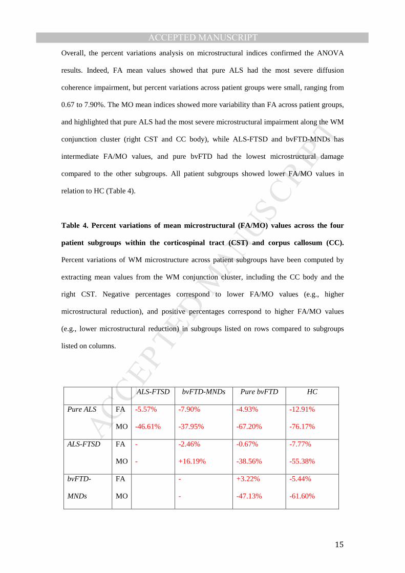
Overall, the percent variations analysis on microstructural indices confirmed the ANOVA
results. Indeed, FA mean values showed that pure ALS had the most severe diffusion
coherence impairment, but percent variations across patient groups were small, ranging from
0.67 to 7.90%. The MO mean indices showed more variability than FA across patient groups,
and highlighted that pure ALS had the most severe microstructural impairment along the WM
conjunction cluster (right CST and CC body), while ALS-FTSD and bvFTD-MNDs has
intermediate FA/MO values, and pure bvFTD had the lowest microstructural damage
compared to the other subgroups. All patient subgroups showed lower FA/MO values in
relation to HC (Table 4).
Table 4. Percent variations of mean microstructural (FA/MO) values across the four
patient subgroups within the corticospinal tract (CST) and corpus callosum (CC).
Percent variations of WM microstructure across patient subgroups have been computed by
extracting mean values from the WM conjunction cluster, including the CC body and the
right CST. Negative percentages correspond to lower FA/MO values (e.g., higher
microstructural reduction), and positive percentages correspond to higher FA/MO values
(e.g., lower microstructural reduction) in subgroups listed on rows compared to subgroups
listed on columns.
ALS-FTSD bvFTD-MNDs Pure bvFTD HC
Pure ALS FA
MO
-5.57%
-46.61%
-7.90%
-37.95%
-4.93%
-67.20%
-12.91%
-76.17%
ALS-FTSD FA
MO
-
-
-2.46%
+16.19%
-0.67%
-38.56%
-7.77%
-55.38%
bvFTD-
MNDs
FA
MO
-
-
+3.22%
-47.13%
-5.44%
-61.60%
MANUSCRIP
T
ACCEPTED
ACCEPTED MANUSCRIPT
16
4. DISCUSSION
In the present study, we explored the pattern of shared neurostructural alterations
within the bvFTD-ALS disease spectrum and their relationship with clinical phenotype. Our
results highlighted an overlap of brain changes in bvFTD and ALS in regions encompassing
both motor and cognitive networks. The overall findings concerning brain atrophy and
microstructural changes are in line with those reported previously (Lillo et al., 2012).
However, the overlap patterns reported here suggest that common alterations between ALS
and bvFTD can be less widespread in case of pure motor neuron or cognitive diseases.
Indeed, we did not find extra-motor damages (e.g., inferior longitudinal fasciculus) in the
conjunction pattern between microstructural white-matter features, and nor did we identify
commonalities in grey-matter atrophy in the motor cortex. However, in Lillo et al. (2012) the
subjects in pure ALS group showed a longer disease course (i.e., disease duration median = 3
years, interquartile range = 1.1-5 years) compared to that of ALS patients in the present study
(i.e., median = 18 months, interquartile range = 7.5-29.75 months). This may have crucially
affected both GM and WM conjunction patterns. Moreover, the presence of an additional
patient group including subjects with combined FTD and ALS diseases may be the reason
why they observed larger conjunction patterns.
Specifically, we provided evidence that while the GM conjunction cluster encompasses
high-level cognitive fronto-limbic areas (i.e., bilateral ACC and right medial orbital gyrus),
the conjunction cluster of WM microstructural alterations involves bundles within the motor
system (i.e., the upper portion of the CST and the body of the CC). In a broader perspective,
the presence of GM atrophy within the orbital and mesial portions of the prefrontal cortex in
Pure bvFTD FA
MO
-
-
-8.39%
-27.37%
ALS = amyotrophic lateral sclerosis; ALS-FTSD = ALS with cognitive and/or
behavioral disorders; bvFTD = behavioral variant of frontotemporal dementia; MNDs
= motor neuron dysfunctions; HC = healthy controls

MANUSCRIP
T
ACCEPTED
ACCEPTED MANUSCRIPT
17
ALS, as well as the WM microstructural changes of motor-related fibers in bvFTD, support
the notion of converging pathological processes in ALS and bvFTD (Ahmed et al., 2016). In
addition, our study results complement recent findings reporting shared patterns of functional
connectivity in ALS and bvFTD (Trojsi et al., 2015). Indeed, the authors described a
decreased functional connectivity common to ALS and bvFTD in GM areas involving both
sensorimotor (i.e., primary and supplementary motor areas) and cognitive networks, included
the salience (i.e., ACC, orbitofrontal and insular regions), as well the executive and the right
fronto-parietal networks.
Beside showing the spatial convergence of brain neurostructural degeneration in
bvFTD and ALS patients, we evaluated the extent of changes of GM and WM indices within
the conjunction clusters in order to explore the severity of neurostructural impairments in
patient subgroups according to the presence/absence of specific motor and
cognitive/behavioral symptoms. The degree of GM atrophy within the conjunction pattern
including fronto-limbic regions resulted significantly more pronounced in bvFTD than ALS
patients, and encompassed brain areas typically affected by phosphorylated TPD-43
inclusions in the first stages of the progression of Frontotemporal Lobar Degeneration
(Brettschneider et al., 2014). The ACC and right orbitofrontal cortex, consistently detected in
neuroimaging studies, are considered the neurostructural markers of bvFTD (Piguet and
Hodges, 2013). The presence of ACC atrophy in ALS, indeed, has been related to both apathy
(Woolley et al., 2011), and social cognition impairments (Cerami et al., 2013), which are
considered some of the key clinical features of bvFTD (Rascovsky et al., 2011). Moreover,
since the presence of GM and WM damages in extra-motor brain areas has been detected also
in pure ALS (Agosta et al. 2016; Christidi et al., 2017), an early damage of these regions in
ALS might represent an early signature of clinical manifestations, including cognitive and/or
behavioural impairment, which can arise in the later stages of the disease.
The WM microstructural alterations involve a significant decrease in FA and MO of the CST
and CC in both patient groups. These metrics are considered to reflect the orientation of
diffusion coherence and the organization of fibers of WM bundles, respectively (e.g., Douaud

MANUSCRIP
T
ACCEPTED
ACCEPTED MANUSCRIPT
18
et al., 2013, 2011). Within the conjunction clusters we observed different degrees of alteration
for FA (whose reduction was comparable among ALS and bvFTD) and MO (showing a
greater damage in ALS than in bvFTD). Changes in the microstructural properties of CST and
CC are considered early diagnostic biomarkers of ALS (Schuster et al., 2016). Despite the
intrinsic heterogeneity of the ALS population, the degeneration of these WM tracts has been
consistently observed in several DTI investigations (e.g., Douaud et al., 2011a; Filippini et al.,
2010; Turner et al., 2012), and reported in some neuropathological studies (e.g., Smith, 1960).
The reduction of the FA index within the CST is a typical and early DTI signature of
degenerative processes involving the upper motor neurons in ALS patients (Turner et al.,
2009), while the damage of the CC is probably related to either the interhemispheric spread of
the neurodegenerative process or to a secondary and independent degenerative event
(Filippini et al., 2010). Accordingly, the presence of changes in CC and CST in bvFTD
strongly suggests a motor neuron dysfunction also in this group of patients. This evidence is
further supported by previous neuroimaging studies (Daianu et al., 2015; Lillo et al., 2012;
Mahoney et al., 2014; Whitwell et al., 2010; Zhang et al., 2009).
Overall, our results provide further support to the hypothesis of ALS-FTD
pathological continuum. In particular, about a decade ago it has been proposed that a large
part of bvFTD – as well as other clinical manifestations within the Frontotemporal Lobar
Degeneration (FTLD) spectrum – and the majority of sporadic cases of ALS should be
considered two divergent expressions of a unique pathological spectrum centered on the
43kDa TAR DNA-binding protein (TDP-43) proteinopathy (Neumann et al., 2006). This view
has been recently corroborated by neuropathological investigations describing a time-
sequential propagation of neurostructural alterations induced by the spread of phosphorylated
TDP-43 aggregates in sporadic ALS (Brettschneider et al., 2013), which partially converges
with the pathological spread pattern observed in bvFTD (Brettschneider et al., 2014). In
bvFTD the degenerative process starts in the orbital and anterior portions of the prefrontal
cortex, spreads caudally to the lateral and anteromedial parts of the frontal lobe, then to the
limbic structures, brainstem nuclei, sensorimotor cortices, parietal lobes, lower motor
MANUSCRIP
T
ACCEPTED
ACCEPTED MANUSCRIPT
19
neurons, and finally reaches the occipital lobe (Brettschneider et al., 2014). In ALS, the
dissemination of TDP-43 aggregates starts from the motor cortices, the somatomotor nuclei of
the brainstem, and the lower motor neurons. Then, the pathological changes disseminate to
the reticular formation and precerebellar nuclei, and later to the prefrontal (i.e., gyrus rectus
and orbital gyri) and postcentral cortices the striatum, and the medial temporal lobe structures
(Brettschneider et al., 2013). In line with our results, recent MRI studies on large samples of
ALS (Müller et al., 2016; Schulthess et al., 2016) confirmed in vivo the spreading pattern of
neuropathological changes reported by Brettschneider et al. (2013). Indeed, Muller et al.
(2016) proposed a model of DTI-based tract-wise progression pattern in ALS, according to a
neuropathological staging scheme and associated to disease severity, starting from motor
network and disseminating to extra-motor brain regions (i.e., frontal and hippocampal
regions, brainstem). Comparably, Schulthess and colleagues (Schulthess et al., 2016) reported
a sequential involvement of functional networks remarkably resembling the distribution of
TDP-43 pathology.
In the present in vivo study, the neurostructural findings strongly support a
convergence of the TDP-43-like proteinopathy dissemination between specific commonly
involved brain regions in ALS and bvFTD. In particular, the quantification of GM and WM
damages within common altered structures in patient groups unveiled two different trends,
based on the classification of patients in terms of the presence/absence of MNDs and of
cognitive and/or behavioral symptoms. The first trend concerns a higher degree of atrophy in
orbitofrontal and frontomedial regions in patients with more severe cognitive and/or
behavioral symptoms. In particular, the reduction of GM density in the right orbitofrontal
cortex and bilateral ACC resulted more severe in bvFTD-MNDs than in other patient groups,
with ALS-FTSD and pure ALS showing similar values. Conversely, the second trend
highlights a higher degree of degeneration in the motor pathway in patients with more severe
motor neuron disorders. Specifically, the diffusion orientation coherence (FA) within CC and
CST is on average lower in pure ALS, with ALS-FTSD, bvFTD-MNDs and pure bvFTD
having smaller but similar damages. Additionally, microstructural changes affecting the shape

MANUSCRIP
T
ACCEPTED
ACCEPTED MANUSCRIPT
20
of the diffusion tensor (MO), and reflecting an altered organization of WM fibers, revealed a
continuum of alterations ranging from pure ALS (i.e., more severe alteration in relation to
other patient groups) to pure bvFTD (i.e., less severe alteration than other patient groups),
with ALS-FTSD and bvFTD-MNDs having intermediate values.
In the present sample, however, the involvement of the right orbitofrontal cortex and
bilateral ACC in ALS was independent of the presence of cognitive and/or behavioral
symptoms, as half of ALS sample showed a morphometrical alteration of these brain regions
in absence of extra-motor clinical manifestations. Similarly, we found that the presence of
MNDs was independent from the microstructural alterations along the CST/CC. In this
regard, previous investigations identified extra-motor alterations in ALS without cognitive
impairment (e.g., Agosta et al., 2016; Christidi et al., 2017), as well as the presence of motor
alteration in bvFTD without clinical manifestation of MND (e.g., Lillo et al. 2012; Trojsi et
al., 2015). Taken together, these findings might indicate the presence of structural changes
antedating clinical manifestations.
Conversely, the presence of cognitive and/or behavioral impairments or MNDs
without the alteration of the corresponding neurostructural substrate found in some cases (i.e.,
absence of either extra-motor degeneration in ALS-FTSD or CST/CC damage in bvFTD-
MNDs) might be due to the presence of subtle dysfunctions reflected in a defective functional
connectivity within networks regulating cognitive or sensorimotor processes. However,
further structural and functional neuroimaging studies are needed to clarify this issue.
A major limitation of the present study is related to the sample size of patient
subgroups, which did not allow us to perform formal statistical comparisons, but only a
description in terms of percent variations of GM and WM damages (bvFTD, bvFTD-MNDs,
ALS and ALS-FTSD). Of course this can affect the estimate of the actual magnitude of the
difference observed among subgroups, but the consistency between our data and previous
neuropathological (Brettschneider et al., 2013, 2014) and neuroimaging (Müller et al., 2016;
Schulthess et al., 2016) reports strengthens the present results. Nevertheless, although the
small size of our patient subgroups prevents us from drawing robust inferences, the
MANUSCRIP
T
ACCEPTED
ACCEPTED MANUSCRIPT
21
quantification of neurostructural alterations across ALS and bvFTD subgroups can provide
novel clues into the converging anatomical changes within the disease spectrum, and paves
the way for further in-depth investigations.
5. CONCLUSIONS
Altogether, the present data provide new in vivo evidence of converging sequential
pathological dissemination patterns in ALS and bvFTD. These neurodegenerative
proteinopathies share in the majority of cases common pathways of pathology propagation,
entailing both macro- and microstructural alterations, but following different courses.
First, the common pattern of macrostructural damage involving fronto-limbic
structures (i.e., right medial orbital gyrus and bilateral ACC) corroborates the presence of
subtle bvFTD-like disorders in ALS patients. Then, the shared microstructural damage
affecting the corticospinal and commissural (i.e., corpus callosum) fibers indicates a
subclinical ALS-like motor neuron degeneration in bvFTD. In addition, the quantification of
such neurostructural changes reveals the presence of a higher degree of degeneration in the
motor pathway in bvFTD patients with clinical or electromyographical evidence of motor
neuron dysfunction, as well as a higher degree of atrophy of orbitofrontal and fronto-medial
regions in ALS patients with neuropsychological evidence of frontotemporal dysfunction.
In conclusion, although further clinical, neuroimaging and neuropathological studies
based on larger samples are needed to reach more conclusive findings, the present in vivo
imaging study provide a step forward a better comprehension of the bidirectional spreading
pattern of neurodegenerative changes occurring within the ALS-FTD spectrum.
Acknowledgments: We thank Dr. V. Golzi, Dr. A. Marcone, Dr. M.C. Giusti and Dr. M.
Zamboni for patient recruitment.

MANUSCRIP
T
ACCEPTED
ACCEPTED MANUSCRIPT
22
Funding: This work was supported by a Ministry for Instruction, University and Research
(MIUR) grant (PRIN 2010XPMFW4_008; I meccanismi neurocognitivi alla base delle
interazioni sociali).
References
Abrahams, S., Goldstein, L.H., Suckling, J., Ng, V., Simmons, A., Chitnis, X., Atkins, L.,
Williams, S.C.R., Leigh, P.N., 2005. Frontotemporal white matter changes in
amyotrophic lateral sclerosis. J. Neurol. 252, 321–331. doi:10.1007/s00415-005-0646-x
Agosta, F., Ferraro, P.M., Riva, N., Spinelli, E.G., Chiò, A., Canu, E., Valsasina, P., Lunetta,
C., Iannaccone, S., Copetti, M., Prudente, E., Comi, G., Falini, A., Filippi, M., 2016.
Structural brain correlates of cognitive and behavioral impairment in MND. Hum. Brain
Mapp. 37, 1614–1626. doi:10.1002/hbm.23124
Ahmed, R.M., Irish, M., Piguet, O., Halliday, G.M., Ittner, L.M., Farooqi, S., Hodges, J.R.,
Kiernan, M.C., 2016. Amyotrophic lateral sclerosis and frontotemporal dementia:
distinct and overlapping changes in eating behaviour and metabolism. Lancet. Neurol.
doi:10.1016/S1474-4422(15)00380-4
Ashburner, J., 2009. Computational anatomy with the SPM software. Magn. Reson. Imaging.
doi:10.1016/j.mri.2009.01.006
Ashburner, J., 2007. A fast diffeomorphic image registration algorithm. Neuroimage 38, 95–
113. doi:10.1016/j.neuroimage.2007.07.007
Brettschneider, J., Del Tredici, K., Irwin, D.J., Grossman, M., Robinson, J.L., Toledo, J.B.,
Fang, L., Van Deerlin, V.M., Ludolph, A.C., Lee, V.M.Y., Braak, H., Trojanowski, J.Q.,
2014. Sequential distribution of pTDP-43 pathology in behavioral variant
frontotemporal dementia (bvFTD). Acta Neuropathol. 127, 423–439.
doi:10.1007/s00401-013-1238-y
Brettschneider, J., Del Tredici, K., Toledo, J.B., Robinson, J.L., Irwin, D.J., Grossman, M.,
Suh, E., Van Deerlin, V.M., Wood, E.M., Baek, Y., Kwong, L., Lee, E.B., Elman, L.,
McCluskey, L., Fang, L., Feldengut, S., Ludolph, A.C., Lee, V.M.Y., Braak, H.,

MANUSCRIP
T
ACCEPTED
ACCEPTED MANUSCRIPT
23
Trojanowski, J.Q., 2013. Stages of pTDP-43 pathology in amyotrophic lateral sclerosis.
Ann. Neurol. 74, 20–38. doi:10.1002/ana.23937
Brooks, B.R., Miller, R.G., Swash, M., Munsat, T.L., 2000. El Escorial revisited: revised
criteria for the diagnosis of amyotrophic lateral sclerosis. Amyotroph. Lateral Scler.
Other Motor Neuron Disord. 1, 293–299. doi:DOI 10.1080/146608200300079536
Burrell, J.R., Kiernan, M.C., Vucic, S., Hodges, J.R., 2011. Motor Neuron dysfunction in
frontotemporal dementia. Brain 134, 2582–2594. doi:10.1093/brain/awr195
Cedarbaum, J.M., Stambler, N., Malta, E., Fuller, C., Hilt, D., Thurmond, B., Nakanishi, A.,
1999. The ALSFRS-R: A revised ALS functional rating scale that incorporates
assessments of respiratory function. J. Neurol. Sci. 169, 13–21. doi:10.1016/S0022-
510X(99)00210-5
Cerami, C., Dodich, A., Canessa, N., Crespi, C., Iannaccone, S., Corbo, M., Lunetta, C.,
Consonni, M., Scola, E., Falini, A., Cappa, S.F., 2013. Emotional empathy in
amyotrophic lateral sclerosis: a behavioural and voxel-based morphometry study.
doi:10.3109/21678421.2013.785568
Cerami, C., Marcone, A., Crespi, C., Iannaccone, S., Marangoni, C., Dodich, A., Giusti,
M.C., Zamboni, M., Golzi, V., Cappa, S.F., 2015a. Motor neuron dysfunctions in the
frontotemporal lobar degeneration spectrum: A clinical and neurophysiological study. J.
Neurol. Sci. doi:10.1016/j.jns.2015.02.039
Cerami, C., Marcone, A., Crespi, C., Iannaccone, S., Marangoni, C., Dodich, A., Giusti,
M.C., Zamboni, M., Golzi, V., Cappa, S.F., 2015b. Motor neuron dysfunctions in the
frontotemporal lobar degeneration spectrum: a clinical and neurophysiological study.
doi:10.1016/j.jns.2015.02.039
Consonni, M., Iannaccone, S., Cerami, C., Frasson, P., Lacerenza, M., Lunetta, C., Corbo, M.,
Cappa, S.F., 2013. The cognitive and behavioural profile of Amyotrophic Lateral
Sclerosis: Application of the consensus criteria. Behav. Neurol. 27, 143–153.
doi:10.3233/BEN-2012-110202
Daianu, M., Mendez, M.F., Baboyan, V.G., Jin, Y., Melrose, R.J., Jimenez, E.E., Thompson,

MANUSCRIP
T
ACCEPTED
ACCEPTED MANUSCRIPT
24
P.M., 2015. An advanced white matter tract analysis in frontotemporal dementia and
early-onset Alzheimer???s disease. Brain Imaging Behav. doi:10.1007/s11682-015-
9458-5
Devenney, E., Vucic, S., Hodges, J.R., Kiernan, M.C., 2015. Motor neuron disease-
frontotemporal dementia: a clinical continuum. Expert Rev. Neurother. 15, 509–522.
doi:10.1586/14737175.2015.1034108
Douaud, G., Filippini, N., Knight, S., Talbot, K., Turner, M.R., 2011a. Integration of
structural and functional magnetic resonance imaging in amyotrophic lateral sclerosis.
Brain 134, 3470–9. doi:10.1093/brain/awr279
Douaud, G., Jbabdi, S., Behrens, T.E.J., Menke, R.A., Gass, A., Monsch, A.U., Rao, A.,
Whitcher, B., Kindlmann, G., Matthews, P.M., Smith, S., 2011b. DTI measures in
crossing-fibre areas: Increased diffusion anisotropy reveals early white matter alteration
in MCI and mild Alzheimer’s disease. Neuroimage 55, 880–890.
doi:10.1016/j.neuroimage.2010.12.008
Douaud, G., Menke, R. a L., Gass, A., Monsch, A.U., Rao, A., Whitcher, B., Zamboni, G.,
Matthews, P.M., Sollberger, M., Smith, S., 2013. Brain microstructure reveals early
abnormalities more than two years prior to clinical progression from mild cognitive
impairment to Alzheimer’s disease. J. Neurosci. 33, 2147–55.
doi:10.1523/JNEUROSCI.4437-12.2013
Eickhoff, S.B., Stephan, K.E., Mohlberg, H., Grefkes, C., Fink, G.R., Amunts, K., Zilles, K.,
2005. A new SPM toolbox for combining probabilistic cytoarchitectonic maps and
functional imaging data. Neuroimage 25, 1325–1335.
doi:10.1016/j.neuroimage.2004.12.034
Ferrari, R., Kapogiannis, D., Huey, E.D., Momeni, P., 2011. FTD and ALS: a tale of two
diseases. Curr. Alzheimer Res. 8, 273–94. doi:10.2174/156720511795563700
Filippini, N., Douaud, G., MacKay, C.E., Knight, S., Talbot, K., Turner, M.R., 2010. Corpus
callosum involvement is a consistent feature of amyotrophic lateral sclerosis. Neurology
75, 1645–1652. doi:10.1212/WNL.0b013e3181fb84d1
MANUSCRIP
T
ACCEPTED
ACCEPTED MANUSCRIPT
25
Goldstein, L.H., Abrahams, S., 2013. Changes in cognition and behaviour in amyotrophic
lateral sclerosis: Nature of impairment and implications for assessment. Lancet Neurol.
doi:10.1016/S1474-4422(13)70026-7
Hua, K., Zhang, J., Wakana, S., Jiang, H., Li, X., Reich, D.S., Calabresi, P.A., Pekar, J.J., van
Zijl, P.C.M., Mori, S., 2008. Tract probability maps in stereotaxic spaces: Analyses of
white matter anatomy and tract-specific quantification. Neuroimage 39, 336–347.
doi:10.1016/j.neuroimage.2007.07.053
Lattante, S., Ciura, S., Rouleau, G.A., Kabashi, E., 2015. Defining the genetic connection
linking amyotrophic lateral sclerosis (ALS) with frontotemporal dementia (FTD).
Trends Genet. doi:10.1016/j.tig.2015.03.005
Lillo, P., Mioshi, E., Burrell, J.R., Kiernan, M.C., Hodges, J.R., Hornberger, M., 2012. Grey
and White Matter Changes across the Amyotrophic Lateral Sclerosis-Frontotemporal
Dementia Continuum. PLoS One 7. doi:10.1371/journal.pone.0043993
Ling, S.C., Polymenidou, M., Cleveland, D.W., 2013. Converging mechanisms in als and
FTD: Disrupted RNA and protein homeostasis. Neuron.
doi:10.1016/j.neuron.2013.07.033
Lomen-Hoerth, C., 2011. Clinical phenomenology and neuroimaging correlates in ALS-FTD,
in: Journal of Molecular Neuroscience. pp. 656–662. doi:10.1007/s12031-011-9636-x
Lomen-Hoerth, C., Anderson, T., Miller, B., 2002. The overlap of amyotrophic lateral
sclerosis and frontotemporal dementia. Neurology 59, 1077–1079.
doi:10.1097/WCO.0b013e3283168d1d
Mahoney, C.J., Ridgway, G.R., Malone, I.B., Downey, L.E., Beck, J., Kinnunen, K.M.,
Schmitz, N., Golden, H.L., Rohrer, J.D., Schott, J.M., Rossor, M.N., Ourselin, S., Mead,
S., Fox, N.C., Warren, J.D., 2014. Profiles of white matter tract pathology in
frontotemporal dementia. Hum. Brain Mapp. 35, 4163–4179. doi:10.1002/hbm.22468
Müller, H.-P., Turner, M.R., Grosskreutz, J., Abrahams, S., Bede, P., Govind, V., Prudlo, J.,
Ludolph, A.C., Filippi, M., Kassubek, J., 2016. A large-scale multicentre cerebral
diffusion tensor imaging study in amyotrophic lateral sclerosis. J. Neurol. Neurosurg.
MANUSCRIP
T
ACCEPTED
ACCEPTED MANUSCRIPT
26
Psychiatry jnnp-2015-311952. doi:10.1136/jnnp-2015-311952
Neumann, M., Sampathu, D.M., Kwong, L.K., Truax, A.C., Micsenyi, M.C., Chou, T.T.,
Bruce, J., Schuck, T., Grossman, M., Clark, C.M., McCluskey, L.F., Miller, B.L.,
Masliah, E., Mackenzie, I.R., Feldman, H., Feiden, W., Kretzschmar, H. a, Trojanowski,
J.Q., Lee, V.M.-Y., 2006. Ubiquitinated TDP-43 in frontotemporal lobar degeneration
and amyotrophic lateral sclerosis. Science 314, 130–3. doi:10.1126/science.1134108
Nichols, T., Brett, M., Andersson, J., Wager, T., Poline, J.B., 2005. Valid conjunction
inference with the minimum statistic. Neuroimage 25, 653–660.
doi:10.1016/j.neuroimage.2004.12.005
Phukan, J., Elamin, M., Bede, P., Jordan, N., Gallagher, L., Byrne, S., Lynch, C., Pender, N.,
Hardiman, O., 2012. The syndrome of cognitive impairment in amyotrophic lateral
sclerosis: a population-based study. J. Neurol. Neurosurg. Psychiatry 83, 102–8.
doi:10.1136/jnnp-2011-300188
Piguet, O., Hodges, J.R., 2013. Behavioural-variant frontotemporal dementia: an update.
Dement. Neuropsychol. 7, 10–18.
Rascovsky, K., Hodges, J.R., Knopman, D., Mendez, M.F., Kramer, J.H., Neuhaus, J., Van
Swieten, J.C., Seelaar, H., Dopper, E.G.P., Onyike, C.U., Hillis, A.E., Josephs, K.A.,
Boeve, B.F., Kertesz, A., Seeley, W.W., Rankin, K.P., Johnson, J.K., Gorno-Tempini,
M.L., Rosen, H., Prioleau-Latham, C.E., Lee, A., Kipps, C.M., Lillo, P., Piguet, O.,
Rohrer, J.D., Rossor, M.N., Warren, J.D., Fox, N.C., Galasko, D., Salmon, D.P., Black,
S.E., Mesulam, M., Weintraub, S., Dickerson, B.C., Diehl-Schmid, J., Pasquier, F.,
Deramecourt, V., Lebert, F., Pijnenburg, Y., Chow, T.W., Manes, F., Grafman, J.,
Cappa, S.F., Freedman, M., Grossman, M., Miller, B.L., 2011. Sensitivity of revised
diagnostic criteria for the behavioural variant of frontotemporal dementia. Brain 134,
2456–2477. doi:10.1093/brain/awr179
Ringholz, G.M., Appel, S.H., Bradshaw, M., Cooke, N.A., Mosnik, D.M., Schulz, P.E., 2005.
Prevalence and patterns of cognitive impairment in sporadic ALS. Neurology 65, 586–
590. doi:10.1212/01.wnl.0000172911.39167.b6
MANUSCRIP
T
ACCEPTED
ACCEPTED MANUSCRIPT
27
Schulthess, I., Gorges, M., Müller, H.-P., Lulé, D., Del Tredici, K., Ludolph, A.C., Kassubek,
J., 2016. Functional connectivity changes resemble patterns of pTDP-43 pathology in
amyotrophic lateral sclerosis. Sci. Rep. 6, 38391. doi:10.1038/srep38391
Schuster, C., Elamin, M., Hardiman, O., Bede, P., 2016. The segmental diffusivity profile of
amyotrophic lateral sclerosis associated white matter degeneration. Eur. J. Neurol. 23,
1361–1371. doi:10.1111/ene.13038
Seeley, W.W., Crawford, R., Rascovsky, K., Kramer, J.H., Weiner, M., Miller, B.L., Gorno-
Tempini, M.L., 2008. Frontal paralimbic network atrophy in very mild behavioral
variant frontotemporal dementia. Arch. Neurol. 65, 249–255.
doi:10.1001/archneurol.2007.38
Smith, S.M., Jenkinson, M., Johansen-Berg, H., Rueckert, D., Nichols, T.E., Mackay, C.E.,
Watkins, K.E., Ciccarelli, O., Cader, M.Z., Matthews, P.M., Behrens, T.E.J., 2006.
Tract-based spatial statistics: Voxelwise analysis of multi-subject diffusion data.
Neuroimage 31, 1487–1505. doi:10.1016/j.neuroimage.2006.02.024
Smith, S.M., Nichols, T.E., 2009. Threshold-free cluster enhancement: Addressing problems
of smoothing, threshold dependence and localisation in cluster inference. Neuroimage
44, 83–98. doi:10.1016/j.neuroimage.2008.03.061
Strong, M.J., Abrahams, S., Goldstein, L.H., Woolley, S., Mclaughlin, P., Snowden, J.,
Mioshi, E., Roberts-South, A., Benatar, M., HortobáGyi, T., Rosenfeld, J., Silani, V.,
Ince, P.G., Turner, M.R., 2017. Amyotrophic lateral sclerosis - frontotemporal spectrum
disorder (ALS-FTSD): Revised diagnostic criteria. Amyotroph. Lateral Scler. Front.
Degener. 0, 1–22. doi:10.1080/21678421.2016.1267768
Strong, M.J., Grace, G.M., Freedman, M., Lomen-Hoerth, C., Woolley, S., Goldstein, L.H.,
Murphy, J., Shoesmith, C., Rosenfeld, J., Leigh, P.N., Bruijn, L., Ince, P., Figlewicz, D.,
2009. Consensus criteria for the diagnosis of frontotemporal cognitive and behavioural
syndromes in amyotrophic lateral sclerosis. Amyotroph. Lateral Scler. 10, 131–146.
doi:Pii 907462414\rDoi 10.1080/17482960802654364
Trojsi, F., Esposito, F., de Stefano, M., Buonanno, D., Conforti, F.L., Corbo, D., Piccirillo,

MANUSCRIP
T
ACCEPTED
ACCEPTED MANUSCRIPT
28
G., Cirillo, M., Monsurrò, M.R., Montella, P., Tedeschi, G., 2015. Functional overlap
and divergence between ALS and bvFTD. Neurobiol. Aging 36, 413–423.
doi:10.1016/j.neurobiolaging.2014.06.025
Tsermentseli, S., Leigh, P.N., Goldstein, L.H., 2012. The anatomy of cognitive impairment in
amyotrophic lateral sclerosis: More than frontal lobe dysfunction. Cortex.
doi:10.1016/j.cortex.2011.02.004
Turner, M.R., Agosta, F., Govind, V., Bede, P., Lulé, D., Verstraete, E., 2012. Neuroimaging
in amyotrophic lateral sclerosis Review. Biomarkers Med. 6, 319–337.
doi:10.2217/bmm.12.26
Turner, M.R., Kiernan, M.C., Leigh, P.N., Talbot, K., 2009. Biomarkers in amyotrophic
lateral sclerosis. Lancet Neurol. doi:10.1016/S1474-4422(08)70293-X
Turner, M.R., Verstraete, E., 2015. What does imaging reveal about the pathology of
amyotrophic lateral sclerosis? Curr. Neurol. Neurosci. Rep. 15, 569.
doi:10.1007/s11910-015-0569-6
Weishaupt, J.H., Hyman, T., Dikic, I., 2016. Common Molecular Pathways in Amyotrophic
Lateral Sclerosis and Frontotemporal Dementia. Trends Mol. Med.
doi:10.1016/j.molmed.2016.07.005
Whitwell, J.L., Avula, R., Senjem, M.L., Kantarci, K., Weigand, S.D., Samikoglu, A.,
Edmonson, H.A., Vemuri, P., Knopman, D.S., Boeve, B.F., Petersen, R.C., Josephs,
K.A., Jack, C.R., 2010. Gray and white matter water diffusion in the syndromic variants
of frontotemporal dementia. Neurology 74, 1279–1287.
doi:10.1212/WNL.0b013e3181d9edde
Woolley, S.C., Zhang, Y., Schuff, N., Weiner, M.W., Katz, J.S., 2011. Neuroanatomical
correlates of apathy in ALS using 4 Tesla diffusion tensor MRI. Amyotroph. Lateral
Scler. 12, 52–58. doi:10.3109/17482968.2010.521842
Zhang, Y., Schuff, N., Du, A.-T., Rosen, H.J., Kramer, J.H., Gorno-Tempini, M.L., Miller,
B.L., Weiner, M.W., 2009. White matter damage in frontotemporal dementia and
Alzheimer’s disease measured by diffusion MRI. Brain 132, 2579–92.

MANUSCRIP
T
ACCEPTED
ACCEPTED MANUSCRIPT
29
doi:10.1093/brain/awp071

MANUSCRIP
T
ACCEPTED
ACCEPTED MANUSCRIPT
30
Figure Legends
Figure 1. Neurostructural profile of bvFTD vs. HC
A: Whole-brain results from VBM highlight, in bvFTD patients compared to HC,
significantly decreased GM density in fronto-temporal and limbic structures bilaterally
(p<0.05, FWE-corrected). The statistical map is superimposed on the MNI T1 template. B:
Whole-brain results from TBSS highlight, in bvFTD patients compared to HC, significantly
decreased FA and MO, as well as significantly increased MD, in the limbic-prefrontal
circuitry (p<0.05, FWE-corrected). The statistical maps are superimposed on FMRIB
standard-space FA template.
Figure 2. Neurostructural profile of ALS vs. HC
A: Whole-brain results from VBM highlight, in ALS patients compared to HC, significantly
decreased GM density in the bilateral ACC and right medial orbital gyrus. The statistical map
is superimposed on the MNI T1 template. B: Whole-brain results from TBSS highlight, in
ALS patients compared to HC, significantly decreased FA and MO in the CST and the body
of CC bilaterally (p<0.05, FWE-corrected). The statistical maps are superimposed on FMRIB
standard-space FA template.
Figure 3. Neurostructural conjunction pattern common to bvFTD and ALS
A: Whole-brain conjunction analysis of GM density highlights, in bvFTD and ALS groups, a
common pattern of alterations involving the bilateral ACC and the right orbitofrontal cortex
(p<0.05, FWE-corrected). The statistical map is superimposed on the MNI T1 template. B:
Whole-brain conjunction analysis of WM microstructural characteristics highlights, in bvFTD
and ALS groups, a common pattern of alterations involving a significant decrease of both FA
and MO indices within the right CST and the body of the CC (p<0.05, FWE-corrected). The
statistical maps are superimposed on FMRIB standard-space FA template.

MANUSCRIP
T
ACCEPTED
ACCEPTED MANUSCRIPT

MANUSCRIP
T
ACCEPTED
ACCEPTED MANUSCRIPT
MANUSCRIP
T
ACCEPTED
ACCEPTED MANUSCRIPT
MANUSCRIP
T
ACCEPTED
ACCEPTED MANUSCRIPT
1
Highlights
ALS and bvFTD share a common pattern of WM and GM alterations
Higher degree of WM degeneration is reflected in more severe motor neuron disorders
GM atrophy is greater in presence of more severe executive and/or behavioral symptoms
Motor pathway damage in bvFTD may indicate a subtle ALS-like motor neuron dysfunction
Fronto-limbic atrophy in ALS may suggest the presence of subtle bvFTD-like symptoms


![Monitoria multimodal cerebral multimodal monitoring[2]](https://static.fdocuments.us/doc/165x107/552957004a79599a158b46fd/monitoria-multimodal-cerebral-multimodal-monitoring2.jpg)