Quantification of circulating Mycobacterium tuberculosis ... · Quantification of circulating...
6
Quantification of circulating Mycobacterium tuberculosis antigen peptides allows rapid diagnosis of active disease and treatment monitoring Chang Liu a,b,c , Zhen Zhao d , Jia Fan a,c , Christopher J. Lyon a,c , Hung-Jen Wu a,e , Dobrin Nedelkov f , Adrian M. Zelazny d , Kenneth N. Olivier g , Lisa H. Cazares h , Steven M. Holland i , Edward A. Graviss j , and Ye Hu a,b,c,1 a Department of Nanomedicine, Houston Methodist Research Institute, Houston, TX 77030; b School of Biological and Health Systems Engineering, Arizona State University, Tempe, AZ 85287; c Virginia G. Piper Biodesign Center for Personalized Diagnostics, The Biodesign Institute, Arizona State University, Tempe, AZ 85287; d Department of Laboratory Medicine, Clinical Center, National Institutes of Health, Bethesda, MD 20892; e Department of Chemical Engineering, Texas A&M University, College Station, TX 77843; f Molecular Biomarkers Laboratory, The Biodesign Institute, Arizona State University, Tempe, AZ 85287; g Cardiovascular and Pulmonary Branch, National Heart, Lung, and Blood Institute, Bethesda, MD 20892; h Molecular and Translational Sciences, US Army Medical Research Institute of Infectious Diseases, Frederick, MD 21702; i Laboratory of Clinical Infectious Diseases, National Institute of Allergy and Infectious Diseases, Bethesda, MD 20892; and j Department of Pathology and Genomic Medicine, Houston Methodist Research Institute, Houston, TX 77030 Edited by William R. Jacobs Jr., Albert Einstein College of Medicine, Howard Hughes Medical Institute, Bronx, NY, and approved February 17, 2017 (received for review January 6, 2017) Tuberculosis (TB) is a major global health threat, resulting in an urgent unmet need for a rapid, non–sputum-based quantitative test to detect active Mycobacterium tuberculosis (Mtb) infections in clinically diverse populations and quickly assess Mtb treatment responses for emerging drug-resistant strains. We have identified Mtb-specific peptide fragments and developed a method to rap- idly quantify their serum concentrations, using antibody-labeled and energy-focusing porous discoidal silicon nanoparticles (nano- disks) and high-throughput mass spectrometry (MS) to enhance sensitivity and specificity. NanoDisk-MS diagnosed active Mtb cases with high sensitivity and specificity in a case-control study with cohorts reflecting the complexity of clinical practice. Similar robust sensitivities were obtained for cases of culture-positive pul- monary TB (PTB; 91.3%) and extrapulmonary TB (EPTB; 92.3%), and the sensitivities obtained for culture-negative PTB (82.4%) and EPTB (75.0%) in HIV-positive patients significantly outper- formed those reported for other available assays. NanoDisk-MS also exhibited high specificity (87.1–100%) in both healthy and high-risk groups. Absolute quantification of serum Mtb antigen concentration was informative in assessing responses to antimy- cobacterial treatment. Thus, a NanoDisk-MS assay approach could significantly improve the diagnosis and management of active TB cases, and perhaps other infectious diseases as well. tuberculosis | blood test | nanodisk | rapid diagnosis | treatment monitoring D espite international efforts and initiatives, tuberculosis (TB) remains a major public health concern worldwide, associ- ated with high morbidity and mortality (1, 2). Detecting active TB cases and monitoring their responses to therapy are fraught with challenges, relying predominantly on microbiologic tech- niques that use sputum samples, including acid-fast Bacillus (AFB) smear microscopy—widely used as an initial test for TB diagnosis (3, 4)—and Mycobacterium tuberculosis (Mtb) culture, both of which have only moderate sensitivity and specificity and a long turnaround time (5, 6). Moreover, sputum samples are difficult to obtain after symptom improvement, and often are not diagnostically useful for extrapulmonary TB (EPTB) cases. The PCR-based Xpert MTB/RIF sputum assay was introduced to improve the speed and specificity of TB diagnosis, but this assay has poor sensitivity under low bacterial loads and cannot distin- guish live and nonviable Mtb contributions (7, 8). A recent World Health Organization (WHO) policy update acknowledged the low quality of evidence supporting the use of Xpert MTB/RIF to di- agnose EPTB (9). Diagnostic challenges can be further magnified in patients coinfected with HIV and TB (10). In addition, none of these techniques provides quantitative results that can be used to monitor treatment effects (11, 12). Consequently, there is an urgent need, highlighted as a high priority in a recent WHO consensus report (13), for the devel- opment of rapid, quantitative, non–sputum-based biomarker tests that do not require bacterial isolation to detect active TB (13). Non–sputum-based IFN-γ release assays (IGRAs), which measure ex vivo immune responses to Mtb antigens, have re- ceived negative policy recommendations for active TB diagnosis owing to their inability to distinguish active TB and latent TB infection (LTBI), as well as their poor diagnostic performance in HIV/TB-coinfected patients and EPTB patients (14). One recent report used the expression of a set of host innate immune re- sponse genes in blood to diagnose pulmonary TB (PTB) cases, but that retrospective study did not examine blood samples of EPTB patients, and could not identify culture-negative TB cases (15). Detection of Mtb antigens in patient blood samples can provide direct evidence of TB, but no currently available method has demonstrated adequate diagnostic sensitivity and specificity, Significance Active Mycobacterium tuberculosis (Mtb) infections represent a significant global health threat, but can be difficult to diagnose and manage owing to the nonquantitative nature and rela- tively poor performance of current frontline sputum-based di- agnostic assays, which can be further degraded by certain Mtb manifestations. This study describes the development of a rapid and quantitative blood-based assay with high sensitivity and specificity for active Mtb infections that can be used to monitor responses to antimycobacterial therapy. Our method combines antibody-labeled, energy-focusing nanodisks with high-throughput mass spectrometry to enhance the detection of Mtb-specific peptides in digested serum samples, and should allow rapid clinical translation. This approach also should be applicable to other infectious diseases, particu- larly those for which conventional immunoassays exhibit suboptimal performance. Author contributions: C.L., Z.Z., and Y.H. designed research; C.L., J.F., H.-J.W., and D.N. performed research; E.A.G. contributed clinical samples; C.L., Z.Z., J.F., C.J.L., A.M.Z., K.N.O., L.H.C., S.M.H., E.A.G., and Y.H. analyzed data; and C.L., Z.Z., C.J.L., A.M.Z., L.H.C., S.M.H., E.A.G., and Y.H. wrote the paper. The authors declare no conflict of interest. This article is a PNAS Direct Submission. Freely available online through the PNAS open access option. 1 To whom correspondence should be addressed. Email: [email protected]. This article contains supporting information online at www.pnas.org/lookup/suppl/doi:10. 1073/pnas.1621360114/-/DCSupplemental. www.pnas.org/cgi/doi/10.1073/pnas.1621360114 PNAS | April 11, 2017 | vol. 114 | no. 15 | 3969–3974 MEDICAL SCIENCES ENGINEERING
Transcript of Quantification of circulating Mycobacterium tuberculosis ... · Quantification of circulating...
Quantification of circulating Mycobacteriumtuberculosis antigen peptides allows rapid diagnosisof active disease and treatment monitoringChang Liua,b,c, Zhen Zhaod, Jia Fana,c, Christopher J. Lyona,c, Hung-Jen Wua,e, Dobrin Nedelkovf, Adrian M. Zelaznyd,Kenneth N. Olivierg, Lisa H. Cazaresh, Steven M. Hollandi, Edward A. Gravissj, and Ye Hua,b,c,1
aDepartment of Nanomedicine, Houston Methodist Research Institute, Houston, TX 77030; bSchool of Biological and Health Systems Engineering, ArizonaState University, Tempe, AZ 85287; cVirginia G. Piper Biodesign Center for Personalized Diagnostics, The Biodesign Institute, Arizona State University,Tempe, AZ 85287; dDepartment of Laboratory Medicine, Clinical Center, National Institutes of Health, Bethesda, MD 20892; eDepartment of ChemicalEngineering, Texas A&M University, College Station, TX 77843; fMolecular Biomarkers Laboratory, The Biodesign Institute, Arizona State University, Tempe,AZ 85287; gCardiovascular and Pulmonary Branch, National Heart, Lung, and Blood Institute, Bethesda, MD 20892; hMolecular and Translational Sciences,US Army Medical Research Institute of Infectious Diseases, Frederick, MD 21702; iLaboratory of Clinical Infectious Diseases, National Institute of Allergy andInfectious Diseases, Bethesda, MD 20892; and jDepartment of Pathology and Genomic Medicine, Houston Methodist Research Institute, Houston, TX 77030
Edited by William R. Jacobs Jr., Albert Einstein College of Medicine, Howard Hughes Medical Institute, Bronx, NY, and approved February 17, 2017 (receivedfor review January 6, 2017)
Tuberculosis (TB) is a major global health threat, resulting in anurgent unmet need for a rapid, non–sputum-based quantitativetest to detect active Mycobacterium tuberculosis (Mtb) infectionsin clinically diverse populations and quickly assess Mtb treatmentresponses for emerging drug-resistant strains. We have identifiedMtb-specific peptide fragments and developed a method to rap-idly quantify their serum concentrations, using antibody-labeledand energy-focusing porous discoidal silicon nanoparticles (nano-disks) and high-throughput mass spectrometry (MS) to enhancesensitivity and specificity. NanoDisk-MS diagnosed active Mtbcases with high sensitivity and specificity in a case-control studywith cohorts reflecting the complexity of clinical practice. Similarrobust sensitivities were obtained for cases of culture-positive pul-monary TB (PTB; 91.3%) and extrapulmonary TB (EPTB; 92.3%),and the sensitivities obtained for culture-negative PTB (82.4%)and EPTB (75.0%) in HIV-positive patients significantly outper-formed those reported for other available assays. NanoDisk-MSalso exhibited high specificity (87.1–100%) in both healthy andhigh-risk groups. Absolute quantification of serum Mtb antigenconcentration was informative in assessing responses to antimy-cobacterial treatment. Thus, a NanoDisk-MS assay approach couldsignificantly improve the diagnosis and management of active TBcases, and perhaps other infectious diseases as well.
tuberculosis | blood test | nanodisk | rapid diagnosis |treatment monitoring
Despite international efforts and initiatives, tuberculosis (TB)remains a major public health concern worldwide, associ-
ated with high morbidity and mortality (1, 2). Detecting activeTB cases and monitoring their responses to therapy are fraughtwith challenges, relying predominantly on microbiologic tech-niques that use sputum samples, including acid-fast Bacillus(AFB) smear microscopy—widely used as an initial test for TBdiagnosis (3, 4)—and Mycobacterium tuberculosis (Mtb) culture,both of which have only moderate sensitivity and specificity anda long turnaround time (5, 6). Moreover, sputum samples aredifficult to obtain after symptom improvement, and often are notdiagnostically useful for extrapulmonary TB (EPTB) cases. ThePCR-based Xpert MTB/RIF sputum assay was introduced toimprove the speed and specificity of TB diagnosis, but this assayhas poor sensitivity under low bacterial loads and cannot distin-guish live and nonviable Mtb contributions (7, 8). A recent WorldHealth Organization (WHO) policy update acknowledged the lowquality of evidence supporting the use of Xpert MTB/RIF to di-agnose EPTB (9). Diagnostic challenges can be further magnifiedin patients coinfected with HIV and TB (10). In addition, none of
these techniques provides quantitative results that can be used tomonitor treatment effects (11, 12).Consequently, there is an urgent need, highlighted as a high
priority in a recent WHO consensus report (13), for the devel-opment of rapid, quantitative, non–sputum-based biomarkertests that do not require bacterial isolation to detect active TB(13). Non–sputum-based IFN-γ release assays (IGRAs), whichmeasure ex vivo immune responses to Mtb antigens, have re-ceived negative policy recommendations for active TB diagnosisowing to their inability to distinguish active TB and latent TBinfection (LTBI), as well as their poor diagnostic performance inHIV/TB-coinfected patients and EPTB patients (14). One recentreport used the expression of a set of host innate immune re-sponse genes in blood to diagnose pulmonary TB (PTB) cases,but that retrospective study did not examine blood samples ofEPTB patients, and could not identify culture-negative TB cases(15). Detection of Mtb antigens in patient blood samples canprovide direct evidence of TB, but no currently available methodhas demonstrated adequate diagnostic sensitivity and specificity,
Significance
ActiveMycobacterium tuberculosis (Mtb) infections represent asignificant global health threat, but can be difficult to diagnoseand manage owing to the nonquantitative nature and rela-tively poor performance of current frontline sputum-based di-agnostic assays, which can be further degraded by certain Mtbmanifestations. This study describes the development of arapid and quantitative blood-based assay with high sensitivityand specificity for active Mtb infections that can be used tomonitor responses to antimycobacterial therapy. Our methodcombines antibody-labeled, energy-focusing nanodisks withhigh-throughput mass spectrometry to enhance the detectionof Mtb-specific peptides in digested serum samples, andshould allow rapid clinical translation. This approach alsoshould be applicable to other infectious diseases, particu-larly those for which conventional immunoassays exhibitsuboptimal performance.
Author contributions: C.L., Z.Z., and Y.H. designed research; C.L., J.F., H.-J.W., and D.N.performed research; E.A.G. contributed clinical samples; C.L., Z.Z., J.F., C.J.L., A.M.Z., K.N.O.,L.H.C., S.M.H., E.A.G., and Y.H. analyzed data; and C.L., Z.Z., C.J.L., A.M.Z., L.H.C., S.M.H., E.A.G.,and Y.H. wrote the paper.
The authors declare no conflict of interest.
This article is a PNAS Direct Submission.
Freely available online through the PNAS open access option.1To whom correspondence should be addressed. Email: [email protected].
This article contains supporting information online at www.pnas.org/lookup/suppl/doi:10.1073/pnas.1621360114/-/DCSupplemental.
www.pnas.org/cgi/doi/10.1073/pnas.1621360114 PNAS | April 11, 2017 | vol. 114 | no. 15 | 3969–3974
MED
ICALSC
IENCE
SEN
GINEE
RING
likely owing to the epitope-masking effects of host proteins (16)and homology with related antigens of several nontuberculousmycobacteria (NTM) (17, 18).Here we report a blood-based assay for rapid, specific, and high-
sensitivity quantification of active TB infections in patients, whichuses antibody-conjugated nanodisks to enrich Mtb-specific peptidesof 10-kDa culture filtrate protein (CFP-10) and 6-kDa early secre-tory antigenic target (ESAT-6) from trypsin-digested serum samples.These factors demonstrate homology with those expressed by otherMycobacterium species, but have tryptic peptides that exhibit strongMtb selectivity. Thus, Mtb-derived CFP-10 and ESAT-6 serumconcentrations appear likely to be strong predictors of active TBdisease, because they are actively secreted by virulent mycobacterialstrains, can be detected early afterMtb infection, and have activitiesthat attenuate mycobacterial clearance (19), implying that theirpresence in serum can be used to diagnose active Mtb infections.We have incorporated several advances to allow the diagnosis of
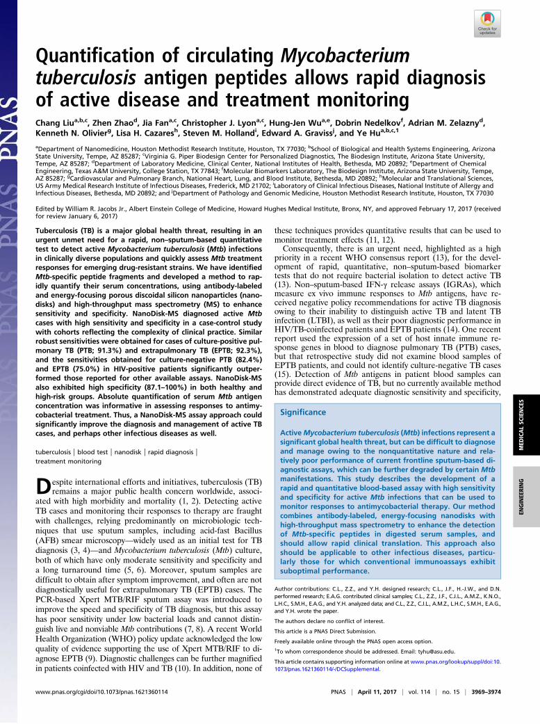
TB from patient blood samples: identification of Mtb-selective CFP-10 and ESAT-6 peptides, and development of antibody-conjugatednanodisks that dramatically increase both target peptide enrichmentand matrix-assisted laser desorption/ionization (MALDI) of boundpeptides to enhance detection by high-throughput MALDI time-of-flight mass spectrometry (MALDI-TOF MS). Furthermore, trypsindigestion is thought to disrupt protein complexes to release Mtbantigens that might be undetectable in conventional immunoassaystargeting intact Mtb proteins. The NanoDisk-MS method (Fig. 1)permits rapid quantification of serum markers specific for activeTB, overcoming obstacles associated with current methodologies,and uses accepted clinical instrumentation to enhance its potentialfor clinical translation.We evaluated the diagnostic performance of NanoDisk-MS in
HIV-negative and HIV-positive patients drawn from the HoustonTuberculosis Initiative (HTI), a large, population-based TBsurveillance study. Results from case-control groups in thesepopulations and longitudinal samples from patients undergoinganti-TB therapy provide strong proof-of-principle evidence sup-porting the clinical utility of this detection platform.
ResultsSensitive Nanoparticle-Mediated Detection of Mtb-Specific SerumPeptides. Serum CFP-10 and ESAT-6 expression theoreticallycan be used to diagnose all activeMtb infections, including EPTBcases (20); however, some NTM strains express homologs thatmay reduce the utility of these proteins as biomarkers (21). Sincepeptide sequence is the gold standard for protein discrimination,we examined whether tryptic peptides could distinguish Mtb-derived ESAT-6 and CFP-10 from homologs produced by otherspecies. MALDI-TOF MS analysis of recombinant protein trypticdigests detected CFP-10 (TDAATLAQEAGNFER;m/z 1,593.75)and ESAT-6 (WDATATELNNALQNLAR; m/z 1,900.95) pep-tides with high signal-to-noise ratios that were subsequently con-firmed by liquid chromatography-tandem mass spectrometry, andwhich showed strong Mtb specificity when aligned with homologsfrom 12 NTM species (17, 18) (SI Appendix, Figs. S1–S3). Bothpeptides demonstrated perfect homology with Mycobacterium bovis,a relatively rare form of TB, but diverged markedly from two speciesresponsible for the majority of NTM infections, Mycobacteriumavium and Mycobacterium intracellulare (22), as well as most otherNTM species. The ESAT-6 peptide showed little homology to anyNTM, whereas the CFP-10 peptide demonstrated homology onlyto some strains of Mycobacterium kansasii, Mycobacterium mar-inum, and Mycobacterim ulcerans that were not expected to sig-nificantly interfere with diagnostic specificity in clinical use.The wide dynamic range of serum protein expression can com-
plicate the cleavage and subsequent detection of low-abundanceserum proteins (23). We found that supplemental microwave irra-diation allowed serum CFP-10 and ESAT-6 digestion within 20 mininstead of overnight, as is normally required for complex proteinsamples (24), reducing the “sample-to-answer” time to 4 h whileincreasing the MS signal for target peptides by more than threefold(SI Appendix, Fig. S4).We next analyzed several potential nanoparticle enrichment
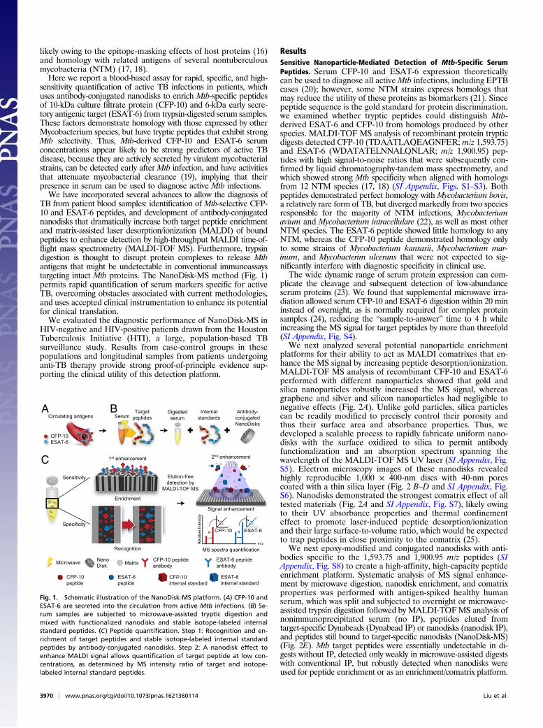
platforms for their ability to act as MALDI comatrixes that en-hance the MS signal by increasing peptide desorption/ionization.MALDI-TOF MS analysis of recombinant CFP-10 and ESAT-6performed with different nanoparticles showed that gold andsilica nanoparticles robustly increased the MS signal, whereasgraphene and silver and silicon nanoparticles had negligible tonegative effects (Fig. 2A). Unlike gold particles, silica particlescan be readily modified to precisely control their porosity andthus their surface area and absorbance properties. Thus, wedeveloped a scalable process to rapidly fabricate uniform nano-disks with the surface oxidized to silica to permit antibodyfunctionalization and an absorption spectrum spanning thewavelength of the MALDI-TOF MS UV laser (SI Appendix, Fig.S5). Electron microscopy images of these nanodisks revealedhighly reproducible 1,000 × 400-nm discs with 40-nm porescoated with a thin silica layer (Fig. 2 B–D and SI Appendix, Fig.S6). Nanodisks demonstrated the strongest comatrix effect of alltested materials (Fig. 2A and SI Appendix, Fig. S7), likely owingto their UV absorbance properties and thermal confinementeffect to promote laser-induced peptide desorption/ionizationand their large surface-to-volume ratio, which would be expectedto trap peptides in close proximity to the comatrix (25).We next epoxy-modified and conjugated nanodisks with anti-
bodies specific to the 1,593.75 and 1,900.95 m/z peptides (SIAppendix, Fig. S8) to create a high-affinity, high-capacity peptideenrichment platform. Systematic analysis of MS signal enhance-ment by microwave digestion, nanodisk enrichment, and comatrixproperties was performed with antigen-spiked healthy humanserum, which was split and subjected to overnight or microwave-assisted trypsin digestion followed by MALDI-TOFMS analysis ofnonimmunoprecipitated serum (no IP), peptides eluted fromtarget-specific Dynabeads (Dynabead IP) or nanodisks (nanodisk IP),and peptides still bound to target-specific nanodisks (NanoDisk-MS)(Fig. 2E). Mtb target peptides were essentially undetectable in di-gests without IP, detected only weakly in microwave-assisted digestswith conventional IP, but robustly detected when nanodisks wereused for peptide enrichment or as an enrichment/comatrix platform.
Circulating antigensA B
C
TargetpeptidesSerum
Internalstandards
Enrichment
1st enhancement
Sensitivity
Specificity
Digestedserum
2nd enhancement
Signal enhancement
CFP-10ESAT-6
Antibody-conjugated NanoDisks
Elution-freedetection by
MALDI-TOF MS
Recognition MS spectra quantification
NanoDisk Matrix CFP-10 peptide
antibodyESAT-6 peptide antibody
CFP-10peptide
ESAT-6peptide
CFP-10internal standard
ESAT-6internal standard
Microwave
CFP-10 ESAT-6
Fig. 1. Schematic illustration of the NanoDisk-MS platform. (A) CFP-10 andESAT-6 are secreted into the circulation from active Mtb infections. (B) Se-rum samples are subjected to microwave-assisted tryptic digestion andmixed with functionalized nanodisks and stable isotope-labeled internalstandard peptides. (C) Peptide quantification. Step 1: Recognition and en-richment of target peptides and stable isotope-labeled internal standardpeptides by antibody-conjugated nanodisks. Step 2: A nanodisk effect toenhance MALDI signal allows quantification of target peptide at low con-centrations, as determined by MS intensity ratio of target and isotope-labeled internal standard peptides.
3970 | www.pnas.org/cgi/doi/10.1073/pnas.1621360114 Liu et al.

The mean Mtb peptide signal was increased by 2.5-fold for CFP-10and 2.6-fold for ESAT-6 by microwave-assisted digestion, by anadditional 6.6-fold for both CFP-10 and ESAT-6 by nanodisk-mediated enrichment, and then by an additional 9.9-fold forCFP-10 and 10.2-fold for ESAT-6 by nanodisk comatrix activity.
Quantification of Mtb-Specific Peptides in Human Serum. TB-freehuman serum spiked with recombinant CFP-10 and ESAT-6standards was digested under microwave irradiation, spiked withstable isotope-labeled internal standards, and analyzed byNanoDisk-MS. Calibration curves for antigen quantitation weregenerated by plotting the MS spectra intensity ratio betweentarget peptides and internal standards against their respectiveinput recombinant protein concentrations. Excellent correlation(R2 >0.98) was observed for curves made with different nanodiskbatches (Fig. 2F), with values exhibiting 14–22% within-run and16–23% between-run coefficients of variation. CFP-10 showed a50 pM limit of detection (LOD) and a 200 pM limit of quanti-fication (LOQ), ESAT-6 had a 200 pM LOD and a 500 pMLOQ, and measurement accuracies ranged from ∼74% (1 nM)to ∼90% (20 nM) (SI Appendix, Table S1). NanoDisk-MS alsoreadily distinguished patients with TB and patients without TBin a proof-or-principle multiplex assay (Fig. 2G). Conversely,analyses performed using an advanced MALDI-TOF/TOF MSinstrument failed to directly detect our target peptides in seriallydiluted serum of a TB patient with high CFP-10 (18 nM) andESAT-6 (14 nM) levels, owing to the MALDI-inhibitory effectsof serum sodium and lipids when no IP was performed and onlyweakly detected target signals after conventional IP with peptide-specific Dynabeads, which were lost after 2× serum dilution (Fig.2H). In contrast, the NanoDisk-MS assays robustly detected bothpeptides in 2× serially diluted aliquots down to 32× dilution,demonstrating sufficient LOD in patients with low biomarkerlevels (Fig. 2H).
NanoDisk-MS Diagnostic Sensitivity and Specificity in an HIV-NegativePopulation.We assessed the diagnostic performance of NanoDisk-MS with serum of HIV-negative HTI patients, using a posi-tive signal of either peptide as the TB diagnostic criterion.Cutoff values of CFP-10 concentration (200 pM) and ESAT-6
concentration (650 pM) were established before study initiationbased on the maximum Youden index value in a developmentcohort including 25 active TB cases and 25 non-TB controls (SIAppendix, Fig. S9). Our case-control study contained 27 culture-positive PTB cases, 31 LTBI cases, 32 NTM cases, and 21 healthycontrols. Blinded NanoDisk-MS assays detected target peptidesin 25 of 27 (92.6%) TB cases (Table 1 and Fig. 3A), with 100%sensitivity in smear-positive cases and 91.0% sensitivity in smear-negative cases. No target signal was detected in the healthy con-trols, but false-positive signals were found in 4 of 31 LTBI cases(at risk) and in 3 of 32 NTM cases (disease control), for speci-ficities of 87.1% and 90.6%, respectively. Notably, the LTBI signalmay reveal subclinical TB cases, whereas NTM false-positive re-sults may result from strains of three NTM species (M. kansasii,M. marinum, andM. ulcerans) that account for <5% of NTM cases(22), because the CFP-10 sequence of these two strains matchesthat of our target peptide (SI Appendix, Fig. S3B). LTBI follow-upand NTM strain analyses are needed to address these questions.However, M. kansasii cases were heavily overrepresented in ourNTM group (13 of 32), and two of the three NTM false-positivecases had M. kansasii infection.
NanoDisk-MS Diagnostic Sensitivity and Specificity in an HIV-PositivePopulation. Noninvasive diagnosis of EPTB is challenging owingto the paucibacillary nature of patient sputum samples, and thusMtb cultures are often done using more invasive specimens, in-cluding lymph nodes and pleural or cerebrospinal fluid. EPTB isparticularly common in HIV/TB-coinfected patients, and HIVinfection can disrupt pulmonary granulomas, which may reducethe utility of sputum-based diagnostic tests, whereas the alteredimmune responses in these patients may limit the utility ofT-cell–mediated diagnostic assays (26).We analyzed serum samples from HIV-positive HTI subjects
with culture-positive or -negative PTB or EPTB. Blinded analysesidentified 91.3% (21 of 23) and 82.4% (14 of 17) of the culture-positive and -negative PTB cases, respectively, and 92.3% (12 of13) and 75.0% (6 of 8) of the culture-positive and -negative EPTBcases, respectively (Table 1 and Fig. 3B), while exhibiting 89.7%specificity (26 of 29) with TB-negative/HIV-positive subjects.NanoDisk-MS thus dramatically outperformed Mtb culture-based
1 µm
B CSi
Si
SiO2
SiO2
SiO22 nm
DA
E
***
0
100
200
300
400
500
CFP-10 1593.75 ESAT-6 1900.95
Nor
mal
ized
Inte
nsity
no NPs NanoDisksAuNPs Silica NPsGraphene SiNPsAgNPs
F
HG
1500 1550 1600 1650 1700 1750 1800 1850 1900 1950 m/z0
25
50
75
100
ESAT-6CFP-10
Rel
ativ
e In
tens
ity (%
)
Non-TB Control
TB PatientMtb Culture: positiveClinically confirmed*
1603.60
1593.75
1592 1596 1600 1604 16080255075
100
0255075
100
1900.95
1898 1902 1906 1910 1914
1910.80
0
200
400
600
800
Overnight Microwave
Nor
mal
ized
Inte
nsity
CFP-10 1593.75
No IPDynabeads IPNanoDisk IPNanoDisk-MS
0.001
0.01
0.1
1
10
0.1 1 10
Inte
nsity
Rat
io
(Tar
get P
eptid
e / I
nter
nal
Sta
ndar
d)
Concentration (nM)
CFP-10ESAT-6
R2 = 0.98
R2 = 0.99
0
200
400
600
800
Overnight MicrowaveESAT-6 1900.95
No IPDynabeads IPNanoDisk IPNanoDisk-MS
4
3
2
1
0
Dilution of patient serum
Inte
nsity
Rat
io
CFP-10
1× 2× 8× 32×4×
ESAT-6No IP MALDI-TOF/TOF
Dynabeads IP MALDI-TOF/TOF
NanoDisk-MS
4
3
2
1
0
1× 2× 8× 32×4×
Fig. 2. Method optimization and multiplex quanti-fication of Mtb target peptides. (A) MS signal in-tensity of target peptides analyzed alone (no NPs) orwith graphene, silver (Ag), gold (Au), silicon (Si), silicananoparticles (NPs), or nanodisks. (B) Scanning elec-tron microscopy image of nanodisk structure. (C and D)Transmission electron microscopy images of cross-sectional structure (C) and silica modification (D)of nanodisk inner pore surfaces. (E) CFP-10 (m/z1,593.75; Left) and ESAT-6 (m/z 1,900.95; Right) MSintensity in an antigen-spiked healthy serum samplethat was trypsin-digested overnight (12 h) or by rapidmicrowave-irradiation (20 min) and then analyzedwithout IP, after IP and elution from target-specificDynabeads or nanodisks, or by NanoDisk-MS. (F) Cali-bration curves for CFP-10 and ESAT-6 quantitation inserum (n = 3; R2 >0.98). (G) Representative MS spectraof CFP-10 and ESAT-6 peptides (m/z 1,593.75 and1,900.95, respectively) and their internal standards(m/z 1,603.60 and 1,910.80, respectively) in serum of ahealthy control (blue) and a TB case (red) analyzed byNanoDisk-MS. (H) CFP-10 (Left) and ESAT-6 (Right) MSintensity ratios of 1× (undiluted), 2×, 4×, 8×, 16×, and32× diluted serum of a TB case analyzed by MALDI-TOF/TOF MS without IP, MALDI-TOF/TOF MS withDynabead IP enrichment, and NanoDisk-MS. Datarepresent mean ± SEM. n = 3. ***P < 0.001.
Liu et al. PNAS | April 11, 2017 | vol. 114 | no. 15 | 3971
MED
ICALSC
IENCE
SEN
GINEE
RING

diagnosis of PTB (57.5%; 23 of 40) and EPTB (61.9%; 13 of 21)cases, and exhibited 100% and 84.3% sensitivity for smear-positiveand -negative cases, respectively. These sensitivities also exceedthose found in a study analyzing Xpert MTB/RIF sensitivities forculture-positive PTB cases (86.2%; 50 of 58) and culture-positive(67.7%; 21 of 31) and -negative (29.4%; 5 of 17) EPTB cases in aHIV-positive population (27).
Serum CFP-10 and ESAT-6 Levels in HIV-Infected Patients. HIV/TB-coinfected patients represent a demographically important TBpopulation, because HIV-infected individuals are 20 times morelikely to develop active TB disease (28, 29). Circulating Mtbantigen levels might be increased in these patients, however, ashas been observed for other bacterial antigens (30). Indeed,combined CFP-10 + ESAT-6 levels were significantly higher inHIV-positive patients compared with HIV-negative patients withculture-positive PTB (9.8 nM vs. 3.3 nM) (Fig. 3C). NanoDisk-MSresults are thus expected to permit robust TB diagnosis in HIV-positive patients with sensitivity exceeding that of most conven-tional methods (31).
Longitudinal Quantification of CFP-10 and ESAT-6 in PatientsReceiving Anti-TB Therapy. Serum Mtb antigen concentrationsduring anti-TB therapy may reflect therapeutic efficacy. Weanalyzed serial blood samples from 9 HIV-negative and 12 HIV-positive TB patients during and after 6–12 mo of anti-TB therapyand follow-up. Serum Mtb peptide levels were decreased or un-detectable in most HIV-negative (8 of 9) and HIV-positive (11 of12) TB patients posttherapy (Fig. 4 and SI Appendix, Fig. S10A).The lone nonresponsive HIV-negative patient (ID no. 20020493)was found to have received an incomplete anti-TB regimen(11 of 20 monthly doses) owing to alcohol-induced liver dys-function and to have exhibited consistent culture-positive resultson a review of health records. One HIV-positive patient (ID no.20020282) had a CFP-10 decrease that rebounded 2 mo aftercompletion of therapy, perhaps owing to a lack of leukocytebactericidal activity associated with G6PD deficiency or a decreasein the proportion of CD4+ T lymphocytes (32). All of the otherHIV-positive patients achieved Mtb antigen clearance duringtreatment or showed continued decreases posttreatment andultimately had undetectable antigen levels.We also collected samples from two prospectively enrolled
patients with active TB before and shortly after the start of anti-TBtherapy. Both patients exhibited significant Mtb antigen decreasesby 9 d of treatment (SI Appendix, Fig. S10B), and were symptom-and culture-negative after 1 mo of treatment. Studies with largerlongitudinal cohorts are underway to assess how early changes inantigen level correspond to symptom changes and treatmentoutcomes.
DiscussionSustained and effective TB control is not exclusive to “third-world”countries—a lack of effective vaccines, emergence of drug-resistantTB strains, underperforming diagnostic strategies and slow culture-based therapy evaluation continue to cost millions of lives world-wide. The NanoDisk-MS assay described herein addresses sensi-tivity and speed issues associated with active TB diagnosis, andmeets several criteria for a WHO-mandated noninvasive TB assay,because it (i) uses a small, noninvasive specimen; (ii) does not re-quire bacterial isolation; (iii) has high sensitivity and specificity foractive TB cases in extrapulmonary, culture-negative, and HIV-infected TB patients, for whom diagnosis often requires multipletests, including invasive procedures; (iv) directly quantifies Mtbantigens for rapid monitoring of anti-TB therapy effects; (v) uses astreamlined process amenable to high-throughput operation in bothclinical and research settings; and (vi) can be performed usingequipment already approved by the Food and Drug Administrationfor other diagnostic assays.
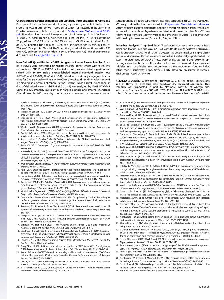
Table 1. Sensitivity and specificity of NanoDisk-MS for active TB detection
HTI cohort (n = 201),group Positive results/total no.
Sensitivity, %(95% CI)
Specificity, %(95% CI)
HIV− groups (n = 111)Pulmonary TB Mtb culture+ 25/27 92.6 (76.6–97.9)LTBI 4/31 87.1 (71.2–94.9)NTM 3/32 90.6 (75.8–96.8)Healthy controls 0/21 100 (84.5–100)
HIV+ groups (n = 90)Pulmonary TB
Mtb culture+ 21/23 91.3 (73.2–97.6)Mtb culture− 14/17 82.4 (59.0–93.8)
Extrapulmonary TBMtb culture+ 12/13 92.3 (66.7–98.6)Mtb culture− 6/8 75.0 (40.9–92.9)
Non-TB 3/29 89.7 (73.6–96.4)
Pulmonary TB Mtb culture+ (n=27)
Healthy Control (n=21)
LTBI (n=31)
NTM (n=32)
A
B
Non-TB Control (n=29)
C
Pulmonary TB Mtb culture+ (n=23) Pulmonary TB Mtb culture- (n=17)
Extrapulmonary TB Mtb culture+ (n=13) Extrapulmonary TB Mtb culture- (n=8)
HIV- Mtb+Pulmonary
(n=27)
4030
10
5
0
HIV+ Mtb+Pulmonary
(n=23)
15
**C
once
ntra
tion
of
Bio
mar
kers
(nM
)
20
25
CFP-10ESAT-6
0 3.5 7nM
HIV-
HIV+
Fig. 3. Identification of active TB in adult patients. (A and B) Serum CFP-10 andESAT-6 concentrations in adult HIV-negative (A) and HIV-positive (B) groups.Each column represents CFP-10 (upper cell, in red) and ESAT-6 (lower cell, inblue) results from a subject, ranked by high to low CFP-10 concentration. An-tigen levels are indicated by the color intensity in the matching gradient bars.(C) Combined Mtb antigen (CFP-10 + ESAT-6) concentrations and means (blackline) for the indicated groups. Data represent mean. n = 3. **P < 0.01.
3972 | www.pnas.org/cgi/doi/10.1073/pnas.1621360114 Liu et al.

We selected CFP-10 and ESAT-6 as biomarkers because theyare known to modulate several key immunologic pathways toreduce mycobacterial clearance and promote host cell lysis,which may enhance Mtb dissemination. Thus, they may be morerelevant than other abundant Mtb proteins, like antigen 85b,which is expressed by virulent and attenuated Mtb complexmycobacteria, including M. bovis bacillus Calmette–Guérin, toaffect proliferation and perhaps uptake. Both CFP-10 andESAT-6 are expressed by the RD-1 locus found in virulent butnot attenuated Mtb complex strains, and RD-1 transfer intoM. bovis bacillus Calmette–Guérin has been shown to increasevirulence and survival (19). CFP-10 and ESAT-6 also appear toplay important roles in the maintenance of Mtb infection byinhibiting mycobacterial clearance (33).NanoDisk-MS demonstrated similar sensitivity for both PTB
cases (87.5%) and clinically challenging EPTB cases (85.7%) inthe HIV-positive study population, where it equaled or signifi-cantly outperformed Xpert MTB/RIF in terms of sensitivity fordetecting EPTB using highly invasive lymph node (84%), pleuralfluid (17%), and cerebrospinal fluid (56%) samples in a meta-analysis (34), and significantly outperformed AFB smear (31%),mycobacteria growth indicator tube culture (69%), and XpertMTB/RIF (66%) in another study (35). Moreover, our resultswere achieved irrespective of Mtb culture status, and althoughgreater sensitivity was achieved in culture-positive samples, re-sults from culture-negative samples markedly outperformed theWHO-defined 66% optimal sensitivity for new high-prioritynonsputum diagnostic tests (13). NanoDisk-MS also accuratelydiagnosed active TB cases in patients with HIV coinfection, whichcan adversely skew the results of blood-based host response im-munoassays (e.g., IGRA and T-cell activation marker–tuberculosisassay) (26, 36), in which serum Mtb peptide concentrations werefound to equal or exceed those found in HIV-negative subjects,supporting robust assay sensitivity. NanoDisk-MS exhibited dif-ferential specificities with healthy (100%) and HIV-positive(89.7%) controls, LTBI cases (87.1%), and NTM cases (90.6%),however. Owing to the increased TB risk in LTBI and HIV-positivegroups and possible strain-specific NTM false-positives, longitudinalstudies of LTBI and HIV-positive populations and strain identifi-cation of NTM cases are needed to further refine the specificity ofthe test in these groups.NanoDisk-MS also was able to precisely quantify serum anti-
gen concentrations, a highly desirable feature for monitoringresponses to anti-TB therapy, given that assays currently used to
monitor treatment response provide only qualitative or semi-quantitative results (smear and culture) or exhibit significantlatency (Xpert). A study evaluating serial sputum samples frompatients with smear-positive TB found that assay positivity andgrade decreased linearly starting at ∼2 wk (smear), 4 wk (cul-ture), or 6 wk (Xpert) posttreatment (37). CD4+ T-cell responsesalso have been reported to differentiate LTBI and active TB casesand to monitor treatment responses, although only HIV-negativeand culture- and/or smear-positive PTB patients were analyzedusing this labor-intensive method, so that its clinical utility remainsunclear (38). Circulating miRNA levels also may serve as TB bio-markers, but this idea has not yet been tested in longitudinal clinicalstudies (39). Results from proof-of-principle NanoDisk-MS studiesanalyzing Mtb peptide responses during anti-TB therapy detectedmarked reductions in both HIV-positive and -negative TB caseson initiation of anti-TB therapy, but these studies were not designedto measure rates of decline or for comparisons with other methods.Future prospective longitudinal studies with frequent sampling areneeded to determine how Mtb peptide clearance corresponds topatient phenotypes and treatment outcomes. Should such a studyconfirm our preliminary results, the NanoDisk-MS assay wouldbe expected improve the speed, accuracy, and efficiency of con-ventional procedures, which require months to estimate therapeuticefficacy or identify treatment resistance.NanoDisk-MS was designed to enable multiplex detection of
serum Mtb antigen concentrations for robust detection of activeTB cases. Serum CFP-10 was detectable at lower concentrationsthan ESAT-6, perhaps owing to variable expression levels ofESAT-6 and CFP-10 in different strains of Mtb (40, 41). How-ever, host β2 microglobulin also has been reported to bindESAT-6 to mask a tryptic cleavage site (amino acids 90–95) requiredfor the 1,900.95 m/z ESAT-6 peptide (16, 42), and thus may inhibitESAT-6 cleavage to decrease the apparent serum ESAT-6 levels inour assay.Nanodisk-MS requires only a simple, low-volume blood draw
rather than invasive biopsies that may be required for othermethods. Many hospitals and microbiology laboratories routinelyuse MALDI-TOF MS for microbial identification (43), whichcould allow rapid clinical translation of a NanoDisk-MS diagnosticassay. Further reductions in operator time and assay cost, alongwith improvements in instrument portability, are needed to meetWHO guidelines for an optimal noninvasive TB assay. Larger,randomized prospective studies are warranted to confirm the re-sults of this proof-of-principle pilot study, but a clinical methodthat shares the advantages of our approach should facilitate earlierinterventions and better patient outcomes (44). The NanoDisk-MSassay also opens up new possibilities for the diagnosis of a widevariety of other infectious diseases, because it should be relativelysimple to generate similar assays to rapidly quantify disease-associatedlow-abundance antigens in blood and other body fluids.
Materials and MethodsClinical Samples. The HTI cohort that served as a source of case and controlsamples in this study were the subjects of a large, population-based TBsurveillance study that performed active surveillance of all confirmed TB casesin Houston/Harris County, Texas between 1995 and 2004. Because of itsmandate to collect all active TB cases, the HTI archived samples from a varietyof TB diseasemanifestations, including HIV-negative and -positive pulmonaryand extrapulmonary TB cases with both positive and negative culture results.Serum samples were obtained from HTI subjects who were notified of thepotential risks of study participation and provided written informed consent.Demographic, microbiology, and diagnostic data are summarized in SI Ap-pendix, Table S2.
Microwave-Assisted and Overnight Tryptic Digestion of Human Serum Samples.Serum samples (100 μL) were mixed with 400 μL of 100 mM NH4HCO3 and10 μL of 1 mg/mL sequencing-grade modified trypsin (Promega) in 1.5-mLEppendorf tubes, placed in a 1,000-mL water bath, irradiated in a 1200 Wmicrowave oven at 20% power for 20 min, and then mixed with a 0.1% finalconcentration of trifluoroacetic acid. Overnight trypsin digestions were in-cubated at 37 °C for 12 h using the same amount of trypsin and the samebuffer conditions without a water bath and microwave irradiation.
0 120 240 360
0 120 240 360
0 120 240 360 0 120 240 360 0 120 240 360
0 120 240 360 0 120 240 360
0 120 240 360 0 120 240 3600
5
1020000450
CFP-10
ESAT-6
0
5
1 020010129
0
5
1 020010244
0
5
1020020224
0
2
420000191
0
2
420010035
0
2
420010170
0
2
420020493
0
2
420030106
Biom
arke
r con
cent
raon
(nM
)
Days a er therapy ini a on
Fig. 4. Evaluation of anti-TB treatment efficacy. CFP-10 and ESAT-6 quan-titation in archived serum samples of HIV-negative TB patients during anti-TB therapy (2–11 mo). Treatment start and end dates are indicated by blueand red dotted lines, respectively. Data represent mean ± SEM. n = 3.
Liu et al. PNAS | April 11, 2017 | vol. 114 | no. 15 | 3973
MED
ICALSC
IENCE
SEN
GINEE
RING
Characterization, Functionalization, and Antibody Immobilization of Nanodisks.Bare nanodisks were fabricated following a previously reported protocol andstored in ACS grade 99.9% isopropyl alcohol for maximum stability (45).Functionalization details are reported in SI Appendix, Materials and Meth-ods. Functionalized nanodisk suspensions (1 mL) were pelleted for 5 min at10,000 × g, vacuum-dried, suspended in 1 mL of PBS (pH 8.6) containing20 μg of anti-1593.75 or anti-1900.95 antibody (GL Biochem), mixed for 2 hat 25 °C, pelleted for 5 min at 10,000 × g, incubated for 30 min in 1 mL of200 mM Tris pH 7/100 mM NaCl solution, washed three times with PBS(pH 7.4), pelleted and suspended in 60 μL of PBS (pH 7.4), and then stored at4 °C until use (SI Appendix, Fig. S7B).
NanoDisk-MS Quantification of Mtb Antigens in Human Serum Samples. Stan-dard curves were generated by spiking healthy donor serum with 0–100 nMrecombinant CFP-10 or ESAT-6, subjected to microwave-assisted digestion andspiked with 10 nM stable isotope-labeled internal standard peptide (m/z1,603.60 and 1,910.80; GenScript USA), mixed with antibody-conjugated nano-disks for 2 h, pelleted for 5 min at 10,000 × g, washed three times with 1 mg/mL1,2-dioleoyl-sn-glycero-3-phospho-L-serine (Avanti Polar Lipids), suspended in6 μL of deionized water and 4.5 μL (1.5 μL × 3) was analyzed by MALDI-TOF-MS,using the MS intensity ratios of each target peptide and internal standards.Clinical sample MS intensity ratios were converted to absolute molar
concentrations through substitution into this calibration curve. The NanoDisk-MS assay is described in more detail in SI Appendix, Materials and Methods.Serum samples used to determine antigen-derived MS signals from trypsinizedserum with or without Dynabead-mediated enrichment or NanoDisk-MS en-richment and comatrix activity were made by serially diluting TB patient serumwith healthy human serum (2×, 4×, 8×, 16×, and 32×).
Statistical Analyses. GraphPad Prism 7 software was used to generate heatmaps and to calculate one-way ANOVA with Bonferroni’s posttest or Kruskal–Wallis one-way ANOVA with Dunn’s posttest as determined by sample distri-bution and variance. Differences were considered statistically significant at P <0.05. The diagnostic accuracy of tests were evaluated using the receiving op-erating characteristic curve. The cutoff values were estimated at various sen-sitivities and specificities and determined at the maximum Youden indexvalue, that is, sensitivity + specificity − 1 (46). Data are presented as mean ±SEM unless noted otherwise.
ACKNOWLEDGMENTS. We thank Professor X. C. Li for helpful discussionsand D. Hawk for excellent mass spectrometry technical assistance. Thisresearch was supported in part by National Institute of Allergy andInfectious Diseases Grants R01 Al113725-01A1 and R01 AI122932-01A1, theJohn S. Dunn Foundation, and the Intramural Research Programs of the NIH.
1. Zumla A, George A, Sharma V, Herbert N, Baroness Masham of Ilton (2013) WHO’s2013 global report on tuberculosis: Successes, threats, and opportunities. Lancet 382(9907):1765–1767.
2. Marais BJ, Schaaf HS, Graham SM (2014) Child health and tuberculosis. Lancet RespirMed 2(4):254–256.
3. Monkongdee P, et al. (2009) Yield of acid-fast smear and mycobacterial culture fortuberculosis diagnosis in people with human immunodeficiency virus. Am J Respir CritCare Med 180(9):903–908.
4. World Health Organization (2013) Systematic Screening for Active Tuberculosis:Principles and Recommendations. (WHO, Geneva).
5. Dunlap NE, et al. (2000) Diagnostic standards and classification of tuberculosis inadults and children. Am J Respir Crit Care Med 161(4 Pt 1):1376–1395.
6. Swaminathan S, Rekha B (2010) Pediatric tuberculosis: Global overview and chal-lenges. Clin Infect Dis 50(Suppl 3):S184–S194.
7. Evans CA (2011) GeneXpert: A game-changer for tuberculosis control? PLoS Med 8(7):e1001064.
8. Ioannidis P, et al. (2011) Cepheid GeneXpert MTB/RIF assay for Mycobacterium tu-berculosis detection and rifampin resistance identification in patients with substantialclinical indications of tuberculosis and smear-negative microscopy results. J ClinMicrobiol 49(8):3068–3070.
9. World Health Organization (2014) Xpert MTB/RIF: WHO Policy Update and ImplementationManual. (WHO, Geneva).
10. Reid MJA, Shah NS (2009) Approaches to tuberculosis screening and diagnosis inpeople with HIV in resource-limited settings. Lancet Infect Dis 9(3):173–184.
11. Horne DJ, et al. (2010) Sputum monitoring during tuberculosis treatment for predictingoutcome: Systematic review and meta-analysis. Lancet Infect Dis 10(6):387–394.
12. Denkinger CM, Pai M, Patel M, Menzies D (2013) Gamma interferon release assay formonitoring of treatment response for active tuberculosis: An explosion in the spa-ghetti factory. J Clin Microbiol 51(2):607–610.
13. World Health Organization (2014) High-Priority Target Product Profiles for New TuberculosisDiagnostics: Report of a Consensus Meeting. (WHO, Geneva).
14. Centers for Disease Control and Prevention (2010) Updated guidelines for using in-terferon gamma release assays to detect Mycobacterium tuberculosis infection—United States. MMWR Recomm Rep 59(RR-5):1–25.
15. Sweeney TE, Braviak L, Tato CM, Khatri P (2016) Genome-wide expression for di-agnosis of pulmonary tuberculosis: A multicohort analysis. Lancet Respir Med 4(3):213–224.
16. Sreejit G, et al. (2014) The ESAT-6 protein of Mycobacterium tuberculosis interactswith beta-2-microglobulin (β2M) affecting antigen presentation function of macro-phage. PLoS Pathog 10(10):e1004446.
17. Simossis VA, Heringa J (2003) The PRALINE online server: Optimising progressivemultiple alignment on the web. Comput Biol Chem 27(4-5):511–519.
18. van Ingen J, de Zwaan R, Dekhuijzen R, Boeree M, van Soolingen D (2009) Region ofdifference 1 in nontuberculous Mycobacterium species adds a phylogenetic andtaxonomical character. J Bacteriol 191(18):5865–5867.
19. Cardona P-J (2012) Understanding Tuberculosis: Deciphering the Secret Life of theBacilli (In Tech, Rijeka, Croatia).
20. Feng TT, et al. (2011) Novel monoclonal antibodies to ESAT-6 and CFP-10 antigens forELISA-based diagnosis of pleural tuberculosis. Int J Tuberc Lung Dis 15(6):804–810.
21. Arend SM, et al. (2002) Tuberculin skin testing and in vitro T cell responses to ESAT-6 andculture filtrate protein 10 after infection with Mycobacterium marinum or M. kansasii.J Infect Dis 186(12):1797–1807.
22. Lai CC, et al. (2010) Increasing incidence of nontuberculous mycobacteria, Taiwan,2000-2008. Emerg Infect Dis 16(2):294–296.
23. Tirumalai RS, et al. (2003) Characterization of the lowmolecular weight human serumproteome. Mol Cell Proteomics 2(10):1096–1103.
24. Sun W, et al. (2006) Microwave-assisted protein preparation and enzymatic digestionin proteomics. Mol Cell Proteomics 5(4):769–776.
25. Wei J, Buriak JM, Siuzdak G (1999) Desorption-ionization mass spectrometry on po-rous silicon. Nature 399(6733):243–246.
26. Portevin D, et al. (2014) Assessment of the novel T-cell activation marker-tuberculosisassay for diagnosis of active tuberculosis in children: A prospective proof-of-conceptstudy. Lancet Infect Dis 14(10):931–938.
27. Zeka AN, Tasbakan S, Cavusoglu C (2011) Evaluation of the GeneXpert MTB/RIF assayfor rapid diagnosis of tuberculosis and detection of rifampin resistance in pulmonaryand extrapulmonary specimens. J Clin Microbiol 49(12):4138–4141.
28. Getahun H, Gunneberg C, Granich R, Nunn P (2010) HIV infection-associated tuber-culosis: The epidemiology and the response. Clin Infect Dis 50(Suppl 3):S201–S207.
29. Abeyewickreme I, Punchihewa N, Maw-Naing A (2012) Success of tuberculosis andHIV collaboration. WHO South-East Asia J Public Health 1(4):359–361.
30. Jiang W, et al. (2009) Plasma levels of bacterial DNA correlate with immune activationand the magnitude of immune restoration in persons with antiretroviral-treated HIVinfection. J Infect Dis 199(8):1177–1185.
31. Theron G, et al. (2011) Evaluation of the Xpert MTB/RIF assay for the diagnosis ofpulmonary tuberculosis in a high HIV prevalence setting. Am J Respir Crit Care Med184(1):132–140.
32. Schilirò G, Minniti C, Sciotto A, Bellino A, Russo A (1986) T-lymphocyte subpopulationchanges during hemolysis in glucose-6-phosphate dehydrogenase (G6PD)-deficientchildren. Am J Hematol 21(2):173–176.
33. Prendergast KA, et al. (2016) The Ag85B protein of the BCG vaccine facilitates mac-rophage uptake but is dispensable for protection against aerosol Mycobacteriumtuberculosis infection. Vaccine 34(23):2608–2615.
34. World Health Organization (2013) Policy Update: Xpert MTB/RIF Assay for the Diagnosisof Pulmonary and Extrapulmonary TB in Adults and Children. (WHO, Geneva).
35. Cavanaugh JS, et al. (2016) Comparative yield of different diagnostic tests for tu-berculosis among people living with HIV in western Kenya. PLoS One 11(3):e0152364.
36. Mandalakas AM, et al. (2008) High level of discordant IGRA results in HIV-infectedadults and children. Int J Tuberc Lung Dis 12(4):417–423.
37. Friedrich SO, et al.; Pan African Consortium for the Evaluation of Anti-tuberculosisAntibiotics (PanACEA) (2013) Assessment of the sensitivity and specificity of XpertMTB/RIF assay as an early sputum biomarker of response to tuberculosis treatment.Lancet Respir Med 1(6):462–470.
38. Adekambi T, et al. (2015) Biomarkers on patient T cells diagnose active tuberculosisand monitor treatment response. J Clin Invest 125(5):1827–1838.
39. Zhang H, et al. (2014) Identification of serum microRNA biomarkers for tuberculosisusing RNA-seq. PLoS One 9(2):e88909.
40. Uplekar S, Heym B, Friocourt V, Rougemont J, Cole ST (2011) Comparative genomicsof Esx genes from clinical isolates of Mycobacterium tuberculosis provides evidencefor gene conversion and epitope variation. Infect Immun 79(10):4042–4049.
41. Arend SM, et al. (2005) ESAT-6 and CFP-10 in clinical versus environmental isolates ofMycobacterium kansasii. J Infect Dis 191(8):1301–1310.
42. Teutschbein J, et al. (2009) A protein linkage map of the ESAT-6 secretion system 1(ESX-1) of Mycobacterium tuberculosis. Microbiol Res 164(3):253–259.
43. Patel R (2013) MALDI-TOF mass spectrometry: Transformative proteomics for clinicalmicrobiology. Clin Chem 59(2):340–342.
44. Denkinger CM, Grenier J, Minion J, Pai M (2012) Promise versus reality: Optimism biasin package inserts for tuberculosis diagnostics. J Clin Microbiol 50(7):2455–2461.
45. Godin B, et al. (2012) Discoidal porous silicon particles: Fabrication and biodistributionin breast cancer bearing mice. Adv Funct Mater 22(20):4225–4235.
46. Youden WJ (1950) Index for rating diagnostic tests. Cancer 3(1):32–35.
3974 | www.pnas.org/cgi/doi/10.1073/pnas.1621360114 Liu et al.