M.Sc.Medical Physics Uni.Dept 2010-11syllabus.b-u.ac.in/unidepts/1011/medical_physics.pdf · Flip...
37
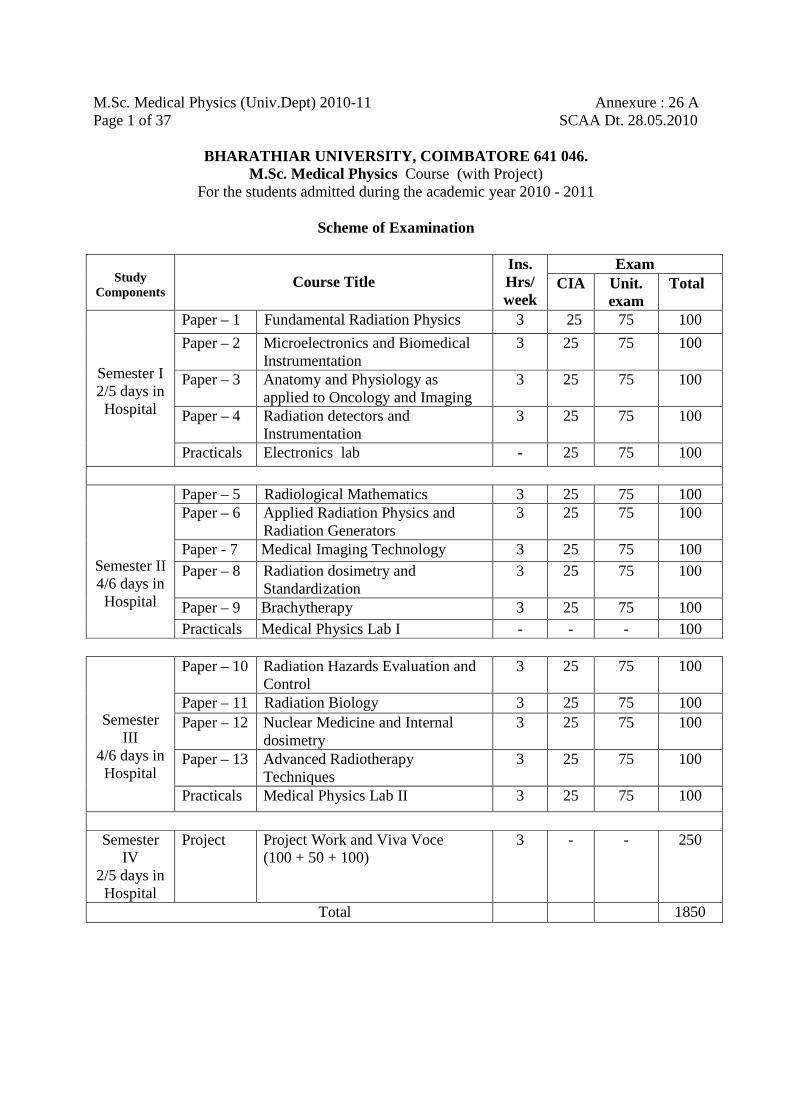
M.Sc. Medical Physics (Univ.Dept) 2010-11 Annexure : 26 A Page 1 of 37 SCAA Dt. 28.05.2010 BHARATHIAR UNIVERSITY, COIMBATORE 641 046. M.Sc. Medical Physics Course (with Project) For the students admitted during the academic year 2010 - 2011 Scheme of Examination Study Components Course Title Ins. Hrs/ week Exam CIA Unit. exam Total Semester I 2/5 days in Hospital Paper – 1 Fundamental Radiation Physics 3 25 75 100 Paper – 2 Microelectronics and Biomedical Instrumentation 3 25 75 100 Paper – 3 Anatomy and Physiology as applied to Oncology and Imaging 3 25 75 100 Paper – 4 Radiation detectors and Instrumentation 3 25 75 100 Practicals Electronics lab - 25 75 100 Semester II 4/6 days in Hospital Paper – 5 Radiological Mathematics 3 25 75 100 Paper – 6 Applied Radiation Physics and Radiation Generators 3 25 75 100 Paper - 7 Medical Imaging Technology 3 25 75 100 Paper – 8 Radiation dosimetry and Standardization 3 25 75 100 Paper – 9 Brachytherapy 3 25 75 100 Practicals Medical Physics Lab I - - - 100 Semester III 4/6 days in Hospital Paper – 10 Radiation Hazards Evaluation and Control 3 25 75 100 Paper – 11 Radiation Biology 3 25 75 100 Paper – 12 Nuclear Medicine and Internal dosimetry 3 25 75 100 Paper – 13 Advanced Radiotherapy Techniques 3 25 75 100 Practicals Medical Physics Lab II 3 25 75 100 Semester IV 2/5 days in Hospital Project Project Work and Viva Voce (100 + 50 + 100) 3 - - 250 Total 1850
Transcript of M.Sc.Medical Physics Uni.Dept 2010-11syllabus.b-u.ac.in/unidepts/1011/medical_physics.pdf · Flip...
M.Sc. Medical Physics (Univ.Dept) 2010-11 Annexure : 26 A Page 1 of 37 SCAA Dt. 28.05.2010
BHARATHIAR UNIVERSITY, COIMBATORE 641 046. M.Sc. Medical Physics Course (with Project)
For the students admitted during the academic year 2010 - 2011
Scheme of Examination
Study
Components
Course Title
Ins. Hrs/ week
Exam CIA Unit.
exam Total
Semester I 2/5 days in Hospital
Paper – 1 Fundamental Radiation Physics 3 25 75 100
Paper – 2 Microelectronics and Biomedical Instrumentation
3 25 75 100
Paper – 3 Anatomy and Physiology as applied to Oncology and Imaging
3 25 75 100
Paper – 4 Radiation detectors and Instrumentation
3 25 75 100
Practicals Electronics lab - 25 75 100
Semester II 4/6 days in Hospital
Paper – 5 Radiological Mathematics 3 25 75 100 Paper – 6 Applied Radiation Physics and
Radiation Generators 3 25 75 100
Paper - 7 Medical Imaging Technology 3 25 75 100 Paper – 8 Radiation dosimetry and
Standardization 3 25 75 100
Paper – 9 Brachytherapy 3 25 75 100 Practicals Medical Physics Lab I - - - 100
Semester III
4/6 days in Hospital
Paper – 10 Radiation Hazards Evaluation and Control
3 25 75 100
Paper – 11 Radiation Biology 3 25 75 100 Paper – 12 Nuclear Medicine and Internal
dosimetry 3 25 75 100
Paper – 13 Advanced Radiotherapy Techniques
3 25 75 100
Practicals Medical Physics Lab II 3 25 75 100
Semester
IV 2/5 days in Hospital
Project Project Work and Viva Voce (100 + 50 + 100)
3 - - 250
Total 1850

M.Sc. Medical Physics (Univ.Dept) 2010-11 Annexure : 26 A Page 2 of 37 SCAA Dt. 28.05.2010
SEMESTER - I
PAPER 1
FUNDAMENTAL RADIATION PHYSICS
Unit 1: Non-ionizing Radiation Physics (9 hours) Electromagnetic spectrum- Different sources of Non Ionising radiation-Radiofrequency, Microwaves, Infrared, Visible and Ultra violet radiation production, physical properties and their interaction with tissues- Electrical Impedance and Biological Impedance - Principle and theory of thermography – applications-Laser: theory and mechanism- interaction of laser radiation with tissues – photothermal -photochemical – photoablation – electro mechanical effect- Lasers in dermatology, oncology and cell biology. Unit 2: Nuclear Physics (9 hours) Radioactivity – General properties of alpha, beta and gamma rays – Laws of radioactivity – Laws of successive transformations – Natural radioactive series – radioactive equilibrium – Alpha ray spectra – Beta ray spectra – Gamma emission – Electron capture – Internal conversion – Nuclear isomerism – Artificial radioactivity – Nuclear cross sections – Elementary ideas of fission and reactors – Fusion. Unit 3: Radiation Quantities and Units (6 hours) Radiation quantities and units – Radiometry – Particle flux and fluence – Energy flux and fluence – Cross section – Linear and mass attenuation coefficients – Mass energy transfer and mass energy absorption coefficients – Stopping power – LET - Radiation chemical yield – W value – Dosimetry – Energy imparted –Absorbed dose- Radiation and tissue weighting factors, equivalent dose, effective dose, committed equivalent dose, committed effected dose – Concepts of collective dose – KERMA-CEMA – Exposure – Air kerma rate constant – Charged particle equilibrium (CPE) – Relationship between kerma, absorbed dose and exposure under CPE – Dose equivalent – Ambient and directional dose equivalents [(H*(d) and H’(d)] – individual dose equivalent penetrating Hp(d) – Individual dose equivalent superficial Hs(d). Unit 4: Radiation Sources (5 hours) Radiation sources – Natural and artificial radioactive sources – Large scale production of isotopes – Reactor produced isotopes – Cyclotron produced isotopes – Fission products – industrial uses – Telecobalt and Brachy Caesium sources – Gold seeds – Tantalum wire – 125 I Sources – Beta ray applicators – Thermal and fast neutron sources – Preparation of tracers and labelled compounds – Preparation of ratio colloids.

M.Sc. Medical Physics (Univ.Dept) 2010-11 Annexure : 26 A Page 3 of 37 SCAA Dt. 28.05.2010
Unit 5: Interaction of Radiation with Matter (11 hours) Interaction of electromagnetic radiation with matter Exponential attenuation – Thomson scattering – Photoelectric and Compton process and energy absorption – Pair production – Attenuation and mass energy absorption coefficients – Relative importance of various processes. Interaction of charged particles with matter – Classical theory of inelastic collisions with atomic electrons – Energy loss per ion pair by primary and secondary ionization – Dependence of collision energy losses on the physical and chemical state of the absorber – Cerenkov radiation – Electron absorption process – Scattering Excitation and Ionization – Radioactive collision – Bremmstrahlung – Range energy relation – Continuous slowing down approximation (CSDA) – straight ahead approximation and detour factors – transmission and depth dependence methods for determination of particle penetration - empirical relations between range and energy – Back scattering. References:
1. J. R. Greening, Medical Physics, North Holland Publishing Co., New York, 1999. 2. Markolf H. Neimz, Laser-Tissue Interactions, Springer Verlag, Germany, 1996. 3. W.R.Hendee, Medical Radiation Physics, Year Book Medical Publishers Inc.,
London, 1981. 4. Lapp R.E. Nuclear Radiation Physics. 5. Oliver R. Radiation Physics in Radiology
M.Sc. Medical Physics (Univ.Dept) 2010-11 Annexure : 26 A Page 4 of 37 SCAA Dt. 28.05.2010
PAPER 2 MICROELETRONICS AND BIOMEDICAL INSTRUMENTATION
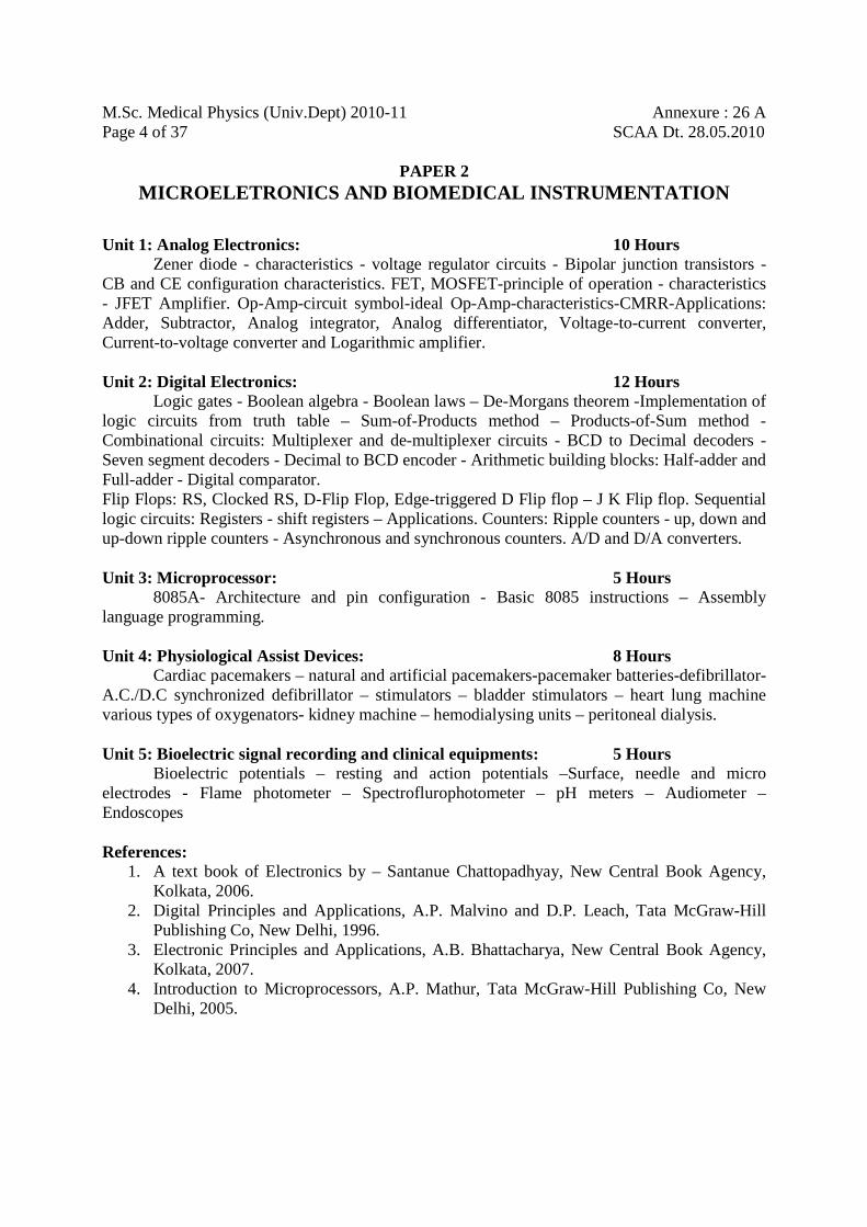
Unit 1: Analog Electronics: 10 Hours Zener diode - characteristics - voltage regulator circuits - Bipolar junction transistors - CB and CE configuration characteristics. FET, MOSFET-principle of operation - characteristics - JFET Amplifier. Op-Amp-circuit symbol-ideal Op-Amp-characteristics-CMRR-Applications: Adder, Subtractor, Analog integrator, Analog differentiator, Voltage-to-current converter, Current-to-voltage converter and Logarithmic amplifier. Unit 2: Digital Electronics: 12 Hours Logic gates - Boolean algebra - Boolean laws – De-Morgans theorem -Implementation of logic circuits from truth table – Sum-of-Products method – Products-of-Sum method - Combinational circuits: Multiplexer and de-multiplexer circuits - BCD to Decimal decoders - Seven segment decoders - Decimal to BCD encoder - Arithmetic building blocks: Half-adder and Full-adder - Digital comparator. Flip Flops: RS, Clocked RS, D-Flip Flop, Edge-triggered D Flip flop – J K Flip flop. Sequential logic circuits: Registers - shift registers – Applications. Counters: Ripple counters - up, down and up-down ripple counters - Asynchronous and synchronous counters. A/D and D/A converters. Unit 3: Microprocessor: 5 Hours 8085A- Architecture and pin configuration - Basic 8085 instructions – Assembly language programming. Unit 4: Physiological Assist Devices: 8 Hours Cardiac pacemakers – natural and artificial pacemakers-pacemaker batteries-defibrillator-A.C./D.C synchronized defibrillator – stimulators – bladder stimulators – heart lung machine various types of oxygenators- kidney machine – hemodialysing units – peritoneal dialysis. Unit 5: Bioelectric signal recording and clinical equipments: 5 Hours Bioelectric potentials – resting and action potentials –Surface, needle and micro electrodes - Flame photometer – Spectroflurophotometer – pH meters – Audiometer – Endoscopes References:
1. A text book of Electronics by – Santanue Chattopadhyay, New Central Book Agency, Kolkata, 2006.
2. Digital Principles and Applications, A.P. Malvino and D.P. Leach, Tata McGraw-Hill Publishing Co, New Delhi, 1996.
3. Electronic Principles and Applications, A.B. Bhattacharya, New Central Book Agency, Kolkata, 2007.
4. Introduction to Microprocessors, A.P. Mathur, Tata McGraw-Hill Publishing Co, New Delhi, 2005.

M.Sc. Medical Physics (Univ.Dept) 2010-11 Annexure : 26 A Page 5 of 37 SCAA Dt. 28.05.2010
PAPER 3
ANATOMY AND PHYSIOLOGY AS APPLIED TO ONCOLOGY AND IMAGING
Unit 1:Structure & function of organs, systems & their common diseases: 12 Hours Skin, Lymphatic system, Bone and muscle, Nervous, Endocrine, Cardiovascular, Respiratory, Digestive (Gastro-Intestinal), Urinary, Reproductive, Eye and ear. Unit 2: Basic, Radiographic anatomy and tumor pathology 8 Hours Anatomy of human body, nomenclature & Surface anatomy, Radiographic Anatomy (including cross sectional anatomy – Identify the different organs/structures on plain x-rays, CT scans and other available imaging modalities. Normal anatomy & deviation for abnormalities. Tumor pathology and carcinogenesis, common pathological features of cancers and interpretation of clinico-pathological data. Unit 3: Clinical aspects of Radiation Oncology 8 Hours Radiation therapy, Surgery, Chemotherapy, Hormone Therapy, Immunotherapy & Radionuclide therapy, Benign and malignant disease, Methods of spread of malignant disease, Staging and grading systems, Treatment intent – Curative & Palliative, Cancer prevention and public education and Early detection & Screening. Patient management on treatment – side effects related to radiation and dose – Acute & Late – Monitoring and common management of side effects – Information and communication. Unit 4: Site specific signs, symptoms, diagnosis and management: 8 Hours Head and Neck, Breast, Gynecological, Gastro-Intestinal tract, Genito-Urinary, Lung & Thorax, Lymphomas & Leukemias & Other cancers including AIDS related cancers. Unit 5: Professional aspects and role of medical physicists: 4 Hours General patient care - Principles of professional practice – Medical terminology – Research & professional writing – patient privacy – Ethical & cultural issues. Legal aspects – Confidentiality, informed consent, Health and safety. References:
1. Meschan. Normal Radiation Anatomy 2. Hollinshead W.H. Text Book of Anatomy

M.Sc. Medical Physics (Univ.Dept) 2010-11 Annexure : 26 A Page 6 of 37 SCAA Dt. 28.05.2010
PAPER 4
RADIATION DETECTORS AND INSTRUMENTATION
Unit 1: Principles of Radiation Detection-Using Ion Chambers 9 Hours Principles of Radiation detection and measurement – Basic Principles of radiation detection – Gas Filled detectors – Ionisation chambers – Theory and design – Construction of condenser type chambers and thimble chambers – Gas multiplication – Proportional and GM Counters – Characteristics of organic and inorganic counters – Dead time and recovery time – quenching. Unit 2: Principles of Radiation Detection-Using other detectors 9 Hours Scintillation detectors – Semiconductor detectors – Chemical systems – Radiographic and Radio chromic films – Thermo luminescent Dosimeters (TLD) – Optically stimulated Luminescence dosimeters (OSLD) – Radiophoto luminescent dosimeters - Neutron Detectors – Nuclear track emulsions for fast neutrons – Solid State Nuclear track (SSNTD) detectors – calorimeters – New Developments. Unit 3: Dosimetry Instruments 8 Hours Dosimeters based on condenser chambers – Pocket chambers – Dosimeters based on current measurement – Different types of electrometers – MOSFET, Vibrating condenser and Varactor bridge types – Secondary standard therapy level dosimeters – Farmers Dosimeters – Radiation field analyzer (RFA) – Radioisotope calibrator – Multipurpose dosimeters – Water phantom dosimetry systems – Brachytheraphy dosimeters – Thermo luminescent dosimeter readers for medical applications – Calibration and maintenance of dosimeters. Unit 4: Radiation Protection instruments 8 Hours Instruments for personnel monitoring – TLD badge readers – PM film densitometers – Glass dosimeters readers - Digital pocket dosimeters using solid state devices and GM counters – Teletector – industrial gamma radiography survey meter – Gamma area (Zone) alarm monitors - Contamination monitors for alpha, beta and gamma radiation – Hand and Foot monitors _Laundry and Portal Monitors - Scintillation monitors for X and gamma radiations - Neutron Monitors, Tissue equivalent survey meters – Flux meter and dose equivalent monitors – Pocket neutron monitors -Teledose systems. Unit 5: Nuclear Medicine instruments 6 Hours Instruments for counting and spectrometry – Portable counting systems for alpha and beta radiation – Gamma ray spectrometers – Multichannel Analyzer – Liquid scintillation counting system – RIA counters – Whole body counters – Air Monitors for radioactive particulates and gases. Details of commercially available instruments and systems.
M.Sc. Medical Physics (Univ.Dept) 2010-11 Annexure : 26 A Page 7 of 37 SCAA Dt. 28.05.2010
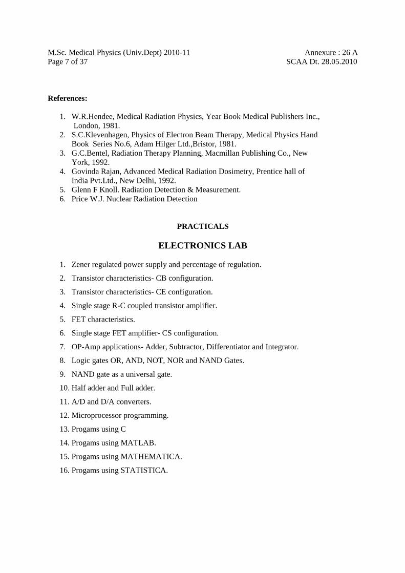
References:
1. W.R.Hendee, Medical Radiation Physics, Year Book Medical Publishers Inc., London, 1981.
2. S.C.Klevenhagen, Physics of Electron Beam Therapy, Medical Physics Hand Book Series No.6, Adam Hilger Ltd.,Bristor, 1981.
3. G.C.Bentel, Radiation Therapy Planning, Macmillan Publishing Co., New York, 1992.
4. Govinda Rajan, Advanced Medical Radiation Dosimetry, Prentice hall of India Pvt.Ltd., New Delhi, 1992.
5. Glenn F Knoll. Radiation Detection & Measurement. 6. Price W.J. Nuclear Radiation Detection
PRACTICALS
ELECTRONICS LAB
1. Zener regulated power supply and percentage of regulation.
2. Transistor characteristics- CB configuration.
3. Transistor characteristics- CE configuration.
4. Single stage R-C coupled transistor amplifier.
5. FET characteristics.
6. Single stage FET amplifier- CS configuration.
7. OP-Amp applications- Adder, Subtractor, Differentiator and Integrator.
8. Logic gates OR, AND, NOT, NOR and NAND Gates.
9. NAND gate as a universal gate.
10. Half adder and Full adder.
11. A/D and D/A converters.
12. Microprocessor programming.
13. Progams using C
14. Progams using MATLAB.
15. Progams using MATHEMATICA.
16. Progams using STATISTICA.

M.Sc. Medical Physics (Univ.Dept) 2010-11 Annexure : 26 A Page 8 of 37 SCAA Dt. 28.05.2010
SEMESTER - II
PAPER 5
RADIOLOGICAL MATHEMATICS
Unit 1: Probability, Statistic and Errors 12 Hours Probability – addition and multiplication laws of probability, conditional probability, population, variates, collection, tabulation and graphical representation of data-Basic ideas of statistical distributions frequency distributions, averages or measures of central tendency, arithmetic mean, properties of arithmetic mean, median, mode, geometric mean, harmonic mean, dispersion, standard deviation, root mean square deviation, standard error and variance, moments, skewness and kurtosis-Application to radiation detection – uncertainty calculations, error propagation, time distribution between background and sample, minimum detectable limit-Binomial distribution, Poisson distribution, Gaussian distribution, exponential distribution – additive property of normal variates, confidence limits, Bivarite distribution, Correlation and Regression, Chi-Square distribution, t-distribution, F-distribution. Unit 2: Solutions of equations and Interpolation: 7 Hours Bisection method – False position method – Newton Raphson method – Basic Gauss elimination method – Forward & Backward differences Gregory Newton forward and backward interpolation formula for equal intervals – Divided differences – properties of divided differences – Newton’s divided difference formula – Lagrange’s interpolation formula for unequal intervals. Unit 3: Monte Carlo Method: 8 Hours History of Monte Carlo simulation, Monte Carlo Method vs Deterministic Method, Random variables, discrete random variables, continuous random variables, probability density function, discrete probability density function, continuous probability distributions, cumulative distribution function, accuracy and precision, law of large number, central limit theorem, random numbers and their generation, tests for randomness, inversion random sampling technique including worked examples, a simple integrals an example of Monte Carlo, sample calculation of neutron transport in tissue, General purpose Monte Carlo codes. Unit 4: Numerical integration and Differentiation: 7 Hours Trapezoidal rule, Simpson’s rule, Simpson’s Three-Eight rule, Boole rule, Weddle rule- Taylor series method for first order differential equations – Basic Euler’s method – Improved Euler’s method – Modified Euler’s method – Runge – Kutta IV order method – RK method for simultaneous first order differential equations – RK method for second order differential equations.

M.Sc. Medical Physics (Univ.Dept) 2010-11 Annexure : 26 A Page 9 of 37 SCAA Dt. 28.05.2010
Unit 5: Computer programming in C 6 Hours Constants – Varibles – Data types – operators and Expression – Input – output statements – control statements functions – arrays – one, two, multidimensional array declarations and initializations – simple applications. References:
1. Hoffman. Numerical Methods for Engineers and scientists – 2nd Edition Revised and Expanded, Marcel Dekker Inc.., 270 Madison Avenue, blew York, NY 10016, Marcel Dekker AG, Hutgasse 4, Postfach 812, CH-4001 Basel, Switzerland.
2. A.C. Bajpai, I.M. calus and J.A. Fairley Numerical Methods for Engineers and scientists – A students course book, John Wiley &sons
3. Band W. Introduction to mathematical physics. 4. Croxton. Elementary Statistics . 5. Dahlberg G. Statistical Method of Medical & Biology students. 6. S.G. Kochan, “Programming in C”, CBS Publishers & Distributors, Delhi, 1991. 7. James Wood, “Computational methods in Reactor shielding”, 1982.
PAPER 6
APPLIED RADIATION PHYSICS AND RADIATION GENERATORS
Unit 1: X- ray Generators 10 Hours Discovery – Production – Properties of X-rays – Characteristics and continuous spectra – Design of hot cathode X-ray tube – Basic requirements of medical diagnostic, therapeutic and industrial radiographic tube – Rotating anode tubes – Hooded anode tubes – Industrial X-ray tubes – X-ray tubes for crystallography – Rating of tubes – Safety devices in X-ray tubes – Ray proof and shockproof tubes – Insulation and cooling of X-ray tubes – Mobile and dental units – Faults in X-ray tubes – Limitations on loading. Electric Accessories for X-ray tubes – Filament and high voltage transformers – High voltage circuits – Half-wave and full-wave rectifiers – Condenser discharge apparatus – Three phase apparatus – Voltage doubling circuits – current and voltage stabilizers – Automatic exposure control – Automatic Brightness Control – Measuring instruments – Measurement of kV and mA – timers – Control panels – Complete X-ray circuit – Image intensifiers and closed circuit TV systems – Modern Trends- Description of low kV therapy x-ray units – spectral distribution of kV x-rays and effect of filtration – thoraeus filter. Unit 2: Particle Accelerators and Telegamma units 9 Hours Particle accelerators for industrial, medical and research applications - The Resonant transformer – Cascade generator – Van De Graff Generator – pelletron – Cyclotron – Betatron - Synchro – Cyclotron – Linear Accelerator – Klystron and magnetron – Traveling and standing wave Acceleration – Microtron - Electron Synchrotron – Proton synchrotron. Details of accelerators facilities in India.
M.Sc. Medical Physics (Univ.Dept) 2010-11 Annexure : 26 A Page 10 of 37 SCAA Dt. 28.05.2010
Construction and working of telecobalt units – source design – beam collimation and penumbra – trimmers and breast cones. Unit 3: Physical basis of Teletherapy 7 Hours
Relative merits and demerits of kV x-rays, gamma rays, MV x-rays and electron beams. Radiotherapy simulator and its applications. CT and virtual simulations-Central axis dosimetry parameters – Tissues air ratio (TAR) Back scatter / peak scatter factor (BSF/PSF) – Percentage depth doses (PDD) – Tissue phantom ratio (TPR) – Tissue maximum ratio (TMR) – collimator, phantom and total scatter factors. Relation between TAR and PDD and its applications – Relation between TMR and PDD and its applications – SAR, SMR, Off axis ratio Field factor. Build-up region and surface dose. Tissue equivalent phantoms. Radiation field analyzer (RFA). Description and measurement of isotope curves/charts. Dosimetry data resources. Unit 4: Teletherapy techniques 6 Hours Treatment planning in teletherepy – target volume definition and dose prescription criteria –SSD and SAD set ups – two and three dimensional localization techniques – contouring – simulation of treatment techniques – field arrangements – single, parallel opposed and multiple fields – corrections for tissue in homogeneity, contour shapes and beam obliquity – integral dose-Clarkson technique for irregular fields – mantle and inverted Y Fields-treatment time and Monitor unit calculations. Unit 5: Computers in Treatment Planning 8 Hours Scope of computers in radiation treatment planning – Review of algorithms used for treatment planning computations – pencil beam, double pencil beam, Clarkson method, convolution superposition, lung interface algorithm, fast Fourier transform, Inverse planning algorithm, Monte Carlo based algorithms. Treatment planning calculations for photon beam, electron beam, and brachytherapy – Factors to be incorporated in computational algorithms. Plan optimization – direct aperture optimization – beamlet optimization – simulated annealing – dose volume histograms – Indices used for plan comparisons – Hardware and software requirements – beam & source library generation. Networking, DICOM and PACS. Acceptances, commissioning and quality assurance of radiotherapy treatment planning systems using IAEA TRS 430 and other protocols. References:
1. W.R.Hendee, Medical Radiation Physics, Year Book Medical Publishers Inc., London, 1981.
2. R.F.Mould, Radiotherapy Treatment Planning, Medical Physics Hand Book Series No.7, Adam Hilger Ltd.,Bristor, 1981.
3. S.C.Klevenhagen, Physics of Electron Beam Therapy, Medical Physics Hand Book Series No.6, Adam Hilger Ltd.,Bristor, 1981.
4. G.C.Bentel, Radiation Therapy Planning, Macmillan Publishing Co.,New York, 1992. 5. Govinda Rajan, Advanced Medical Radiation Dosimetry, Prentice hall of India Pvt.Ltd.,
New Delhi, 1992. 6. H.E. Johns and Cunningham. The physics of radiology.
M.Sc. Medical Physics (Univ.Dept) 2010-11 Annexure : 26 A Page 11 of 37 SCAA Dt. 28.05.2010
7. Faiz M. Khan, The Physics of radiation therapy, Lippincott Williams & Willkins, Philadelphia, 3rd edition, 2003.
8. Faiz M. Khan, Roger A. Potish, Treatment planning in Radiation Oncology, Williams & Willkins, Baltimore, 1998.
9. S.H. Levitt, J.A. Purdy, C.A. Perez and S.Vijayakumar (Editors). Technical Basis of Radiation Therapy practical Clinical Applications – 4th Revised Edition, Springer Berlin Heidelberg New York.
PAPER 7
MEDICAL IMAGING TECHNOLOGY
Unit 1: Physical principles, Radiography techniques, components of diagnostic radiology: 17 hours Physical Principle: Interactions of X-rays with human body, differential transmission of x-ray beam, spatial image formation, visualization of spatial image, factors influencing resolution evaluation of resolution - limitations, of projection imaging technique Viz. superimposition of overlying structures and scatter, application of contrast media and projections at different angles to overcome superimposition of overlying structures. Radiography techniques: Prime factors (kVp, mAs and SID/SFD), influence of prime factors on image quality, selection criterica of prime factors for different types of imaging, different type of projection and slices selected for imaging, objectives of radio-diagnosis, patient dose Vs image quality. Filters: inherent and added filters, purpose of added filters, beryllium filter, filters used for shaping X-ray spectrum (K-edge filters: holmium, gadolinium, molybdenum). Scatter reduction: Factors influencing scatter radiation, objectives of scatter reduction, contrast reduction factor, scatter reduction methods: beam restrictors (diaphragms, cones/cylinders & collimators), grids (grids function, different types of stationary grids, grid performance evaluation parameters, moving grids, artifacts caused by grids, grid selection criteria), air gap technique. Intensifying screens: Function of intensifying screens, function evaluation parameters, emission spectra and screen film matching, conventional screens a Vs rare-earth screens. Radiographic Film: Components of radiographic film, physical principles of image formation on film, double and single emulsion film, sensitometeric parameters of film (density, speed, latitude etc.,). Fluoroscopy, Mammograpy, digital radiography (CR and DR systems), digital subtraction techniques. Unit 2: Computed Tomography: 8 hours Conventional X-ray tomography (Basic principle), Data Accumulation, Original EMI Scanner, Scanning motions or Generations- First, Second, Third and fourth Generations, Principle of Helical CT Scan and Scan Parameters (kV, mA.S and pitch)-Other scan configurations-X-ray tubes, Collimators, Detectors-Scintillation crystal and Xenon Gas Ionization chamber, Image reconstruction, Algorithms for Image reconstruction-Back projection, Iterative method and Analytical methods, Comparison of Mathematical models, CT Numbers,

M.Sc. Medical Physics (Univ.Dept) 2010-11 Annexure : 26 A Page 12 of 37 SCAA Dt. 28.05.2010
Image display, Image quality, Resolution-Spatial and contrast resolution, Patient exposure, Artifacts-Motion artefacts, Streak artefacts, Beam hardening artefacts and Ring artefacts, 3D Imaging –Surface reconstruction and Volumetric reconstruction. Unit 3: Ultrasound Imaging: 4 Hours Interaction of sound waves with body tissues, production of ultrasound – transducers – acoustic coupling – image formation – modes of image display – colour Doppler. Unit 4: Magnetic Resonance Imaging (MRI): 6 Hours Magnetic Resonance image – proton density, relaxation time T1 & T2 images – image characteristics – MRI system components – Magnets, Magnetic fields, Gradients, Magnetic field shielding, Radio Frequency systems, computer functions – Imaging process – image artifacts – MRI safety. Unit 5: Quality assurance in Diagnostic Radiology: 5 Hours Quality assurance of X-ray radiography, Fluoroscopy, Digital X-rays-CT-MRI and Mammography. References:
1. R.S.Khandpur, Hand book of Biomedical instrumentation, Tata McGraw Hill Publishing Co., Ltd., New Delhi 1990.
2. R.Aston, Principals of Biomedical Instrumentation and Measurement, Merril publishing co., London, 1990.
3. A.Maccuski, Medical Imaging Systems, England Cliffs, N.J.Prentice Hall, 1996. 4. M.Arumugam, Biomedical Instrumentation, Anuradha Publishing Co., Kumbakonam,
Tamilnadu, 1992. 5. Curry, T.S., Dowdey, J.E., Murry, R.C., (1990), Christensen’s introduction to the physics
of diagnostic radiology (4th ed.), Philadelphia: Lea & Febiger. 6. Bushberg, S.T., Seibert, J.A, Leidholt, E.M. & Boone, J.M. (1994), The essential physics
of medical imaging, Baltimore: Williams & Wilkins. 7. Dendy, P.P. & Heaton, B. (2nd ed), physics for diagnostic radiology, Bristol &
Philadelphia: Institute of physics Publishing. 8. E. Seeram. X-ray imaging equipment, An introduction (1985), Springfield, IL : Charles
C. Thomas. 9. Walter and Miller, “Text book of Radiology”….. 10. G.S.Pant “Advances in diagnostic Medical Physics”.

M.Sc. Medical Physics (Univ.Dept) 2010-11 Annexure : 26 A Page 13 of 37 SCAA Dt. 28.05.2010
PAPER 8
RADIATION DOSIMETRY AND STANDARDIZATION
Unit 1: Dosimetry & Standardization of X and Gamma Rays Beams: 6 Hours Standards – Primary and Secondary Standards, Traceability, Uncertainly in measurement. Charged Particle Equilibrium (CPE), Free Air Ion Chamber (FAIC), Design of parallel plate FAIC, Measurement of Air Kerma/ Exposure with effective SSD. Limitations of FAIC. Bragg-Gray theory, Mathematical expression describing Bragg-Gray principle and its derivation. Burlin and Spencer Attix Cavity theories. Transient Charged particle Equilibrium (TCPE), Concept of Dgas, Cavity ion chambers, Derivation of an expression for sensitivity of a cavity ion chamber. General definition of calibration factor – Nx, Nk, ND, air, ND, w. IAEA TRS277: Various steps to arrive at the expression for Dw starting from Nx. TRS398: ND, w, Q : ND, W : KQ,Q0 : KQ, Derivation of an expression for KQ,Q0. Calorimetric standards –AAPM TG 51 and other dosimetric protocols-Intercomparison of standard. Unit 2: Measurement of Dw for External beams: 6 Hours Reference conditions for measurement, Type of ion chambers, phantom, Waterproof sleeve, Derivation of an expression for Machine Timing error, Procedure for evolution of Temperature and pressure correction : Thermometers and pressure gauges. Measurement of temperature and pressure. Saturation correction: derivation of expression for charge collection efficiency of an ion chamber based on Mie theory. Parallel plate, cylindrical and spherical ion chambers, Ksat, Two voltage method for continuous and pulsed beams Polarity correction. Measurement of Dw for high-energy Electrons beams (TRS 381) from Linear accelerators: Beam quality, beam quality index, beam quality correction coefficient, Cross calibration using intermediate beam quality and depth dose characteristics. Quality Audit Programmes in Reference and Non-Reference conditions. Unit 3: Neutron Standards & Dosimetry: 8 Hours Neutron classification, neutron sources, Neutron standards – primary standards, secondary standards, Neutron yield and fluence rate measurements, Manganese sulpate bath system, precision long counter, Activation method. Neutron spectrometry, threshold detectors, scintillation detectors & multispheres, Neutron dosimetry, Neutron survey meters, calibration, neutron field around medical accelerators. Unit 4: Standardization of Radionuclide: 8 Hours Methods of Measurement of radioactivity – Defined solid angle and 4Л counting – Beta gamma coincidence counting – Standardization of beat emitters and electron capture nuclides with proportional, GM and scintillation counters – Standardization of gamma emitters with scintillation spectrometers – Ionization chamber methods – Extrapolation chamber – Routine sample measurements – Liquid counter – Windowless counting of liquid samples – scintillation

M.Sc. Medical Physics (Univ.Dept) 2010-11 Annexure : 26 A Page 14 of 37 SCAA Dt. 28.05.2010
counting methods for alpha, beta and gamma emitter – Reentrant ionization chamber methods – Methods using (n, ŕ) and (n, p) reactions – Determination of yields of neutron sources – Space integration methods – Solids state detectors. Unit 5: Radiation Chemistry and Chemical Dosimetry: 12 Hours Definitions of free radicals and G-Values-Kinetics of radiation chemical transformations – LET and dose-rate effects – Radiation chemistry of water and aqueous solutions, peroxy radicals, pH effects – Radiation Chemistry of gases and reactions of dosimetry interest – Radiation polymerization, effects of radiation on polymers and their applications in dosimetry – Description of irradiators from dosimetric view point – Dosimetry principles – Definitions of optical density, molar absorption coefficient, Beer – Lamberts law, spectrophotometry – Dose calculations - – Laboratory techniques – Reagents and procedures _ Requirements for an ideal chemical dosimeter – Fricke dosimeter – FBX dosimeter – Free radical dosimeter – Ceric sulphate dosimeter – Other high and low level dosimeters – Applications of chemical dosimeters in Radiotherapy and industrial irradiators. References:
1. IAEA TRS 374, Calibration of Dosimeters used in Radiation Therapy. 2. F.H. Attix. Introduction to Radiological Physics and Radiation Dosimetry, Viley - VCH,
Verlog, 2004. 3. W.R.Hendee, Medical Radiation Physics, Year Book Medical Publishers Inc.,
London, 1981. 4. S.C.Klevenhagen, Physics of Electron Beam Therapy, Medical Physics Hand Book
Series No.6, Adam Hilger Ltd., Bristol, 1981. 5. G.C.Bentel, Radiation Therapy Planning, Macmillan Publishing Co.,New York, 1992. 6. Govinda Rajan, Advanced Medical Radiation Dosimetry, Prentice hall of India Pvt.Ltd.,
New Delhi, 1992. 7. IAEA TRS 277, “Absorbed dose determination in Photon and Electron beams”.

M.Sc. Medical Physics (Univ.Dept) 2010-11 Annexure : 26 A Page 15 of 37 SCAA Dt. 28.05.2010
PAPER 9 BRACHYTHERAPY
Unit 1: Physics of Brachytheraphy: 7 Hours Definition and classification of brachytherphy techniques – surface mould, intracavitary, interstitial and intraluminal techniques. Dose rate considerations and classification of brachytheraphy techniques – Low dose rate (LDR), high dose rate (HDR) and pulsed dose rate (PDR)- Intracavity, interstitial, intraluminal and surface mould - Afterloading techniques – Advantages and disadvantages of manual and remote afterloading techniques- temporary and permanent implants Unit 2: Brachytherapy sources and dosimetry: 7 Hours Requirement for brachytherapy sources – Description of radium and radium substitutes -
137 Cs, 60 Co, 192Ir, 125I and other commonly used brachytherapy sources. Paterson parker and Manchester Dosage systems. ICRU 38 and 58 protocols. Specification and calibration of brachytheraphy sources - RAKR and AKS – IAEA TECDOC 1274 and ICRU 72 recommendations. Point and line source dosimetry formalisms – Sievert Integral – AAPM TG-43/43U1 and other dosimetry formalisms. Unit 3: BT equipments: 8 Hours Introduction - Manual pre loading systems- manual after loading systems - remote after loading systems -source trains (fixed and programmable) - stepping source - different types of applicators (gynecological, esophageal, nasopharngeal, bronchial) and templates. Unit 4: Treatment planning and Advances in Brachytherapy: 10 Hours Traditional reconstruction techniques-CT/MR based brachytheraphy planning – forward and inverse planning – DICOM image import/export from OT – Record & Verification. Brachytheraphy treatment for prostate cancer. Partial breast irradiation using balloon catheter - Intra-operative Brachytherapy -Integrated Brachytherapy unit - electronic brachytherapy - micro brachytherapy-Ocular brachytherphy using photon and beta sources. Intravascular brachytheraphy – classification – sources – dosimetry procedures - AAPM TG 60 protocol. Unit 5: Safety in Brachytherapy 8 Hours AAPM and IEC requirements for remote afterloading brachytheraphy equipment. Acceptance, commissioning and quality assurance of remote after loading brachytheraphy equipment. ISO requirements and QA of brachytherpahy sources- room scatter correction. Calibration of protection level instruments and monitors.

M.Sc. Medical Physics (Univ.Dept) 2010-11 Annexure : 26 A Page 16 of 37 SCAA Dt. 28.05.2010
References:
1. D. Baltas, L. Sakelliou and N. Zamboglou The Physics of Modern Brachytherapy for Oncology CRC Press, Taylor and Francis Group, 6000 Brooken Sound Parkway NW Suite 300, Boca Raton – FL 33487-2742.
2. Faiz M. Khan, The Physics of radiation therapy, Lippincott Williams & Willkins, Philadelphia, 3rd edition, 2003.
3. T.J.Godden, “Physical aspects of brachytherapy”.
PRACTICALS
MEDICAL PHYSICS LAB - I
Suggested New Practicals:
(1) Attenuation of Gamma rays through various materials and evaluation of HVL
(2) Study of Voltage-Current Characteristics of a Ion Chamber
(3) Measurement and Verification of PDD, TAR and TMR values
(4) Determination of output of a telecobalt unit - Using TRS 398
(5) Wedge and Tray factor determination
(6) Manual monitor unit calculations of simple and complex treatment plans
(7) Quality Assurance of a Telecobalt unit
(8) Quality Assurance of a Treatment Planning System
(9) Quality Assurance of a Linear Accelerator
(10) Autoradiography test for Brachytherapy source in Remote Afterloader unit
Demonstrations:
(1) Treatment Planning for External Beam with TPS
(2) Treatment Planning for Brachytherapy with TPS
(3) Demonstration of In-air Scanner

M.Sc. Medical Physics (Univ.Dept) 2010-11 Annexure : 26 A Page 17 of 37 SCAA Dt. 28.05.2010
SEMESTER - III
PAPER 10
RADIATION HAZARDS EVALUATION AND CONTROL
Unit 1: Radiation protection standards 7 Hours Radiation dose to individuals from natural radioactivity in the environment and man-made sources. Basic concepts of radiation protection standards – Historical background – International Commission on Radiological protection and its recommendations – The system of Radiological protection – Justification of practice, Optimisation of protection and individual dose limits – potential exposures, dose and constraints – System of protection for intervention – Categories of exposures – Occupational, Public and Medical Exposures – permissible levels for neutron flux – Factors governing internal exposure – Radionuclide concentrations in air and water – ALI, DAC and contamination levels. Unit 2: Principles of Monitoring and Protection 6 Hours Evaluation of external radiation hazards – Effects of distance, time and shielding – shielding calculations – Personnel and area monitoring – Internal radiation hazards – Radio toxicity of different radionuclide and classification of laboratories – Control of contamination – Bioassay and air monitoring – chemical protection – Radiation accidents – disaster monitoring. Unit 3: Safety in the Medical Uses of Radiation 15 Hours Planning and shielding calculations of medical radiation installation – General considerations – Design of diagnostic, deep therapy, telegamma, accelerators and installations, Brachytherapy facilities, SPECT, PET/CT and Medical Cyclotron in the Nuclear Medicine Department and medical radioisotope laboratories. Evaluation of radiation hazards in medical diagnostic therapeutic installations – Radiation monitoring procedures – Protective measures to reduce radiation exposure to staff and patients – Radiation hazards in brachytherapy department and teletherapy departments and radioisotope laboratories – Particle accelerators protective equipment – Handling of patients – Radiation safety during sources transfer operations special safety features in accelerators, reactors. Unit 4: Radioactive Waste Disposable and Transport of Radioisotopes 7 Hours Radioactive Waste – sources of radioactive waste – Classification of waste – Treatment techniques for solid, liquid and gaseous effluents – Concept of Delay Tank and Various Waste Disposal Methods used in Nuclear Medicine. Permissible limits for disposal of waste – sampling techniques for air, water and solids – Geological, hydrological and meteorological parameters – Ecological considerations. Disposal of radioactive wastes – General methods of disposal. Transportation of radioactive substances – Historical background – General packing requirements – Transports documents – Labeling and marking of packages – Regulations applicable for different modes of transport – Transports by post –Transport emergencies –

M.Sc. Medical Physics (Univ.Dept) 2010-11 Annexure : 26 A Page 18 of 37 SCAA Dt. 28.05.2010
Special requirements for transport of large radioactive sources and fissile materials – Exemptions from regulations – shipments approval – Shipment exclusive use – Transports under special arrangement – Consignors and carriers responsibilities. Unit 5: Radiation safety Legislation and Radiation Emergencies and their Medical Management (Seminar) 5 Hours Atomic Energy Act-1962, RPR-2004 and applicable safety codes. Radiation accidents and emergencies in the use of radiation sources and equipment industry and medicine - Radiographic cameras and teletherapy units – Loading and unloading of sources – Loss of Radiation sources and their tracing – Typical accidents cases, Radiation injuries, their treatment and medical management – Case his histories. References:
1. Herman Cember. Introduction to Health Physics 2. Atomic Energy Act 1962 3. AERB Radiation Production Rules 2004 4. ICRP 1990 Recommendations 5. ICRP 2007 Recommendations 6. IAEA Basis Safety Standards 115, 1997 7. Shapiro J. Radiation Protection 8. Mckenzie. Radiation protection in Radiotherapy

M.Sc. Medical Physics (Univ.Dept) 2010-11 Annexure : 26 A Page 19 of 37 SCAA Dt. 28.05.2010
PAPER 11
RADIATION BIOLOGY
Unit 1: Cell Biology 7 Hours Cell Physiology and biochemistry – Structures of the cell _ Types of cells and tissue, their structures and functions - Organic constituents of cells – Carbohydrates, fats, proteins and nucleic acids – Enzymes and their functions – Functions of mitochondria, ribosomes, golgi bodies and Iysosomes – Cell metabolism – DNA as concepts of gene and gene action – Mitotic and meiotic cell division – Semi conservative DNA synthesis, Genetic variation Crossing over, mutation, chromosome segregation – Heredity and its mechanisms. Unit 2: Interaction of Radiation with Cells 9 Hours Action of radiation on living cells – Radiolytic products of water and their interaction with biomolecule – Nucleic acids, proteins, enzymes, fats – Influence of oxygen, temperature – Cellular effects of radiation – Mitotic delay, chromosome aberrations, mutations and recombinations – Giant cell formation, cell death Recovery from radiation damage – Potentially lethal damage and sublethal damage recovery - Pathways for repair of radiation damage. Law of Bergonie and Tribondeau. Repair misrepair hypothesis – Dual action hypothesis – Modification of radiation damage – LET,RBE, dose rate, dose fractionation – Oxygen and other chemical sensitizers – Anoxic, hypoxic, base analogs, folic acid, and energy metabolism inhibitors – Hyperthermic sensitization – Radio-protective agents. Unit 3: Biological Basis of Radiotherapy 6 Hours Physical and biological factors affecting cell survival, tumor regrowth and normal tissue response – Non-conventional fractionation scheme and their effect of reoxygenation, repair, redistribution in the cell cycle – High LET radiation therapy. Unit 4: Radiobiological models 8 Hours Cell population kinetic models- Survival curve parameters – Model for radiation action – Target theory – Multihit, Multitarget –Time dose fractionation – Basis for dose fractionation in beam therapy – Concepts for Nominal Standard Dose (NSD), Roentgen equivalent therapy (RET) – Time dose fractionation (TDF) factors and cumulative radiation effects (CRE) – Gap correction, Linear and Linear Quadratic models, TCP and NTCP evaluation.

M.Sc. Medical Physics (Univ.Dept) 2010-11 Annexure : 26 A Page 20 of 37 SCAA Dt. 28.05.2010
Unit 5: Biological Effects of Radiation 10 Hours Somatic effects of radiation – Physical factors influencing somatic effects – Dependence on dose, dose rate, type and energy of radiation, temperature, anoxia, - Acute radiation sickness – LD 50 dose – Effects of radiation on skin and blood forming organs, digestive track – Sterility and cataract formation – Effects of chronic exposure to radiation – Induction of leukemia – Radiation Carcinogenesis – Risk of carcinogenesis – Animal and human data – Shortening of life span – In-utero exposure – Genetic effects of radiation – Factors affecting frequency of radiation induced mutations – Dose-effects relationship – first generation effects – Effects due to mutation of recessive characteristics – Genetic burden – Prevalence of hereditary diseases and defects – Spontaneous mutation rate – Concept of doubling dose and genetic risk estimate. References:
1. E.J.Hall, Radiobiology for Radiologists, J.B.Lippincott Co., Philadelphia, 1987. 2. S.P.Yarmonenko, Radiology of Humans and animals, MIR,Publishers, Moscow, 1990.
PAPER 12
NUCLEAR MEDICINE AND INTERNAL DOSIMETRY
Unit 1: Physics of Nuclear Medicine 6 Hours Introduction to nuclear Medicine, Unsealed Sources, production of Radionuclide used in Nuclear Medicine; Reactor based Radionuclide, Accelerators based Radionuclide, photonuclear activation, Equations for Radionuclide production, Radionuclide Generators and their operation principles. Various usages of Radiopharmaceuticals. Unit 2: In-vivo and In-vitro techniques: 4 Hours Thyroid Uptake Measurements, Reno gram, Life Span of RBC, Blood Volume studies, life Span of RBC etc. General concept of Radionuclide, imaging and Historical developments. In-vitro Techniques: RIA/IRMA techniques and its principles. Unit 3: Emission Tomography techniques: 12 Hours
Radionuclide Imaging: Other techniques and Instruments; The Rectilinear Scanner and its
operational principles, Basic Principles and Design of the Anger Camera / Scintillation Camera; System components, Detector System and Electronics, Different types of Collimators, Design and performance Characteristic of the Parallel hole, Converging, Diverging and Pin hole collimator, Image Display and Recording Systems, Digital Image processing Systems, Scanning Camera, Limitation of the Detector System and Electronics.

M.Sc. Medical Physics (Univ.Dept) 2010-11 Annexure : 26 A Page 21 of 37 SCAA Dt. 28.05.2010
Different Imaging Techniques: Basic Principles, Two dimensional Imaging Techniques, Three Dimensional Imaging techniques – Basic principles and Problems, Focal plane Tomography, Emission Computed Tomography, Single Photon Emission Computed Tomography, Positron Emission Tomography. Various Image Reconstruction Techniques during Image formation such as Back projection and Fourier based Techniques, Iterative Reconstruction method and their drawbacks. Attenuation Correction, Scatter Correction, Resolution Correction, Other requirements or Sources of Error. Image Quality Parameters: Spatial Resolution, Factor affecting spatial Resolution, Methods of Evaluation of spatial Resolution, Contrast, Noise. NEMA protocols followed for quality Assurance / Quality Control of Imaging Instruments. Unit 4: Applied PET imaging: 8 Hours Principles of PET, PET Instrumentations, Annihilation Coincidence Detection, PET Detector Scanner Design, Data Acquisition for PET, Data corrections and Quantitative Aspect of PET, Working of Medical Cyclotron, Radioisotopes produced and their characteristic. Treatment of Thyrotoxicosis, Thyroid cancer with I-131, use of P-32 and Y-90 for palliative treatment, Radiation Synovectomy and the isotopes used. Unit 5: Internal Radiation Dosimetry 10 Hours Different Compartmental Model; Single Compartmental Model, Two Compartmental Model with Back Transference, Two Compartmental Model without Back Transference. Classical Methods of Dose Evaluation: Beta particle Dosimetry; Equilibrium Dose Rate Equation, Beta Dose Calculation Specific Gamma Ray constant, Gamma Ray Dosimetry, Geometrical Factor Calculation, Dosimetry of Low energy Electromagnetic Radiation. MIRD Technique for Dose calculations; Basic producer and some practical problems, Cumulative Activity, Equilibrium Dose Constant, Absorbed Fraction, Specific Absorbed Fraction, Dose Reciprocity Theorem, Mean Dose per unit Cumulative Activity and problems related to the Dose calculations. Limitation of MIRD Technique. Reference:
1. G.S.Pant “Advances in diagnostic Medical Physics”. 2. W.H.Blahd, Nuclear medicine, McGraw Hill Co., New Delhi 1980. 3. W.N.Wagner, Principles of Nuclear Medicine, W.B.Saunders Co., London
1970. 4. J.Herbert and D.A.Rocha, Text Book of Nuclear Medicine, Vol 2 and 6,
Lea and Febiger Co., Philadelphia, 1984. 5. S.Webb, The Physics of Medical Imaging, Medical Science Series, Adam
Hilgers Publications, Bristol, 1984.

M.Sc. Medical Physics (Univ.Dept) 2010-11 Annexure : 26 A Page 22 of 37 SCAA Dt. 28.05.2010
PAPER 13
ADVANCED RADIOTHERAPY TECHNIQUES
Unit 1: Special Techniques in radiation therapy: 8 Hours Total body irradiation (TBI) – large field dosimetry – total skin electron therapy (TSET) – electron are treatment and dosimetry – intraoperative radiotherapy. Unit 2: Treatment aids: 6 Hours Block Cutting machines - 3-D planning and its importance - Conformal Radiotherapy – Wedge filters-Motorized wedge - Universal Wedges - Dynamic wedge - Virtual Simulation Process - Portal vision - QA of portal vision –shielding blocks and Compensators- inhomogenity compensator - Planar Compensators - Electronic Compensators Unit 3: Intensity modulated radiation therapy (IMRT): 10 Hours IMRT principles – MLC based IMRT – step and shoot and sliding window techniques – Compensator based IMRT – planning process – inverse treatment planning – immobilization for IMRT – dose verification phantoms, dosimeters, protocols and procedures – machine and patient specific QA. Intensity Modulated Arc therapy (IMAT e.g. Rapid Arc). Image Guided Radiotherapy (IGRT) – concept, imaging modality, kV cone beam CT (kVCT), MV cone beam CT (MVCT), image registration, plan adaptation, QA protocol and procedures – special phantom, 4DCT. Tomotheraphy – principle – commissioning – imaging – planning and dosimetry – delivery – plan adaptation – QA protocol and procedures. Unit 4: Stereotactic radiosurgry/radiotherophy (SRS/SRT): 8 Hours Cone and mMLC based X-Knife – Gamma Knife – immobilization devices for SRS/SRT – dosimetry and planning procedures – Evaluation of SRS/SRT treatment plans – QA protocols and procedure for X- and Gamma Knife units – Patient specific QA. Physical, planning clinical aspects and quality assurance of stereotactic body radiotherapy (SBRT) and Cyber Knife based therapy. Unit 5: Other advanced techniques : 8 Hours Particulate beam therapy – Relative merits of electrons, neutron, x-ray and gamma ray beams – Neutron capture therapy: History, Principle, Radiobiology, Dosimetry, Advantages and difficulties – Heavy ion therapy. Reference: 1) S. Webb. The physics of three dimensional radiation therapy, Institute of Physics publishing, Philadelphia, 1993. 2) S. Webb. The physics of conformal radiotherapy, Institute of Physics publishing, Philadelphia, 1997.
M.Sc. Medical Physics (Univ.Dept) 2010-11 Annexure : 26 A Page 23 of 37 SCAA Dt. 28.05.2010
3) S. Webb. Intensity Modulated radiation therapy, Institute of Physics publishing, Philadelphia, 2001. 4) Faiz M. Khan, The Physics of Radiation Therapy, Lippincott Williams & Wilkins, Philadelphia, 3rd edition, 2003 5) J. Van Dyk, The Modern Technology of Radiation Oncology, Medical Physics Publishing, Madison, WI, 1999. 6. J.F.Fowler, Nuclear Particles in Cancer Treatment, Adam Hilger Ltd., Philadelphia, Pa, 1981.
PRACTICALS
MEDICAL PHYSICS LAB - II
Suggested New Practicals:
1. Cross Calibration of Ion Chambers 2. Absolute Calibration of Photon and Electron beams - using TRS 398 3. Evaluation of Profile parameters using Radiation Field Analyzer 4. Manual Treatment Planning of Single and Parallel Opposed fields 5. Manual Treatment Planning of Three and Four fields 6. Quality Assurance of Multileaf Collimator 7. Quality Assurance of a Brachytherapy unit 8. Calibration of Film Scanner 9. Pretreatment IMRT Quality Assurance 10. Radiation Protection survey of Teletherapy and Brachytherapy installations
Demonstrations:
(1) Nuclear Medicine uptake studies (2) Gamma Camera demonstration (3) Demonstration of Linear Detector Array

M.Sc. Medical Physics (Univ.Dept) 2010-11 Annexure : 26 A Page 24 of 37 SCAA Dt. 28.05.2010
QUESTION PATTERN
(Based on the weightage given to each unit)
Time: 3 hours Max. Marks = 75 PART A (10 x 1 Mark = 10 Marks) Answer all questions: PART B (5 x 5 Marks = 25 Marks) Answer any five questions out of seven: PART C (5 x 8 Marks = 40 Marks) Answer all questions:

M.Sc. Medical Physics (Univ.Dept) 2010-11 Annexure : 26 A Page 25 of 37 SCAA Dt. 28.05.2010
PAPER 1- FUNDAMENTAL RADIATION PHYSICS Time: 3 hours Max. Marks = 75
PART A - Answer all questions: (10 x 1 Mark = 10 Marks) 1. Unit 1 2. Unit 2 3. Unit 3 4. Unit 4 5. Unit 5 6. Unit 1 7. Unit 2 8. Unit 3 9. Unit 5 10. Unit 5
PART B- Answer any five questions: (5 x 5 Marks = 25 Marks) 11. Unit 1 12. Unit 2 13. Unit 3 14. Unit 4 15. Unit 5 16. Unit 1 17. Unit 2 18. Unit 5
PART C - Answer all questions: (5 x 8 Marks = 40 Marks) 19. a. Unit 1
(OR) b. Unit 1
20. a. Unit 2 (OR) b. Unit 2
21. a. Unit 3 (OR) b. Unit 3
22. a. Unit 4 (OR) b. Unit 4
23. a. Unit 5 (OR) b. Unit 5

M.Sc. Medical Physics (Univ.Dept) 2010-11 Annexure : 26 A Page 26 of 37 SCAA Dt. 28.05.2010
PAPER 2- MICROELETRONICS AND BIOMEDICAL INSTRUMENTATION Time: 3 hours Max. Marks = 75 PART A - Answer all questions: (10 x 1 Mark = 10 Marks)
1. Unit 1 2. Unit 2 3. Unit 3 4. Unit 4 5. Unit 5 6. Unit 1 7. Unit 2 8. Unit 4 9. Unit 1 10. Unit 2
PART B- Answer any five questions: (5 x 5 Marks = 25 Marks)
11. Unit 1 12. Unit 2 13. Unit 3 14. Unit 4 15. Unit 5 16. Unit 1 17. Unit 2 18. Unit 4
PART C - Answer all questions: (5 x 8 Marks = 40 Marks) 19. a. Unit 1
(OR) b. Unit 1
20. a. Unit 2 (OR) b. Unit 2
21. a. Unit 3 (OR) b. Unit 5
22. a. Unit 4 (OR) b. Unit 4
23. a. Unit 1 (OR) b. Unit 2

M.Sc. Medical Physics (Univ.Dept) 2010-11 Annexure : 26 A Page 27 of 37 SCAA Dt. 28.05.2010
PAPER 3 -ANATOMY AND PHYSIOLOGY AS APPLIED TO ONCOLOGY AND IMAGING
Time: 3 hours Max. Marks = 75 PART A - Answer all questions: (10 x 1 Mark = 10 Marks)
1. Unit 1 2. Unit 2 3. Unit 3 4. Unit 4 5. Unit 5 6. Unit 1 7. Unit 2 8. Unit 3 9. Unit 4 10. Unit 1
PART B- Answer any five questions: (5 x 5 Marks = 25 Marks)
11. Unit 1 12. Unit 2 13. Unit 3 14. Unit 4 15. Unit 5 16. Unit 5 17. Unit 1 18. Unit 3
PART C - Answer all questions: (5 x 8 Marks = 40 Marks) 19. a. Unit 1
(OR) b. Unit 1
20. a. Unit 2 (OR) b. Unit 2
21. a. Unit 3 (OR) b. Unit 3
22. a. Unit 4 (OR) b. Unit 4
23. a. Unit 1 (OR) b. Unit 2

M.Sc. Medical Physics (Univ.Dept) 2010-11 Annexure : 26 A Page 28 of 37 SCAA Dt. 28.05.2010
PAPER 4- RADIATION DETECTORS AND INSTRUMENTATION Time: 3 hours Max. Marks = 75 PART A - Answer all questions: (10 x 1 Mark = 10 Marks)
1. Unit 1 2. Unit 2 3. Unit 3 4. Unit 4 5. Unit 5 6. Unit 1 7. Unit 2 8. Unit 3 9. Unit 4 10. Unit 4
PART B- Answer any five questions: (5 x 5 Marks = 25 Marks)
11. Unit 1 12. Unit 2 13. Unit 3 14. Unit 4 15. Unit 5 16. Unit 1 17. Unit 2 18. Unit 3
PART C - Answer all questions: (5 x 8 Marks = 40 Marks) 19. a. Unit 1
(OR) b. Unit 1
20. a. Unit 2 (OR) b. Unit 2
21. a. Unit 3 (OR) b. Unit 3
22. a. Unit 4 (OR) b. Unit 4
23. a. Unit 5 (OR) b. Unit 5
M.Sc. Medical Physics (Univ.Dept) 2010-11 Annexure : 26 A Page 29 of 37 SCAA Dt. 28.05.2010
PAPER 5 -RADIOLOGICAL MATHEMATICS
Time: 3 hours Max. Marks = 75 PART A - Answer all questions: (10 x 1 Mark = 10 Marks)
1. Unit 1 2. Unit 2 3. Unit 3 4. Unit 4 5. Unit 5 6. Unit 1 7. Unit 2 8. Unit 3 9. Unit 4 10. Unit 5
PART B- Answer any five questions : (5 x 5 Marks = 25 Marks)
11. Unit 1 12. Unit 2 13. Unit 3 14. Unit 4 15. Unit 5 16. Unit 1 17. Unit 2 18. Unit 3
PART C - Answer all questions: (5 x 8 Marks = 40 Marks) 19. a. Unit 1
(OR) b. Unit 1
20. a. Unit 2 (OR) b. Unit 2
21. a. Unit 3 (OR) b. Unit 3
22. a. Unit 4 (OR) b. Unit 4
23. a. Unit 5 (OR) b. Unit 1

M.Sc. Medical Physics (Univ.Dept) 2010-11 Annexure : 26 A Page 30 of 37 SCAA Dt. 28.05.2010
PAPER 6 APPLIED RADIATION PHYSICS AND RADIATION GENERATORS
Time: 3 hours Max. Marks = 75 PART A - Answer all questions: (10 x 1 Mark = 10 Marks)
1. Unit 1 2. Unit 2 3. Unit 3 4. Unit 4 5. Unit 5 6. Unit 1 7. Unit 2 8. Unit 3 9. Unit 5 10. Unit 1
PART B- Answer any five questions: (5 x 5 Marks = 25 Marks)
11. Unit 1 12. Unit 2 13. Unit 3 14. Unit 4 15. Unit 5 16. Unit 1 17. Unit 2 18. Unit 5
PART C - Answer all questions: (5 x 8 Marks = 40 Marks) 19. a. Unit 1
(OR) b. Unit 1
20. a. Unit 2 (OR) b. Unit 2
21. a. Unit 3 (OR) b. Unit 3
22. a. Unit 4 (OR) b. Unit 1
23. a. Unit 5 (OR) b. Unit 5
M.Sc. Medical Physics (Univ.Dept) 2010-11 Annexure : 26 A Page 31 of 37 SCAA Dt. 28.05.2010
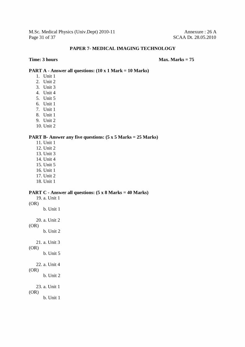
PAPER 7- MEDICAL IMAGING TECHNOLOGY Time: 3 hours Max. Marks = 75 PART A - Answer all questions: (10 x 1 Mark = 10 Marks)
1. Unit 1 2. Unit 2 3. Unit 3 4. Unit 4 5. Unit 5 6. Unit 1 7. Unit 1 8. Unit 1 9. Unit 2 10. Unit 2
PART B- Answer any five questions: (5 x 5 Marks = 25 Marks)
11. Unit 1 12. Unit 2 13. Unit 3 14. Unit 4 15. Unit 5 16. Unit 1 17. Unit 2 18. Unit 1
PART C - Answer all questions: (5 x 8 Marks = 40 Marks) 19. a. Unit 1
(OR) b. Unit 1
20. a. Unit 2 (OR) b. Unit 2
21. a. Unit 3 (OR) b. Unit 5
22. a. Unit 4 (OR) b. Unit 2
23. a. Unit 1 (OR) b. Unit 1

M.Sc. Medical Physics (Univ.Dept) 2010-11 Annexure : 26 A Page 32 of 37 SCAA Dt. 28.05.2010
PAPER 8- RADIATION DOSIMETRY AND STANDARDIZATION Time: 3 hours Max. Marks = 75 PART A - Answer all questions: (10 x 1 Mark = 10 Marks)
1. Unit 1 2. Unit 2 3. Unit 3 4. Unit 4 5. Unit 5 6. Unit 1 7. Unit 2 8. Unit 3 9. Unit 4 10. Unit 5
PART B- Answer any five questions: (5 x 5 Marks = 25 Marks)
11. Unit 1 12. Unit 2 13. Unit 3 14. Unit 4 15. Unit 5 16. Unit 3 17. Unit 4 18. Unit 5
PART C - Answer all questions: (5 x 8 Marks = 40 Marks) 19. a. Unit 1
(OR) b. Unit 1
20. a. Unit 2 (OR) b. Unit 2
21. a. Unit 3 (OR) b. Unit 3
22. a. Unit 4 (OR) b. Unit 4
23. a. Unit 5 (OR) b. Unit 5

M.Sc. Medical Physics (Univ.Dept) 2010-11 Annexure : 26 A Page 33 of 37 SCAA Dt. 28.05.2010
PAPER 9- BRACHYTHERAPY Time: 3 hours Max. Marks = 75 PART A - Answer all questions: (10 x 1 Mark = 10 Marks)
1. Unit 1 2. Unit 2 3. Unit 3 4. Unit 4 5. Unit 5 6. Unit 1 7. Unit 2 8. Unit 3 9. Unit 4 10. Unit 5
PART B- Answer any five questions: (5 x 5 Marks = 25 Marks)
11. Unit 1 12. Unit 2 13. Unit 3 14. Unit 4 15. Unit 5 16. Unit 3 17. Unit 4 18. Unit 5
PART C - Answer all questions: (5 x 8 Marks = 40 Marks) 19. a. Unit 1
(OR) b. Unit 1
20. a. Unit 2 (OR) b. Unit 2
21. a. Unit 3 (OR) b. Unit 3
22. a. Unit 4 (OR) b. Unit 4
23. a. Unit 5 (OR) b. Unit 5

M.Sc. Medical Physics (Univ.Dept) 2010-11 Annexure : 26 A Page 34 of 37 SCAA Dt. 28.05.2010
PAPER 10- RADIATION HAZARDS EVALUATION AND CONTROL Time: 3 hours Max. Marks = 75 PART A - Answer all questions: (10 x 1 Mark = 10 Marks)
1. Unit 1 2. Unit 2 3. Unit 3 4. Unit 4 5. Unit 5 6. Unit 1 7. Unit 2 8. Unit 3 9. Unit 4 10. Unit 3
PART B- Answer any five questions: (5 x 5 Marks = 25 Marks)
11. Unit 1 12. Unit 2 13. Unit 3 14. Unit 4 15. Unit 5 16. Unit 1 17. Unit 3 18. Unit 4
PART C - Answer all questions: (5 x 8 Marks = 40 Marks) 19. a. Unit 1
(OR) b. Unit 1
20. a. Unit 2 (OR) b. Unit 5
21. a. Unit 3 (OR) b. Unit 3
22. a. Unit 4 (OR) b. Unit 4
23. a. Unit 3 (OR) b. Unit 3

M.Sc. Medical Physics (Univ.Dept) 2010-11 Annexure : 26 A Page 35 of 37 SCAA Dt. 28.05.2010
PAPER 11- RADIATION BIOLOGY Time: 3 hours Max. Marks = 75 PART A - Answer all questions: (10 x 1 Mark = 10 Marks)
1. Unit 1 2. Unit 2 3. Unit 3 4. Unit 4 5. Unit 5 6. Unit 1 7. Unit 2 8. Unit 3 9. Unit 4 10. Unit 5
PART B- Answer any five questions: (5 x 5 Marks = 25 Marks)
11. Unit 1 12. Unit 2 13. Unit 3 14. Unit 4 15. Unit 5 16. Unit 2 17. Unit 4 18. Unit 5
PART C - Answer all questions: (5 x 8 Marks = 40 Marks) 19. a. Unit 1
(OR) b. Unit 1
20. a. Unit 2 (OR) b. Unit 2
21. a. Unit 3 (OR) b. Unit 3
22. a. Unit 4 (OR) b. Unit 4
23. a. Unit 5 (OR) b. Unit 5

M.Sc. Medical Physics (Univ.Dept) 2010-11 Annexure : 26 A Page 36 of 37 SCAA Dt. 28.05.2010
PAPER 12- NUCLEAR MEDICINE AND INTERNAL DOSIMETRY Time: 3 hours Max. Marks = 75 PART A - Answer all questions: (10 x 1 Mark = 10 Marks)
1. Unit 1 2. Unit 2 3. Unit 3 4. Unit 4 5. Unit 5 6. Unit 3 7. Unit 5 8. Unit 3 9. Unit 4 10. Unit 5
PART B- Answer any five questions: (5 x 5 Marks = 25 Marks)
11. Unit 1 12. Unit 2 13. Unit 3 14. Unit 4 15. Unit 5 16. Unit 3 17. Unit 4 18. Unit 5
PART C - Answer all questions: (5 x 8 Marks = 40 Marks) 19. a. Unit 1
(OR) b. Unit 2
20. a. Unit 3 (OR) b. Unit 3
21. a. Unit 4 (OR) b. Unit 4
22. a. Unit 5 (OR) b. Unit 5
23. a. Unit 3 (OR) b. Unit 3

M.Sc. Medical Physics (Univ.Dept) 2010-11 Annexure : 26 A Page 37 of 37 SCAA Dt. 28.05.2010
PAPER 13- ADVANCED RADIOTHERAPY TECHNIQUES Time: 3 hours Max. Marks = 75 PART A - Answer all questions: (10 x 1 Mark = 10 Marks)
1. Unit 1 2. Unit 2 3. Unit 3 4. Unit 4 5. Unit 5 6. Unit 1 7. Unit 2 8. Unit 3 9. Unit 4 10. Unit 5
PART B- Answer any five questions: (5 x 5 Marks = 25 Marks)
11. Unit 1 12. Unit 2 13. Unit 3 14. Unit 4 15. Unit 5 16. Unit 3 17. Unit 4 18. Unit 5
PART C - Answer all questions: (5 x 8 Marks = 40 Marks) 19. a. Unit 1
(OR) b. Unit 1
20. a. Unit 2 (OR) b. Unit 2
21. a. Unit 3 (OR) b. Unit 3
22. a. Unit 4 (OR) b. Unit 4
23. a. Unit 5 (OR) b. Unit 5