MR of Hereditary Hemorrhagic Telangiectasia: Prevalence ... · PDF fileMR of Hereditary...
8
MR of Hereditary Hemorrhagic Telangiectasia: Prevalence and Spectrum of Cerebrovascular Malformations Robert K. Fulbright, John C. Chaloupka, Christopher M. Putman, Gordon K. Sze, Michael M. Merriam, Graham K. Lee, Pierre B. Fayad, Issam A. Awad, and Robert I. White, Jr PURPOSE: Our goal was to describe the prevalence and types of cerebral vascular malfor- mations (CVMs) seen with MR imaging in patients with hereditary hemorrhagic telangiectasia (HHT). METHODS: We reviewed retrospectively the brain MR images of 184 consecutive patients with HHT. Catheter angiography was performed in 17 patients with CVMs detected on MR images. RESULTS: MR imaging revealed 63 CVMs in 42 patients. Classic arteriovenous malforma- tions (n 5 10) had a conspicuous network of vessels with flow voids and enlarged adjacent pial vessels. Apparent venous malformations (n 5 5) were best seen after administration of contrast material as a prominent vessel coursing through normal brain parenchyma. Indeterminate vascular malformations (n 5 48) had a spectrum of appearances characterized by variable combinations of heterogeneous signal intensity, enhancement, or hemosiderin. Angiography in 17 patients revealed 47 CVMs. Forty-six were arteriovenous malformations (AVMs), including 25 CVMs not seen with MR imaging and 21 CVMs that by MR criteria included 8 AVMs and 13 indeterminate vascular malformations. Angiography confirmed 1 venous malformation seen with MR imaging but failed to detect 3 indeterminate lesions revealed by MR imaging. CONCLUSION: MR imaging of a large cohort of consecutive patients with HHT revealed a CVM prevalence of 23% (42/184). Most CVMs (48/63) have an atypical appearance for vascular malformations on MR images. Angiographic correlation suggests that MR imaging underesti- mates the prevalence of CVMs and that the majority of indeterminate CVMs, despite their variable MR appearance, are AVMs. Hereditary hemorrhagic telangiectasia (HHT), also known as Osler-Weber-Rendu disease, is an autoso- mal dominant vascular dysplasia. Although the orig- inal descriptions of this syndrome emphasized the association of epistaxis with mucocutaneous telangi- ectasia (1– 4), subsequent reports have shown that multiple organs can harbor vascular abnormalities, including the brain, lungs, and gastrointestinal tract (5–10). Ten percent of patients are estimated to suf- fer serious morbidity or die prematurely from arte- riovenous malformations (AVMs) of these three or- gan systems (7–9, 11). Recent genetic studies have linked HHT to chromosome 9q33-q34 in some fami- lies and to chromosome 12q in others (12–17). It remains unknown, however, how gene defects or other factors interact to cause vascular malforma- tions. Cerebral vascular malformations (CVMs) in pa- tients with HHT are a manifestation of the underlying vascular dysplasia. These lesions represent abnormal arteriovenous connections that fail to differentiate properly into arteriolar, capillary, and venular chan- nels (18, 19). Because CVMs can lead to neurologic deficits when they hemorrhage, it is important to recognize their imaging features, especially since ev- idence suggests a higher prevalence of HHT than previously recognized (9, 20 –23). Our institution established a screening program for patients with HHT by using standard magnetic reso- nance (MR) imaging techniques to test the hypothesis Received June 3, 1997; accepted after revision September 15. Presented at the annual meeting of the American Society of Neuroradiology, Chicago, Ill, April 1995. From the Departments of Neuroradiology (R.K.F., G.K.S., M.M.M., G.K.L.), Interventional Neuroradiology (J.C.C., C.M.P.), Neurosurgery (J.C.C., I.A.A.), Neurology (P.B.F.), and Peripheral Vascular and Interventional Radiology (R.I.W.), Yale University School of Medicine, New Haven, Conn. Address reprint requests to Robert K. Fulbright, MD, Depart- ment of Radiology, Section of Neuroradiology, Yale University School of Medicine, Box 208042, New Haven, CT 06520. © American Society of Neuroradiology AJNR Am J Neuroradiol 19:477–484, March 1998 477
Transcript of MR of Hereditary Hemorrhagic Telangiectasia: Prevalence ... · PDF fileMR of Hereditary...
AJNR Am J Neuroradiol 19:477–484, March 1998
MR of Hereditary Hemorrhagic Telangiectasia:Prevalence and Spectrum of Cerebrovascular
Malformations
Robert K. Fulbright, John C. Chaloupka, Christopher M. Putman, Gordon K. Sze,Michael M. Merriam, Graham K. Lee, Pierre B. Fayad, Issam A. Awad, and Robert I. White, Jr
PURPOSE: Our goal was to describe the prevalence and types of cerebral vascular malfor-mations (CVMs) seen with MR imaging in patients with hereditary hemorrhagic telangiectasia(HHT).
METHODS: We reviewed retrospectively the brain MR images of 184 consecutive patientswith HHT. Catheter angiography was performed in 17 patients with CVMs detected on MRimages.
RESULTS: MR imaging revealed 63 CVMs in 42 patients. Classic arteriovenous malforma-tions (n 5 10) had a conspicuous network of vessels with flow voids and enlarged adjacent pialvessels. Apparent venous malformations (n 5 5) were best seen after administration of contrastmaterial as a prominent vessel coursing through normal brain parenchyma. Indeterminatevascular malformations (n 5 48) had a spectrum of appearances characterized by variablecombinations of heterogeneous signal intensity, enhancement, or hemosiderin. Angiography in17 patients revealed 47 CVMs. Forty-six were arteriovenous malformations (AVMs), including25 CVMs not seen with MR imaging and 21 CVMs that by MR criteria included 8 AVMs and13 indeterminate vascular malformations. Angiography confirmed 1 venous malformation seenwith MR imaging but failed to detect 3 indeterminate lesions revealed by MR imaging.
CONCLUSION: MR imaging of a large cohort of consecutive patients with HHT revealed aCVM prevalence of 23% (42/184). Most CVMs (48/63) have an atypical appearance for vascularmalformations on MR images. Angiographic correlation suggests that MR imaging underesti-mates the prevalence of CVMs and that the majority of indeterminate CVMs, despite theirvariable MR appearance, are AVMs.
Hereditary hemorrhagic telangiectasia (HHT), alsoknown as Osler-Weber-Rendu disease, is an autoso-mal dominant vascular dysplasia. Although the orig-inal descriptions of this syndrome emphasized theassociation of epistaxis with mucocutaneous telangi-ectasia (1–4), subsequent reports have shown thatmultiple organs can harbor vascular abnormalities,including the brain, lungs, and gastrointestinal tract(5–10). Ten percent of patients are estimated to suf-
Received June 3, 1997; accepted after revision September 15.Presented at the annual meeting of the American Society of
Neuroradiology, Chicago, Ill, April 1995.From the Departments of Neuroradiology (R.K.F., G.K.S.,
M.M.M., G.K.L.), Interventional Neuroradiology (J.C.C., C.M.P.),Neurosurgery (J.C.C., I.A.A.), Neurology (P.B.F.), and PeripheralVascular and Interventional Radiology (R.I.W.), Yale UniversitySchool of Medicine, New Haven, Conn.
Address reprint requests to Robert K. Fulbright, MD, Depart-ment of Radiology, Section of Neuroradiology, Yale UniversitySchool of Medicine, Box 208042, New Haven, CT 06520.
© American Society of Neuroradiology
47
fer serious morbidity or die prematurely from arte-riovenous malformations (AVMs) of these three or-gan systems (7–9, 11). Recent genetic studies havelinked HHT to chromosome 9q33-q34 in some fami-lies and to chromosome 12q in others (12–17). Itremains unknown, however, how gene defects orother factors interact to cause vascular malforma-tions.
Cerebral vascular malformations (CVMs) in pa-tients with HHT are a manifestation of the underlyingvascular dysplasia. These lesions represent abnormalarteriovenous connections that fail to differentiateproperly into arteriolar, capillary, and venular chan-nels (18, 19). Because CVMs can lead to neurologicdeficits when they hemorrhage, it is important torecognize their imaging features, especially since ev-idence suggests a higher prevalence of HHT thanpreviously recognized (9, 20–23).
Our institution established a screening program forpatients with HHT by using standard magnetic reso-nance (MR) imaging techniques to test the hypothesis
7
478 FULBRIGHT AJNR: 19, March 1998
that CVMs are common in this patient population.We report our findings as to the prevalence and typesof CVMs seen with MR imaging in a large number ofHHT patients, and compare the MR findings withthose of conventional cerebral angiography in a sub-set of patients.
MethodsFrom 1988 to 1995, 184 consecutive patients were examined
in a specialized multidisciplinary clinic at a single institution.They were referred for screening MR imaging as part of theirinitial clinical assessment. Two of the three following criterianeeded to be present to establish the diagnosis of HHT: nose-bleeds at least four times a month, telangiectasia of the skin,and a mother or father with HHT (10). All patients had MRimaging consisting of axial T1-weighted sequences with param-eters of 500/12/1 (repetition time [TR]/echo time/excitation), amatrix of 256 3 192, a 24-cm field of view (FOV), and 5-mm-thick sections with a 1-mm gap; dual-echo long-TR sequences(2000/30,80/1) with a 256 3 192 matrix, 5-mm-thick sectionswith a 2.5-mm gap, and a 24-mm FOV; axial or coronal gradi-ent-recalled echo sequences (750/50) with a 10° flip angle, a20-mm FOV, and 5-mm-thick sections with a 1-mm gap; and128 patients had an axial T1-weighted sequence following ad-ministration of a standard dose of intravenous contrast mate-rial. All cases were reviewed retrospectively by three neurora-diologists to identify and classify vascular malformations. Aconsensus reading determined final categorization.
The AVMs were defined as those CVMs with one or moreof the following MR features: a collection of enlarged vesselseasily seen on both T2- and T1-weighted (without and withcontrast enhancement) sequences; associated enlargement orectasia of adjacent pial arteries and draining veins; a well-defined, relatively large nidus (.15 mm); and such secondaryfeatures as dilated proximal intracranial or extracranial arteries(angiomatous change) (24–27). Brain adjacent to AVM mightshow old hemorrhage, gliosis, or both.
Venous malformations were defined as those CVMs with allthe following MR imaging features: a single enlarged vascularchannel usually in a subcortical location (periventricular whitematter, mesencephalon, deep cerebellum); no discernible en-largement of adjacent arteries; best seen after administrationof contrast material; could be associated with smaller linearvascular channels orthogonal to the axial plane of the singleenlarged vascular channel (caput medusae appearance); andnormal adjacent brain (28, 29).
Cavernous malformations were lesions with a complete, con-centric ring of curvilinear hypointensity on long-TR sequencesthat appeared to enlarge or “bloom” on gradient-recalled echosequences; a complex multilobular center with septa and aspeckled or reticulated appearance on T2-weighted sequencesowing to a combination of hypointensities and hyperintensities;and no or minimal enhancement after administration of con-trast material (30–33).
Indeterminate CVMs were lesions that did not fit into one ofthe three categories of cerebral AVMs, venous malformations,or cavernous malformations. Their MR features included asmall, localized tangle of curvilinear structures (10 to 15 mm)without a discernible nidus; no enlargement of adjacent arter-ies and veins; curvilinear or punctate structures with variablecombinations of localized hypointensity or hyperintensity onshort- and long-TR images; curvilinear or punctate hypointen-sity on long-TR sequences that could bloom on gradient-recalled echo sequences but that did not form a complete,concentric ring; and improved visualization on contrast-en-hanced T1-weighted sequences.
Conventional cerebral angiography was indicated in 17 pa-tients with CVMs seen at MR imaging and who had focalneurologic (motor, sensory, visual, or language) deficits, sei-
zures, or both. Catheter angiography was performed by selec-tive hand injection of iohexol (4 to 6 mL) into each internalcarotid artery, and at least one vertebral artery. High-resolu-tion (1024 3 1024 matrix) digital subtraction angiography withmagnification views or film-screen technique was used. AVMswere defined as lesions with a focal collection of abnormalvessels (nidus) located within the brain parenchyma that wereassociated with early visualization of veins during either thearterial or late capillary phase (ie, arteriovenous shunting).Venous malformations were lesions defined as aberrant, smallbranching vessels that coalesced into a larger single vessel thatin turn connected to either a subcortical or subependymal vein.These malformations were always seen during the normal ve-nous phase of angiography. Cavernous malformations usuallydo not have angiographic findings, although focal pooling ofcontrast material in the late capillary or venous phase can beseen, without evidence of arteriovenous shunting.
Results
MR Imaging FeaturesMR imaging revealed 63 CVMs in 42 patients. The
types of CVMs included 10 AVMs in nine patients;five apparent venous malformations in five patients;and 48 indeterminate CVMs in 31 patients. Ninepatients had multiple CVMs, ranging in number fromtwo to five. Six of these patients had multiple inde-terminate lesions and three had a combination ofCVMs: one patient had two AVMs and one indeter-minate lesion, and two patients had three indetermi-nate lesions and one AVM. No patient had classicalMR imaging characteristics of cavernous malforma-tions.
AVMs (Fig 1) had prominent dilatation of afferentand efferent vessels and nidus components (.15 mm)consisting of serpentine flow voids of various sizes onT1- and T2-weighted sequences. The observed flowvoids were most likely caused by a combination ofhigh-velocity blood flow and the dephasing effectsrelated to laminar flow disturbance. The abnormalvessels of these cerebral AVMs tended to insinuatethemselves within brain tissue rather than cause anencapsulated mass. Evidence of associated hemor-rhage was occasionally identified in the form of he-mosiderosis.
Apparent venous malformations had imaging fea-tures that most closely resembled those described forclassic venous malformations of the brain. They werebest seen or only seen on T1-weighted contrast-enhanced sequences and manifested as enhancinglinear or punctate structures within a subcortical orsubpial location (Fig 2). Occasionally, smaller vascu-lar structures radiating into a larger, central vesselwere observed. These CVMs were not associated withany signal abnormalities of adjacent brain. Since theyhad an MR imaging appearance very much like thatdescribed for venous malformations yet did not al-ways have conventional angiographic correlation, wedesignated them as apparent venous malformations.
Indeterminate CVMs (n 5 48) were a heteroge-neous group of vascular lesions with a spectrum ofMR appearances: some had features that resembledsmall cavernous malformations, others resembled ve-nous malformations, mixed vascular malformations,
AJNR: 19, March 1998 TELANGIECTASIA 479
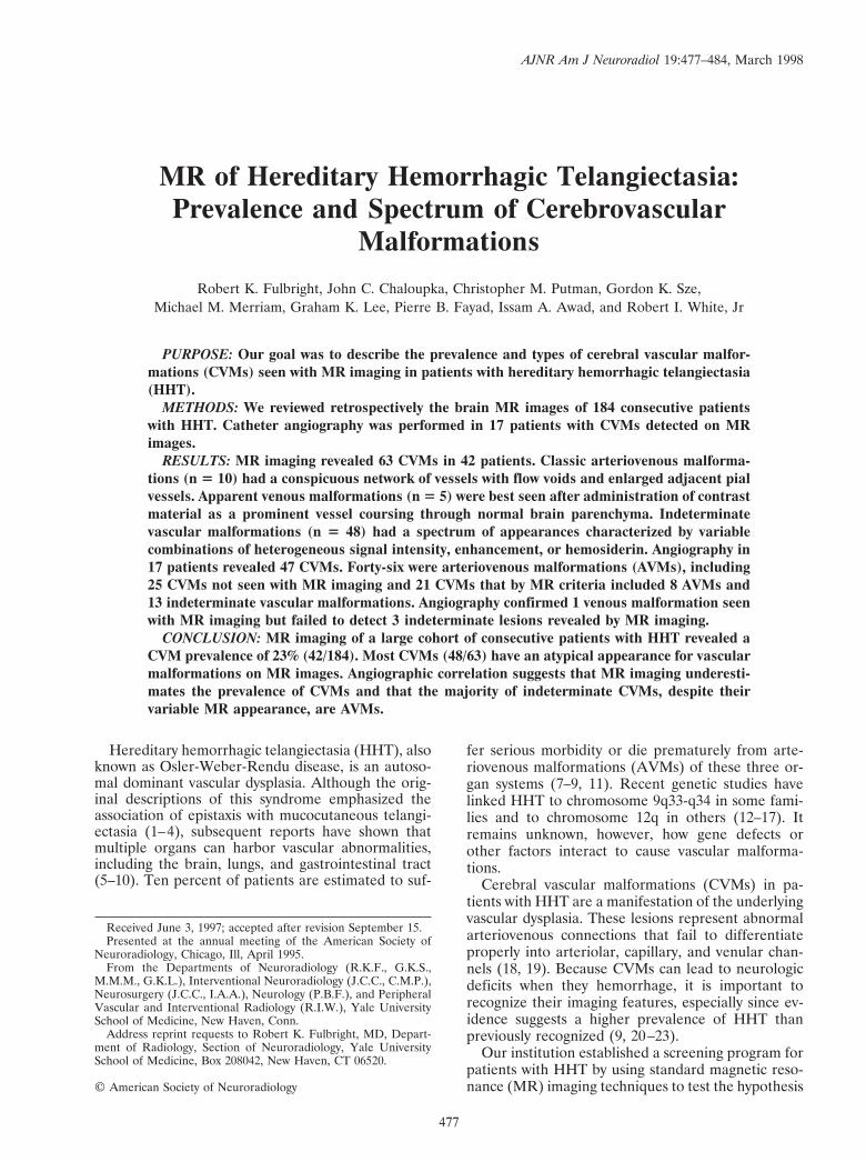
FIG 2. At MR imaging, this lesion was classified as an indeterminate malformation, but angiographic imaging revealed an AVM.Axial T2-weighted (2000/80/1) (A ) and axial contrast-enhanced T1-weighted (500/20/1) (B ) images reveal a partially enhancing lesion
with heterogeneous signal in the right perimesencephalic cistern (arrows).C, Frontal oblique angiogram shows that this is an AVM with a feeding artery (straight arrow), nidus (curved arrow), and early draining
vein (arrowhead ).
FIG 1. A lesion classified as an AVM atboth MR imaging and angiography.
Axial T1-weighted (500/20/1) (A ),axial T2-weighted (2000/80/1) (B ),and coronal contrast-enhanced T1-weighted (500/20/1) (C ) MR imagesshow large vessels in middle frontal,precentral, and postcentral gyrus onthe left.
Lateral (D ) and frontal (E ) projectionsfrom conventional angiography showlarge arterial feeder (short straight ar-row, D ), nidus (curved arrow, D ), drain-ing vein (long straight arrow, D ), andvenous varix (curved arrow, E ).
or poorly defined vascular anomalies. Thirteen inde-terminate lesions had angiographic correlation (seebelow). Indeterminate CVMs were small (5 to 15mm) and appeared as ovoid lesions or as focal, cur-vilinear or patchy structures (Figs 3–6). The signalintensity on T2-weighted images was variable, appear-ing bright, dark, or both, and distributed in no par-ticular pattern. Lesions were surrounded partially orinterspersed by focal or curvilinear structures thathad decreased signal intensity on both T1- and T2-
weighted sequences. These hypointense structuressometimes bloomed on gradient-recalled echo se-quences, but a complete ring of hypointensity was notseen. Indeterminate lesions had variable enhance-ment with contrast administration, with small lesionsbest seen on contrast-enhanced images as enhancingcurvilinear structures (Figs 3 and 5). Although someindeterminate malformations had features suggestiveof cavernous malformations (Fig 4), they did not havethe classic pattern of a reticulated center of variable
480 FULBRIGHT AJNR: 19, March 1998
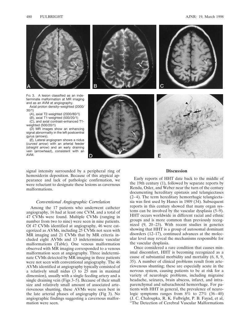
FIG 3. A lesion classified as an inde-terminate malformation at MR imagingand as an AVM at angiography.
Axial proton density–weighted (2000/30/1)
(A ), axial T2-weighted (2000/80/1)(B ), axial T1-weighted (500/20/1)(C ), and axial contrast-enhanced T1-
weighted (500/20/1)(D ) MR images show an enhancing
signal abnormality in the left postcentralgyrus (arrows).
(E), Lateral angiogram shows a nidus(curved arrow) with an arterial feeder(straight arrow) and an early drainingvein (arrowhead ), consistent with anAVM.
signal intensity surrounded by a peripheral ring ofhemosiderin deposition. Because of this atypical ap-pearance and lack of pathologic confirmation, wewere reluctant to designate these lesions as cavernousmalformations.
Conventional Angiographic CorrelationAmong the 17 patients who underwent catheter
angiography, 16 had at least one CVM, and a total of47 CVMs were found. Multiple CVMs (ranging innumber from two to nine) were seen in nine patients.Of 47 CVMs identified at angiography, 46 were cat-egorized as AVMs, including 25 CVMs not seen withMR imaging and 21 CVMs that by MR criteria in-cluded eight AVMs and 13 indeterminate vascularmalformations (Table). One venous malformationobserved with MR imaging corresponded to a venousmalformation seen at angiography. Three indetermi-nate CVMs detected by MR imaging in three patientswere not seen with conventional angiography. The 46AVMs identified at angiography typically consisted ofa relatively small nidus (3 to 25 mm in maximaldimension), usually with a single feeding artery and asingle draining vein (Figs 3–5). Because of their smallsize and relatively small amount of associated arte-riovenous shunting, these AVMs were seen best inthe late arterial phases of angiography (Fig 3). Noangiographic findings suggesting a cavernous malfor-mation were seen.
Discussion
Early reports of HHT date back to the middle ofthe 19th century (1), followed by separate reports byRendu, Osler, and Weber near the turn of the centurydocumenting hereditary epistaxis and telangiectases(2–4). The term hereditary hemorrhagic telangiecta-sia was first used by Hanes in 1909 (34). Subsequentreports in this century showed that many organ sys-tems can be involved by the vascular dysplasia (5–9).HHT occurs worldwide in different racial and ethnicgroups and is more common than previously recog-nized (9, 20–23). With recent studies in geneticsshowing that HHT is a group of autosomal dominantdisorders (12–17), continued advances at the molec-ular level may reveal the mechanisms responsible forthe vascular dysplasia.
Once considered a rare condition that causes min-imal discomfort, HHT is becoming recognized as acause of substantial morbidity and mortality (6, 8, 9,35). A number of clinical problems result from arte-riovenous shunting; these are especially acute in thenervous system, causing patients to be at risk for avariety of neurologic problems, including migraineheadache, seizures, brain abscess, infarct, and intra-parenchymal and subarachnoid hemorrhage. For pa-tients with HHT in general, the prevalence of neuro-logic symptoms ranges from 8% to 27% (36–40)(J. C. Chaloupka, R. K. Fulbright, P. B. Fayad, et al,“The Detection of Cerebral Vascular Malformations
AJNR: 19, March 1998 TELANGIECTASIA 481
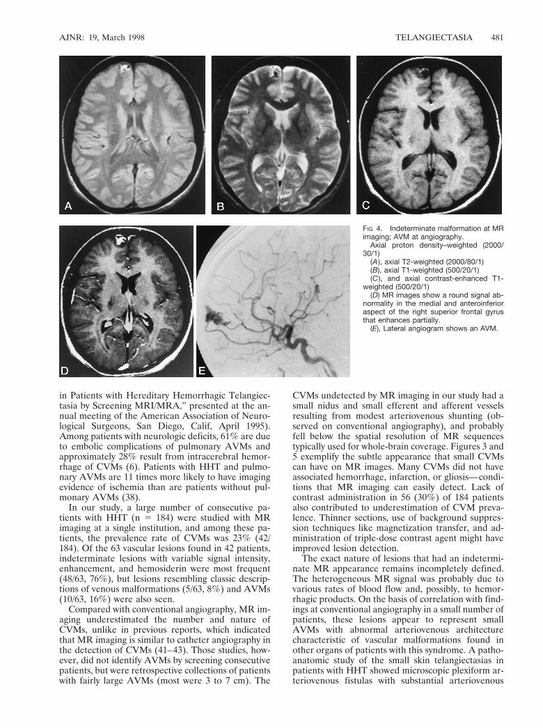
FIG 4. Indeterminate malformation at MRimaging; AVM at angiography.
Axial proton density–weighted (2000/30/1)
(A ), axial T2-weighted (2000/80/1)(B), axial T1-weighted (500/20/1)(C ), and axial contrast-enhanced T1-
weighted (500/20/1)(D) MR images show a round signal ab-
normality in the medial and anteroinferioraspect of the right superior frontal gyrusthat enhances partially.
(E), Lateral angiogram shows an AVM.
in Patients with Hereditary Hemorrhagic Telangiec-tasia by Screening MRI/MRA,” presented at the an-nual meeting of the American Association of Neuro-logical Surgeons, San Diego, Calif, April 1995).Among patients with neurologic deficits, 61% are dueto embolic complications of pulmonary AVMs andapproximately 28% result from intracerebral hemor-rhage of CVMs (6). Patients with HHT and pulmo-nary AVMs are 11 times more likely to have imagingevidence of ischemia than are patients without pul-monary AVMs (38).
In our study, a large number of consecutive pa-tients with HHT (n 5 184) were studied with MRimaging at a single institution, and among these pa-tients, the prevalence rate of CVMs was 23% (42/184). Of the 63 vascular lesions found in 42 patients,indeterminate lesions with variable signal intensity,enhancement, and hemosiderin were most frequent(48/63, 76%), but lesions resembling classic descrip-tions of venous malformations (5/63, 8%) and AVMs(10/63, 16%) were also seen.
Compared with conventional angiography, MR im-aging underestimated the number and nature ofCVMs, unlike in previous reports, which indicatedthat MR imaging is similar to catheter angiography inthe detection of CVMs (41–43). Those studies, how-ever, did not identify AVMs by screening consecutivepatients, but were retrospective collections of patientswith fairly large AVMs (most were 3 to 7 cm). The
CVMs undetected by MR imaging in our study had asmall nidus and small efferent and afferent vesselsresulting from modest arteriovenous shunting (ob-served on conventional angiography), and probablyfell below the spatial resolution of MR sequencestypically used for whole-brain coverage. Figures 3 and5 exemplify the subtle appearance that small CVMscan have on MR images. Many CVMs did not haveassociated hemorrhage, infarction, or gliosis—condi-tions that MR imaging can easily detect. Lack ofcontrast administration in 56 (30%) of 184 patientsalso contributed to underestimation of CVM preva-lence. Thinner sections, use of background suppres-sion techniques like magnetization transfer, and ad-ministration of triple-dose contrast agent might haveimproved lesion detection.
The exact nature of lesions that had an indetermi-nate MR appearance remains incompletely defined.The heterogeneous MR signal was probably due tovarious rates of blood flow and, possibly, to hemor-rhagic products. On the basis of correlation with find-ings at conventional angiography in a small number ofpatients, these lesions appear to represent smallAVMs with abnormal arteriovenous architecturecharacteristic of vascular malformations found inother organs of patients with this syndrome. A patho-anatomic study of the small skin telangiectasias inpatients with HHT showed microscopic plexiform ar-teriovenous fistulas with substantial arteriovenous
482 FULBRIGHT AJNR: 19, March 1998
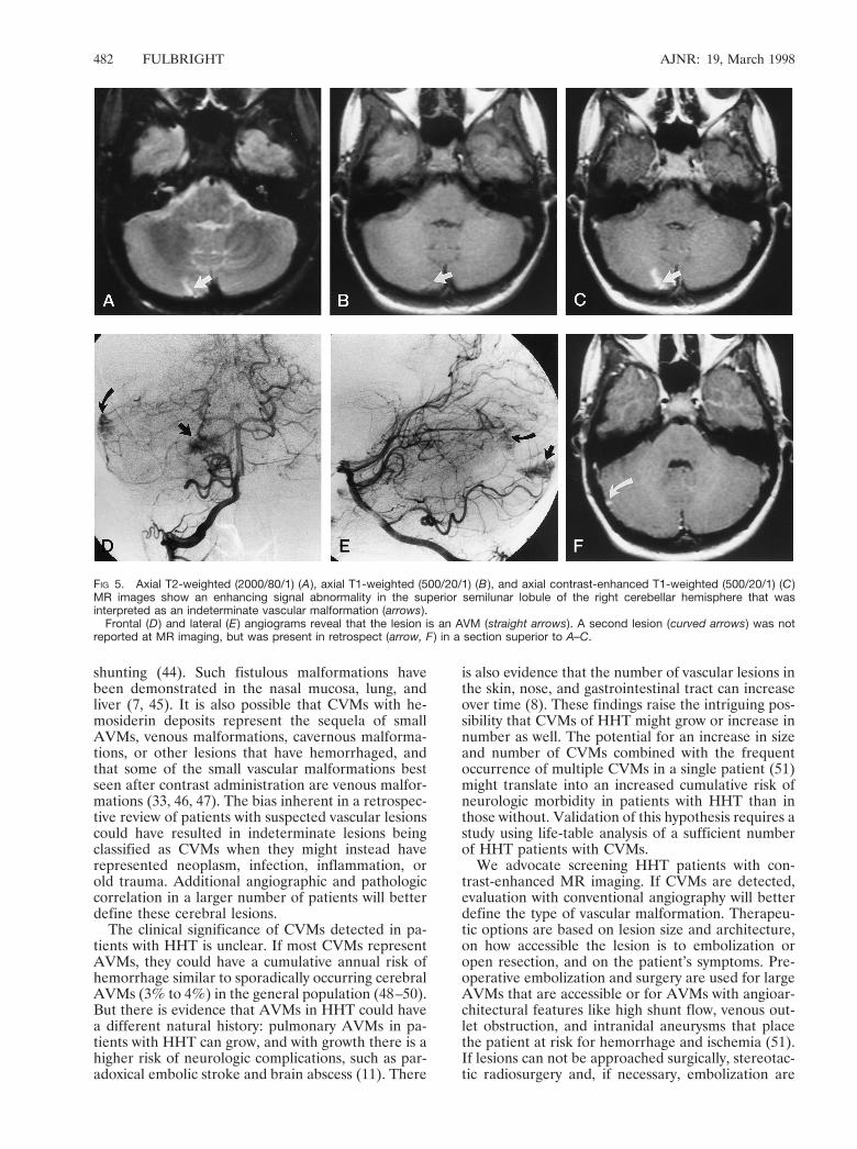
FIG 5. Axial T2-weighted (2000/80/1) (A ), axial T1-weighted (500/20/1) (B ), and axial contrast-enhanced T1-weighted (500/20/1) (C )MR images show an enhancing signal abnormality in the superior semilunar lobule of the right cerebellar hemisphere that wasinterpreted as an indeterminate vascular malformation (arrows).
Frontal (D ) and lateral (E ) angiograms reveal that the lesion is an AVM (straight arrows). A second lesion (curved arrows) was notreported at MR imaging, but was present in retrospect (arrow, F ) in a section superior to A–C.
shunting (44). Such fistulous malformations havebeen demonstrated in the nasal mucosa, lung, andliver (7, 45). It is also possible that CVMs with he-mosiderin deposits represent the sequela of smallAVMs, venous malformations, cavernous malforma-tions, or other lesions that have hemorrhaged, andthat some of the small vascular malformations bestseen after contrast administration are venous malfor-mations (33, 46, 47). The bias inherent in a retrospec-tive review of patients with suspected vascular lesionscould have resulted in indeterminate lesions beingclassified as CVMs when they might instead haverepresented neoplasm, infection, inflammation, orold trauma. Additional angiographic and pathologiccorrelation in a larger number of patients will betterdefine these cerebral lesions.
The clinical significance of CVMs detected in pa-tients with HHT is unclear. If most CVMs representAVMs, they could have a cumulative annual risk ofhemorrhage similar to sporadically occurring cerebralAVMs (3% to 4%) in the general population (48–50).But there is evidence that AVMs in HHT could havea different natural history: pulmonary AVMs in pa-tients with HHT can grow, and with growth there is ahigher risk of neurologic complications, such as par-adoxical embolic stroke and brain abscess (11). There
is also evidence that the number of vascular lesions inthe skin, nose, and gastrointestinal tract can increaseover time (8). These findings raise the intriguing pos-sibility that CVMs of HHT might grow or increase innumber as well. The potential for an increase in sizeand number of CVMs combined with the frequentoccurrence of multiple CVMs in a single patient (51)might translate into an increased cumulative risk ofneurologic morbidity in patients with HHT than inthose without. Validation of this hypothesis requires astudy using life-table analysis of a sufficient numberof HHT patients with CVMs.
We advocate screening HHT patients with con-trast-enhanced MR imaging. If CVMs are detected,evaluation with conventional angiography will betterdefine the type of vascular malformation. Therapeu-tic options are based on lesion size and architecture,on how accessible the lesion is to embolization oropen resection, and on the patient’s symptoms. Pre-operative embolization and surgery are used for largeAVMs that are accessible or for AVMs with angioar-chitectural features like high shunt flow, venous out-let obstruction, and intranidal aneurysms that placethe patient at risk for hemorrhage and ischemia (51).If lesions can not be approached surgically, stereotac-tic radiosurgery and, if necessary, embolization are
AJNR: 19, March 1998 TELANGIECTASIA 483
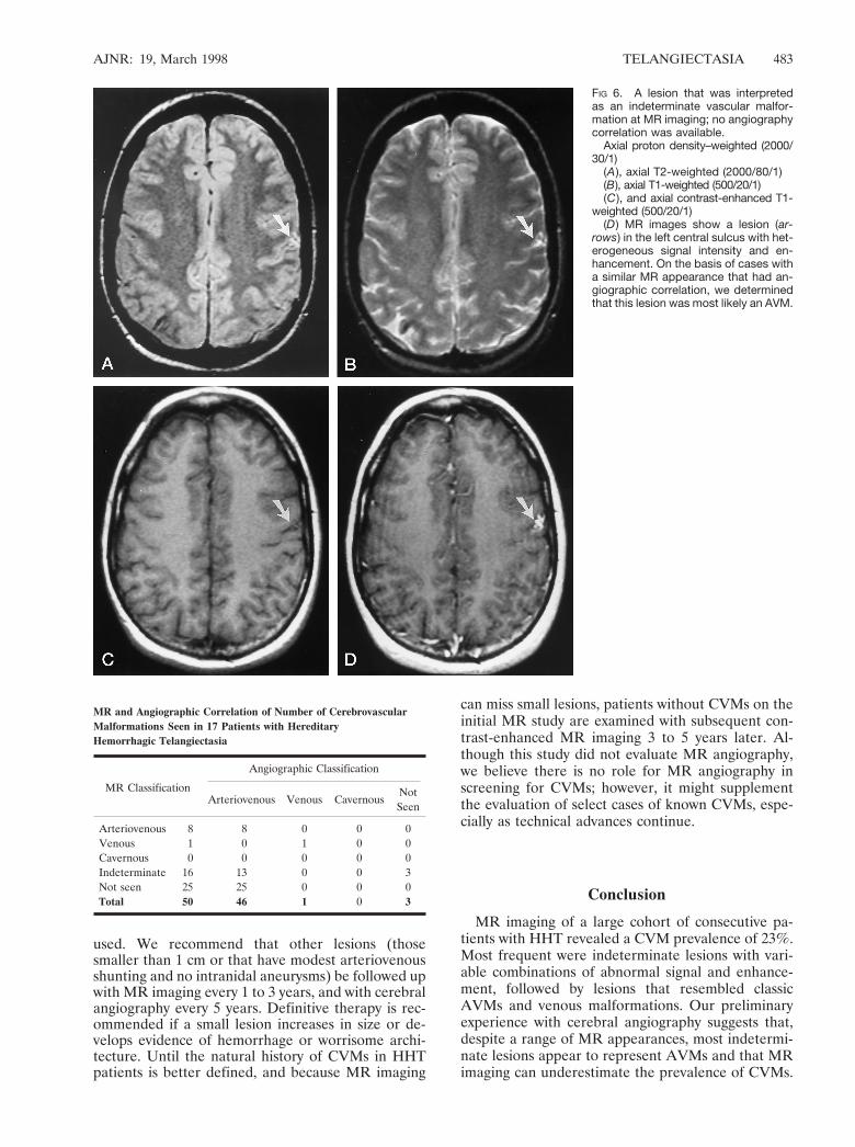
FIG 6. A lesion that was interpretedas an indeterminate vascular malfor-mation at MR imaging; no angiographycorrelation was available.
Axial proton density–weighted (2000/30/1)
(A ), axial T2-weighted (2000/80/1)(B), axial T1-weighted (500/20/1)(C ), and axial contrast-enhanced T1-
weighted (500/20/1)(D ) MR images show a lesion (ar-
rows) in the left central sulcus with het-erogeneous signal intensity and en-hancement. On the basis of cases witha similar MR appearance that had an-giographic correlation, we determinedthat this lesion was most likely an AVM.
used. We recommend that other lesions (thosesmaller than 1 cm or that have modest arteriovenousshunting and no intranidal aneurysms) be followed upwith MR imaging every 1 to 3 years, and with cerebralangiography every 5 years. Definitive therapy is rec-ommended if a small lesion increases in size or de-velops evidence of hemorrhage or worrisome archi-tecture. Until the natural history of CVMs in HHTpatients is better defined, and because MR imaging
MR and Angiographic Correlation of Number of CerebrovascularMalformations Seen in 17 Patients with HereditaryHemorrhagic Telangiectasia
MR Classification
Angiographic Classification
Arteriovenous Venous CavernousNotSeen
Arteriovenous 8 8 0 0 0Venous 1 0 1 0 0Cavernous 0 0 0 0 0Indeterminate 16 13 0 0 3Not seen 25 25 0 0 0Total 50 46 1 0 3
can miss small lesions, patients without CVMs on theinitial MR study are examined with subsequent con-trast-enhanced MR imaging 3 to 5 years later. Al-though this study did not evaluate MR angiography,we believe there is no role for MR angiography inscreening for CVMs; however, it might supplementthe evaluation of select cases of known CVMs, espe-cially as technical advances continue.
Conclusion
MR imaging of a large cohort of consecutive pa-tients with HHT revealed a CVM prevalence of 23%.Most frequent were indeterminate lesions with vari-able combinations of abnormal signal and enhance-ment, followed by lesions that resembled classicAVMs and venous malformations. Our preliminaryexperience with cerebral angiography suggests that,despite a range of MR appearances, most indetermi-nate lesions appear to represent AVMs and that MRimaging can underestimate the prevalence of CVMs.
484 FULBRIGHT AJNR: 19, March 1998
References1. Sutton HG. Epistaxis as an indication of impaired nutrition, and of
degeneration of the vascular system. Med Mirror 1864;:769–7812. Rendu HJ. Epistaxis repetees chez un sujet porteir depetits angio-
mes cutanes et muqueux. Gax Hop 1896;:1322–13233. Osler W. On a family form of recurring epistaxis, associated with
multiple telangiectases of the skin and mucous membranes. BullJohns Hopkins Hosp 1901;12:333–337
4. Weber RP. Multiple hereditary developmental angiomata (telangi-ectases) of the skin and mucous membranes associated with recur-ring haemorrhages. Lancet 1907;2:160–162
5. Sobel D, Norman D. CNS manifestations of hereditary hemor-rhagic telangiectasia. AJNR Am J Neuroradiol 1984;5:569–573
6. Roman G, Fisher M, Perl DP, Poser CM. Neurological manifesta-tions of hereditary hemorrhagic telangiectasia (Rendu-Osler-We-ber disease): report of 2 cases and review of the literature. AnnNeurol 1978;4:130–144
7. Peery WH. Clinical spectrum of hereditary hemorrhagic telangiec-tasia (Osler-Weber-Rendu disease). Am J Med 1987;82:989–997
8. Plauchu H, de Chadarevian JP, Bideau A, Robert JM. Age-relatedclinical profile of hereditary hemorrhagic telangiectasia in an epi-demiologically recruited population. Am J Med Genet 1989;32:291–297
9. Porteous ME, Burn J, Proctor SJ. Hereditary haemorrhagic telan-giectasia: a clinical analysis. J Med Genet 1992;29:527–530
10. Guttmacher AE, Marchuk DA, White RI, Jr. Current concepts:hereditary hemorrhagic telangiectasia. N Engl J Med 1995;333:913–924
11. White RI Jr. Pulmonary arteriovenous malformations: how do wediagnose them and why is it important to do so? Radiology 1992;182:633–635
12. Shovlin CL, Hughes JM, Tuddenham EG, et al. A gene for hered-itary haemorrhagic telangiectasia maps to chromosome 9q3. NatGenet 1994;6:205–209
13. Porteous MEM, Curtis A, Williams O, Marchuk D, BhattacharyaSS, Burn J. Genetic heterogeneity in hereditary haemorrhagic tel-angiectasia. J Med Genet 1994;31:925–926
14. McAllister KA, Lennon F, Bowles-Biesecker B, et al. Geneticheterogeneity in hereditary haemorrhagic telangiectasia: possiblecorrelation with clinical phenotype. J Med Genet 1994;31:823–829
15. Heutink P, Haitjema T, Breedveld GJ. Linkage of hereditary hae-morrhagic telangiectasia to chromosome k9q34 and evidence forlocus heterogeneity. J Med Genet 1994;31:933–936
16. Vincent P, Plauchu H, Hazan J, Faure S, Weissenbach J, Godte J.A third locus for hereditary haemorrhagic telangiectasia maps tochromosome 12q. Hum Mol Genet 1995;31:945–950
17. McAllister KA, Grogg KM, Johnson DW, et al. Endoglin, a TGF-Bbinding protein of endothelial cells, is the gene for hereditaryhaemorrhagic telangiectasia type 1. Nat Genet 1994;8:345–351
18. Bird RM, Jaques WE. Vascular lesions of hereditary hemorrhagictelangiectasia. N Engl J Med 1959;260:597–599
19. Albert P. Personal experience in the treatment of 178 cases ofarteriovenous malformations of the brain. Acta Neurochir (Wein)1982;61:207–226
20. Plauchu H, Bideau A. Epidemiologie et constitution d’un registrede population a propos d’une concentration geographcique d’unemaladie hereditaire rare. Population 1984;4–5:765–786
21. Vase P, Grove O. Gastrointestinal lesions in hereditary hemor-rhagic telangiectasia. Gastroenterology 1986;91:1079–1083
22. Jessurun GA, Kamphuis DJ, van der Zande FH, Nossent JC.Cerebral arteriovenous malformations in The Netherlands Antilles:high prevalence of hereditary hemorrhagic telangiectasia–relatedsingle and multiple cerebral arteriovenous malformations. ClinNeurol Neurosurg 1993;95:193–198
23. Guttmacher AE, McKinnon WC, Upton MD. Hereditary hemor-rhagic telangiectasia: a disorder in search of the genetics commu-nity. Am J Med Genet 1994;52:252–253
24. Marks MP, Lane B, Steinberg GK, Chang P. Vascular characteris-tics of intracerebral arteriovenous malformations in patients withclinical steal. AJNR Am J Neuroradiol 1991;12:489–496
25. Smith HJ, Strother CM, Kikuchi Y, et al. MR imaging in themanagement of supratentorial intracranial AVMs. AJNR Am JNeuroradiol 1988;9:225–235
26. Kumar AJ, Vinuela F, Fox AJ, Rosenbaum AE. Unruptured intra-cranial arteriovenous malformations do cause mass effect. AJNRAm J Neuroradiol 1985;6:29–32
27. Chappell PM, Steinberg GK, Marks MP. Clinically documentedhemorrhage in cerebral arteriovenous malformations: MR charac-teristics. AJNR Am J Neuroradiol 1992;12:489–496
28. Wilms G, Marchal G, Vas Hecke P, et al. Cerebral venous angio-mas: MR imaging at 1.5 Tesla. Neuroradiology 1990;32:81–85
29. Osterton B, Solymosi L. Magnetic resonance angiography of cere-bral developmental anomalies: its role in differential diagnosis.Neuroradiology 1993;35:97–104
30. Rigamonti D, Drayer BP, Johnson PC, Hadley MN, Zabramski J,Spetzler RF. The MRI appearance of cavernous malformations(angiomas). J Neurosurg 1987;67:518–524
31. Gomori JM, Grossman RI. Occult cerebral vascular malformations:high field MR imaging. Radiology 1986;158:707–713
32. Rapacki TFX, Brantly MJ, Furlow TW, Geyer CA, Toro VE,George ED. Heterogeneity of cerebral cavernous hemangiomasdiagnosed by MR imaging. J Comput Assist Tomogr 1990;14:18–25
33. Robinson JR, Awad IA, Little JR. Natural history of the cavernousangioma. J Neurosurg 1991;75:709–714
34. Hanes FM. Multiple hereditary telangiectases causing hemorrhage(hereditary hemorrhagic telangiectasia). Bull Johns Hopkins Hosp1909;20:63–73
35. Ference BA, Shannon TM, White RI Jr, Zawin M, Burdge CM.Life-threatening pulmonary hemorrhage with pulmonary arterio-venous malformations and hereditary hemorrhagic telangiectasia.Chest 1994;106:1387–1390
36. Davidson CH. Hereditary haemorrhagic telangiectasia: a report ona family of seven generations. Scott Med J 1959;4:260–262
37. Hodgson CH, Kaye RL. Hereditary hemorrhagic telangiectasia andpulmonary arteriovenous fistula. N Engl J Med 1959;261:625–636
38. Fulbright RK, Merriam MM, Fayad PB, Sze GK, Egglin TK, WhiteRI Jr. Hereditary hemorrhagic telangiectasia: correlation of MRimaging and clinical findings in 130 patients. Radiology 1994;193(P):211
39. Boczko ML. Neurological implications of HHT. J Nerv Ment Dis1964;139:525–536
40. Fayad PB. Neurologic manifestations of hereditary hemorrhagictelangiectasia. In: Gilman S, Goldstein GW, Waxman SG, eds.Neurobase. La Jolla, Calif: Arbor; 1995
41. Nussel F, Wegmuller H, Huber P. Comparison of magnetic reso-nance angiography, magnetic resonance imaging and conventionalangiography in cerebral arteriovenous malformation. Neuroradiol-ogy 1991;33:56–61
42. Pott M, Huber M, Assheuer J, Bewermeyer H. Comparison ofMRI, CT and angiography in cerebral arteriovenous malforma-tions. Bildgebung 1992;59:98–102
43. Prayer L, Wimberger D, Stiglbauer R, et al. Haemorrhage inintracerebral arteriovenous malformations: detection with MRIand comparison with clinical history. Neuroradiology 1993;35:424–427
44. Braverman IM, Keh A, Jacobson BS. Ultrastructure and three-dimensional organization of the telangiectases of hereditary hem-orrhagic telangiectasia. J Invest Dermatol 1990;95:422–427
45. White RI Jr, Mitchell SE, Barth KH, et al. Angioarchitecture ofpulmonary arteriovenous malformations: an important consider-ation before embolotherapy. AJR Am J Roentgenol 1983;:681–686
46. Awad IA, Robinson JR, Mohanty S, Estes ML. Mixed VascularMalformations of the brain: clinical and pathogenetic consider-ations. Neurosurgery 1993;33:179–188
47. Latchaw RE. Commentary: venous angioma, cavernous angioma,and hemorrhage. AJNR Am J Neuroradiol 1994;15:1255–1257
48. Crawford PM, West CR, Chadwick DW, Shaw MDM. Arterio-venous malformations of the brain: natural history in unoperatedpatients. J Neurol Neurosurg Psychiatry 1986;49:1–10
49. Jane JA, Kassell NF, Torner JC, Winn HR. The natural history ofaneurysms and arteriovenous malformations. J Neurosurg 1985;62:321–323
50. Minakawa T, Tanaka R, Koike T, Takeuchi S, Sasaki O. Angio-graphic follow-up study of cerebral arteriovenous malformationswith reference to their enlargement and regression. Neurosurgery1989;24:68–74
51. Putman CM, Chaloupka JC, Fulbright RK, Awad IA, White RI, Jr,Fayad PB. Exceptional multiplicity of cerebral arteriovenous mal-formations associated with hereditary hemorrhagic telangiectasia(Osler-Weber-Rendu syndrome). AJNR Am J Neuroradiol 1996;17:1733–1742