Molecular Pathways Involved in Colorectal Cancer: Implications
21
Int. J. Mol. Sci. 2013, 14, 16365-16385; doi:10.3390/ijms140816365 International Journal of Molecular Sciences ISSN 1422-0067 www.mdpi.com/journal/ijms Review Molecular Pathways Involved in Colorectal Cancer: Implications for Disease Behavior and Prevention Dora Colussi 1 , Giovanni Brandi 2 , Franco Bazzoli 1 and Luigi Ricciardiello 1, * 1 Department of Medical and Surgical Sciences, University of Bologna, Via Massarenti 9, Pad 5, Bologna 40138, Italy; E-Mails: [email protected] (D.C.); [email protected] (F.B.) 2 Department of Experimental, Diagnostic and Specialty Medicine, University of Bologna, Via Massarenti 9, Pad 5, Bologna 40138, Italy; E-Mail: [email protected] * Author to whom correspondence should be addressed; E-Mail: [email protected]; Tel.: +39-51-6363-381; Fax: +39-51-343-926. Received: 22 July 2013; in revised form: 25 July 2013 / Accepted: 26 July 2013 / Published: 7 August 2013 Abstract: Research conducted during the past 30 years has increased our understanding of the mechanisms involved in colorectal cancer initiation and development. The findings have demonstrated the existence of at least three pathways: chromosomal instability, microsatellite instability and CpG island methylator phenotype. Importantly, new studies have shown that inflammation and microRNAs contribute to colorectal carcinogenesis. Recent data have demonstrated that several genetic and epigenetic changes are important in determining patient prognosis and survival. Furthermore, some of these mechanisms are related to patients’ response to drugs, such as aspirin, which could be used for both chemoprevention and treatment in specific settings. Thus, in the near future, we could be able to predict disease behavior based on molecular markers found on tumors, and direct the best treatment options for patients. Keywords: colorectal cancer; chromosomal instability; microsatellite instability; DNA methylation; aspirin OPEN ACCESS
Transcript of Molecular Pathways Involved in Colorectal Cancer: Implications

Int. J. Mol. Sci. 2013, 14, 16365-16385; doi:10.3390/ijms140816365
International Journal of
Molecular Sciences ISSN 1422-0067
www.mdpi.com/journal/ijms
Review
Molecular Pathways Involved in Colorectal Cancer: Implications for Disease Behavior and Prevention
Dora Colussi 1, Giovanni Brandi 2, Franco Bazzoli 1 and Luigi Ricciardiello 1,*
1 Department of Medical and Surgical Sciences, University of Bologna, Via Massarenti 9, Pad 5,
Bologna 40138, Italy; E-Mails: [email protected] (D.C.);
[email protected] (F.B.) 2 Department of Experimental, Diagnostic and Specialty Medicine, University of Bologna,
Via Massarenti 9, Pad 5, Bologna 40138, Italy; E-Mail: [email protected]
* Author to whom correspondence should be addressed; E-Mail: [email protected];
Tel.: +39-51-6363-381; Fax: +39-51-343-926.
Received: 22 July 2013; in revised form: 25 July 2013 / Accepted: 26 July 2013 /
Published: 7 August 2013
Abstract: Research conducted during the past 30 years has increased our understanding of
the mechanisms involved in colorectal cancer initiation and development. The findings
have demonstrated the existence of at least three pathways: chromosomal instability,
microsatellite instability and CpG island methylator phenotype. Importantly, new studies
have shown that inflammation and microRNAs contribute to colorectal carcinogenesis.
Recent data have demonstrated that several genetic and epigenetic changes are important in
determining patient prognosis and survival. Furthermore, some of these mechanisms are
related to patients’ response to drugs, such as aspirin, which could be used for both
chemoprevention and treatment in specific settings. Thus, in the near future, we could be
able to predict disease behavior based on molecular markers found on tumors, and direct
the best treatment options for patients.
Keywords: colorectal cancer; chromosomal instability; microsatellite instability;
DNA methylation; aspirin
OPEN ACCESS

Int. J. Mol. Sci. 2013, 14 16366
1. Introduction
Colorectal cancer (CRC) is one of the major causes of morbidity and mortality, representing the
second major cause of cancer incidence among females and the third among males. Epidemiologists
reported that in 2008 the annual worldwide incidence of CRC cases was 1.2 million, almost equally
split between males and females [1]. Worldwide incidence appears to be highly variable with
increasing trends in countries historically considered at lower risk.
The pathogenesis of CRC is very complex and diverse and is also influenced by multiple factors,
some of which are related to diet and lifestyle, while others are related to genetic predisposition.
Another risk factor is the presence of long-standing inflammatory bowel diseases (IBD), either
Crohn’s or ulcerative colitis [2]. Several epidemiological studies have confirmed the involvement of
numerous environmental and dietary factors, such as cigarette smoking, alcohol abuse, a diet high in
fat and low in fiber, a sedentary lifestyle and obesity [3]. Physical activity [4], long-term therapy with
low-dose aspirin [5], and the Mediterranean diet [6] have proved to have possible preventive effects.
The pathogenesis of CRC varies according to genetic or epigenetic changes, which are related to
each other in varying degrees. They follow the multiple stages pattern theorized by Fearon and
Vogelstein [7]. Such genetic and epigenetic alterations are directly responsible for a specific event
within the sequence that leads to CRC, by contributing to the “initiation” of neoplastic transformation
of healthy epithelium and/or determining the “progression” towards more malignant stages of the illness.
The different pathways are characterized by distinctive models of genetic instability, subsequent
clinical manifestations, and pathological behavior characteristics. Most CRC follows the chromosomal
instability (CIN) pathway, characterized by widespread loss of heterozygosis (LOH) and gross
chromosomal abnormalities [8,9]. The second involves approximately 15% of CRC and is due to
derangement of the DNA Mismatch Repair (MMR) system and consequential microsatellite instability
(MSI). The MMR system is responsible for the production of proteins that recognize and direct repair
of single nucleotide mismatches at microsatellite sequences that escape the proofreading system of
DNA polymerase.
In recent years, it has been established that other systems and pathways are involved in the
pathogenesis of colorectal cancer, including abnormal DNA methylation, inflammation and, more
recently the discovery that microRNA (miRNA) can actively contribute to the carcinogenic process.
These, along with the aforementioned CIN, MSI and DNA methylation will be discussed.
2. CIN Pathway
2.1. The WNT Signaling Pathway
CIN is the most well characterized type of colorectal pathway and the most common. The
tumorigenic process involves different mitotic spindle checkpoint regulators and proteins that mutually
influence mitotic chromosome stability [10,11]. A “key” initial mutation is the early mutation of the
adenomatous polyposis coli (APC) tumor suppressor gene, involved in both sporadic CIN and, when
germline mutated, in all Familial Adenomatous Polyposis (FAP) [12,13]. In FAP syndrome, an
autosomal-dominant genetic disorder characterized by the development of hundreds to thousands
adenomas in the colorectum during adolescence and young adulthood, there is a germline mutation of
Int. J. Mol. Sci. 2013, 14 16367
the APC gene that has been identified in 60%–80% of families with FAP [14]. An attenuated form of
FAP (AFAP), characterized by less than 100 adenomas, occurs with APC germline mutations
involving the 5' or 3' region of the gene. Importantly, 16%–40% of patients with less than 100 polyps
carry the bi-allelic inactivation of the MUTYH based-excision repair gene, a condition called
MUTYH-associated polyposis (MAP). Phenotypically AFAP and MAP are very similar [15].
The APC tumor suppressor gene is involved in APC/β-catenin/Tcf pathway. Its inactivation results
in increased WNT pathway signaling, through the failure to degrade β-catenin. The β-catenin
cytoplasmic accumulation leads to its translocation into the nucleus and stimulates the TCF-targets,
with increased proliferation, differentiation, migration and adhesion of colorectal cells. Mutations in
genes implicated in APC/β-catenin/Tcf pathway in CRC lacking APC mutations are also found in
sporadic CIN tumors, in particular mutations of β-catenin in 48% of tumors without APC
mutations [16], indicating that CTNNB1 mutations are present in the early stages of the colorectal
pathogenesis and possibly substitutes the APC mutations in the stages of initiation [16,17].
Also, different components of the WNT/APC/β-cat pathway can be directly or indirectly
altered—for example via constitutively activating β-catenin or Tcf. Among the various regulatory
genes that interact with the APC suppressor gene, the mitotic checkpoint protein BubR1 was found to
play a crucial role. BubR1 is a component of the mitotic checkpoint machinery along with Bub1,
Bub3, Mad1, Mad2, Mad3, Mps-1 and CENP-E. By binding to Cdc20 it inhibits APC activity by
stimulating a “wait anaphase” signal [18]. Its downregulation and consequent inactivation contribute to
the formation of polyploid cells, prolonged cell survival, and excess proliferation, indicating a potential
pathogenic mechanism in the initiation of chromosomal instability in CRC sporadic forms [19].
The activity of β-catenin can be indirectly increased by mutations in oncogenes that regulate its
activity at various levels. β-Catenin mutually interacts with different members of the Notch pathway,
fundamental regulators of cellular differentiation and recently found involved in colorectal
carcinogenesis [20]. Kwon and colleagues found that Notch1 increases the accumulation of active
β-Catenin protein without needing ligand-receptor activation [21]. They also found that the chronic use
of non-steroidal anti-inflammatory drugs (NSAIDs), specifically Ibuprofen, induce a dose-dependent
decrease of Notch pathway activity. This confirms the protective effects that have been extensively
studied regarding the role of NSAIDs on colorectal cancer [21].
Additional genetic perturbations that modulate β-Catenin activity include CDK8 (cyclin dependent
kinase-8) gene amplification, located at 13q12.13, that is present in approximately 60% of CRC cases.
Increased CDK8 kinase activity acts as an oncogene in colorectal cancer by stimulating both
β-Catenin [22] and Notch1, thus increasing transcription and cell differentiation [23]. Consistent with
previous studies, Firestein et al. reported a significant association between CDK8 expression and
β-catenin activation, fatty acid synthase (FASN) overexpression and p53 expression. CDK8
over-expression was also significantly correlated with a poor CRC prognosis [24].
Recently, activation of orphan receptors LGR-4 and LGR-5, G-protein-coupled receptors, was
found to increase signaling by binding with proteins in the R-respondin family, known potentiators of
the WNT signaling pathway. The authors found that the increased Wnt/β-Catenin activity is obtained
through enhanced WNT co-receptor LRP6 phosphorylation [25].
Cyclin D1 (CCND1) was also found to be implicated in APC signaling. CCND1, together with
other cyclin-dependent kinases that inhibit CCND1, such as p27 (CDKN1B) and p21 (CDKN1A), are

Int. J. Mol. Sci. 2013, 14 16368
central to cell cycle control—especially in the transition from G1 to S phase. [26] Excessive CCND1
activation by APC mutation contributes to the development of colonic neoplasia by allowing the cell to
escape apoptosis. Arber and colleagues evaluated the presence of CCND1 in normal colonic mucosa,
adenoma and adenocarcinoma and confirm its increased expression only in mucosa from individuals
affected by CRC [27].
Finally, Morikawa and colleagues - through studies of CRC prevention in obese individuals - found
that obesity and physical inactivity increases the risk of developing colorectal cancer without affecting
the WNT/beta-catenin pathway [28].
2.2. RAS Pathway
The above-mentioned early mutations of CIN pathway, are then followed by subsequent events that
promote new mutations and facilitate the tumor’s progression from benign to malignant stages. The
adenoma to carcinoma transition is determined firstly by the K-ras gene, a proto-oncogene that
encodes for the GTPase protein involved in the transduction and propagation of extracellular
signals—e.g., mitogen-activated protein kinase (MAPKs). Mutations of K-ras lead to a permanently
active state that permits the cell to evade apoptosis and acquire a growth advantage. More than 90% of
mutations in the K-ras gene happen at codon 12 and at codon 13 [29]. Mutations at codon 12 confer a
more oncogenic phenotype than the mutations at codon 13, suggesting that codon 13 mutations are
more involved in the adenoma-carcinoma transition; whereas codon 12 mutations predispose colorectal
tumor cells to local invasion and metastasis [30]. Imamura and colleagues reinforced this hypothesis
by confirming that the respective malignancy of the codon 12 and 13 mutations is independent of the
BRAF mutations that are often associated with a poorer CRC prognosis: even after eliminating BRAF
as a confounding factor, codon 12 mutations were implicated in significantly higher colorectal cancer
specific mortality codon 13 mutations [31]. Studying the mutation in K-ras codon 12 and 13, in
patients affected by CRC allows for the simultaneous evaluation of CRC prognosis and choice of
chemotherapeutic strategies to pursue.
The RAS pathway is also involved with other signals critical for initiation of carcinogenesis. Horst
and colleagues demonstrated that high WNT activity was connected with increased MAPK signaling,
in K-ras mutated CRC samples [32]. Furthermore, Baba and colleagues saw an interaction between the
AMP-activated protein kinase (AMPK) and MAPK. AMPK is a cellular energy balance status sensor, and
plays a role in the regulation of cell proliferation and growth through the inhibition of the mTOR pathway
and activation of the CDKN1A (p21) pathway and p53. Increased expression of phosphorylated-AMPK
is associated with a good prognosis among p-ERK-activated CRC patients [33].
2.3. The p53 System
p53 loss of function is frequently present in the later stages of colorectal tumorigenesis [34]. The
p53 gene is located on chromosome 17p and its mutation is one of the key steps in colorectal
carcinogenesis and stimulates high proliferative activity through the loss of cell cycle control and
apoptosis. Oikawa demonstrated that p53 largely controlled BubR1 transcription and expression and, in
patients with 17p Loss of Heterozygosity (LOH), BubR1 activity was significantly downregulated [35].
El-Deiry and colleagues identified a wild-type p53-activated fragment 1 (WAF-1), a highly induced

Int. J. Mol. Sci. 2013, 14 16369
gene (directly regulated by p53) that suppresses tumor cell growth in the p53 pathway. When p53 is
mutated, the protective role of WAF-1 is not expressed [36]. Another function of p53 is to regulate
energy balance, through activation of the AMPK pathway. Morikawa and colleagues further explored
this role of p53 in energy balance and described that among non-obese patients, p53 positivity was
associated with reduced cancer-specific survival while an adverse effect of obesity on CRC patient
mortality was observed in p53 negative subjects [37].
During the progression of CRC pathogenesis, mutations in different cyclin-dependent
kinases (CDKs) are also involved. P53, through the AMPK pathway, up-regulates the CDK inhibitor
1A (CDKN1A or p21), which is involved in regulating the cell cycle (energy balance status, cellular
senescence and stem cell aging). Ogino and colleagues observed p21 loss of function in 79% of CRC
and found this to be significantly associated with p53 expression. They demonstrated a positive
correlation between p21 loss and CRC survival with increasing patient age, specifically for
patients >60 years. Moreover, the adverse effect of obesity in CRC is not observed in p21 loss
CRC [38]. Another CDK associated with p53 mutations is with CDK inhibitor 1B (CDKN1B or
p27) [39]. P27 is involved in the control of the progression into S phase of the cell cycle and its
degradation is associated with CRC progression [40]. CDKN1B expression is inversely associated
with the MSI-H and CIMP-H types of CRC, and more in p53-negative cancers [39].
P53 also interacts with Cyclooxygenase-2 (COX-2), which plays a role in promoting inflammation
and cell proliferation in CRC [41]. Interestingly, COX-2-positive tumors were found to be associated
with an increased cancer-specific mortality regardless of p53 status, indicating that COX-2 could be an
independent prognostic factor of colorectal cancers [42].
2.4. Other Pathways Involved in CIN
An often co-occurring molecular alteration with the p53 loss is the LOH of chromosome 18q
(65.4%) [43], where the genes Smad2, Smad4 and DCC genes are located. LOH of 18q has been associated
with a strong negative prognosis in colon cancer, in particular with high metastatic potential [44].
Ogino studied non-MSI-high CRC tumors associated with 18q LOH, and found an association with a
decreased survival in colorectal cancer patients [45]. In accordance with the previous hypothesis,
Lanza and colleagues showed that there was a favorable clinical outcome in negative CRC 18q allelic
loss. They demonstrated that in these patients the 5-years disease-free survival rate was 96% [43].
An alteration that often occurs simultaneously with APC gene mutations is found in the
phosphoinositide-3 kinase (PI3KCA) signaling pathway [46], which triggers the activation of different
nuclear transcriptional factors through a kinase cascade. Mutations in the PI3KCA gene stimulate cell
growth as well as the production of fatty acid synthase via the AKT pathway [47]. PI3KCA mutations
also interact with a central regulator of cell growth and metabolism, mTOR, and with K-ras. Deming and
colleagues demonstrated that the simultaneous presence of APC and PI3KCA gene mutations in animal
models are associated with increased tumor multiplicity and size, and a more aggressive behavior [46].
Finally, Liao and colleagues found that the chronic use of aspirin lengthens survival in individuals with
mutated-PIK3CA colorectal cancer, possibly by inducing apoptosis via PIK3CA inhibition [48].
Recently, it has been hypothesized that LINE-1 (long interspersed nucleotide element-1) extreme
hypomethylation is associated with the CIN pathway. LINE-1 (L1) retrotransposon facilitates genomic

Int. J. Mol. Sci. 2013, 14 16370
and chromosomal instability via global DNA hypomethylation and contributes to the expression of
non-coding RNA. L1 elements represent approximately 17% of the human genome [49]. Ogino
revealed an inverse association between the MSI pathway and LINE-1 hypomethylation [50], and
Baba and colleagues also confirmed a direct association between LINE-1 hypomethylation and
chromosomal instability (CIN) [51]. Researchers have also found that this pathway presents specific
clinical features, characterized by a young age of onset of CRC (<60 years old) and a more aggressive
behavior [51,52]. This suggests that LINE-1 extreme hypomethylation may produce a distinct subtype
of colorectal cancers with a unique pathogenic mechanism.
Other mechanisms that develop early during tumorigenesis and that contribute to making
aneuploidy in the CIN pathway are represented by the alterations of the kinetochore, a multiprotein
complex essential for chromosome segregation during mitosis. Centromere protein (CENP)-A and
CENP-H are two kinetochore components, whose overexpression was found in a high percentage of
CRC; their high expression causes an altered association with non-centromeric regions of chromatin,
leading to disruptions of the kinetochore complex [53,54]. Tomonaga et al. demonstrated a more
prominent induction of aneuploidy by CENP-H overexpression than CENP-A, and a characteristic
CENP-H overexpression in CIN colorectal cell lines compared with MSI-H colorectal cell lines [53].
Other components implicated in the CIN pathway include the Hypoxia Inducible Factor (HIF)-1
and HIF-2, which function as essential mediators of cellular response to hypoxia and increase the
expression of different genes involved in angiogenesis, cell survival, and glucose metabolism, by
influencing different pathways, including mTOR. An over-expression of HIF1α, a key regulatory
subunit of HIF1 and HIF2, has been reported to directly upregulate COX-2 expression in CRC by
binding with COX-2 [55,56]. Importantly HIF1α over-expression was significantly associated with
shorter colorectal cancer-specific survival and overall survival [55].
Finally, the expression of cathepsin B (CTSB), a lysosomal cysteine protease, was found elevated in
all stages of CRC, from early tumor initiation to metastatic lesions. Chan and colleagues demonstrated
that CTSB expression was not associated with CRC stage, but strongly associated with a significant
increase in risk of CRC-specific mortality and an increase in overall mortality [57].
3. MSI Pathway
The MSI pathway represents a form of genomic instability involved in the genesis of approximately
15% of sporadic colorectal cancer and >95% of Hereditary Non Polyposis Colorectal Cancer
(HNPCC) syndrome. MSI is caused by the inactivity of the DNA Mismatch Repair (MMR) system.
Disabled DNA MMR causes a 100-fold increase in the mutation rate in colorectal mucosa cells [58].
The MMR system is a multi-protein system, which acts like a proofing machine to increase the fidelity
of DNA replications by identifications and direct repair of mismatched nucleotides [59,60]. The MMR
system acts only when an error eludes the intrinsic error checking system of DNA polymerase [59]. In
human cells, the functioning MMR system is composed of multiple interacting proteins including the
human MutS homologue (MSH) 2, and human MutL homologue (MLH) 1.
CRC that develops through the MSI pathway presents peculiar clinical features: more often located
in the proximal colon, with a poorly differentiated and a mucinous or medullary histotype, and often
presents intense peritumoral and intratumoral lymphocytic infiltrations [61]. In general, the prognosis

Int. J. Mol. Sci. 2013, 14 16371
and survival of patients affected by MSI-high CRC is better and longer than that of patients with
CIN positive CRC [61]. Importantly, MSI-high CRC does not respond to 5-fluorouracyl-based
chemotherapies [62].
In the HNPCC syndrome, CRC development is determined by germline mutation in one of the
MMR components. HNPCC is an autosomal dominant genetic disorder characterized by a young age
of onset (<50 years old) of colorectal cancer as well as other malignant tumors, including endometrial
and ovarian cancers. In 95% of HNPCCs, mutations are present in hMLH1 and hMSH2 [63]. The
clinical manifestations can be diverse, depending upon which gene is involved and where the
mutations occur [64]. Defective hMSH2 is associated with a 40%–60% increased risk of developing
endometrial cancer, while defective hMLH1 with a 50%–80% increased risk of developing
CRC [65,66]. Furthermore mutations in hMSH6 are associated with 11%–19% increased risk of
developing gastric cancer [67] while mutations in hPMS2 with a 9%–12% increased risk of develop
ovarian cancer [68]. Recently, a subclass of the MMR deficient HNPCC families have been found to
carry germline deletions of the Epithelial Cell Adhesion Molecule (EpCAM) resulting in hMSH2 gene
silencing [69]. EPCAM carriers show a lower risk of developing endometrial cancers. HNPCC is a
good example of a genotype-phenotype association, and the identification of mutation carriers is
critical for implementing optimum screening and follow-up procedures [63]. Also, in families with
high suspected HNPCC, clinical parameters can help direct new suspected cases toward targeted
genetic testing [70].
In sporadic settings, MSI-high CRCs are mostly due to epigenetic silencing of the hMLH1 gene
promoter [71–74]. The resulting mutant phenotype, as in HNPCC settings, leads to inactivation of
target genes, in particular tumor suppressors having a microsatellite sequence in their coding region.
Importantly, sporadic MSI-high CRC cases harbor the V600E mutation of the BRAF oncogene, a
member of the RAF family involved in the mediation of cellular response to the growth signal through
the RAS-RAF-MAP kinase [75]. MSI-high sporadic CRCs display CIMP features (a combination of
two pathways), and will be described further in the CIMP pathway section.
More than 80% of MSI-CRC harbor mutations of the TGF-β Receptor II (TGF-βRII) [76].
TGF-βRII mutations are found in adenomas either featuring high-grade dysplasia or progressing to
adenocarcinoma, and represent a common cause of neoplastic progression in the late and metastatic
steps of MSI-High CRCs [77]. Additionally, mutations in the Smad2 and Smad4 genes, part of the
TGF-β pathway, are common in MSI-high CRCs [78]. Smad4 mutations facilitate the switch to the
tumor-promoting role of TGF-β signaling [79]. Eppert and colleagues demonstrated that the loss of
function of Smad2 contributes, independently of Smad4, to deactivated TGF-β signaling [80]. Another
mutational target in the genesis of MSI-high CRCs is the alteration of the 2 polyadenine (A8) tracts in
exon 10 of the activin type 2 receptor (ACVR2). The ACVR2 gene encodes for a transmembrane
receptor, whose activation causes differentiation and growth suppression signaling through the
phosphorylation of Smad2 and Smad3 proteins. Jung and colleagues identified these mutations only in
MSI-high CRCs—further demonstrating that the ACVR2 mutation occurred frequently with TGF-βR2
mutations [81].
Another target gene in the MSI-high CRC pathway is the pro-apoptotic tumor suppressor gene BAX.
Homozygous frameshift mutations of BAX occur in 50% of CRCs cases and promote the cell’s escape
from intrinsic apoptosis mechanisms [82,83]. BAX gene mutations, like TGF-βRII mutations, can be

Int. J. Mol. Sci. 2013, 14 16372
present in neoplastic progressions despite early adenoma mutations [84]. However, Shima and
colleagues studied the co-occurring mutations of TGF-βRII and BAX in a large cohort of patients, and
demonstrated that MSI-high CRCs were associated with a better prognosis than MS stable CRCs,
regardless of the presence of mutations of TGF-βRII and BAX [85].
In addition to the above-mentioned genes frequently present in MSI-high CRC, other genes are
present at a lower frequency (around 20%) including mutations in the MMR genes hMSH3 (36.5%)
and hMSH6 (17.5%), Insulin Growth Factor Type 2 Receptor (IGFIIR) (22%), BLM gene (16%),
PIK3CA (15%), G protein-coupled receptor of Prostaglandin-endoperoxide synthase 2 (PTGS2) (33%)
and Cyclin D1 gene (28%) [86–90].
Recently, Baba and colleagues found that G protein-coupled receptor PTGER2 overexpression, the
downstream target of PGE2, which is involved in inflammation and cancer, is strongly associated with
MSI [88].
Finally, Ogino demonstrated that the presence of cyclin D1 in the colon neoplastic mucosa was
found not only in patients with the altered CIN pathway, but also in those with the altered MSI pathway.
Its overexpression was associated with lower colon cancer–specific, and overall, mortality [91].
4. CIMP and the “Serrated” Pathway
A third pathway through which CRC progresses is the CpG island methylator phenotype
(CIMP) [92,93]. It consists of the aberrant hypermethylation of CpG dinucleotide sequences localized
in the promoter regions of genes involved in cell cycle regulation, apoptosis, angiogenesis, DNA
repair, invasion and adhesion. The promoter hypermethylations cause the loss of gene expression.
CIMP is found in approximately 20%–30% of CRC and it was reported that clinical features of CIMP
CRCs are similar to those associated with MSI [94]. An early event that is correlated with the
progression of histologic grades is the silencing of the p16INK4a tumor suppressor gene, whose loss of
function causes uncontrolled cell proliferation, leading to neoplastic transformation [95–98].
Based on the number of methylated markers, the CIMP phenotype can be also divided into
CIMP-high and CIMP-low. The BRAF oncogene mutation is often identified in CIMP-high CRC and
is associated with increased cell growth, progression of carcinogenesis, and high colon cancer specific
mortality [99]. However CIMP-high tumors, regardless of BRAF mutation, are associated with
reduced colon cancer mortality [99].
Importantly BRAF V600E mutations were found in 90% of CRC cases with sessile serrated
adenoma (SSA) lesions and never in the conventional adenomas. The BRAF mutation is an early event
in the serrated pathway and its forced expression will lead to a state of dormancy known as
senescence. In SSA, BRAF mutations were found either in early hyperplastic polyps (the serrated
precursors) or in the advanced dysplastic serrated polyps, confirming its role in neoplastic
progression [100–102]. The SSA polyps and the BRAF mutation frequently have CIMP-high and
MSI-high features; thus, researchers established that, in sporadic settings, CIMP-high microsatellite
unstable CRCs derive from the serrated pathway [101].
BRAF and KRAS mutations are mutually exclusive [103]. Recently, researchers discovered that
when KRAS mutation was found in CIMP CRCs, it is associated with lower markers of methylation,
called CIMP-low. This is also frequently associated with mutations in the DNA repair gene

Int. J. Mol. Sci. 2013, 14 16373
Methylguanine Methyltransferase (MGMT) and with the loss of function of the PIK3CA [98,104,105].
CIMP-low, in contrast with CIMP-high, appears to have different phenotype, with a low-level of DNA
methylation [106]. An alternative serrated pathway was extensively studied by Jass and colleagues,
who described polyps in an “alternative serrated pathway”, as a hybrid of adenomatous and serrated
polyps. They hypothesized that these polyps, carrying K-ras mutation, represent only 2% of CRC, but
present an extremely aggressive malignant potential, through inactivations of MGMT [103,107,108].
The progression out of the senescence state can also be determined by the loss of p53 function and
by the silencing of insulin-like growth factor binding protein 7 (IGFBP7), an important mediator of the
p53-induced senescence [109]. Ogino S. and colleagues also found that the silencing, and subsequently
the downregulation, of cyclin-dependent kinase inhibitor-1B (CDKM1B or p27) was associated
with CIMP-high CRC and, like IGFBP7, was associated with dysfunctions in p53 [110]. DNA
methyltransferase-3B (DMT3B) overexpression seems to play a role in establishing and maintaining
the aformentioned methylation patterns [111].
5. Other CRC Pathways
5.1. MicroRNA
Recently, microRNAs (miRNAs) have been found to be involved in CRC pathogenesis. miRNA are
a class of short (20–22 nucleotide) non-coding RNAs which regulate protein expression by inhibiting
mRNA translation, in particular of genes involved in cell differentiation, development, proliferation
and apoptosis. The number of miRNAs involved in CRC pathogenesis is very large and still
expanding, as new miRNAs are continuously being identified. They can be upregulated or
downregulated in CRC, operating like oncogenes and tumor suppressor genes. For example, Bandres
and collegues found the altered expression of 13 miRNAs in patients affected by CRC and an
interesting, divergent expression of miRNAs in CRCs with either KRAS or BRAF mutations—
indicating that these altered expressions may be related to miRNAs’ regulatory action in the RAS
pathway [112]. Upregulation of miR-31 was found to be associated with stage IV CRC [112].
Downregulation of miR-145 and miR-143 was demonstrated by other studies, showing that their
expression is reduced in precancerous adenomatous polyps, as compared to normal tissue; thus,
researchers suggest these miRNAs play a key role in the early development of the tumors [113–115].
Interestingly, Lanza et al. found significant upregulations of miR-17-92, miR-17-5p, miR-20, miR25,
miR-92-1, miR-92-2, miR-93-1 and miR-106a in the microsatellite stable (MSS) CRC and not in MSI
CRC [116]. Furthermore, Motoyama et al. demonstrated an increased expression of miR-31, miR-183,
miR-17-5p, miR18a, miR-20a and miR-92 in tumoral tissue as compared to normal colorectal mucosa,
and saw an association between overexpression of miR-18a and a worse CRC prognosis [115].
Recently, high expression of miR-203 was associated with poor survival among Caucasians with stage
IV colorectal cancers: and interestingly, it was an indicator of poor survival in blacks with either
stages I or II colorectal cancers. Finally, expression of miR-21 expression predicted a poor prognosis in
patients with stage IV cancer [117].

Int. J. Mol. Sci. 2013, 14 16374
5.2. Inflammatory Pathway
Chronic inflammation is a critical component of CRC initiation and progression. This is supported
by finding of strong associations between IBD and CRC, and by findings supporting the positive
effects of chronic NSAIDs use in CRC. Multiple different markers of inflammation predispose an
individual to CRC. This happens by enhancing stimulation, by sustaining cell growth through
promoting anti-apoptotic system, and by increasing DNA-damage through the activation of the
mutagenic reactive oxygen and nitrogen species. Other mechanisms include the production of
angiogenic and lymphangiogenic growth factors, and changes of the membrane systems to facilitate
invasion and altering cell adhesion [118]. In support of the role of chronic inflammation in CRCs,
researchers studied the role of the pro-inflammatory cytokine tumor necrosis factor (TNF)-α, the
transcription factor Signal Transducer and Activator of Transcription 3 (STAT3) protein, Interleukin
(IL)-6 and the C-reactive protein (CRP). Chronically elevated levels of TNF-α promote tumor growth,
proliferation and metastasis. IL-6 is a cytokine involved in the regulation of the acute phase of
inflammation and, in its own transduction pathway, stimulates the transcription of STAT3 [119].
STAT3 activation stimulates its translocation into the nucleus and then stimulates cell proliferation,
differentiation, apoptosis and promotes metastasis by inducing the expression of different gene
targets—such as VEGFR2 (vascular endothelial growth factor receptor 2), Bcl-2, CyclinD1, MMP2-9,
ICAM-1, and COX-2 [120,121].
CRP is a biomarker of inflammation, both in the acute phase and in the chronic low phase of
inflammation [122,123]. The role of this inflammatory mediator was controversial, as researchers
obtained discordant results. Chan and colleagues, who investigated the influence of CRP, Interleukin-6
(IL-6) and Soluble Tumor Necrosis Factor Receptor 2 (TNFR-2, a TNF-α receptor superfamily
member) in CRC, in a cohort of 33,000 women, found an increased risk of CRC in woman having high
levels of sTNFR-2 (p = 0.03), but found no correlation with the other two markers [124]. Interestingly,
those with high baseline levels of sTNFR-2 who took aspirin had lower risk of developing colorectal
cancer. On the other hand Song and colleagues researched the same inflammatory markers, and did not
find any correlation, only a positive association between IL-6 and increased risk of CRC in lean
individuals (p = 0.03) [125]. Moreover, Knupfer and colleagues found higher levels of IL-6 in
neoplastic colorectal mucosa than in normal mucosa and strong associations between advanced CRC
stage, tumor size and a worse prognosis [126]. Finally, Belluco et al. also found a significant
association between elevated IL-6 serum levels and worse 5-years survival CRC [127].
Ma and colleagues found an increased level of STAT3 in the abnormal CRC tissue compared with
the normal mucosa, and its correlation with CRC metastasis and stage and also its association with
cyclin D1 overexpression [120]. However these results were obtained in a very small group of patients.
Interestingly, Otani and colleagues, in a nested case-control study of 38,000 people during an
11 year period, demonstrated CRP to be significantly associated with CRC in the early stages of tumor
growth [122], while Gunter found a 25% increase in CRP levels in CRC patients compared to controls,
and a stronger association in lean patients [123]. In contrast, in a nested case-control study of 141
patients affected by CRC, no association between CRP levels and the risk of CRC was found [128].

Int. J. Mol. Sci. 2013, 14 16375
6. Conclusions
The findings that different molecular pathways are involved in colorectal cancer development have
helped researchers build different models and understand how colorectal cancer initiates and
progresses. However, the application of molecular markers on large-scale populations is now
facilitating the understanding of the peculiar role of these alterations on disease behavior, prognosis
and response to treatments. Among them, the CIMP pathway and the contribution from miRNA require
further examination and investigation by researchers for a better and more complete understanding.
The results from the cited studies (Table 1) will be useful for developing strategies, possibly with
the use of multiple molecular markers, to predict future disease behavior in newly cancer-diagnosed
patients. Importantly, this will help define therapeutic strategies, even with anti-inflammatory drugs,
for each individual patient based on their molecular tumor profile. Interestingly, several molecular
markers (BRAF and PI3KCA, to cite some) have been found to be predictors of colon cancer risk and
mortality in relation to aspirin and anti-inflammatory drugs consumption. As stated above,
inflammation is a key contributor to colorectal carcinogenesis and anti-inflammatory drugs have been
extensively explored also as chemopreventive agents. The recent findings that long-term use of low-dose
Aspirin is protective against colorectal cancer development, clearly indicates that anti-inflammatory
drugs could be effectively used to prevent colorectal cancer [5]. However, results from the CAPP-1 [129]
and CAPP-2 [130] trials have yielded negative results on the use of aspirin in FAP and HNPCC
populations, and the reasons are unclear. Thus selection of patients suitable for chemoprevention should
be performed, and possibly baseline inflammatory markers could be of help for the selection process.
Table 1. Molecular markers and implications for disease behavior.
Gene Effect on disease Ref.
CDK8 overexpression Poor prognosis [24]
K-ras cod. 12 mutation Metastatic disease; poor prognosis; increased cancer specific mortality
[30,31]
p-AMPK Better survival among p-ERK positive [33]
p53 expression Better survival among non obese [37]
p21 loss Better survival for patients >60 yrs [38]
COX-2-positive tumors Increased cancer specific mortality [42]
18q Loss in non MSI → decreased survival No loss → 5 year survival 96%
[43,45]
PI3KCA mutations Increased survival among chronic aspirin users [48]
Line-1 Hypomethylation Young age of onset and increased cancer and overall mortality [50,51]
HIF1 High colorectal cancer-specific mortality [55]
Cathepsin B expression High colorectal cancer and overall mortality [57]
MSI Better prognosis and survival than CIN/MSS [61,85,99]
Cyclin D1 overexpression Low colon cancer and overall mortality [91]
BRAF V600E High cancer-specific mortality [99]
CIMP-High Low colon cancer-specific mortality [99]
miR-203 Poor survival among caucasians with stage IV and poor survival in blacks with stages I and II CRC
[117]
miR-21 Poor prognosis in patients with stage IV CRC [117]
Int. J. Mol. Sci. 2013, 14 16376
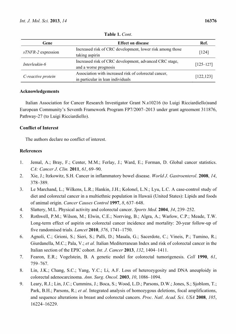
Table 1. Cont.
Gene Effect on disease Ref.
sTNFR-2 expression Increased risk of CRC development, lower risk among those taking aspirin
[124]
Interleukin-6 Increased risk of CRC development, advanced CRC stage, and a worse prognosis
[125–127]
C-reactive protein Association with increased risk of colorectal cancer, in particular in lean individuals
[122,123]
Acknowledgements
Italian Association for Cancer Research Investigator Grant N.�10216 (to Luigi Ricciardiello)�and
European Community’s Seventh Framework Program FP7/2007–2013 under grant agreement 311876,
Pathway-27 (to Luigi Ricciardiello).
Conflict of Interest
The authors declare no conflict of interest.
References
1. Jemal, A.; Bray, F.; Center, M.M.; Ferlay, J.; Ward, E.; Forman, D. Global cancer statistics.
CA: Cancer J. Clin. 2011, 61, 69–90.
2. Xie, J.; Itzkowitz, S.H. Cancer in inflammatory bowel disease. World J. Gastroenterol. 2008, 14,
378–389.
3. Le Marchand, L.; Wilkens, L.R.; Hankin, J.H.; Kolonel, L.N.; Lyu, L.C. A case-control study of
diet and colorectal cancer in a multiethnic population in Hawaii (United States): Lipids and foods
of animal origin. Cancer Causes Control 1997, 8, 637–648.
4. Slattery, M.L. Physical activity and colorectal cancer. Sports Med. 2004, 34, 239–252.
5. Rothwell, P.M.; Wilson, M.; Elwin, C.E.; Norrving, B.; Algra, A.; Warlow, C.P.; Meade, T.W.
Long-term effect of aspirin on colorectal cancer incidence and mortality: 20-year follow-up of
five randomised trials. Lancet 2010, 376, 1741–1750.
6. Agnoli, C.; Grioni, S.; Sieri, S.; Palli, D.; Masala, G.; Sacerdote, C.; Vineis, P.; Tumino, R.;
Giurdanella, M.C.; Pala, V.; et al. Italian Mediterranean Index and risk of colorectal cancer in the
Italian section of the EPIC cohort. Int. J. Cancer 2013, 132, 1404–1411.
7. Fearon, E.R.; Vogelstein, B. A genetic model for colorectal tumorigenesis. Cell 1990, 61,
759–767.
8. Lin, J.K.; Chang, S.C.; Yang, Y.C.; Li, A.F. Loss of heterozygosity and DNA aneuploidy in
colorectal adenocarcinoma. Ann. Surg. Oncol. 2003, 10, 1086–1094.
9. Leary, R.J.; Lin, J.C.; Cummins, J.; Boca, S.; Wood, L.D.; Parsons, D.W.; Jones, S.; Sjoblom, T.;
Park, B.H.; Parsons, R.; et al. Integrated analysis of homozygous deletions, focal amplifications,
and sequence alterations in breast and colorectal cancers. Proc. Natl. Acad. Sci. USA 2008, 105,
16224–16229.

Int. J. Mol. Sci. 2013, 14 16377
10. Bardi, G.; Johansson, B.; Pandis, N.; Mandahl, N.; Bak-Jensen, E.; Lindstrom, C.; Tornqvist, A.;
Frederiksen, H.; Andren-Sandberg, A.; Mitelman, F.; et al. Cytogenetic analysis of 52 colorectal
carcinomas—Non-random aberration pattern and correlation with pathologic parameters.
Int. J. Cancer 1993, 55, 422–428.
11. Bardi, G.; Sukhikh, T.; Pandis, N.; Fenger, C.; Kronborg, O.; Heim, S. Karyotypic
characterization of colorectal adenocarcinomas. Genes Chromosomes Cancer 1995, 12, 97–109.
12. Shih, I.M.; Zhou, W.; Goodman, S.N.; Lengauer, C.; Kinzler, K.W.; Vogelstein, B. Evidence that
genetic instability occurs at an early stage of colorectal tumorigenesis. Cancer Res. 2001, 61,
818–822.
13. Sieber, O.M.; Lamlum, H.; Crabtree, M.D.; Rowan, A.J.; Barclay, E.; Lipton, L.; Hodgson, S.;
Thomas, H.J.; Neale, K.; Phillips, R.K.; et al. Whole-gene APC deletions cause classical familial
adenomatous polyposis, but not attenuated polyposis or “multiple” colorectal adenomas.
Proc. Natl. Acad. Sci. USA 2002, 99, 2954–2958.
14. Powell, S.M.; Petersen, G.M.; Krush, A.J.; Booker, S.; Jen, J.; Giardiello, F.M.; Hamilton, S.R.;
Vogelstein, B.; Kinzler, K.W. Molecular diagnosis of familial adenomatous polyposis.
N. Engl. J. Med. 1993, 329, 1982–1987.
15. Sieber, O.M.; Lipton, L.; Crabtree, M.; Heinimann, K.; Fidalgo, P.; Phillips, R.K.; Bisgaard,
M.L.; Orntoft, T.F.; Aaltonen, L.A.; Hodgson, S.V.; et al. Multiple colorectal adenomas, classic
adenomatous polyposis, and germ-line mutations in MYH. N. Engl. J. Med. 2003, 348, 791–799.
16. Sparks, A.B.; Morin, P.J.; Vogelstein, B.; Kinzler, K.W. Mutational analysis of the
APC/beta-catenin/Tcf pathway in colorectal cancer. Cancer Res. 1998, 58, 1130–1134.
17. Morin, P.J.; Sparks, A.B.; Korinek, V.; Barker, N.; Clevers, H.; Vogelstein, B.; Kinzler, K.W.
Activation of beta-catenin-Tcf signaling in colon cancer by mutations in beta-catenin or APC.
Science 1997, 275, 1787–1790.
18. Chan, G.K.; Jablonski, S.A.; Sudakin, V.; Hittle, J.C.; Yen, T.J. Human BUBR1 is a mitotic
checkpoint kinase that monitors CENP-E functions at kinetochores and binds the
cyclosome/APC. J. Cell Biol. 1999, 146, 941–954.
19. Shin, H.J.; Baek, K.H.; Jeon, A.H.; Park, M.T.; Lee, S.J.; Kang, C.M.; Lee, H.S.; Yoo, S.H.;
Chung, D.H.; Sung, Y.C.; et al. Dual roles of human BubR1, a mitotic checkpoint kinase, in the
monitoring of chromosomal instability. Cancer Cell 2003, 4, 483–497.
20. van Es, J.H.; van Gijn, M.E.; Riccio, O.; van den Born, M.; Vooijs, M.; Begthel, H.; Cozijnsen, M.;
Robine, S.; Winton, D.J.; Radtke, F.; et al. Notch/gamma-secretase inhibition turns proliferative
cells in intestinal crypts and adenomas into goblet cells. Nature 2005, 435, 959–963.
21. Kwon, C.; Cheng, P.; King, I.N.; Andersen, P.; Shenje, L.; Nigam, V.; Srivastava, D. Notch
post-translationally regulates beta-catenin protein in stem and progenitor cells. Nat. Cell. Biol.
2011, 13, 1244–1251.
22. Firestein, R.; Bass, A.J.; Kim, S.Y.; Dunn, I.F.; Silver, S.J.; Guney, I.; Freed, E.; Ligon, A.H.;
Vena, N.; Ogino, S.; et al. CDK8 is a colorectal cancer oncogene that regulates beta-catenin
activity. Nature 2008, 455, 547–551.
23. Fryer, C.J.; White, J.B.; Jones, K.A. Mastermind recruits CycC:CDK8 to phosphorylate the
Notch ICD and coordinate activation with turnover. Mol. Cell 2004, 16, 509–520.

Int. J. Mol. Sci. 2013, 14 16378
24. Firestein, R.; Shima, K.; Nosho, K.; Irahara, N.; Baba, Y.; Bojarski, E.; Giovannucci, E.L.;
Hahn, W.C.; Fuchs, C.S.; Ogino, S.; et al. CDK8 expression in 470 colorectal cancers in relation
to beta-catenin activation, other molecular alterations and patient survival. Int. J. Cancer 2010,
126, 2863–2873.
25. Carmon, K.S.; Gong, X.; Lin, Q.; Thomas, A.; Liu, Q. R-spondins function as ligands of the
orphan receptors LGR4 and LGR5 to regulate Wnt/beta-catenin signaling. Proc. Natl. Acad. Sci.
USA 2011, 108, 11452–11457.
26. Alao, J.P. The regulation of cyclin D1 degradation: Roles in cancer development and the
potential for therapeutic invention. Mol. Cancer 2007, 6, 24.
27. Arber, N.; Hibshoosh, H.; Moss, S.F.; Sutter, T.; Zhang, Y.; Begg, M.; Wang, S.; Weinstein, I.B.;
Holt, P.R. Increased expression of cyclin D1 is an early event in multistage colorectal
carcinogenesis. Gastroenterology 1996, 110, 669–674.
28. Morikawa, T.; Kuchiba, A.; Lochhead, P.; Nishihara, R.; Yamauchi, M.; Imamura, Y.; Liao, X.;
Qian, Z.R.; Ng, K.; Chan, A.T.; et al. Prospective analysis of body mass index, physical activity,
and colorectal cancer risk associated with beta-catenin (CTNNB1) status. Cancer Res. 2013, 73,
1600–1610.
29. Malumbres, M.; Barbacid, M. RAS oncogenes: The first 30 years. Nat. Rev. Cancer 2003, 3,
459–465.
30. Guerrero, S.; Casanova, I.; Farre, L.; Mazo, A.; Capella, G.; Mangues, R. K-ras codon 12
mutation induces higher level of resistance to apoptosis and predisposition to
anchorage-independent growth than codon 13 mutation or proto-oncogene overexpression.
Cancer Res. 2000, 60, 6750–6756.
31. Imamura, Y.; Morikawa, T.; Liao, X.; Lochhead, P.; Kuchiba, A.; Yamauchi, M.; Qian, Z.R.;
Nishihara, R.; Meyerhardt, J.A.; Haigis, K.M.; et al. Specific mutations in KRAS codons 12 and
13, and patient prognosis in 1075 BRAF wild-type colorectal cancers. Clin. Cancer Res. 2012,
18, 4753–4763.
32. Horst, D.; Chen, J.; Morikawa, T.; Ogino, S.; Kirchner, T.; Shivdasani, R.A. Differential WNT
activity in colorectal cancer confers limited tumorigenic potential and is regulated by MAPK
signaling. Cancer Res. 2012, 72, 1547–1556.
33. Baba, Y.; Nosho, K.; Shima, K.; Meyerhardt, J.A.; Chan, A.T.; Engelman, J.A.; Cantley, L.C.;
Loda, M.; Giovannucci, E.; Fuchs, C.S.; et al. Prognostic significance of AMP-activated protein
kinase expression and modifying effect of MAPK3/1 in colorectal cancer. Br. J. Cancer 2010,
103, 1025–1033.
34. Baker, S.J.; Preisinger, A.C.; Jessup, J.M.; Paraskeva, C.; Markowitz, S.; Willson, J.K.;
Hamilton, S.; Vogelstein, B. p53 gene mutations occur in combination with 17p allelic deletions
as late events in colorectal tumorigenesis. Cancer Res. 1990, 50, 7717–7722.
35. Oikawa, T.; Okuda, M.; Ma, Z.; Goorha, R.; Tsujimoto, H.; Inokuma, H.; Fukasawa, K.
Transcriptional control of BubR1 by p53 and suppression of centrosome amplification by
BubR1. Mol. Cell Biol. 2005, 25, 4046–4061.
36. el-Deiry, W.S.; Tokino, T.; Velculescu, V.E.; Levy, D.B.; Parsons, R.; Trent, J.M.; Lin, D.;
Mercer, W.E.; Kinzler, K.W.; Vogelstein, B. WAF1, a potential mediator of p53 tumor
suppression. Cell 1993, 75, 817–825.

Int. J. Mol. Sci. 2013, 14 16379
37. Morikawa, T.; Kuchiba, A.; Liao, X.; Imamura, Y.; Yamauchi, M.; Qian, Z.R.; Nishihara, R.;
Sato, K.; Meyerhardt, J.A.; Fuchs, C.S.; et al. Tumor TP53 expression status, body mass index
and prognosis in colorectal cancer. Int. J. Cancer 2012, 131, 1169–1178.
38. Ogino, S.; Nosho, K.; Shima, K.; Baba, Y.; Irahara, N.; Kirkner, G.J.; Hazra, A.; de Vivo, I.;
Giovannucci, E.L.; Meyerhardt, J.A.; et al. p21 expression in colon cancer and modifying effects
of patient age and body mass index on prognosis. Cancer Epidemiol. Biomark. Prev. 2009, 18,
2513–2521.
39. Ogino, S.; Kawasaki, T.; Ogawa, A.; Kirkner, G.J.; Loda, M.; Fuchs, C.S. Cytoplasmic
localization of p27 (cyclin-dependent kinase inhibitor 1B/KIP1) in colorectal cancer: Inverse
correlations with nuclear p27 loss, microsatellite instability, and CpG island methylator
phenotype. Hum. Pathol. 2007, 38, 585–592.
40. Loda, M.; Cukor, B.; Tam, S.W.; Lavin, P.; Fiorentino, M.; Draetta, G.F.; Jessup, J.M.;
Pagano, M. Increased proteasome-dependent degradation of the cyclin-dependent kinase
inhibitor p27 in aggressive colorectal carcinomas. Nat. Med. 1997, 3, 231–234.
41. Swamy, M.V.; Herzog, C.R.; Rao, C.V. Inhibition of COX-2 in colon cancer cell lines by
celecoxib increases the nuclear localization of active p53. Cancer Res. 2003, 63, 5239–5242.
42. Ogino, S.; Kirkner, G.J.; Nosho, K.; Irahara, N.; Kure, S.; Shima, K.; Hazra, A.; Chan, A.T.;
Dehari, R.; Giovannucci, E.L.; et al. Cyclooxygenase-2 expression is an independent predictor of
poor prognosis in colon cancer. Clin. Cancer Res. 2008, 14, 8221–8227.
43. Lanza, G.; Matteuzzi, M.; Gafa, R.; Orvieto, E.; Maestri, I.; Santini, A.; del Senno, L.
Chromosome 18q allelic loss and prognosis in stage II and III colon cancer. Int. J. Cancer 1998,
79, 390–395.
44. Kern, S.E.; Fearon, E.R.; Tersmette, K.W.; Enterline, J.P.; Leppert, M.; Nakamura, Y.;
White, R.; Vogelstein, B.; Hamilton, S.R. Clinical and pathological associations with allelic loss
in colorectal carcinoma [corrected]. JAMA 1989, 261, 3099–3103.
45. Ogino, S.; Nosho, K.; Irahara, N.; Shima, K.; Baba, Y.; Kirkner, G.J.; Meyerhardt, J.A.;
Fuchs, C.S. Prognostic significance and molecular associations of 18q loss of heterozygosity: A
cohort study of microsatellite stable colorectal cancers. J. Clin. Oncol. 2009, 27, 4591–4598.
46. Deming, D.A.; Leystra, A.A.; Nettekoven, L.; Sievers, C.; Miller, D.; Middlebrooks, M.;
Clipson, L.; Albrecht, D.; Bacher, J.; Washington, M.K.; et al. PIK3CA and APC mutations are
synergistic in the development of intestinal cancers. Oncogene 2013, doi:10.1038/onc.2013.167.
47. Samuels, Y.; Velculescu, V.E. Oncogenic mutations of PIK3CA in human cancers. Cell Cycle
2004, 3, 1221–1224.
48. Liao, X.; Lochhead, P.; Nishihara, R.; Morikawa, T.; Kuchiba, A.; Yamauchi, M.; Imamura, Y.;
Qian, Z.R.; Baba, Y.; Shima, K.; et al. Aspirin use, tumor PIK3CA mutation, and
colorectal-cancer survival. N. Engl. J. Med. 2012, 367, 1596–1606.
49. Cordaux, R.; Batzer, M.A. The impact of retrotransposons on human genome evolution.
Nat. Rev. Genet. 2009, 10, 691–703.
50. Ogino, S.; Kawasaki, T.; Nosho, K.; Ohnishi, M.; Suemoto, Y.; Kirkner, G.J.; Fuchs, C.S.
LINE-1 hypomethylation is inversely associated with microsatellite instability and CpG island
methylator phenotype in colorectal cancer. Int. J. Cancer 2008, 122, 2767–2773.

Int. J. Mol. Sci. 2013, 14 16380
51. Baba, Y.; Huttenhower, C.; Nosho, K.; Tanaka, N.; Shima, K.; Hazra, A.; Schernhammer, E.S.;
Hunter, D.J.; Giovannucci, E.L.; Fuchs, C.S.; et al. Epigenomic diversity of colorectal cancer
indicated by LINE-1 methylation in a database of 869 tumors. Mol. Cancer 2010, 9, 125.
52. Ogino, S.; Nishihara, R.; Lochhead, P.; Imamura, Y.; Kuchiba, A.; Morikawa, T.; Yamauchi, M.;
Liao, X.; Qian, Z.R.; Sun, R.; et al. Prospective study of family history and colorectal cancer risk
by tumor LINE-1 methylation level. J. Natl. Cancer Inst. 2013, 105, 130–140.
53. Tomonaga, T.; Matsushita, K.; Ishibashi, M.; Nezu, M.; Shimada, H.; Ochiai, T.; Yoda, K.;
Nomura, F. Centromere protein H is up-regulated in primary human colorectal cancer and its
overexpression induces aneuploidy. Cancer Res. 2005, 65, 4683–4689.
54. Tomonaga, T.; Matsushita, K.; Yamaguchi, S.; Oohashi, T.; Shimada, H.; Ochiai, T.; Yoda, K.;
Nomura, F. Overexpression and mistargeting of centromere protein-A in human primary
colorectal cancer. Cancer Res. 2003, 63, 3511–3516.
55. Baba, Y.; Nosho, K.; Shima, K.; Irahara, N.; Chan, A.T.; Meyerhardt, J.A.; Chung, D.C.;
Giovannucci, E.L.; Fuchs, C.S.; Ogino, S. HIF1A overexpression is associated with poor
prognosis in a cohort of 731 colorectal cancers. Am. J. Pathol. 2010, 176, 2292–2301.
56. Kaidi, A.; Qualtrough, D.; Williams, A.C.; Paraskeva, C. Direct transcriptional up-regulation of
cyclooxygenase-2 by hypoxia-inducible factor (HIF)-1 promotes colorectal tumor cell survival
and enhances HIF-1 transcriptional activity during hypoxia. Cancer Res. 2006, 66, 6683–6691.
57. Chan, A.T.; Baba, Y.; Shima, K.; Nosho, K.; Chung, D.C.; Hung, K.E.; Mahmood, U.; Madden,
K.; Poss, K.; Ranieri, A.; et al. Cathepsin B expression and survival in colon cancer: Implications
for molecular detection of neoplasia. Cancer Epidemiol. Biomark. Prev. 2010, 19, 2777–2785.
58. Thomas, D.C.; Umar, A.; Kunkel, T.A. Microsatellite instability and mismatch repair defects in
cancer. Mutat. Res. 1996, 350, 201–205.
59. Fishel, R. Mismatch repair, molecular switches, and signal transduction. Genes Dev. 1998, 12,
2096–2101.
60. Boland, C.R.; Thibodeau, S.N.; Hamilton, S.R.; Sidransky, D.; Eshleman, J.R.; Burt, R.W.;
Meltzer, S.J.; Rodriguez-Bigas, M.A.; Fodde, R.; Ranzani, G.N.; et al. A National Cancer
Institute Workshop on Microsatellite Instability for cancer detection and familial predisposition:
Development of international criteria for the determination of microsatellite instability in
colorectal cancer. Cancer Res. 1998, 58, 5248–5257.
61. Lanza, G.; Gafa, R.; Maestri, I.; Santini, A.; Matteuzzi, M.; Cavazzini, L. Immunohistochemical
pattern of MLH1/MSH2 expression is related to clinical and pathological features in colorectal
adenocarcinomas with microsatellite instability. Mod. Pathol. 2002, 15, 741–749.
62. Sinicrope, F.A.; Sargent, D.J. Molecular pathways: Microsatellite instability in colorectal cancer,
prognostic, predictive, and therapeutic implications. Clin. Cancer Res. 2012, 18, 1506–1512.
63. Vasen, H.F.; Moslein, G.; Alonso, A.; Bernstein, I.; Bertario, L.; Blanco, I.; Burn, J.; Capella, G.;
Engel, C.; Frayling, I.; et al. Guidelines for the clinical management of Lynch syndrome
(hereditary non-polyposis cancer). J. Med. Genet. 2007, 44, 353–362.
64. Boland, C.R.; Koi, M.; Chang, D.K.; Carethers, J.M. The biochemical basis of microsatellite
instability and abnormal immunohistochemistry and clinical behavior in Lynch syndrome: From
bench to bedside. Fam. Cancer 2008, 7, 41–52.

Int. J. Mol. Sci. 2013, 14 16381
65. Aaltonen, L.A.; Peltomaki, P.; Mecklin, J.P.; Jarvinen, H.; Jass, J.R.; Green, J.S.; Lynch, H.T.;
Watson, P.; Tallqvist, G.; Juhola, M.; et al. Replication errors in benign and malignant tumors
from hereditary nonpolyposis colorectal cancer patients. Cancer Res. 1994, 54, 1645–1648.
66. Fishel, R.; Lescoe, M.K.; Rao, M.R.; Copeland, N.G.; Jenkins, N.A.; Garber, J.; Kane, M.;
Kolodner, R. The human mutator gene homolog MSH2 and its association with hereditary
nonpolyposis colon cancer. Cell 1993, 75, 1027–1038.
67. Kolodner, R.D.; Tytell, J.D.; Schmeits, J.L.; Kane, M.F.; Gupta, R.D.; Weger, J.; Wahlberg, S.;
Fox, E.A.; Peel, D.; Ziogas, A.; et al. Germ-line msh6 mutations in colorectal cancer families.
Cancer Res. 1999, 59, 5068–5074.
68. Liu, B.; Parsons, R.; Papadopoulos, N.; Nicolaides, N.C.; Lynch, H.T.; Watson, P.; Jass, J.R.;
Dunlop, M.; Wyllie, A.; Peltomaki, P.; et al. Analysis of mismatch repair genes in hereditary
non-polyposis colorectal cancer patients. Nat. Med. 1996, 2, 169–174.
69. Perez-Cabornero, L.; Sanz, M.I.; Sampedro, E.V.; Aras, E.L.; Becares, A.A.; Pino, C.M.;
Dominguez, M.D. Frequency of rearrangements in Lynch syndrome cases associated with
MSH2: Characterization of a new deletion involving both EPCAM and the 5' part of MSH2.
Cancer Prev. Res. (Phila) 2011, 4, 1556–1562.
70. Kastrinos, F.; Steyerberg, E.W.; Balmana, J.; Mercado, R.; Gallinger, S.; Haile, R.; Casey, G.;
Hopper, J.L.; LeMarchand, L.; Lindor, N.M.; et al. Comparison of the clinical prediction model
PREMM(1,2,6) and molecular testing for the systematic identification of Lynch syndrome in
colorectal cancer. Gut 2013, 62, 272–279.
71. Kane, M.F.; Loda, M.; Gaida, G.M.; Lipman, J.; Mishra, R.; Goldman, H.; Jessup, J.M.;
Kolodner, R. Methylation of the hMLH1 promoter correlates with lack of expression of hMLH1
in sporadic colon tumors and mismatch repair-defective human tumor cell lines. Cancer Res.
1997, 57, 808–811.
72. Herman, J.G.; Umar, A.; Polyak, K.; Graff, J.R.; Ahuja, N.; Issa, J.P.; Markowitz, S.; Willson,
J.K.; Hamilton, S.R.; Kinzler, K.W.; et al. Incidence and functional consequences of hMLH1
promoter hypermethylation in colorectal carcinoma. Proc. Natl. Acad. Sci. USA 1998, 95,
6870–6875.
73. Veigl, M.L.; Kasturi, L.; Olechnowicz, J.; Ma, A.H.; Lutterbaugh, J.D.; Periyasamy, S.;
Li, G.M.; Drummond, J.; Modrich, P.L.; Sedwick, W.D.; et al. Biallelic inactivation of hMLH1
by epigenetic gene silencing, a novel mechanism causing human MSI cancers. Proc. Natl. Acad.
Sci. USA 1998, 95, 8698–8702.
74. Ricciardiello, L.; Ceccarelli, C.; Angiolini, G.; Pariali, M.; Chieco, P.; Paterini, P.; Biasco, G.;
Martinelli, G.N.; Roda, E.; Bazzoli, F. High thymidylate synthase expression in colorectal cancer
with microsatellite instability: Implications for chemotherapeutic strategies. Clin. Cancer Res.
2005, 11, 4234–4240.
75. Parsons, M.T.; Buchanan, D.D.; Thompson, B.; Young, J.P.; Spurdle, A.B. Correlation of tumour
BRAF mutations and MLH1 methylation with germline mismatch repair (MMR) gene mutation
status: A literature review assessing utility of tumour features for MMR variant classification.
J. Med. Genet. 2012, 49, 151–157.
76. Takayama, T.; Miyanishi, K.; Hayashi, T.; Sato, Y.; Niitsu, Y. Colorectal cancer: Genetics of
development and metastasis. J. Gastroenterol. 2006, 41, 185–192.

Int. J. Mol. Sci. 2013, 14 16382
77. Grady, W.M.; Rajput, A.; Myeroff, L.; Liu, D.F.; Kwon, K.; Willis, J.; Markowitz, S. Mutation
of the type II transforming growth factor-beta receptor is coincident with the transformation of
human colon adenomas to malignant carcinomas. Cancer Res. 1998, 58, 3101–3104.
78. Riggins, G.J.; Kinzler, K.W.; Vogelstein, B.; Thiagalingam, S. Frequency of Smad gene
mutations in human cancers. Cancer Res. 1997, 57, 2578–2580.
79. Zhang, B.; Halder, S.K.; Kashikar, N.D.; Cho, Y.J.; Datta, A.; Gorden, D.L.; Datta, P.K.
Antimetastatic role of Smad4 signaling in colorectal cancer. Gastroenterology 2010, 138, 969–980.
80. Eppert, K.; Scherer, S.W.; Ozcelik, H.; Pirone, R.; Hoodless, P.; Kim, H.; Tsui, L.C.; Bapat, B.;
Gallinger, S.; Andrulis, I.L.; et al. MADR2 maps to 18q21 and encodes a TGFbeta-regulated
MAD-related protein that is functionally mutated in colorectal carcinoma. Cell 1996, 86, 543–552.
81. Jung, B.; Doctolero, R.T.; Tajima, A.; Nguyen, A.K.; Keku, T.; Sandler, R.S.; Carethers, J.M.
Loss of activin receptor type 2 protein expression in microsatellite unstable colon cancers.
Gastroenterology 2004, 126, 654–659.
82. Rampino, N.; Yamamoto, H.; Ionov, Y.; Li, Y.; Sawai, H.; Reed, J.C.; Perucho, M. Somatic
frameshift mutations in the BAX gene in colon cancers of the microsatellite mutator phenotype.
Science 1997, 275, 967–969.
83. Yamamoto, H.; Sawai, H.; Perucho, M. Frameshift somatic mutations in gastrointestinal cancer
of the microsatellite mutator phenotype. Cancer Res. 1997, 57, 4420–4426.
84. Yagi, O.K.; Akiyama, Y.; Nomizu, T.; Iwama, T.; Endo, M.; Yuasa, Y. Proapoptotic gene BAX
is frequently mutated in hereditary nonpolyposis colorectal cancers but not in adenomas.
Gastroenterology 1998, 114, 268–274.
85. Shima, K.; Morikawa, T.; Yamauchi, M.; Kuchiba, A.; Imamura, Y.; Liao, X.; Meyerhardt, J.A.;
Fuchs, C.S.; Ogino, S. TGFBR2 and BAX mononucleotide tract mutations, microsatellite
instability, and prognosis in 1072 colorectal cancers. PLoS One 2011, 6, e25062.
86. Calin, G.A.; Gafa, R.; Tibiletti, M.G.; Herlea, V.; Becheanu, G.; Cavazzini, L.;
Barbanti-Brodano, G.; Nenci, I.; Negrini, M.; Lanza, G. Genetic progression in microsatellite
instability high (MSI-H) colon cancers correlates with clinico-pathological parameters: A study
of the TGRbetaRII, BAX, hMSH3, hMSH6, IGFIIR and BLM genes. Int. J. Cancer 2000, 89,
230–235.
87. Nosho, K.; Kawasaki, T.; Ohnishi, M.; Suemoto, Y.; Kirkner, G.J.; Zepf, D.; Yan, L.;
Longtine, J.A.; Fuchs, C.S.; Ogino, S. PIK3CA mutation in colorectal cancer: Relationship with
genetic and epigenetic alterations. Neoplasia 2008, 10, 534–541.
88. Baba, Y.; Nosho, K.; Shima, K.; Goessling, W.; Chan, A.T.; Ng, K.; Chan, J.A.; Giovannucci,
E.L.; Fuchs, C.S.; Ogino, S. PTGER2 overexpression in colorectal cancer is associated with
microsatellite instability, independent of CpG island methylator phenotype. Cancer Epidemiol.
Biomark. Prev. 2010, 19, 822–831.
89. Nosho, K.; Kawasaki, T.; Chan, A.T.; Ohnishi, M.; Suemoto, Y.; Kirkner, G.J.; Fuchs, C.S.;
Ogino, S. Cyclin D1 is frequently overexpressed in microsatellite unstable colorectal cancer,
independent of CpG island methylator phenotype. Histopathology 2008, 53, 588–598.
90. Souza, R.F.; Appel, R.; Yin, J.; Wang, S.; Smolinski, K.N.; Abraham, J.M.; Zou, T.T.; Shi, Y.Q.;
Lei, J.; Cottrell, J.; et al. Microsatellite instability in the insulin-like growth factor II receptor
gene in gastrointestinal tumours. Nat. Genet. 1996, 14, 255–257.

Int. J. Mol. Sci. 2013, 14 16383
91. Ogino, S.; Nosho, K.; Irahara, N.; Kure, S.; Shima, K.; Baba, Y.; Toyoda, S.; Chen, L.;
Giovannucci, E.L.; Meyerhardt, J.A.; Fuchs, C.S. A cohort study of cyclin D1 expression and
prognosis in 602 colon cancer cases. Clin. Cancer Res. 2009, 15, 4431–4438.
92. Samowitz, W.S.; Albertsen, H.; Herrick, J.; Levin, T.R.; Sweeney, C.; Murtaugh, M.A.;
Wolff, R.K.; Slattery, M.L. Evaluation of a large, population-based sample supports a CpG
island methylator phenotype in colon cancer. Gastroenterology 2005, 129, 837–845.
93. Shen, L.; Toyota, M.; Kondo, Y.; Lin, E.; Zhang, L.; Guo, Y.; Hernandez, N.S.; Chen, X.;
Ahmed, S.; Konishi, K.; et al. Integrated genetic and epigenetic analysis identifies three different
subclasses of colon cancer. Proc. Natl. Acad. Sci. USA 2007, 104, 18654–18659.
94. Ogino, S.; Odze, R.D.; Kawasaki, T.; Brahmandam, M.; Kirkner, G.J.; Laird, P.W.; Loda, M.;
Fuchs, C.S. Correlation of pathologic features with CpG island methylator phenotype (CIMP) by
quantitative DNA methylation analysis in colorectal carcinoma. Am. J. Surg. Pathol. 2006, 30,
1175–1183.
95. Dong, S.M.; Lee, E.J.; Jeon, E.S.; Park, C.K.; Kim, K.M. Progressive methylation during the
serrated neoplasia pathway of the colorectum. Mod. Pathol. 2005, 18, 170–178.
96. Shima, K.; Nosho, K.; Baba, Y.; Cantor, M.; Meyerhardt, J.A.; Giovannucci, E.L.; Fuchs, C.S.;
Ogino, S. Prognostic significance of CDKN2A (p16) promoter methylation and loss of
expression in 902 colorectal cancers: Cohort study and literature review. Int. J. Cancer 2011,
128, 1080–1094.
97. Myohanen, S.K.; Baylin, S.B.; Herman, J.G. Hypermethylation can selectively silence individual
p16ink4A alleles in neoplasia. Cancer Res. 1998, 58, 591–593.
98. Petko, Z.; Ghiassi, M.; Shuber, A.; Gorham, J.; Smalley, W.; Washington, M.K.;
Schultenover, S.; Gautam, S.; Markowitz, S.D.; Grady, W.M. Aberrantly methylated CDKN2A,
MGMT, and MLH1 in colon polyps and in fecal DNA from patients with colorectal polyps.
Clin. Cancer Res. 2005, 11, 1203–1209.
99. Ogino, S.; Nosho, K.; Kirkner, G.J.; Kawasaki, T.; Meyerhardt, J.A.; Loda, M.;
Giovannucci, E.L.; Fuchs, C.S. CpG island methylator phenotype, microsatellite instability,
BRAF mutation and clinical outcome in colon cancer. Gut 2009, 58, 90–96.
100. Spring, K.J.; Zhao, Z.Z.; Karamatic, R.; Walsh, M.D.; Whitehall, V.L.; Pike, T.; Simms, L.A.;
Young, J.; James, M.; Montgomery, G.W.; et al. High prevalence of sessile serrated adenomas
with BRAF mutations: A prospective study of patients undergoing colonoscopy. Gastroenterology
2006, 131, 1400–1407.
101. Kambara, T.; Simms, L.A.; Whitehall, V.L.; Spring, K.J.; Wynter, C.V.; Walsh, M.D.;
Barker, M.A.; Arnold, S.; McGivern, A.; Matsubara, N.; et al. BRAF mutation is associated with
DNA methylation in serrated polyps and cancers of the colorectum. Gut 2004, 53, 1137–1144.
102. Torlakovic, E.; Snover, D.C. Serrated adenomatous polyposis in humans. Gastroenterology
1996, 110, 748–755.
103. Chan, T.L.; Zhao, W.; Leung, S.Y.; Yuen, S.T. BRAF and KRAS mutations in colorectal
hyperplastic polyps and serrated adenomas. Cancer Res. 2003, 63, 4878–4881.
104. Ogino, S.; Kawasaki, T.; Kirkner, G.J.; Suemoto, Y.; Meyerhardt, J.A.; Fuchs, C.S. Molecular
correlates with MGMT promoter methylation and silencing support CpG island methylator
phenotype-low (CIMP-low) in colorectal cancer. Gut 2007, 56, 1564–1571.

Int. J. Mol. Sci. 2013, 14 16384
105. Shima, K.; orikawa, T.; Baba, Y.; Nosho, K.; Suzuki, M.; Yamauchi, M.; Hayashi, M.;
Giovannucci, E.; Fuchs, C.S.; Ogino, S. MGMT promoter methylation, loss of expression and
prognosis in 855 colorectal cancers. Cancer Causes Control 2011, 22, 301–309.
106. Kawasaki, T.; Ohnishi, M.; Nosho, K.; Suemoto, Y.; Kirkner, G.J.; Meyerhardt, J.A.;
Fuchs, C.S.; Ogino, S. CpG island methylator phenotype-low (CIMP-low) colorectal cancer
shows not only few methylated CIMP-high-specific CpG islands, but also low-level methylation
at individual loci. Mod. Pathol. 2008, 21, 245–255.
107. O’Brien, M.J.; Yang, S.; Mack, C.; Xu, H.; Huang, C.S.; Mulcahy, E.; Amorosino, M.;
Farraye, F.A. Comparison of microsatellite instability, CpG island methylation phenotype,
BRAF and KRAS status in serrated polyps and traditional adenomas indicates separate pathways
to distinct colorectal carcinoma end points. Am. J. Surg. Pathol. 2006, 30, 1491–1501.
108. Jass, J.R.; Baker, K.; Zlobec, I.; Higuchi, T.; Barker, M.; Buchanan, D.; Young, J. Advanced
colorectal polyps with the molecular and morphological features of serrated polyps and adenomas:
Concept of a “fusion” pathway to colorectal cancer. Histopathology 2006, 49, 121–131.
109. Suzuki, H.; Igarashi, S.; Nojima, M.; Maruyama, R.; Yamamoto, E.; Kai, M.; Akashi, H.;
Watanabe, Y.; Yamamoto, H.; Sasaki, Y.; et al. IGFBP7 is a p53-responsive gene specifically
silenced in colorectal cancer with CpG island methylator phenotype. Carcinogenesis 2010, 31,
342–349.
110. Ogino, S.; Kawasaki, T.; Kirkner, G.J.; Yamaji, T.; Loda, M.; Fuchs, C.S. Loss of nuclear p27
(CDKN1B/KIP1) in colorectal cancer is correlated with microsatellite instability and CIMP.
Mod. Pathol. 2007, 20, 15–22.
111. Nosho, K.; Shima, K.; Irahara, N.; Kure, S.; Baba, Y.; Kirkner, G.J.; Chen, L.; Gokhale, S.;
Hazra, A.; Spiegelman, D.; et al. DNMT3B expression might contribute to CpG island
methylator phenotype in colorectal cancer. Clin. Cancer Res. 2009, 15, 3663–3671.
112. Bandres, E.; Cubedo, E.; Agirre, X.; Malumbres, R.; Zarate, R.; Ramirez, N.; Abajo, A.;
Navarro, A.; Moreno, I.; Monzo, M.; Garcia-Foncillas, J. Identification by Real-time PCR of 13
mature microRNAs differentially expressed in colorectal cancer and non-tumoral tissues.
Mol. Cancer 2006, 5, 29.
113. Cummins, J.M.; He, Y.; Leary, R.J.; Pagliarini, R.; Diaz, L.A., Jr.; Sjoblom, T.; Barad, O.;
Bentwich, Z.; Szafranska, A.E.; Labourier, E.; et al. The colorectal microRNAome. Proc. Natl.
Acad. Sci. USA 2006, 103, 3687–3692.
114. Michael, M.Z.; SM, O.C.; van Holst Pellekaan, N.G.; Young, G.P.; James, R.J. Reduced
accumulation of specific microRNAs in colorectal neoplasia. Mol. Cancer Res. 2003, 1, 882–891.
115. Motoyama, K.; Inoue, H.; Takatsuno, Y.; Tanaka, F.; Mimori, K.; Uetake, H.; Sugihara, K.;
Mori, M. Over- and under-expressed microRNAs in human colorectal cancer. Int. J. Oncol.
2009, 34, 1069–1075.
116. Lanza, G.; Ferracin, M.; Gafa, R.; Veronese, A.; Spizzo, R.; Pichiorri, F.; Liu, C.G.; Calin, G.A.;
Croce, C.M.; Negrini, M. mRNA/microRNA gene expression profile in microsatellite unstable
colorectal cancer. Mol. Cancer 2007, 6, 54.
117. Bovell, L.C.; Shanmugam, C.; Putcha, B.D.; Katkoori, V.R.; Zhang, B.; Bae, S.; Singh, K.P.;
Grizzle, W.E.; Manne, U. The prognostic value of microRNAs varies with patient race/ethnicity
and stage of colorectal cancer. Clin. Cancer Res. 2013, 19, 3955–3965.

Int. J. Mol. Sci. 2013, 14 16385
118. Coussens, L.M.; Werb, Z. Inflammation and cancer. Nature 2002, 420, 860–867.
119. Terzic, J.; Grivennikov, S.; Karin, E.; Karin, M. Inflammation and colon cancer.
Gastroenterology 2010, 138, 2101–2114.
120. Ma, X.T.; Wang, S.; Ye, Y.J.; Du, R.Y.; Cui, Z.R.; Somsouk, M. Constitutive activation of Stat3
signaling pathway in human colorectal carcinoma. World J. Gastroenterol. 2004, 10, 1569–1573.
121. Corvinus, F.M.; Orth, C.; Moriggl, R.; Tsareva, S.A.; Wagner, S.; Pfitzner, E.B.; Baus, D.;
Kaufmann, R.; Huber, L.A.; Zatloukal, K.; et al. Persistent STAT3 activation in colon cancer is
associated with enhanced cell proliferation and tumor growth. Neoplasia 2005, 7, 545–555.
122. Otani, T.; Iwasaki, M.; Sasazuki, S.; Inoue, M.; Tsugane, S.; Japan Public Health Center-Based
Prospective Study Group. Plasma C-reactive protein and risk of colorectal cancer in a nested
case-control study: Japan Public Health Center-based prospective study. Cancer Epidemiol.
Biomark. Prev. 2006, 15, 690–695.
123. Gunter, M.J.; Stolzenberg-Solomon, R.; Cross, A.J.; Leitzmann, M.F.; Weinstein, S.; Wood,
R.J.; Virtamo, J.; Taylor, P.R.; Albanes, D.; Sinha, R. A prospective study of serum C-reactive
protein and colorectal cancer risk in men. Cancer Res. 2006, 66, 2483–2487.
124. Chan, A.T.; Ogino, S.; Giovannucci, E.L.; Fuchs, C.S. Inflammatory markers are associated with
risk of colorectal cancer and chemopreventive response to anti-inflammatory drugs.
Gastroenterology 2011, 140, 799–808.
125. Song, M.; Wu, K.; Ogino, S.; Fuchs, C.S.; Giovannucci, E.L.; Chan, A.T. A prospective study of
plasma inflammatory markers and risk of colorectal cancer in men. Br. J. Cancer 2013, 108,
1891–1898.
126. Knupfer, H.; Preiss, R. Serum interleukin-6 levels in colorectal cancer patients—A summary of
published results. Int. J. Colorectal. Dis. 2010, 25, 135–140.
127. Belluco, C.; Nitti, D.; Frantz, M.; Toppan, P.; Basso, D.; Plebani, M.; Lise, M.; Jessup, J.M.
Interleukin-6 blood level is associated with circulating carcinoembryonic antigen and prognosis
in patients with colorectal cancer. Ann. Surg. Oncol. 2000, 7, 133–138.
128. Ito, Y.; Suzuki, K.; Tamakoshi, K.; Wakai, K.; Kojima, M.; Ozasa, K.; Watanabe, Y.;
Kawado, M.; Hashimoto, S.; Suzuki, S.; et al. Colorectal cancer and serum C-reactive protein
levels: A case-control study nested in the JACC Study. J. Epidemiol. 2005, 15, S185–189.
129. Burn, J.; Bishop, D.T.; Chapman, P.D.; Elliott, F.; Bertario, L.; Dunlop, M.G.; Eccles, D.; Ellis,
A.; Evans, D.G.; Fodde, R.; et al. A randomized placebo-controlled prevention trial of aspirin
and/or resistant starch in young people with familial adenomatous polyposis. Cancer Prev. Res.
(Phila) 2011, 4, 655–665.
130. Burn, J.; Bishop, D.T.; Mecklin, J.P.; Macrae, F.; Moslein, G.; Olschwang, S.; Bisgaard, M.L.;
Ramesar, R.; Eccles, D.; Maher, E.R.; et al. Effect of aspirin or resistant starch on colorectal
neoplasia in the Lynch syndrome. N. Engl. J. Med. 2008, 359, 2567–2578.
© 2013 by the authors; licensee MDPI, Basel, Switzerland. This article is an open access article
distributed under the terms and conditions of the Creative Commons Attribution license
(http://creativecommons.org/licenses/by/3.0/).