Mitochondrial Serine Acetyltransferase Functions as a · Mitochondrial Serine Acetyltransferase...
13
Mitochondrial Serine Acetyltransferase Functions as a Pacemaker of Cysteine Synthesis in Plant Cells 1[C][W][OA] Florian H. Haas, Corinna Heeg, Rafael Queiroz, Andrea Bauer, Markus Wirtz, and Ru ¨ diger Hell* Heidelberg Institute for Plant Sciences, Heidelberg University, 69120 Heidelberg, Germany (F.H.H., C.H., M.W., R.H.); and German Cancer Research Center, 69120 Heidelberg, Germany (R.Q., A.B.) Cysteine (Cys) synthesis in plants is carried out by two sequential reactions catalyzed by the rate-limiting enzyme serine acetyltransferase (SAT) and excess amounts of O-acetylserine(thiol)lyase. Why these reactions occur in plastids, mitochondria, and cytosol of plants remained unclear. Expression of artificial microRNA (amiRNA) against Sat3 encoding mitochondrial SAT3 in transgenic Arabidopsis (Arabidopsis thaliana) plants demonstrates that mitochondria are the most important compartment for the synthesis of O-acetylserine (OAS), the precursor of Cys. Reduction of RNA levels, protein contents, SAT enzymatic activity, and phenotype strongly correlate in independent amiSAT3 lines and cause significantly retarded growth. The expression of the other four Sat genes in the Arabidopsis genome are not affected by amiRNA-SAT3 according to quantitative real-time polymerase chain reaction and microarray analyses. Application of radiolabeled serine to leaf pieces revealed severely reduced incorporation rates into Cys and even more so into glutathione. Accordingly, steady-state levels of OAS are 4-fold reduced. Decrease of sulfate reduction-related genes is accompanied by an accumulation of sulfate in amiSAT3 lines. These results unequivocally show that mitochondria provide the bulk of OAS in the plant cell and are the likely site of flux regulation. Together with recent data, the cytosol appears to be a major site of Cys synthesis, while plastids contribute reduced sulfur as sulfide. Thus, Cys synthesis in plants is significantly different from that in nonphotosynthetic eukaryotes at the cellular level. Cys synthesis in plants is a fundamental process for protein biosynthesis and all anabolic pathways that require reduced sulfur. Bacteria and fungi are able to reduce sulfate by assimilatory sulfate reduction to sulfide and to integrate it into Cys in the cytosol. In contrast, mammals need to take up reduced sulfur as Met and can synthesize Cys via trans-sulfurylation in the cytosol. Protein biosynthesis in the mitochondria in animals and fungi receives Cys, therefore, from the cytosol. In plants, the assimilatory sulfate reduction pathway is localized to plastids, whereas Cys synthe- sis is found in the cytosol, plastids, and mitochondria. Why Cys synthesis in plants is localized in three compartments has long been speculated. Serine acetyltransferase (SAT; also termed Serat; EC 2.3.1.30) catalyzes the activation of Ser by acetyl-CoA to O-acetylserine (OAS). In Arabidopsis (Arabidopsis thaliana), five genes encoding SAT1 to SAT5 are local- ized on the five chromosomes (Hell et al., 2002). SAT1 is plastid localized, SAT3 is found in mitochondria, and SAT5 is cytosolic (Kawashima et al., 2005). SAT2 and SAT4 are also cytosolic proteins, but they differ substantially from the other SATs in their amino acid sequences and are much less expressed (Kawashima et al., 2005). The Arabidopsis Sat genes are almost constitutively expressed (www.genevestigator.ethz. ch), and the encoded proteins are of low abundance in comparison with O-acetylserine(thiol)lyase (OAS- TL; EC 2.5.1.47), since the activity of the latter is 300- fold higher than SAT activity in leaves (Ruffet et al., 1994). SAT activity is generally regarded as the rate- limiting step of Cys synthesis (Wirtz and Droux, 2005; Wirtz and Hell, 2006). Fractionation of pea (Pisum sativum) leaf cells showed that about 80% of extractable SAT activity under substrate-saturating conditions re- sides in mitochondria, while about 10% each is found in plastids and cytosol (Ruffet et al., 1995; Droux, 2003). The second step of Cys synthesis consists of the b-substitution of the acetyl moiety in OAS by sulfide and is catalyzed by OAS-TL. OAS-TLs are members of the pyridoxal phosphate-dependent b-substituted Ala synthase family (Hatzfeld et al., 2000b). Of the nine OAS-TL-like genes in Arabidopsis, one is a pseu- dogene, three encode the cytosolic OAS-TL A, plastid OAS-TL B, and mitochondrial OAS-TL C enzymes, and one is a b-cyanoalanine synthase that localizes to mitochondria (Hatzfeld et al., 2000b; Jost et al., 2000; 1 This work was supported by the Stiftung der deutschen Wirt- schaft and the Schmeil-Foundation, Heidelberg (grants to F.F.H.), and by the Bioquant Landesgraduiertenkolleg Baden-Wu ¨ rttemberg (grant to C.H.). * Corresponding author; e-mail [email protected]. The author responsible for distribution of materials integral to the findings presented in this article in accordance with the policy described in the Instructions for Authors (www.plantphysiol.org) is: Ru ¨ diger Hell ([email protected]). [C] Some figures in this article are displayed in color online but in black and white in the print edition. [W] The online version of this article contains Web-only data. [OA] Open Access articles can be viewed online without a sub- scription. www.plantphysiol.org/cgi/doi/10.1104/pp.108.125237 Plant Physiology, October 2008, Vol. 148, pp. 1055–1067, www.plantphysiol.org Ó 2008 American Society of Plant Biologists 1055 www.plantphysiol.org on July 20, 2018 - Published by Downloaded from Copyright © 2008 American Society of Plant Biologists. All rights reserved.
Transcript of Mitochondrial Serine Acetyltransferase Functions as a · Mitochondrial Serine Acetyltransferase...
Mitochondrial Serine Acetyltransferase Functions as aPacemaker of Cysteine Synthesis in Plant Cells1[C][W][OA]
Florian H. Haas, Corinna Heeg, Rafael Queiroz, Andrea Bauer, Markus Wirtz, and Rudiger Hell*
Heidelberg Institute for Plant Sciences, Heidelberg University, 69120 Heidelberg, Germany (F.H.H., C.H.,M.W., R.H.); and German Cancer Research Center, 69120 Heidelberg, Germany (R.Q., A.B.)
Cysteine (Cys) synthesis in plants is carried out by two sequential reactions catalyzed by the rate-limiting enzyme serineacetyltransferase (SAT) and excess amounts of O-acetylserine(thiol)lyase. Why these reactions occur in plastids, mitochondria,and cytosol of plants remained unclear. Expression of artificial microRNA (amiRNA) against Sat3 encoding mitochondrialSAT3 in transgenic Arabidopsis (Arabidopsis thaliana) plants demonstrates that mitochondria are the most importantcompartment for the synthesis of O-acetylserine (OAS), the precursor of Cys. Reduction of RNA levels, protein contents,SAT enzymatic activity, and phenotype strongly correlate in independent amiSAT3 lines and cause significantly retardedgrowth. The expression of the other four Sat genes in the Arabidopsis genome are not affected by amiRNA-SAT3 according toquantitative real-time polymerase chain reaction and microarray analyses. Application of radiolabeled serine to leaf piecesrevealed severely reduced incorporation rates into Cys and even more so into glutathione. Accordingly, steady-state levels ofOAS are 4-fold reduced. Decrease of sulfate reduction-related genes is accompanied by an accumulation of sulfate in amiSAT3lines. These results unequivocally show that mitochondria provide the bulk of OAS in the plant cell and are the likely site offlux regulation. Together with recent data, the cytosol appears to be a major site of Cys synthesis, while plastids contributereduced sulfur as sulfide. Thus, Cys synthesis in plants is significantly different from that in nonphotosynthetic eukaryotes atthe cellular level.
Cys synthesis in plants is a fundamental process forprotein biosynthesis and all anabolic pathways thatrequire reduced sulfur. Bacteria and fungi are able toreduce sulfate by assimilatory sulfate reduction tosulfide and to integrate it into Cys in the cytosol. Incontrast, mammals need to take up reduced sulfur asMet and can synthesize Cys via trans-sulfurylation inthe cytosol. Protein biosynthesis in the mitochondriain animals and fungi receives Cys, therefore, from thecytosol. In plants, the assimilatory sulfate reductionpathway is localized to plastids, whereas Cys synthe-sis is found in the cytosol, plastids, and mitochondria.Why Cys synthesis in plants is localized in threecompartments has long been speculated.Serine acetyltransferase (SAT; also termed Serat; EC
2.3.1.30) catalyzes the activation of Ser by acetyl-CoA
to O-acetylserine (OAS). In Arabidopsis (Arabidopsisthaliana), five genes encoding SAT1 to SAT5 are local-ized on the five chromosomes (Hell et al., 2002). SAT1is plastid localized, SAT3 is found in mitochondria,and SAT5 is cytosolic (Kawashima et al., 2005). SAT2and SAT4 are also cytosolic proteins, but they differsubstantially from the other SATs in their amino acidsequences and are much less expressed (Kawashimaet al., 2005). The Arabidopsis Sat genes are almostconstitutively expressed (www.genevestigator.ethz.ch), and the encoded proteins are of low abundancein comparison with O-acetylserine(thiol)lyase (OAS-TL; EC 2.5.1.47), since the activity of the latter is 300-fold higher than SAT activity in leaves (Ruffet et al.,1994). SAT activity is generally regarded as the rate-limiting step of Cys synthesis (Wirtz and Droux, 2005;Wirtz and Hell, 2006). Fractionation of pea (Pisumsativum) leaf cells showed that about 80% of extractableSAT activity under substrate-saturating conditions re-sides in mitochondria, while about 10% each is foundin plastids and cytosol (Ruffet et al., 1995; Droux, 2003).The second step of Cys synthesis consists of theb-substitution of the acetyl moiety in OAS by sulfideand is catalyzed by OAS-TL. OAS-TLs are membersof the pyridoxal phosphate-dependent b-substitutedAla synthase family (Hatzfeld et al., 2000b). Of thenine OAS-TL-like genes in Arabidopsis, one is a pseu-dogene, three encode the cytosolic OAS-TL A, plastidOAS-TL B, and mitochondrial OAS-TL C enzymes,and one is a b-cyanoalanine synthase that localizes tomitochondria (Hatzfeld et al., 2000b; Jost et al., 2000;
1 This work was supported by the Stiftung der deutschen Wirt-schaft and the Schmeil-Foundation, Heidelberg (grants to F.F.H.),and by the Bioquant Landesgraduiertenkolleg Baden-Wurttemberg(grant to C.H.).
* Corresponding author; e-mail [email protected] author responsible for distribution of materials integral to the
findings presented in this article in accordance with the policydescribed in the Instructions for Authors (www.plantphysiol.org) is:Rudiger Hell ([email protected]).
[C] Some figures in this article are displayed in color online but inblack and white in the print edition.
[W] The online version of this article contains Web-only data.[OA] Open Access articles can be viewed online without a sub-
scription.www.plantphysiol.org/cgi/doi/10.1104/pp.108.125237
Plant Physiology, October 2008, Vol. 148, pp. 1055–1067, www.plantphysiol.org � 2008 American Society of Plant Biologists 1055 www.plantphysiol.orgon July 20, 2018 - Published by Downloaded from
Copyright © 2008 American Society of Plant Biologists. All rights reserved.
Hell et al., 2002; Watanabe et al., 2008). According tocell fractionation of spinach (Spinacia oleracea), cauli-flower (Brassica oleracea), and Datura innoxia, 40% to45% of maximal OAS-TL activity is localized in plas-tids and cytosol, while about 5% is attributed to mito-chondria (Lunn et al., 1990; Rolland et al., 1992; Kuskeet al., 1996). As a result of the low mitochondrial OAS-TL activity, this activity is believed to be a side activityof b-cyanoalanine synthase in spinach (Warrilow andHawkesford, 2000). The other four OAS-TL-like genesin Arabidopsis are only weakly expressed and char-acterized and show little or no Cys synthase activities(Wirtz and Hell, 2006; Heeg et al., 2008).
The three major SATs, SAT5, SAT1, and SAT3, formthe Cys synthase protein complex with the majorOAS-TLs, A, B, and C, in the cytosol, plastids, andmitochondria, according to interaction studies of theArabidopsis proteins (Bogdanova and Hell, 1997; Jostet al., 2000). The Cys synthase complex was hypoth-esized to function in the cellular control of Cys syn-thesis, because SATrequires the presence of OAS-TL togain full activity (Droux et al., 1998). At the same time,complex formation inactivates OAS-TL, and thus thereaction intermediate OAS dissociates from the com-plex and is freely available. OAS is known fromfeeding experiments to activate the expression ofnumerous genes, including genes related to sulfateuptake and reduction (Hirai et al., 2003; Maruyama-Nakashita et al., 2005). At the same time, OAS is able todissociate the Cys synthase complex, at least in vitro,which is antagonized through stabilization by sulfide(Droux et al., 1998; Berkowitz et al., 2002; Wirtz et al.,2004; Wirtz and Hell, 2006). This effector-driven, re-versible protein-protein interaction supposedly func-tions in cytosol, plastids, and mitochondria, althoughcell fractionations suggest considerably different ratiosof SAT and OAS-TL activities in these compartments(Droux, 2003; Wirtz and Droux, 2005).
The role of OAS-TL in the three compartments withindependent protein biosynthesis was recently ad-dressed using Arabidopsis T-DNA insertion lines.Knockout of cytosolic OAS-TL A apparently causedoxidative stress and lesions as well as sensitivity toheavy metals (Lopez-Martin et al., 2008). While thisphenotype was not reported by Watanabe et al. (2008)and Heeg et al. (2008), the latter studies confirmed themajor roles of OAS-TL A, B, and C. OAS-TL A and Btogether contribute 95% of OAS-TL activity in leaves,and OAS-TL C contributes only about 5%. Despite thissmall contribution, a growth rate retardation of ap-proximately 20% was observed for the oastlC mutantline. However, an oastlAB double mutant with OAS-TLC as the only Cys-forming activity was fully viable,with only a small reduction in growth rate (Heeg et al.,2008). OAS contents in the oastlC mutant were de-creased, but in the oastlAB mutant they were found tobe elevated, while labeling studies with [35S]sulfaterevealed lowered incorporation of 35S into Cys only inoastlA mutants. From these findings and the highspecific SAT activity in isolated mitochondria, it was
concluded that mitochondria are the major site of OASsynthesis and that the cytosol, but not chloroplasts,provides most of the cellular Cys (Heeg et al., 2008).Despite the partial redundancy of Cys synthesis be-tween these compartments, the results point toward aspecific role of SAT inmitochondria as a pacemaker forthe rate of cellular Cys synthesis. The investigationspresented here use specific down-regulation of thegene encoding mitochondrial SAT3 by artificial micro-RNA (amiRNA) expression in Arabidopsis. Directevidence for the dominant role of mitochondrial OASformation for cellular Cys synthesis and its significancefor plant growth is provided.
RESULTS
Expression of amiRNA against Sat3 Causes a Severe
Growth Phenotype
According to in vitro analyses, the majority of SATactivity resides in mitochondria in pea leaves andheterotrophic Arabidopsis cell cultures (Ruffet et al.,1995; Heeg et al., 2008). The expression of the gene thatencodes the mitochondrial isoform SAT3 (Sat3, equiv-alent to Serat2;2; At3g13110) was down-regulated byRNA interference to determine (1) if mitochondriaindeed provide most of the OAS in vivo and (2) if thisstep limits Cys biosynthesis in the plant cell. Highestspecificity of the designed amiRNA construct to Sat3mRNA compared with the other four Sat messageswas achieved using the WMD 2 - Web MicroRNADesigner (www.weigelworld.org). Furthermore, thesequence homology and hybridization energy of ami-SAT3 were carefully analyzed and revealed specifictargeting of amiSAT3 to Sat3 mRNA compared withRNAs of Sat1, Sat2, Sat4, and Sat5 genes as well as therest of the Arabidopsis genome (Supplemental Fig.S1). The amiSAT3 construct was expressed under thecontrol of the cauliflower mosaic virus 35S promoterand used to transform Arabidopsis ecotype Columbia(Col-0; amiSAT3 lines).
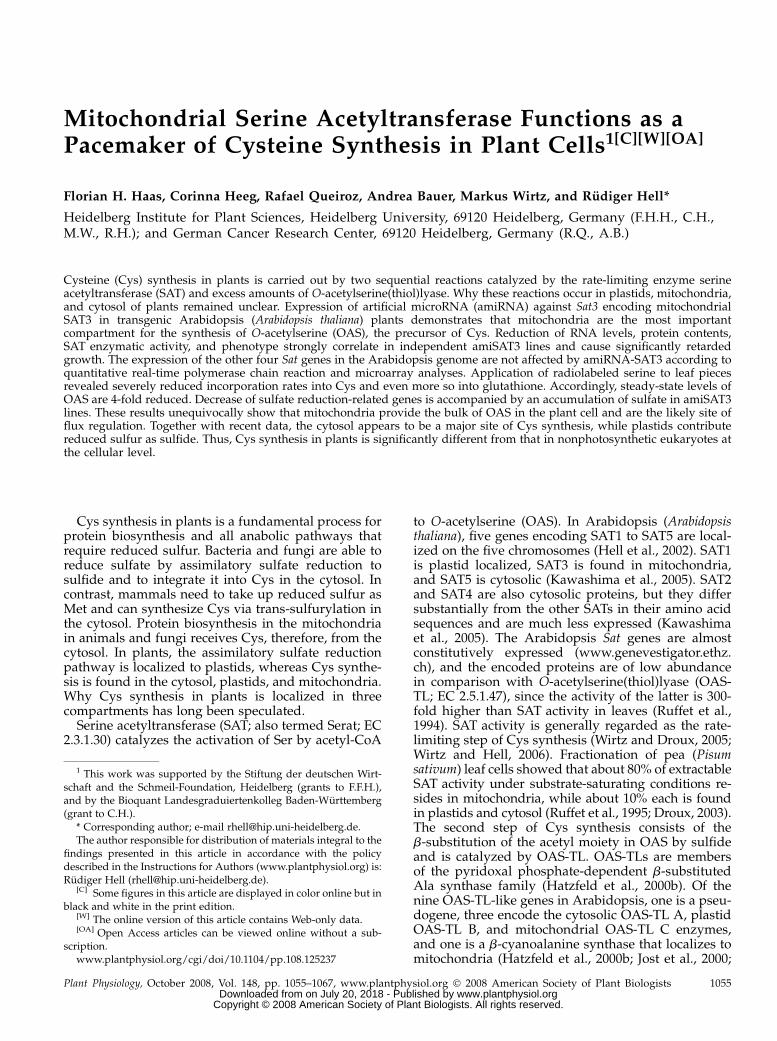
Several independent transformants (T1) were iden-tified using kanamycin as a selectable marker. Nineplants of quite variable size survived (Fig. 1, A and B),while numerous others failed to develop any furtherand died at the early rosette stage before production offlowers (data not shown). Determination of SAT3 pro-tein levels in the surviving amiSAT3 lines by specificantibodies (Wirtz and Hell, 2003) revealed decreasingamounts with declining plant size. In comparison,protein contents of the major OAS-TL forms A, B, andC, which can be simultaneously detected by an OAS-TL-specific antibody (Fig. 1C; Heeg et al., 2008), werenot affected at all. Indeed, SAT3 protein contents androsette sizes of amiSAT3 lines showed a strong linearcorrelation (r = 0.97; Fig. 1D).
Lines 4 and 5 showed intermediate growth retarda-tion and were selected for further analyses. The T2generation of amiSAT3 lines 4 and 5 confirmed the
Haas et al.
1056 Plant Physiol. Vol. 148, 2008 www.plantphysiol.orgon July 20, 2018 - Published by Downloaded from
Copyright © 2008 American Society of Plant Biologists. All rights reserved.
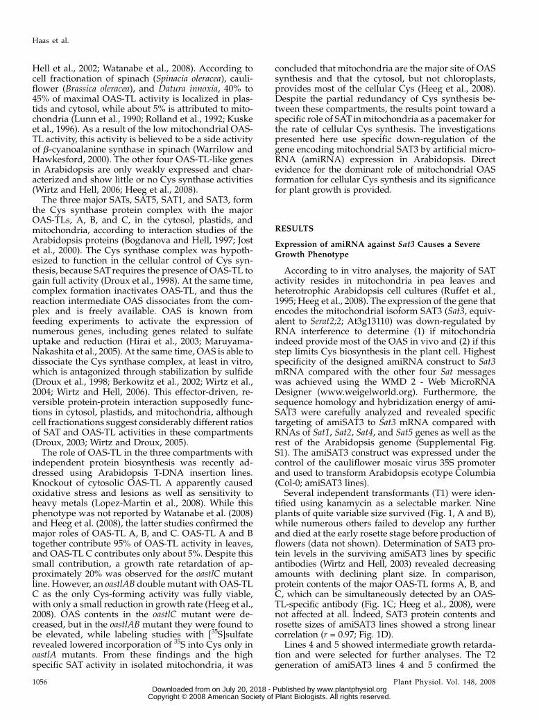
growth phenotypes of the T1 generation (Fig. 2).Growth rates of lines 4 and 5 differed significantlyfrom those of the wild type from week 4 on aftergermination, and fresh weights of rosettes were 40%64% and 33% 6 4%, respectively, of wild-type levelsafter 8 weeks.
Specific Down-Regulation of Mitochondrial SAT Causes
Growth Retardation
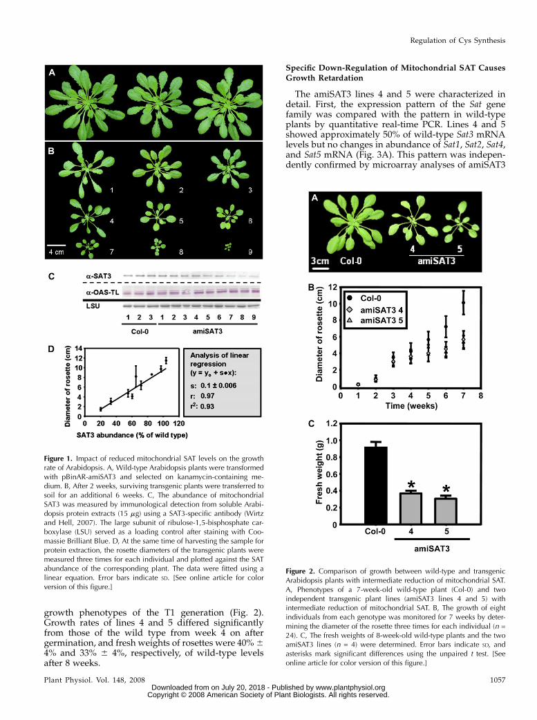
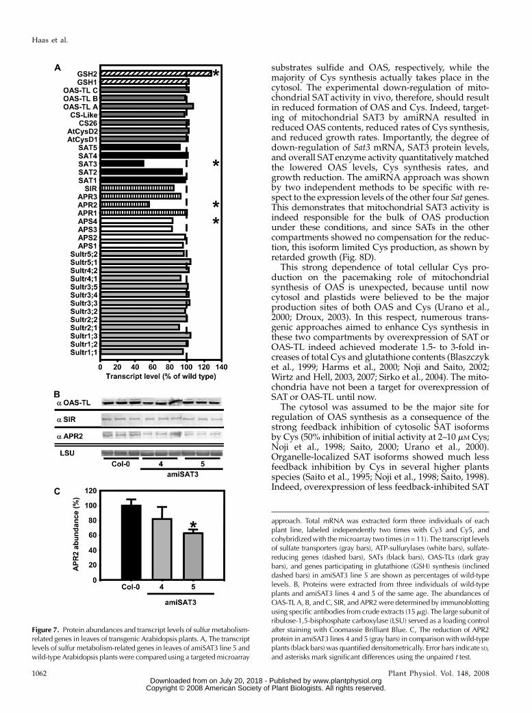
The amiSAT3 lines 4 and 5 were characterized indetail. First, the expression pattern of the Sat genefamily was compared with the pattern in wild-typeplants by quantitative real-time PCR. Lines 4 and 5showed approximately 50% of wild-type Sat3 mRNAlevels but no changes in abundance of Sat1, Sat2, Sat4,and Sat5 mRNA (Fig. 3A). This pattern was indepen-dently confirmed by microarray analyses of amiSAT3
Figure 1. Impact of reduced mitochondrial SAT levels on the growthrate of Arabidopsis. A, Wild-type Arabidopsis plants were transformedwith pBinAR-amiSAT3 and selected on kanamycin-containing me-dium. B, After 2 weeks, surviving transgenic plants were transferred tosoil for an additional 6 weeks. C, The abundance of mitochondrialSAT3 was measured by immunological detection from soluble Arabi-dopsis protein extracts (15 mg) using a SAT3-specific antibody (Wirtzand Hell, 2007). The large subunit of ribulose-1,5-bisphosphate car-boxylase (LSU) served as a loading control after staining with Coo-massie Brilliant Blue. D, At the same time of harvesting the sample forprotein extraction, the rosette diameters of the transgenic plants weremeasured three times for each individual and plotted against the SATabundance of the corresponding plant. The data were fitted using alinear equation. Error bars indicate SD. [See online article for colorversion of this figure.]
Figure 2. Comparison of growth between wild-type and transgenicArabidopsis plants with intermediate reduction of mitochondrial SAT.A, Phenotypes of a 7-week-old wild-type plant (Col-0) and twoindependent transgenic plant lines (amiSAT3 lines 4 and 5) withintermediate reduction of mitochondrial SAT. B, The growth of eightindividuals from each genotype was monitored for 7 weeks by deter-mining the diameter of the rosette three times for each individual (n =24). C, The fresh weights of 8-week-old wild-type plants and the twoamiSAT3 lines (n = 4) were determined. Error bars indicate SD, andasterisks mark significant differences using the unpaired t test. [Seeonline article for color version of this figure.]
Regulation of Cys Synthesis
Plant Physiol. Vol. 148, 2008 1057 www.plantphysiol.orgon July 20, 2018 - Published by Downloaded from
Copyright © 2008 American Society of Plant Biologists. All rights reserved.
line 5 (see below). These findings confirmed the spec-ificity of the amiRNA approach and demonstrated thatno compensatory up-regulation of other Sat genesoccurred in the transgenic lines. Second, SAT enzymeactivities were determined from whole leaf proteinextracts (Fig. 3B). The down-regulation of Sat3 mRNAwas mirrored by decreases of SAT enzyme activity inthe two lines. Finally, the same extent of decrease inlines 4 and 5 was observed with respect to proteinlevels using a SAT3-specific antiserum for immuno-blotting and subsequent densitometric quantification(Fig. 3, C and D). Thus, the phenotype of growthretardation is caused by gene-specific down-regulationof the gene encoding mitochondrial SAT.
Flux of Ser into Cys and Glutathione Is Reduced inamiSAT3 Lines
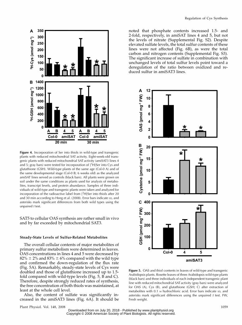
The causal relationship between reduced growthand specific down-regulation of the gene encodingmitochondrial SAT3 implies a reduced rate of OASformation and ultimately of Cys synthesis, unlessother SAT isoforms compensated for the decreasedactivity of the major SAT location in the cell in otherways than transcriptional increases. This assumptionwas tested by incubation of leaf discs in the light withradiolabeled [3H]Ser and quantification of label in Cysand glutathione (Fig. 4). Application of labeled Serrather than sulfate was chosen to monitor flux via OASbut not of the assimilatory sulfate reduction pathway.[3H]Ser uptake of wild-type plants and amiSAT3 lines4 and 5 was compared after 20 and 30 min, respec-tively. Radioactive labeling of Cys formed from [3H]OAS corresponded to half of the flux rate comparedwith the wild type at both time points due to down-regulation of mitochondrial SAT3 activity. An evenstronger reduction of incorporation, down to 30% to40%, was observed for glutathione. This differencesuggests that, after uptake into the leaf cells, [3H]Serwas transported into the mitochondria to be used forthe synthesis of [3H]OAS. This entered the cytosol toserve for the synthesis of Cys, which finally movedinto the chloroplasts as substrate for the initial step ofglutathione formation.
It is concluded that decreased mitochondrial SATactivity has a profound effect on flux into Cys andglutathione due to reduced overall OAS synthesis. Asexpected from the constant expression levels of thefour unaffected SAT genes in Arabidopsis, no signif-icant flux compensation could be observed, since thereductions in Sat3 mRNA and SAT3 protein levelswere in the same range as the reductions of incorpo-ration rates into Cys. Furthermore, these matchingquantitative molecular and metabolic changes due toamiSAT3 expression imply that the contributions ofthe potentially important plastid SAT1 and cytosolic
Figure 3. Molecular and biochemical characterization of transgenicArabidopsis amiSAT3 plants. A, Transcript levels for all members of theSAT gene family in Arabidopsis were determined by quantitative real-time PCR after extraction of total mRNA from leaves of 8-week-oldplants (n . 5). Proteins were extracted from three individuals of wild-type plants (black bars) and amiSAT3 plants of lines 4 and 5 (gray bars)of the same age. B, The crude protein extracts (60–100 mg) were testedthree times for SAT activity (n = 9). C and D, The abundance ofmitochondrial SAT3 was analyzed (n = 9) by immunological detection(C) and quantified by densitometry (D). The large subunit of ribulose-1,5-bisphosphate carboxylase (LSU) served as a loading control after
staining with Coomassie Brilliant Blue. Error bars indicate SD, andasterisks mark significant differences using the unpaired t test.
Haas et al.
1058 Plant Physiol. Vol. 148, 2008 www.plantphysiol.orgon July 20, 2018 - Published by Downloaded from
Copyright © 2008 American Society of Plant Biologists. All rights reserved.
SAT5 to cellular OAS synthesis are rather small in vivoand by far exceeded by mitochondrial SAT3.
Steady-State Levels of Sulfur-Related Metabolites
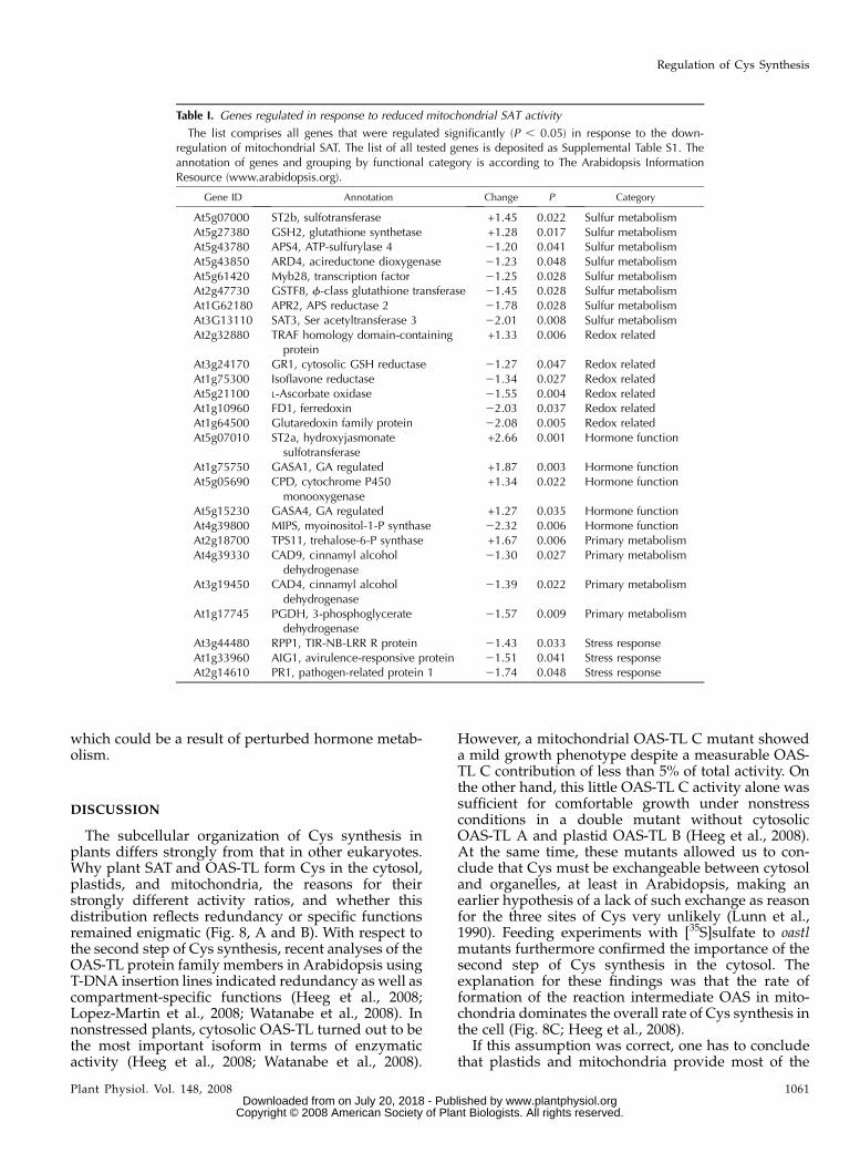
The overall cellular contents of major metabolites ofprimary sulfur metabolism were determined in leaves.OAS concentrations in lines 4 and 5 were decreased by82%6 2% and 83%6 6% compared with the wild typeand confirmed the down-regulation of the flux rate(Fig. 5A). Remarkably, steady-state levels of Cys weredoubled and those of glutathione increased up to 1.5-fold compared with wild-type levels (Fig. 5, B and C).Therefore, despite strongly reduced rates of synthesis,the free concentration of both thiols was maintained, atleast at the whole cell level.Also, the content of sulfate was significantly in-
creased in the amiSAT3 lines (Fig. 6A). It should be
noted that phosphate contents increased 1.5- and2-fold, respectively, in amiSAT lines 4 and 5, but notthe levels of nitrate (Supplemental Fig. S2). Despiteelevated sulfate levels, the total sulfur contents of theselines were not affected (Fig. 6B), as were the totalcarbon and nitrogen contents (Supplemental Fig. S3).The significant increase of sulfate in combination withunchanged levels of total sulfur levels point toward aderegulation of the ratio between oxidized and re-duced sulfur in amiSAT3 lines.
Figure 4. Incorporation of Ser into thiols in wild-type and transgenicplants with reduced mitochondrial SAT activity. Eight-week-old trans-genic plants with reduced mitochondrial SAT activity (amiSAT3 lines 4and 5; gray bars) were tested for incorporation of [3H]Ser into Cys andglutathione (GSH). Wild-type plants of the same age (Col-0 A) and ofthe same developmental stage (Col-0 B; 6 weeks old) as the analyzedamiSAT lines served as controls (black bars). All plants were grown onsoil under the same conditions as plants used for analysis of metabo-lites, transcript levels, and protein abundance. Samples of three indi-viduals of wild-type and transgenic plants were taken and analyzed forincorporation of the radioactive label from [3H]Ser into thiols after 20and 30 min according to Heeg et al. (2008). Error bars indicate SD, andasterisks mark significant differences from both wild types using theunpaired t test.
Figure 5. OAS and thiol contents in leaves of wild-type and transgenicArabidopsis plants. Rosette leaves of three Arabidopsis wild-type plants(black bars) and three individuals of each independent transgenic plantline with reduced mitochondrial SATactivity (gray bars) were analyzedfor OAS (A), Cys (B), and glutathione (GSH; C) after extraction ofmetabolites with 0.1 M hydrochloric acid. Error bars indicate SD, andasterisks mark significant differences using the unpaired t test. FW,Fresh weight.
Regulation of Cys Synthesis
Plant Physiol. Vol. 148, 2008 1059 www.plantphysiol.orgon July 20, 2018 - Published by Downloaded from
Copyright © 2008 American Society of Plant Biologists. All rights reserved.

Impact of Reduced OAS Synthesis on Expression ofSulfur Metabolism-Related Genes
The transcriptome response of amiSAT3 lines wasinvestigated with an array containing 912 genes de-signed for the detection of even small changes ofmRNA abundance, as is often observed with genes ofprimary metabolism. Sixty genes with different ex-pression intensity by robust constitutive patterns ac-cording to Czechowski et al. (2005) were included tooptimize the evaluation of all genes of sulfur metab-olism and key regulatory genes of other metabolicpathways, as well as stress response, redox homeosta-sis, hormone metabolism, and membrane transport. Inaddition, 152 candidate genes were included, whichare reported to be regulated in response to sulfuravailability (Hirai et al., 2003; Higashi et al., 2006;Maruyama-Nakashita et al., 2006). The whole list ofinvestigated genes is shown in Supplemental Table S1.
Based on three biological repetitions of the wild typeand amiSAT3 line 5 with four technical replicates, eachincluding dye swaps for each set, 26 genes were found
to be significantly up- or down-regulated according toP values lower than 0.05 (Table I). Most of the signif-icantly regulated genes (eight) belong to the categoryof sulfur metabolism, while a lower number of redox-related genes (six) and genes involved in hormonefunction (five), primary metabolism (four), and stressresponse (three) were also found to be significantlyaltered in abundance.
Microarray analyses of amiSAT3 line 5 confirmedthe specificity and extent of down-regulation of Sat3expression and the unchanged mRNA levels of theother four Sat genes that had been observed by real-time PCR (Fig. 3). Most interestingly, two genes up-stream of SAT in assimilatory sulfate reduction weredown-regulated: Aps4, encoding a ubiquitously ex-pressed plastidic isoform of ATP-sulfurylase, which isresponsible for the activation of sulfate prior to reduc-tion (Hatzfeld et al., 2000a); and Apr2, encoding aden-osine 5#-phosphosulfate reductase (APR), the majorplastid isoform for the first reduction step. None of theother genes of primary sulfur metabolism was signif-icantly affected (Fig. 7A). The mRNA data of selectedgenes were complemented by immunoblotting of leafproteins. Protein contents of sulfite reductase (SIR) andOAS-TL A, B, and C were unchanged, while APRprotein was decreased by 40% in amiSAT3 line 5 (Fig.7B). Thus, all three protein species precisely reflectedthe mRNA contents determined by microarray analy-sis, although it should be noted that the APR2 antise-rum can probably cross-react with all three APRisoforms (Koprivova et al., 2000).
Only two genes out of the 152 candidate genesknown for sulfur-dependent regulation (Hirai et al.,2003) were found to be significantly regulated inamiSAT3 line 5. Both genes, At1g33960 (avirulence-responsive protein) and At1g75300 (putative isofla-vone reductase), are known to be transcriptionallyregulated also by other environmental factors or hor-mone stimuli. Especially At1g75300 is regulated morestrongly by methyl jasmonate (MJ) than by sulfuravailability (www.genevestigator.ethz.ch). In this re-spect, it is noteworthy that the gene with the highestup-regulation (2.6-fold) is ST2a, a hydroxyjasmonatesulfotransferase (At5g0710), which is supposed toinactivate MJ by sulfonation (Gidda et al., 2003).Reponses to disease and pathogen attack have beenknown for a long time to be coordinated by MJ. Allsignificantly regulated genes of the category stressresponse in this experiment (PR1, AIG1, and PRP1) areinvolved in responses to diseases or pathogens. Inagreement with the idea of inactivation of MJ byinduction of ST2a, all of them are down-regulated.Besides genes responding to MJ, two genes (GASA1and GASA4) that were known as markers for GAstimulus are induced. Taken together, the expressiondata point toward a down-regulation of the sulfateassimilation pathway in amiSAT3 lines in order tocoordinate plastid sulfate reduction with the reducedsynthesis of OAS in mitochondria. Additionally, aninactivation of stress-responsive genes was observed,
Figure 6. Sulfate and total sulfur contents in leaves of wild-type andtransgenic Arabidopsis plants. A, Rosette leaves of three Arabidopsiswild-type plants (black bars) and three individuals of each independenttransgenic plant line with reduced mitochondrial SAT activity (graybars) were analyzed for sulfate contents. FW, Fresh weight. B, The totalsulfur content in dried rosette leaves of 8-week-old wild-type plants(black bars) and the amiSAT3 lines 4 and 5 (gray bars) was analyzedusing an elemental analyzer (n = 4). Error bars indicate SD, and asterisksmark significant differences using the unpaired t test. DW, Dry weight.
Haas et al.
1060 Plant Physiol. Vol. 148, 2008 www.plantphysiol.orgon July 20, 2018 - Published by Downloaded from
Copyright © 2008 American Society of Plant Biologists. All rights reserved.
which could be a result of perturbed hormone metab-olism.
DISCUSSION
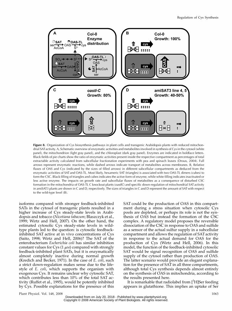
The subcellular organization of Cys synthesis inplants differs strongly from that in other eukaryotes.Why plant SAT and OAS-TL form Cys in the cytosol,plastids, and mitochondria, the reasons for theirstrongly different activity ratios, and whether thisdistribution reflects redundancy or specific functionsremained enigmatic (Fig. 8, A and B). With respect tothe second step of Cys synthesis, recent analyses of theOAS-TL protein family members in Arabidopsis usingT-DNA insertion lines indicated redundancy as well ascompartment-specific functions (Heeg et al., 2008;Lopez-Martin et al., 2008; Watanabe et al., 2008). Innonstressed plants, cytosolic OAS-TL turned out to bethe most important isoform in terms of enzymaticactivity (Heeg et al., 2008; Watanabe et al., 2008).
However, a mitochondrial OAS-TL C mutant showeda mild growth phenotype despite a measurable OAS-TL C contribution of less than 5% of total activity. Onthe other hand, this little OAS-TL C activity alone wassufficient for comfortable growth under nonstressconditions in a double mutant without cytosolicOAS-TL A and plastid OAS-TL B (Heeg et al., 2008).At the same time, these mutants allowed us to con-clude that Cys must be exchangeable between cytosoland organelles, at least in Arabidopsis, making anearlier hypothesis of a lack of such exchange as reasonfor the three sites of Cys very unlikely (Lunn et al.,1990). Feeding experiments with [35S]sulfate to oastlmutants furthermore confirmed the importance of thesecond step of Cys synthesis in the cytosol. Theexplanation for these findings was that the rate offormation of the reaction intermediate OAS in mito-chondria dominates the overall rate of Cys synthesis inthe cell (Fig. 8C; Heeg et al., 2008).
If this assumption was correct, one has to concludethat plastids and mitochondria provide most of the
Table I. Genes regulated in response to reduced mitochondrial SAT activity
The list comprises all genes that were regulated significantly (P , 0.05) in response to the down-regulation of mitochondrial SAT. The list of all tested genes is deposited as Supplemental Table S1. Theannotation of genes and grouping by functional category is according to The Arabidopsis InformationResource (www.arabidopsis.org).
Gene ID Annotation Change P Category
At5g07000 ST2b, sulfotransferase +1.45 0.022 Sulfur metabolismAt5g27380 GSH2, glutathione synthetase +1.28 0.017 Sulfur metabolismAt5g43780 APS4, ATP-sulfurylase 4 21.20 0.041 Sulfur metabolismAt5g43850 ARD4, acireductone dioxygenase 21.23 0.048 Sulfur metabolismAt5g61420 Myb28, transcription factor 21.25 0.028 Sulfur metabolismAt2g47730 GSTF8, f-class glutathione transferase 21.45 0.028 Sulfur metabolismAt1G62180 APR2, APS reductase 2 21.78 0.028 Sulfur metabolismAt3G13110 SAT3, Ser acetyltransferase 3 22.01 0.008 Sulfur metabolismAt2g32880 TRAF homology domain-containing
protein+1.33 0.006 Redox related
At3g24170 GR1, cytosolic GSH reductase 21.27 0.047 Redox relatedAt1g75300 Isoflavone reductase 21.34 0.027 Redox relatedAt5g21100 L-Ascorbate oxidase 21.55 0.004 Redox relatedAt1g10960 FD1, ferredoxin 22.03 0.037 Redox relatedAt1g64500 Glutaredoxin family protein 22.08 0.005 Redox relatedAt5g07010 ST2a, hydroxyjasmonate
sulfotransferase+2.66 0.001 Hormone function
At1g75750 GASA1, GA regulated +1.87 0.003 Hormone functionAt5g05690 CPD, cytochrome P450
monooxygenase+1.34 0.022 Hormone function
At5g15230 GASA4, GA regulated +1.27 0.035 Hormone functionAt4g39800 MIPS, myoinositol-1-P synthase 22.32 0.006 Hormone functionAt2g18700 TPS11, trehalose-6-P synthase +1.67 0.006 Primary metabolismAt4g39330 CAD9, cinnamyl alcohol
dehydrogenase21.30 0.027 Primary metabolism
At3g19450 CAD4, cinnamyl alcoholdehydrogenase
21.39 0.022 Primary metabolism
At1g17745 PGDH, 3-phosphoglyceratedehydrogenase
21.57 0.009 Primary metabolism
At3g44480 RPP1, TIR-NB-LRR R protein 21.43 0.033 Stress responseAt1g33960 AIG1, avirulence-responsive protein 21.51 0.041 Stress responseAt2g14610 PR1, pathogen-related protein 1 21.74 0.048 Stress response
Regulation of Cys Synthesis
Plant Physiol. Vol. 148, 2008 1061 www.plantphysiol.orgon July 20, 2018 - Published by Downloaded from
Copyright © 2008 American Society of Plant Biologists. All rights reserved.
substrates sulfide and OAS, respectively, while themajority of Cys synthesis actually takes place in thecytosol. The experimental down-regulation of mito-chondrial SAT activity in vivo, therefore, should resultin reduced formation of OAS and Cys. Indeed, target-ing of mitochondrial SAT3 by amiRNA resulted inreduced OAS contents, reduced rates of Cys synthesis,and reduced growth rates. Importantly, the degree ofdown-regulation of Sat3 mRNA, SAT3 protein levels,and overall SATenzyme activity quantitatively matchedthe lowered OAS levels, Cys synthesis rates, andgrowth reduction. The amiRNA approach was shownby two independent methods to be specific with re-spect to the expression levels of the other four Sat genes.This demonstrates that mitochondrial SAT3 activity isindeed responsible for the bulk of OAS productionunder these conditions, and since SATs in the othercompartments showed no compensation for the reduc-tion, this isoform limited Cys production, as shown byretarded growth (Fig. 8D).
This strong dependence of total cellular Cys pro-duction on the pacemaking role of mitochondrialsynthesis of OAS is unexpected, because until nowcytosol and plastids were believed to be the majorproduction sites of both OAS and Cys (Urano et al.,2000; Droux, 2003). In this respect, numerous trans-genic approaches aimed to enhance Cys synthesis inthese two compartments by overexpression of SAT orOAS-TL indeed achieved moderate 1.5- to 3-fold in-creases of total Cys and glutathione contents (Blaszczyket al., 1999; Harms et al., 2000; Noji and Saito, 2002;Wirtz and Hell, 2003, 2007; Sirko et al., 2004). The mito-chondria have not been a target for overexpression ofSAT or OAS-TL until now.
The cytosol was assumed to be the major site forregulation of OAS synthesis as a consequence of thestrong feedback inhibition of cytosolic SAT isoformsby Cys (50% inhibition of initial activity at 2–10 mM Cys;Noji et al., 1998; Saito, 2000; Urano et al., 2000).Organelle-localized SAT isoforms showed much lessfeedback inhibition by Cys in several higher plantsspecies (Saito et al., 1995; Noji et al., 1998; Saito, 1998).Indeed, overexpression of less feedback-inhibited SAT
Figure 7. Protein abundances and transcript levels of sulfur metabolism-related genes in leaves of transgenic Arabidopsis plants. A, The transcriptlevels of sulfur metabolism-related genes in leaves of amiSAT3 line 5 andwild-type Arabidopsis plants were compared using a targeted microarray
approach. Total mRNA was extracted form three individuals of eachplant line, labeled independently two times with Cy3 and Cy5, andcohybridizedwith the microarray two times (n = 11). The transcript levelsof sulfate transporters (gray bars), ATP-sulfurylases (white bars), sulfate-reducing genes (dashed bars), SATs (black bars), OAS-TLs (dark graybars), and genes participating in glutathione (GSH) synthesis (inclineddashed bars) in amiSAT3 line 5 are shown as percentages of wild-typelevels. B, Proteins were extracted from three individuals of wild-typeplants and amiSAT3 lines 4 and 5 of the same age. The abundances ofOAS-TL A, B, and C, SIR, and APR2were determined by immunoblottingusing specific antibodies from crude extracts (15mg). The large subunit ofribulose-1,5-bisphosphate carboxylase (LSU) served as a loading controlafter staining with Coomassie Brilliant Blue. C, The reduction of APR2protein in amiSAT3 lines 4 and 5 (gray bars) in comparisonwithwild-typeplants (black bars) was quantified densitometrically. Error bars indicate SD,and asterisks mark significant differences using the unpaired t test.
Haas et al.
1062 Plant Physiol. Vol. 148, 2008 www.plantphysiol.orgon July 20, 2018 - Published by Downloaded from
Copyright © 2008 American Society of Plant Biologists. All rights reserved.
isoforms compared with stronger feedback-inhibitedSATs in the cytosol of transgenic plants resulted in ahigher increase of Cys steady-state levels in Arabi-dopsis and tobacco (Nicotiana tabacum; Blaszczyk et al.,1999; Wirtz and Hell, 2007). On the other hand, theestimated cytosolic Cys steady-state levels in wild-type plants led to the question: is cytosolic feedback-inhibited SAT active at in vivo concentrations of Cys(Saito, 1998; Wirtz and Hell, 2006)? The SAT of theenterobacterium Escherichia coli has similar inhibitionconstant values for Cys (1 mM) compared with stronglyfeedback-inhibited plant SATs, but it is enzymaticallyalmost completely inactive during normal growth(Kredich and Becker, 1971). In the case of E. coli, sucha strict down-regulation makes sense due to the life-style of E. coli, which supports the organism withexogenous Cys. It remains unclear why cytosolic SAT,which contributes less than 10% of the total SAT ac-tivity (Ruffet et al., 1995), would be potently inhibitedby Cys. Possible explanations for the presence of this
SAT could be the production of OAS in this compart-ment during a stress situation when cytosolic Cyspools are depleted, or perhaps its role is not the syn-thesis of OAS but instead the formation of the CSCcomplex. A regulatory model proposes the reversibledissociation of the CSC in response to OAS and sulfideas a sensor of the actual sulfur supply in a subcellularcompartment and allows the regulation of SATactivityin response to the actual demand for OAS for theproduction of Cys (Wirtz and Hell, 2006). In thismodel, the function of the feedback-inhibited cytosolicSAT would be signal recognition of OAS and sulfidesupply of the cytosol rather than production of OAS.The latter scenario would provide an elegant explana-tion for the presence of SAT in all three compartments,although total Cys synthesis depends almost entirelyon the synthesis of OAS in mitochondria, according tothe results presented here.
It is remarkable that radiolabel from [3H]Ser feedingappears in glutathione. This implies an uptake of Ser
Figure 8. Organization of Cys biosynthesis pathways in plant cells and transgenic Arabidopsis plants with reduced mitochon-drial SATactivity. A, Schematic overview of enzymatic activities andmetabolites involved in synthesis of Cys in the cytosol (whitepanel), the mitochondrion (light gray panel), and the chloroplast (dark gray panel). Enzymes are indicated in boldface letters.Black fields of pie charts show the ratio of enzymatic activities present inside the respective compartment as percentages of totalextractable activity calculated from subcellular fractionation experiments with pea and spinach leaves (Droux, 2004). Fullarrows represent enzymatic reactions, while dashed arrows indicate transport of metabolites across membranes. B, Relativefluxes of OAS and Cys (indicated by the sizes of filled arrows) in different subcellular compartments as deduced from theenzymatic activities of SAT and OAS-TL. Most likely, hexameric SAT (triangles) is associated with two OAS-TL dimers (cubes) toform the CSC. Black filling of triangles and cubes indicates the active form of enzyme, while white filling indicates inactivated orless active enzyme. The impacts on growth rate and subcellular fluxes of metabolites as a consequence of disturbed CSCformation in the mitochondria of OAS-TL C knockout plants (oastlC) and specific down-regulation of mitochondrial SATactivityin amiSAT3 plants are shown in C and D, respectively. The sizes of triangles in C and D represent the amount of SATwith respectto the wild-type level (B).
Regulation of Cys Synthesis
Plant Physiol. Vol. 148, 2008 1063 www.plantphysiol.orgon July 20, 2018 - Published by Downloaded from
Copyright © 2008 American Society of Plant Biologists. All rights reserved.
into mitochondria, formation of OAS, transport to thecytosol for Cys synthesis, and import into plastids toserve as substrate of g-glutamylcysteine, the precursorof glutathione that is exclusively formed in plastids inArabidopsis (Wachter et al., 2005). The possibility ofgrowth retardation in amiSAT3 lines due to deficiencyof Cys in the mitochondria themselves can be ex-cluded, since Cys can be imported into mitochondriafrom the cytosol (Heeg et al., 2008). Consequently, theretarded growth phenotype must be a consequence ofreduced total Cys synthesis rates in the cell, althoughcytosolic SAT5 and plastid SAT1 are still present.
OAS contents were decreased in Arabidopsis ami-SAT3 lines 4 and 5, but surprisingly, steady-state levelsof Cys and glutathione were increased despite low-ered flux into these thiols. Maintenance of Cys andglutathione contents was also observed in the Arabi-dopsis oastlC T-DNA mutant line, which lacked mito-chondrial OAS-TL C and showed reduced growth(Heeg et al., 2008). In this respect, the different func-tions of OAS and thiols for cell metabolism have tobe considered: OAS functions as reaction intermedi-ate and potential signal for gene expression duringsulfate starvation (Kim et al., 1999; Hirai et al., 2003),Cys is essential for protein biosynthesis and as a donorof reduced sulfur in numerous reactions (Hell andBergmann, 1990; Pilon-Smits et al., 2002; Picciocchiet al., 2003; Wirtz and Droux, 2005), and glutathione isan integral component of redox homeostasis (MeyerandHell, 2005; Meyer et al., 2007). It is conceivable thatthe maintenance of thiol levels for these reasons is aprerequisite for the proper function of cell metabolism.Nonetheless, this would not fully explain the elevationof Cys steady-state levels. More likely, in amiSAT3lines, Cys steady-state levels are up-regulated as asignal to serve as a signal to adjust cell metabolism toreduced fluxes of OAS.
The microarray analysis of leaves of the ArabidopsisamiSAT3 line 5 in comparison with wild-type plantsrevealed that none of the OAS-TL genes and none ofthe genes of sulfate uptake and primary sulfur metab-olism were changed in expression, with the exceptionof one member of the Aps and Apr gene family each.Down-regulation of APR2 protein was confirmed byimmunoblotting. Reduced activation and reductionof sulfate would explain the increased contents ofsulfate in amiSAT3 lines. Especially the strong down-regulation of APR2 could be of particular importance,since lower APR2 activity in leaves was shown to beresponsible for the higher sulfate content of the Arabi-dopsis ecotype Shahdara in comparison with ecotypeBay-0 (Loudet et al., 2007). High levels of Cys as wellas glutathione were shown to down-regulate genes forsulfate activation and reduction in roots of severalhigher plants (Koprivova et al., 2000; Vauclare et al.,2002). It seems conceivable, therefore, that the highsteady-state levels of Cys and glutathione in amiSAT3lines 4 and 5 are needed to efficiently down-regulatesulfate reduction in order to avoid the accumulation oftoxic sulfide, which may not be entirely scavenged by
Cys synthesis in the transgenic lines. In agreement withthis hypothesis, sulfate accumulates 2-fold in amiSAT3lines 4 and 5 in comparison with the wild type.
Further putative signals for the down-regulation ofAPR2 are OAS and MJ. It has been repeatedly ob-served that OAS, possibly due to the dissociation ofthe CSC, acts as a signal for the entire sulfur assimi-lation pathway. Feeding of OAS regulates more than650 and 850 genes in leaves and roots of Arabidopsis,respectively (Hirai et al., 2003). At least in roots ofArabidopsis, all APR isoforms are up-regulated inresponse to exogenous application of OAS (Koprivovaet al., 2000). Induction of APR by application of OASwas also shown in Lemna minor, indicating that regu-lation of APR by OAS is a general mechanism inhigher plants (Kopriva et al., 2002). Thus, the 4-foldlowered OAS and doubled thiol steady-state levels inamiSAT3 lines may, independently or in combination,act as signals for the repression of Apr2 in amiSAT3lines. MJ has been shown to modulate expressionlevels of sulfate assimilation-related genes (Jost et al.,2005; Sasaki-Sekimoto et al., 2005). According tomicro-array analysis of the amiSAT3 line 5 ST2a, a hydroxy-jasmonate sulfotransferase, which is supposed toinactivate jasmonic acid signals (Gidda et al., 2003),is one of the strongest responding genes. In agreementwith such a possible mechanism, several genes ofthe stress response to pathogens and diseases weredown-regulated and none was up-regulated on themicroarray in this study. Therefore, a diminished octa-decanoic acid signal may have contributed to the re-duced Aps4 and Apr2 expression.
CONCLUSION
Specific down-regulation of mitochondrial SAT3activity in Arabidopsis by amiRNA expression dem-onstrated that mitochondria, but not chloroplasts orthe cytosol, are the dominant source of OAS in vivo.Despite strongly reduced flux into Cys in amiSAT3plants, the steady-state levels of Cys and glutathionewere elevated and presumably contributed to therepression of assimilatory sulfate reduction and accu-mulation of free sulfate. This, together with previousevidence, strongly suggests that OAS from mitochon-dria and sulfide from chloroplasts serve as substratesfor the bulk of Cys synthesis in the cytosol.
MATERIALS AND METHODS
General Cloning
Standard molecular biology technologies like growth of bacteria, plasmid
isolation, and PCR were performed as described by Sambrook et al. (1989)
according to GLP standards. The amiRNA-SAT3-specific sequences were
selected with the WMD 2 -WebMicroRNA Designer (www.weigelworld.org).
The amiRNA of SAT3 was constructed using an overlapping PCR approach
with primers P1 (5#-gaTAAAATACAAGTCCCAGCCCCtctctcttttgtattcc-3#),P2 (5#-gaGGGGCTGGGACTTGTATTTTAtcaaagagaatcaatga-3#), P3 (5#-gaG-
GAGCTGGGACTTCTATTTTTtcacaggtcgtgatatg-3#), P4 (5#-gaAAAAATA-
GAAGTCCCAGCTCCtctacatatatattcct-3#), pRS300-A (5#-CTGCAAGGCGAT-
Haas et al.
1064 Plant Physiol. Vol. 148, 2008 www.plantphysiol.orgon July 20, 2018 - Published by Downloaded from
Copyright © 2008 American Society of Plant Biologists. All rights reserved.
TAAGTTCGGTAAC-3#), and pRS300-B (5#-GCGGATAACAATTTCACA-
CAGGAAACAG-3#) according to Schwab et al. (2006), which resulted in the
amplification of the amiRNA loop structure, the SAT-specific sequence, and
terminal restriction sites for KpnI and BamHI at the 5# and 3# ends, respec-
tively. The resulting PCR product was digested with KpnI and BamHI and
cloned in the plant transformation vector pBINAR under the control of the
cauliflower mosaic virus 35S promoter, resulting in pBINAR-amiSAT3 (Sup-
plemental Fig. S1). The following cDNAwas employed in this study: AtSAT3
(X80938; Bogdanova et al., 1995).
Plant Growth and Transformation
Transformation of Agrobacterium tumefaciens C58 with binary vectors and
subsequent transformation and selection of Arabidopsis (Arabidopsis thaliana
Col-0) were carried out as described by Clough and Bent (1998). Wild-type
plants and plants expressing amiRNA-SAT3 (F1 or F2 generation) were grown
in climate chambers in growth medium containing one-half soil and one-half
substrate 2 (Klasmann-Deilmann) under controlled conditions: 8.5 h of light,
100 mE light photon flux density, 24�C and 18�C at day and night, respectively,
and 50% humidity. Rosette leaves of each 8-week-old plant were pooled and
used to analyze metabolites as well as the expression of sulfur-related genes at
the transcript and protein levels.
Determination of Metabolites
Hydrophilic metabolites were extracted from leaves of Arabidopsis plants
according to Wirtz and Hell (2003). Thiols, OAS, anions, and total contents of
carbon, nitrogen, and sulfur were quantified as described by Heeg et al. (2008)
in order to ensure the comparability of data sets.
Determination of Enzyme Activities
Total soluble proteins were isolated with 0.5 mL of 50 mM HEPES, pH 7.4,
10 mM KCl, 1 mM EDTA, 10% glycerin, 30 mM dithiothreitol, and 0.5%
phenylmethylsulfonyl fluoride from 0.2 g of leaf material that was ground to a
fine powder in liquid nitrogen. Cell debris was removed by centrifugation at
16,000g and 4�C for 10 min. Proteins were quantified as described by Bradford
(1976) using bovine serum albumin as a standard. The enzymatic activities of
SAT and OAS-TL were determined according to Nakamura et al. (1987): SAT
activity was assayed by coupling to the OAS-TL reaction. All SAT activity
determinations were supplemented with 2 units of purified recombinant
OAS-TL (Wirtz et al., 2004) to ensure high excess of OAS-TL activity during
coupling of both reactions.
Immunological Detection of Proteins
Total proteins from leaves were separated according to Laemmli (1970) by
discontinuous SDS-PAGE in Mini-Protean II cells (Bio-Rad). Transfer of
proteins, washing, and development of signals were performed as described
by Heeg et al. (2008) using the alkaline phosphatase-anti-rabbit IgG conjugate
(Promega; 1:10,000) detection of SATand OAS-TL proteins. APR and SIR were
detected with primary antibodies (1:2,000; incubation at 4�C overnight) after
blocking of the nitrocellulose membrane with 3% milk powder. The primary
antibodies were detected with a 1:5,000 dilution of the above-mentioned
secondary antibody. The resulting signals were quantified by densitometry
using the INTAS gel documentation system in combination with Gel-Pro
Express 4.0 software (INTAS).
Determination of Incorporation Rates Using3H-Labeled Ser
Twelve hours prior to the labeling experiment, soil-grown plants were
enclosed in transparent plastic bags to ensure opening of the stomata. Leaf
pieces (30 mg) were cut out from the interveinal fields of leaves and floated on
the labeling solution (half-strength Hoagland medium containing 2.5 mM [3H]
Ser [185–925 GBq mmol21; Hartmann Analytic]) on a horizontal shaker with
60 rpm in the light (17 mE). Samples were taken after 20 and 30 min, washed
twice with half-strength Hoagland medium [2.5 mM Ca(NO3)2, 2.5 mM KNO3,
0.5 mM MgSO4, 0.5 mM KH2PO4, 40 mM Fe-EDTA, 25 mMH3BO3, 2.25 mM
MnCl2, 1.9 mM ZnSO4, 0.15 mM, CuSO4, and 0.05 mM (NH4)6MO7O24, pH 5.9],
and frozen in liquid nitrogen. A negative control for contamination of the leaf
surface with [3H]Ser was taken by dipping leaf pieces into the labeling
solution for 1 s, prior to washing and harvesting like the samples. The samples
were powdered using the Bio101 ThermoSavant Fast Prep system (Qbiogene)
according to the manufacturer’s instructions. The metabolites were extracted
and separated prior to quantification of the incorporated 3H label as described
by Heeg et al. (2008).
Determination of Transcript Levels Using aCustom-Made Microarray
Design and Production of the Microarray
50mer oligonucleotides of 912 selected genes were synthesized by Ocimum
Biosolutionswith anN-terminal amino linker and checked for cross-hybridization
with the Smith-Waterman alignment. Oligonucleotides were diluted to a con-
centrationof0.01 to1mM in53SSC (0.75Msodiumchlorideand0.075Msodium
citrate) containing 2.5 M betaine and spotted on epoxy-coatedNexterionE slides
(Peqlab) with a MicroGRid robot (Biorobotics) using SMP3 pins (TeleChem
International). A microarray contains all probes in four replicates, while the
assembly of replicates on the microarray was randomized to avoid positional
effects.
Sample Preparation, Hybridization, and Evaluation
Fluorescently labeled cDNA samples were prepared from 10 mg of total
RNA that had been isolated from Arabidopsis leaf tissue with the RNeasy
Plant Kit (Qiagen) according to the manufacturer’s protocol. Routinely, Cy3/
Cy5-dCTP was directly incorporated during first-strand synthesis according
to The Institute for Genomic Research direct labeling protocol (www.tigr.org).
The labeled cDNA probe was purified using the QIAquick Purification Kit
(Qiagen) according to the manufacturer’s instructions, and the incorporation
rate of label was determined by photometric measurement. A Cy3-labeled
wild-type cDNA sample and a Cy5-labeled amiSAT3 cDNA sample were
mixed after concentration of each sample in a vacuum concentrator to a final
volume of 20 mL. The resulting sample was immediately denatured by the
addition of 20 mL of prewarmed hybridization buffer h (53 SSC, 0.1% [v/v]
SDS, 0.06% [w/v] DNA from fish sperm, MB grade, and 40% [v/v] form-
amide) followed by incubation at 100�C for 5 min. The microarray was
prehybridized with buffer p (53 SSC, 0.2% SDS, and 0.5% bovine serum
albumin) for 45 min and washed three times with 250 mL of double-distilled
water, before the denatured sample was applied and incubated for 20 h at
50�C in a Slide Booster (Advalytix). The hybridized microarray was washed
at 30�C by subsequently applying buffer w1 (23 SSC and 0.5% SDS), w2 (0.2%
SSC and 0.5% SDS), and w3 (0.1% SSC) for 10 min each. Signals were detected
using the ScanArray5000 confocal laser scanner (Packard BioChip Technolo-
gies) and quantified with the GenePix Pro 4.1 analysis software (Axon
Instruments). Data quality assessment, normalization, and correspondence
cluster analysis were performedwith the analysis and data warehouse software
M-CHiPS (DKFZ) according to Fellenberg et al. (2001; www.mchips.org). Signal
intensities of repeated hybridizations were normalized by the majority of spots,
and the minimal signal intensity was at least above twice the SD of the back-
ground signal.
Determination of Transcript Levels by Real-Time PCR
Total RNA from leaf tissue was extracted with the RNeasy Plant Kit
(Qiagen) according to the manufacturer’s protocol. Total RNAwas transcribed
into cDNA and analyzed by quantitative real-time PCR as described by Talke
et al. (2006). The following primers were used for specific amplification of
actin7 and all members of the SAT gene family from Arabidopsis: SAT1-f
(5#-CACATGCCGAACCGGTAATAC-3#), SAT1-r (5#-GGTGAATCTTCCGGT-
TTACAGAGA-3#), SAT2-f (5#-ACGCTAAGGGAACTCATAAGTCAGA-3#),SAT2-r (5#-TCTTCTCTTATAGCATCCCAAATAGGA-3#), SAT3-f (5#-AAT-
GGAACCCAGACCAAAACC-3#), SAT3-r (5#-GCCCAAACATCATCGACT-
TCA-3#), SAT4-f (5#-CTCTTCCAATGATTGTCTCCCG-3#), SAT4-r (5#-CCT-CTCGAAAGGAAACTCGTCA-3#), SAT5-f (5#-TGGACACAGATCAAGG-
CGG-3#), SAT5-r (5#-ATGAGAAAGAATCGTCGAATATAGATAGC-3#), actin7-f(5#-CAACCGGTATTGTGCTCGATTC-3#), and actin7-r (5#-GAGTGAGTCTGT-
GAGATCCCG-3#).
Regulation of Cys Synthesis
Plant Physiol. Vol. 148, 2008 1065 www.plantphysiol.orgon July 20, 2018 - Published by Downloaded from
Copyright © 2008 American Society of Plant Biologists. All rights reserved.
Statistical Analysis
Regression analyses of data sets were performed with SigmaPlot 8.0 that
uses the Marquardt-Levenberg algorithm for determination of independent
variables. Comparison of means from different sets of data was analyzed for
statistical significance with the unpaired t test. Constant variance and normal
distribution of data were carefully checked with SigmaStat 3.0 prior to
statistical analysis.
Supplemental Data
The following materials are available in the online version of this article.
Supplemental Figure S1. Specificity of the amiRNA-SAT3 approach.
Supplemental Figure S2. Nitrate and phosphate contents in leaves of
wild-type and transgenic Arabidopsis plants.
Supplemental Figure S3. Carbon and nitrogen contents in leaves of wild-
type and transgenic Arabidopsis plants.
Supplemental Table S1. List of genes that were analyzed by the targeted
microarray approach.
ACKNOWLEDGMENTS
We are indebted to Dr. S. Kopriva (John Innes Institute, Norwich, UK) and
Prof. Dr. D. Weigel (Max Planck Institute, Tubingen, Germany) for the kind
gifts of the APR2 antiserum and the pRS300 vector, respectively. We thank S.
Hassel (University of Heidelberg) for excellent technical assistance, Dr. Maria
Bernal (University of Heidelberg) for support of quantitative real-time PCR
analysis of the SAT gene family, and Dr. A. Meyer (University of Heidelberg)
for critically reading the manuscript.
Received June 24, 2008; accepted August 25, 2008; published August 27, 2008.
LITERATURE CITED
Berkowitz O, Wirtz M, Wolf A, Kuhlmann J, Hell R (2002) Use of
biomolecular interaction analysis to elucidate the regulatory mecha-
nism of the cysteine synthase complex from Arabidopsis thaliana. J Biol
Chem 277: 30629–30634
Blaszczyk A, Brodzik R, Sirko A (1999) Increased resistance to oxidative
stress in transgenic tobacco plants overexpressing bacterial serine
acetyltransferase. Plant J 20: 237–243
Bogdanova N, Bork C, Hell R (1995) Cysteine biosynthesis in plants:
isolation and functional identification of a cDNA encoding a serine
acetyltransferase from Arabidopsis thaliana. FEBS Lett 358: 43–47
Bogdanova N, Hell R (1997) Cysteine synthesis in plants: protein-protein
interactions of serine acetyltransferase from Arabidopsis thaliana. Plant J
11: 251–262
Bradford MM (1976) A rapid and sensitive method for the quantitation of
microgram quantities of protein utilizing the principle of protein-dye
binding. Anal Biochem 72: 248–254
Clough SJ, Bent AF (1998) Floral dip: a simplified method for Agro-
bacterium-mediated transformation of Arabidopsis thaliana. Plant J 16:
735–743
Czechowski T, Stitt M, Altmann T, Udvardi MK, Scheible WR (2005)
Genome-wide identification and testing of superior reference genes for
transcript normalization in Arabidopsis. Plant Physiol 139: 5–17
Droux M (2003) Plant serine acetyltransferase: new insights for regulation
of sulphur metabolism in plant cells. Plant Physiol Biochem 41: 619–627
Droux M (2004) Sulfur assimilation and the role of sulfur in plant metab-
olism: a survey. Photosynth Res 79: 331–348
Droux M, Ruffet ML, Douce R, Job D (1998) Interactions between serine
acetyltransferase and O-acetylserine (thiol) lyase in higher plants:
structural and kinetic properties of the free and bound enzymes. Eur J
Biochem 255: 235–245
Fellenberg K, Hauser NC, Brors B, Neutzner A, Hoheisel JD, Vingron M
(2001) Correspondence analysis applied to microarray data. Proc Natl
Acad Sci USA 98: 10781–10786
Gidda SK, Miersch O, Levitin A, Schmidt J, Wasternack C, Varin L (2003)
Biochemical and molecular characterization of a hydroxyjasmonate
sulfotransferase from Arabidopsis thaliana. J Biol Chem 278: 17895–17900
Harms K, von Ballmoos P, Brunold C, Hofgen R, Hesse H (2000) Expres-
sion of a bacterial serine acetyltransferase in transgenic potato plants
leads to increased levels of cysteine and glutathione. Plant J 22: 335–343
Hatzfeld Y, Lee S, Lee M, Leustek T, Saito K (2000a) Functional charac-
terization of a gene encoding a fourth ATP sulfurylase isoform from
Arabidopsis thaliana. Gene 248: 51–58
Hatzfeld Y, Maruyama A, Schmidt A, Noji M, Ishizawa K, Saito K (2000b)
b-Cyanoalanine synthase is a mitochondrial cysteine synthase-like pro-
tein in spinach and Arabidopsis. Plant Physiol 123: 1163–1172
Heeg C, Kruse C, Jost R, Gutensohn M, Ruppert T, Wirtz M, Hell R (2008)
Analysis of the Arabidopsis O-acetylserine(thiol)lyase gene family dem-
onstrates compartment-specific differences in the regulation of cysteine
synthesis. Plant Cell 20: 168–185
Hell R, Bergmann L (1990) g-Glutamylcysteine synthetase in higher plants:
catalytic properties and subcellular localization. Planta 180: 603–612
Hell R, Jost R, Berkowitz O, Wirtz M (2002) Molecular and biochemical
analysis of the enzymes of cysteine biosynthesis in the plant Arabidopsis
thaliana. Amino Acids 22: 245–257
Higashi Y, Hirai MY, Fujiwara T, Naito S, Noji M, Saito K (2006)
Proteomic and transcriptomic analysis of Arabidopsis seeds: molecular
evidence for successive processing of seed proteins and its implication
in the stress response to sulfur nutrition. Plant J 48: 557–571
Hirai M, Fujiwara T, Awazuhara M, Kimura T, Noji M, Saito K (2003)
Global expression profiling of sulfur-starved Arabidopsis by DNA
macroarray reveals the role of O-acetyl-L-serine as a general regulator
of gene expression in response to sulfur nutrition. Plant J 33: 651–663
Jost R, Altschmied L, Bloem E, Bogs J, Gershenzon J, Hahnel U, Hansch
R, Hartmann T, Kopriva S, Kruse C, et al (2005) Expression profiling of
metabolic genes in response to methyl jasmonate reveals regulation of
genes of primary and secondary sulfur-related pathways in Arabidopsis
thaliana. Photosynth Res 86: 491–508
Jost R, Berkowitz O, Wirtz M, Hopkins L, Hawkesford MJ, Hell R (2000)
Genomic and functional characterization of the oas gene family encod-
ing O-acetylserine (thiol) lyases, enzymes catalyzing the final step in
cysteine biosynthesis in Arabidopsis thaliana. Gene 253: 237–247
Kawashima CG, Berkowitz O, Hell R, Noji M, Saito K (2005) Character-
ization and expression analysis of a serine acetyltransferase gene family
involved in a key step of the sulfur assimilation pathway in Arabidop-
sis. Plant Physiol 137: 220–230
Kim H, Hirai MY, Hayashi H, Chino M, Naito S, Fujiwara T (1999) Role of
O-acetyl-L-serine in the coordinated regulation of the expression of a
soybean seed storage-protein gene by sulfur and nitrogen nutrition.
Planta 209: 282–289
Kopriva S, SuterM, von Ballmoos P, Hesse H, Krahenbuhl U, Rennenberg H,
Brunold C (2002) Interaction of sulfate assimilation with carbon and ni-
trogen metabolism in Lemna minor. Plant Physiol 130: 1406–1413
Koprivova A, Suter M, den Camp RO, Brunold C, Kopriva S (2000)
Regulation of sulfate assimilation by nitrogen in Arabidopsis. Plant
Physiol 122: 737–746
Kredich NM, Becker MA (1971) Cysteine biosynthesis: serine transacetylase
andO-acetylserine sulfhydrylase (Salmonella typhimurium). Methods Enzymol
197: 459–471
Kuske CR, Hill KK, Guzman E, Jackson PJ (1996) Subcellular location of
O-acetylserine sulfhydrylase isoenzymes in cell cultures and plant
tissues of Datura innoxia Mill. Plant Physiol 112: 659–667
Laemmli UK (1970) Cleavage of structural proteins during the assembly of
the head of bacteriophage T4. Nature 227: 680–685
Lopez-Martin MC, Becana M, Romero LC, Gotor C (2008) Knocking out
cytosolic cysteine synthesis compromises the antioxidant capacity of the
cytosol to maintain discrete concentrations of hydrogen peroxide in
Arabidopsis. Plant Physiol 147: 562–572
Loudet O, Saliba-Colombani V, Camilleri C, Calenge F, Gaudon V,
Koprivova A, North K, Kopriva S, Daniel-Vedele F (2007) Natural
variation for sulfate content in Arabidopsis thaliana is highly controlled
by APR2. Nat Genet 39: 896–900
Lunn JE, Droux M, Martin J, Douce R (1990) Localization of ATP-
sulfurylase and O-acetylserine(thiol)lyase in spinach leaves. Plant
Physiol 94: 1345–1352
Maruyama-Nakashita A, Nakamura Y, Tohge T, Saito K, Takahashi H
(2006) Arabidopsis SLIM1 is a central transcriptional regulator of plant
sulfur response and metabolism. Plant Cell 18: 3235–3251
Haas et al.
1066 Plant Physiol. Vol. 148, 2008 www.plantphysiol.orgon July 20, 2018 - Published by Downloaded from
Copyright © 2008 American Society of Plant Biologists. All rights reserved.
Maruyama-Nakashita A, Nakamura Y, Watanabe-Takahashi A, Inoue E,
Yamaya T, Takahashi H (2005) Identification of a novel cis-acting
element conferring sulfur deficiency response in Arabidopsis roots.
Plant J 42: 305–314
Meyer AJ, Brach T, Marty L, Kreye S, Rouhier N, Jacquot JP, Hell R (2007)
Redox-sensitive GFP in Arabidopsis thaliana is a quantitative biosensor
for the redox potential of the cellular glutathione redox buffer. Plant J 52:
973–986
Meyer AJ, Hell R (2005) Glutathione homeostasis and redox-regulation by
sulfhydryl groups. Photosynth Res 86: 435–457
Nakamura K, Hayama A, Masada M, Fukushima K, Tamura G (1987)
Measurement of serine acetyltransferase activity in crude plant extracts
by a coupled assay system using cysteine synthase. Plant Cell Physiol
28: 885–891
Noji M, Inoue K, Kimura N, Gouda A, Saito K (1998) Isoform-dependent
differences in feedback regulation and subcellular localization of serine
acetyltransferase involved in cysteine biosynthesis from Arabidopsis
thaliana. J Biol Chem 273: 32739–32745
Noji M, Saito K (2002) Molecular and biochemical analysis of serine
acetyltransferase and cysteine synthase towards sulfur metabolic engi-
neering in plants. Amino Acids 22: 231–243
Picciocchi A, Douce R, Alban C (2003) The plant biotin synthase reaction:
identification and characterization of essential mitochondrial accessory
protein components. J Biol Chem 278: 24966–24975
Pilon-Smits EA, Garifullina GF, Abdel-Ghany S, Kato S, Mihara H, Hale
KL, Burkhead JL, Esaki N, Kurihara T, Pilon M (2002) Characterization
of a NifS-like chloroplast protein from Arabidopsis: implications for its
role in sulfur and selenium metabolism. Plant Physiol 130: 1309–1318
Rolland N, Droux M, Douce R (1992) Subcellular distribution of
O-acetylserine(thiol)lyase in cauliflower (Brassica oleracea L.) inflorescence.
Plant Physiol 98: 927–935
Ruffet ML, Droux M, Douce R (1994) Purification and kinetic properties of
serine acetyltransferase free of O-acetylserine(thiol)lyase from spinach
chloroplasts. Plant Physiol 104: 597–604
Ruffet ML, Lebrun M, Droux M, Douce R (1995) Subcellular distribution
of serine acetyltransferase from Pisum sativum and characterization of
an Arabidopsis thaliana putative cytosolic isoform. Eur J Biochem 227:
500–509
Saito K (1998) Molecular Aspects of Sulfur Assimilation and Acclimation
to Sulfur Supply in Plants. Elsevier Science, Amsterdam
Saito K (2000) Regulation of sulfate transport and synthesis of sulfur-
containing amino acids. Curr Opin Plant Biol 3: 188–195
Saito K, Yokoyama H, Noji M, Murakoshi I (1995) Molecular cloning and
characterization of a plant serine acetyltransferase playing a regulatory
role in cysteine biosynthesis from watermelon. J Biol Chem 270: 16321–
16326
Sambrook J, Fritsch EF, Maniatis T (1989) Molecular Cloning: A Labora-
tory Manual. Cold Spring Harbor Laboratory Press, Cold Spring
Harbor, NY
Sasaki-Sekimoto Y, Taki N, Obayashi T, Aono M, Matsumoto F, Sakurai
N, Suzuki H, Hirai MY, Noji M, Saito K, et al (2005) Coordinated
activation of metabolic pathways for antioxidants and defence com-
pounds by jasmonates and their roles in stress tolerance in Arabidopsis.
Plant J 44: 653–668
Schwab R, Ossowski S, Riester M, Warthmann N, Weigel D (2006) Highly
specific gene silencing by artificial microRNAs in Arabidopsis. Plant Cell
18: 1121–1133
Sirko A, Blaszczyk A, Liszewska F (2004) Overproduction of SAT and/or
OASTL in transgenic plants: a survey of effects. J Exp Bot 55: 1881–1888
Talke IN, Hanikenne M, Kramer U (2006) Zinc-dependent global tran-
scriptional control, transcriptional deregulation, and higher gene copy
number for genes in metal homeostasis of the hyperaccumulator
Arabidopsis halleri. Plant Physiol 142: 148–167
Urano Y, Manabe T, Noji M, Saito K (2000) Molecular cloning and
functional characterization of cDNAs encoding cysteine synthase and
serine acetyltransferase that may be responsible for high cellular cys-
teine content in Allium tuberosum. Gene 257: 269–277
Vauclare P, Kopriva S, Fell D, Suter M, Sticher L, von Ballmoos P,
Krahenbuhl U, den Camp RO, Brunold C (2002) Flux control of sulphate
assimilation inArabidopsis thaliana: adenosine 5#-phosphosulphate reductase
is more susceptible than ATP sulphurylase to negative control by thiols.
Plant J 31: 729–740
Wachter A, Wolf S, Steininger H, Bogs J, Rausch T (2005) Differential
targeting of GSH1 and GSH2 is achieved by multiple transcription
initiation: implications for the compartmentation of glutathione bio-
synthesis in the Brassicaceae. Plant J 41: 15–30
Warrilow AG, Hawkesford MJ (2000) Cysteine synthase (O-acetylserine
(thiol) lyase) substrate specificities classify the mitochondrial isoform as
a cyanoalanine synthase. J Exp Bot 51: 985–993
Watanabe M, Kusano M, Oikawa A, Fukushima A, Noji M, Saito K (2008)
Physiological roles of the b-substituted alanine synthase gene family in
Arabidopsis. Plant Physiol 146: 310–320
Wirtz M, Droux M (2005) Synthesis of the sulfur amino acids: cysteine and
methionine. Photosynth Res 86: 345–362
Wirtz M, Droux M, Hell R (2004) O-Acetylserine (thiol) lyase: an enigmatic
enzyme of plant cysteine biosynthesis revisited in Arabidopsis thaliana.
J Exp Bot 55: 1785–1798
Wirtz M, Hell R (2003) Production of cysteine for bacterial and plant
biotechnology: application of cysteine feedback-insensitive isoforms of
serine acetyltransferase. Amino Acids 24: 195–203
Wirtz M, Hell R (2006) Functional analysis of the cysteine synthase protein
complex from plants: structural, biochemical and regulatory properties.
J Plant Physiol 163: 273–286
Wirtz M, Hell R (2007) Dominant-negative modification reveals the reg-
ulatory function of the multimeric cysteine synthase protein complex in
transgenic tobacco. Plant Cell 19: 625–639
Regulation of Cys Synthesis
Plant Physiol. Vol. 148, 2008 1067 www.plantphysiol.orgon July 20, 2018 - Published by Downloaded from
Copyright © 2008 American Society of Plant Biologists. All rights reserved.