MINISTRY OF PUBLIC HEALTH OF UKRAINE
93
MINISTRY OF PUBLIC HEALTH OF UKRAINE Case studies in Pathomorphology. Self assessment textbook. KROK – 1 (STEP – 1) Part - І
Transcript of MINISTRY OF PUBLIC HEALTH OF UKRAINE

MINISTRY OF PUBLIC HEALTH OF UKRAINE
Case studies in Pathomorphology.
Self assessment textbook.
KROK – 1 (STEP – 1)
Part - І

2
Рекомендовано Центральним методичним кабінетом з вищої ме-
дичної освіти МОЗ України як навчальний посібник для студен-
тів вищих медичних навчальних закладів ІІІ-ІV рівня акредитації
(протокол № 2 від 19.03.2010)
All rights reserved. No part of this publication may be reproduced,
stored in retrieved system, copied or transmitted in any form or by
any means, electronic, mechanical, photocopying, recording or oth-
erwise without written permission from the author.

3
Pathology of cell. Parenchymal dystrophy.
1. During an autopsy a parenchymal fatty dystrophy of the myocardium was diag-
nosed. What is the common or descriptive name of the heart due to this dystrophy?
A. *‘Tabby cat‘ heart (‗Tiger‘s‘ heart)
B. Bovine heart
C. ‗Hairy‘ heart
D. Solder plaque (bony heart)
E. Cor pulmonale
2. A patient with leukemia died from severe chronic anemia. An autopsy revealed
an enlarged heart, with flabby myocardium. It had a dim pale-grey color, yellow
spots and bars. Which pathological process was found in the heart at post-mortem?
A. * Parenchymal fatty dystrophy
B. Vacuolar dystrophy.
C. Hydropic dystrophy.
D. Mesenchymal fatty dystrophy.
E. Mixed dystrophy.
3. A 53 year old patient died with symptoms of liver insufficiency. A post-mortem
examination revealed the enlarged, flabby, yellow-brown liver. Gross examination
of the liver‘s section showed drops of fat. Microscopically: hepatocytes on the pe-
ripheries of the hepatic lobules contained masses of small drops within the cytop-
lasm. Which process most likely took place in the liver?
A. *Fatty dystrophy of the liver
B. Glucosylceramide lipidosis (Gaucher‘s disease)
C. Sphingolopidosis (Niemann-Pick disease)
D. Gangliosidosis (Тay-Sachs disease)
E. Systemic lipoidoses
4. A patient died from chronic cardiovascular insufficiency. At the post-mortem a
‘tabby cat‘ heart was found. From the side of the endocardium, a yellow-white
striped pattern was noticeable. The myocardium was a dim with gray-yellow color.
Which process is most likely diagnosed?
A. * Fatty parenchymal dystrophy.
B. Carbohydrate dystrophy
C. Hydropic dystrophy.
D. Fatty mesenchymal dystrophy.
E. Amyloidosis.
5. A seven-year-old child presented with diphtheria of the pharynx. He subsequent-
ly died from acute cardiac insufficiency. Post-mortem examination of the heart
revealed that the cavities of the heart were extended horizontally. Muscle of the
heart were dim and flabby. Gross section showed motley appearance, with yellow
areas. Microscopically in the cytoplasm of some myocardial cells small vacuoles
were determined. The frozen sections showed vacuoles within cells stained with
sudan-III in orange color. Which type of dystrophy was found in myocardial cells?
A. *Fatty dystrophy

4
B. Carbohydrate dystrophy
C. Vacuolar dystrophy
D. Hyaline dystrophy
E. Hydropic dystrophy
6. A man died due to cardiac insufficiency. At autopsy revealed the heart increased
volume and flabby. A myocardium was a clay-yellow color and dim. From the side
of the endocardium a yellow-white striped pattern was visible (‗tabby cat‘). Under
the microscope the groups of myocardial cells lost their normal structure, their cy-
toplasm contained shallow drops which were black when stained with sudan-IV.
Which one of the following is the correct diagnosis?
A. *Fatty dystrophy of myocardium
B. Cardiosclerosis
C. Rheumatic myocarditis
D. Obesity of the heart
E. Myomalacia
7. A 66-year-old male died from cardiac insufficiency. During the dissection an
increase volume heart was found. Observation of the heart revealed a flabby consis-
tency with stretched chambers. The myocardium section had a dim, clay-yellowish
color. From the side of the endocardium a yellow-white striped pattern was present,
which was especially noted in the papillary muscles. Which pathological process is
the most credible?
A. *Fatty dystrophy of the myocardium
B. Obesity of the heart
C. Dilatation cardiomyopathy
D. Myomalacia
E. Cardiosclerosis
8. A patient died from pulmonary-cardiac insufficiency. During the dissection a
significantly enlarged anemic liver, with yellow doughy consistencies was found. A
liver specimen stained with hematoxylin and eosin exposed various sizes of va-
cuoles in the cytoplasm of the hepatocytes. Which one of the following dystrophies
occurred?
A. *Parenchymal fatty
B. Parenchymal carbohydrate
C. Hyaline
D. Mesenchymal fatty
E. Hydropic
9. A 38-year-old patient, suffering from chronic alcoholism and cirrhosis of the
liver, developed profuse bleeding due to varicose veins of the esophagus which
resulted in death. During the autopsy a liver was noted to be diminished in size
with micronodular tuberosity. The organ was dense and rather yellow in color. A
histological evaluation of the cryostat specimens of the liver, stained with hema-
toxylin and eosin, revealed hepatocytes with large, optically empty vacuoles. These
vacuoles were black when stained with osmium acid. These optically empty va-

5
cuoles hepatocytes indicate:
A. *Fatty dystrophy
B. Inclusions of hyaline.
C. Alcoholic hyaline (Mallory bodies)
D. Vacuolar dystrophy.
E. Carbohydrates dystrophy.
10. A 16-year-old girl presents with the symptoms of sharp pain during swallow-
ing, lymph node enlargement of the neck, and the body temperature of 38°C. The
mucous membrane of the tonsils revealed grayish membranes with yellow tapes
with were not easily separated from the defect. The patient‘s state progressively
worsened which death occurring on the 8th day of the disease due to cardiac insuf-
ficiency. Which of following histological changes in the myocardial cells will be
the most likely finding?
A. *Fatty dystrophy
B. Hydropic dystrophy
C. Hyaline dystrophy
D. Ballooning dystrophy
E. Mucous dystrophy
11. A 44-year-old woman died from chronic alcoholic intoxication. During the au-
topsy a significantly enlarged liver of doughy consistency and rather yellowish
color was found. Microscopically, after staining with hematoxylin and eosin, cytop-
lasm of the hepatocytes contained optically empty vacuoles. Which type of dystro-
phy has taken place?
A. *Parenchymal fatty dystrophy
B. Carbohydrate parenchymal dystrophy
C. Hyaline dystrophy
D. Mesenchymal fatty dystrophy
E. Hydropic dystrophy
12. A patient has died from toxic sepsis. During dissection a ‗tabby cat‘ (‗tiger‘s
heart‘) was found. Microscopically, lipids were detected in the cytoplasm of the
myocardial cells. What is the primary morphological mechanism for development
for this dystrophy?
A. *Decomposition
B. Infiltration
C. Transformation
D. Pathological synthesis.
E. Neoplastic alterations.
13. Ultrastructural investigation of a liver biopsy, revealed that between the mito-
chondria there were numerous flat cisterns and bubbles with secretory granules
circumscribed with membrane. Name a cell structure with the hyperplasic constitu-
ents?
A. * Golgi apparatus
B. Pinocytosis bubbles

6
C. Endoplasmic reticulum
D. Lysosoms
E. Microtubes
14. A 42-year-old female became ill with diphtheria and died from acute cardiac
insufficiency. During dissection it was noted that the heart cavities were extended
and that the muscle of the heart was dim, motley and on a cut surface had yellow
areas. Which process was exposed in the myocardial cells?
A. *Fatty dystrophy
B. Carbohydrate dystrophy
C. Ballooning dystrophy
D. Hyaline dystrophy
E. Hydropic dystrophy
15. During the examination of a newborn, some apparent skin differences are
noted. The skin is dry, with an uneven surface and with the presence of grey plates
which can be removed layer by layer. These changes are related to which type of
dystrophy?
A. *Horny dystrophy
B. Hydropic dystrophy
C. Hyaline dystrophy
D. Fibrinoid swelling
E. Mucoid swelling
16. A male patient had a prosthetic appliance on the lower jaw. The ventral surface
of tongue revealed a dense, gray plaque with a clear boundary. Histology revealed
the thickened of the stratified squamous epithelium due granular and to basal layers
thickening, hyperkeratosis, acanthosis, lymphocyte‘s infiltration of connective tis-
sue. Make a diagnosis.
A. *Leukoplakia
B. Erythroplakia
C. Papilloma
D. Cancer in sity (intra-epithelial neoplasia)
E. Condyloma
17. During the preventive examination of a worker employed in the coal resins
production the areas of thickening and keratinization of the mucous membrane in
the oral cavity were found. This occured mainly on the cheeks areas, showing a
whitish color with a rough surface. They were not painful. Which pathology is this
related to?
A. * Leukoplakia
B. Papillomatosis
C. Glossitis
D. Stomatitis
E. Calcification
18. A 45-year old male is found to have a severe intoxication. A diagnosis of sepsis
is made. Several days later he dies. At autopsy, his myocardium grossly had a ‗tiger

7
heart‘ pattern. . Microscopically, lipids were detected in the cytoplasm of cardiac
hystiocytes. What morphogenetic mechanism prevails in the development of this
dystrophy?
A. *Decomposition..
B. Infiltration.
C. Transformation
D. Abnormal synthesis.
E. Colliquation.
19. A 3-month- old infant dies and autopsy is requested. Electron microscopic ex-
amination of liver tissue revealed a great amount of flat cisterns and vesicles with
secretory granules, surrounded by membrane, scattered among numerous mito-
chondria. Which of the following cell ultra structures has been shown to be hyper-
plastic?
A. *Golgi complex.
B. Pinocytic vesicles.
C. Endoplasmic reticulum.
D. Lysosomes.
E. Filaments.
20. A 36-year-old female develops liver failure followed with lethal outcome. Au-
topsy has shown an enlarged liver of yellow-brown color and soft consistence.
Drops of fat are noticed on the liver cut surface and on the scalpel. Microscopical-
ly: hepatocytes at peripheral zone of a liver lobules contain small drops that fill
cytoplasm and push the nucleus to the periphery. What process in the liver do the
following changes testify to?
A. *Fatty degeneration of liver.
B. Cerebrosidelipidosis (Gaucher's disease).
C. Sphingomyelinlipidosis (Niemann — Pick disease).
D. Gangliosidelipidosis (Tay —Sachs disease).
E. Generalized gangliosidosis (Norman—Landing disease).
21. Autopsy of a menopausal woman with a ling history of a chronic ischemic heart
disease revealed soft and enlarged heart. Its chambers were extended; the myocar-
dium sectional view was lack-luster with grey- yellowish coloring. An endocar-
dium presented with yellow-white banding, most evident in papillary muscles.
What is the most likely pathological process in woman‘s heart?
A. *Fatty degeneration of myocardium.
B. Fatty heart.
C. Dilated cardiomyopathy.
D. Myomalation.
E. Cardiosclerosis.
22. A 77-year-old male with a dental prosthesis on his upper jaw is seen by his
dentist because of a solid gray patch on his tongue. A lesion has irregular contour,
uneven surface, and clear borders. Microscopic investigation of its biopsy revealed
the thickening of stratified squamous epithelium, its hyperkeratosis, and acanthosis

8
accompanied with lymphocytes and macrophages infiltration of subjacent connec-
tive tissue. What is the most likely diagnosis?
A. *Leukoplakia.
B. Erythroplakia.
C. Papilloma.
D. Cancer in situ.
E. Condyloma.
23. An autopsy of a patient, who died of progressive anemia due to leukemia, re-
vealed enlarged and flabby heart. The cut surface the myocardium was dim, pale-
gray with subendocardial yellow spots and streaks. Which of the following patho-
logic processes had developed in the heart?
A. *Parenchymal fatty dystrophy.
B. Vacuolar dystrophy.
C. Hyaline-drop dystrophy.
D. Mesenchymal fatty dystrophy.
E. Workload hypertrophy
Connective tissue’s (mesenchymal) dystrophy
1. A 56 year old female has been ill with chronic fibrocavernous tuberculosis of the
lungs for the past 20 years. She entered the nephrology department with an uremia
syndrome. A test for the presence of amyloid in kidneys was positive. Which form
of amyloid is indicated in this case?
A. *Secondary
B. Primary
C. Localized
D. Familial congenital
E. Senile
2. The dissection of a 49 year old male reveals a deformed mitral valve, which is
thickened and does not completely close. Microscopically the foci of the collagen
fibers are eosinophilic and give a positive reaction on a fibrin test? The most credi-
ble explanation is:
A. *Fibrinoid swelling
B. Fibrinoid inflammation
C. Mucoid swelling
D. Hyalinosis
E. Amyloidosis
3. A 56 year old patient with a six year history of peritonitis has died. During dis-
section the capsule of the liver and the spleen was markedly thickened in places
and was noted as being dense and semi-lucent. The most credible explanation for
this is:
A. *Hyalinosis
B. Necrosis
C. Mucoid swelling

9
D. Fibrinoid swelling
E. Amyloidosis
4. The dissection of a 48 year old patient who suffered with rheumatoid arthritis
reveals an enlarged, dense spleen. A spleen‘s section demonstrates its brown-
reddish color with enlarged follicles which have the appearance of semi-lucent,
grayish-white corns. What is the name of these lesions in the spleen?
A. * Sago-like spleen.
B. Glazed spleen.
C. Sebaceous spleen.
D. Hyalinosis of spleen.
E. Porphyry spleen.
5. During the post-mortem performed on a 72 year old man there are noted some
diminished areas of the spleen with a pinkish color. Microscopic examination re-
vealed that the follicles are diminished in volume and the walls of the arterioles and
trabeculas are thickened as well as containing homogeneous eosinophilic, PAS-
positive masses. Staining with picrofuksin dye reveals the masses to be a red color.
These changes indicate the presence of:
A. *Hyalinosis
B. Amyloidosis
C. Mucoid swelling
D. Fibrinoid swelling
E. Sclerosis
6. A 52 year old male died from a heart attack. At the time of dissection a symme-
tric type of severe obesity discovered. The rupture of the right ventriculum wall
resulted in hemopericardium. Under epicardium an excessive fat tissue formation
discovered. A microscopy of the sample showed the excecive growth of fatty tissue
accompanied with atrophy of myocardial fibers. Which pathological process is
most likely responsible for the patient‘s death?
A. * Simple obesity of the heart.
B. Fatty dystrophy of myocardium.
C. Ischemic heart disease.
D. Hypertension
E. Acute myocardium infarct
7. During dissection of a 65 year old patient, who suffered from a fibrous-
cavernous tuberculosis, an enlarged, dense spleen was found. Spleen section gross-
ly had brown-pinkish color, smooth, waxy-like surface. Which pathological process
listed below is the most credible?
A. * Sebaceous spleen.
B. Glazed spleen.
C. Porphyry spleen.
D. Sago spleen.
E. Cyanotic induration
8. A post mortem performed on a 50 year old male who died of a heart attack indi-

10
cated a symmetric type of obesity of the III degree with rupture of the walls of the
right ventricle and hemopericardium. Under the epicardium surplus deposits of fat
were found. Microscopically, fatty tissue from the epicardium was dispersed in the
myocardium with an atrophy of the muscle fibers. Which process listed below is
the most reliable?
A. *Obesity the heart.
B. Fatty dystrophy of myocardium.
C. Acute infarct of myocardium.
D. Ischemic heart disease.
E. Hypertension.
9. An autopsy of a 45-year-old female revealed the kidneys were dense, yellow in
color and appear to have a greasy brilliance. Which pathological process is most
likely?
A. *Amyloidosis
B. Hyalinosis
C. Fatty dystrophy
D. Mucoid swelling
E. Hemochromatosis
10. Macroscopic examination of a stomach delivered from surgery, revealed a
round lesion 1.5cm in diameter which extended by the muscle layer at the antral
zone of a small curvature. A semilucent dense area on the bottom of the defect was
also determined. It resembled hyaline cartilage. Which process developed in the
bottom of the stomach lesion?
A. *Localized hyalinosis
B. Amyloidosis
C. Mucoid swelling
D. Fibrinoid changes
E. Generalized hyalinosis
11. A skin biopsy of a patient with allergic vasculitis was submitted for examina-
tion. It is discovered that the vessel walls were thickened and homogeneous. Picro-
fuxin stained a tissues a yellow color. They were Shiff-positive. Which pathologi-
cal process developed in the walls of the vessels?
A. *Fibrinoid swelling
B. Amyloidosis
C. Mucoid swelling
D. Hyalinosis
E. Lipidosis
12. The post-mortem of a patient revealed feature of chronic kidney insufficiency.
Grossly, kidneys were enlarged, dense, wax-like, with foci of irregular depressed
scars on their surface. Microscopically, the mesangeal areas were expanded and the
glomerular capillaries obstructed by Congo red stain-positive amorphous acellular
material. In some sections the deposits took on nodular appearance. Which of the
following diagnoses is most reliable?

11
A. *Amyloidosis of the kidneys (Amyloid nephropathy)
B. Acute glomerulonephritis
C. Chronic glomerulonephritis
D. Subacute glomerulonephritis
E. Lipoid nephrosis
13. At autopsy a 76-year-old male, with a history of peritonitis 10 years ago, is
found to have thickened and dense both liver and spleen capsules. They were trans-
lucent on a sectional view. What is the most likely pathology of the described or-
gans capsules?
A. *Hyalinosis.
B. Necrosis.
C. Mucoid swelling.
D. Fibrinoid swelling.
E. Amyloidosis.
14. A 55-year-old female, with a long history of rheumatoid arthritis, develops ren-
al failure and dies. An autopsy revealed an enlarged solid spleen. On the sectional
view, its tissue had brown-reddish coloring with enlarged follicles that look like
translucent grayish-white grains. What is the most likely pathological process?
A. *Sago spleen.
B. Frosted spleen.
C. Lardaceous spleen.
D. Spleen hyalinosis.
E. Porphyry spleen.
15. A 66-year-old female, with a long history of post-traumatic osteomyelitis, is
admitted to the hospital for treatment of nephrotic syndrome. On the night pf ad-
mission she suddenly dies. Autopsy revealed dense, white kidneys with scars in the
cortical layer; they had a sebaceous glow on the cut surface. What is the most likely
kidneys pathology?
A. *Secondary amyloidosis.
B. Primary amyloidosis.
С. Idiopathic amyloidosis.
D. Chronic glomerulonephritis.
E. Chronic pyelonephritis.
16. A 55-year-old man, with a long history of a symmetrical type of severe obesity,
developed acute heart insufficiency followed with lethal outcome. An autopsy re-
vealed right ventricle wall burst with hemopericardium and excessive amount of
fatty tissue under epicardium. Microscopically: adipose tissue from epicardium
penetrates myocardium with muscle fibers atrophy. Name the pathological process?
A. *Simple fatty heart.
B. Fatty degeneration of myocardium.
C. Ischemic disease.
D. Essential hypertension.
E. Acute myocardial infarction.

12
17. An autopsy of a 56-year-old man with cavernous tuberculosis of the lungs re-
vealed enlarged dense spleen. The cut surface of the spleen tissue had brown-red,
smooth, and wax-like appearance. Which of the following is the most likely pa-
thology in the spleen?
A. * Lardaceous spleen. .
B. Glaze spleen.
C. Porphyric spleen.
D. Sago spleen.
E. Cyanotic induration.
18. A patient had been suffering from bronchoectases for a long time and died of
uremia. Autopsy revealed enlarged, dense kidneys with greasy cut surfaces. Which
of the following was the most likely disease?
A. *Renal amyloidosis.
B. Glomerulonephritis.
C. Acute tubular necrosis.
D. Pyelonephritis.
E. Nephosclerosis
Mixed dystrophy
1. The necrotic Peyer‘s patches of the ileum from the patient with typhoid fever are
stained in a yellow-brown color. Which pigment impregnates the necrotic tissue?
A. * Bilirubin
B. Hemoglobin
C. Lipofuscin
D. Indol
E. Melanin
2. During post-mortem of a patient arrived from a tropical country, it is discovered
that there is a hemomelanosis of a liver, spleen and elements of the reticuloendo-
thelial stroma. These changes are characteristic for which disease?
A. *Malaria
B. Dysentery
C. Diabetes mellitus
D. Exanthematic typhus
E. Grippe
3. A patient who suffered from cancer of the stomach died from cachexia. During
the post-mortem the characteristic alteration in the heart were found. How would
this condition be termed in the heart?
A. *‘Brown‘ atrophy
B. ‗Hairy‘ heart
C. Solder plaque (bony heart)
D. Tiger‘s heart (‗tabby cat‘)
E. Bovine heart
4. A man with insufficiency of the mitral valve complained of a cough and sputum
with a brownish colouring. Which pigment results in this color of the sputum?

13
A. *Hemosiderin
B. Melanin
С. Hemoglobin
D. Hemomelanin
E. Iron sulfate
5. The post-mortem of a man who presented in the hospital with a history of a
snakebite reveals expressed intravessels hemolysis. During dissection it is noted
that the spleen, bone marrow and lymphatic nodes had a brown colouring. Micro-
scopic examination showed that the cytoplasm of macrophages got a brown pig-
ment. Which pigment accumulated in the tissues?
A. * Hemosiderin
B. Hematoidin
C. Hematin
D. Lipofuscin
E. Bilirubin.
6. The dissection of a patient who suffered from rheumatism and chronic rheumatic
valvulitis revealed that mitral valve leaflet was thickened with rough stony depo-
sits. Name the pathology presented with stony appearance of the valves?
A. * Dystrophy calcification
B. Metastatic calcification
C. Metabolic calcification.
D. Fibrinoid
E. Amyloidosis.
7. An endoscopy was performed on a patient with a chronic stomach ulcer compli-
cated with hemorrhage. This procedure revealed a brownish (coffee-like) liquid in
the stomach. Which pigment results in the color of the stomach contents?
A. * Hematin hydrochloride
B. Hemosiderin
C. Bilirubin
D. Ferritin
E. Porphyrin
8. A 46-year-old man has an acute stomach ulcer complicated by gastric bleeding
and vomiting. Gastric masses had a brown color and ―coffee-like‖ appearance.
Which pigment created such colouring?
A. * Hematin hydrochloride
B. Hemoglobin
C. Bilirubin
D. Hemomelanin
E. Iron sulfide
9. A 66-year-old patient complained of pain in the hands and feet joints. Physical
examination revealed a deformation and painful of the joints. Laboratory tests
showed the increased level of uric acid salts in the blood and urine. Which one of
following is not being fully metabolized?

14
A. *Nucleoprotein.
B. Calcium.
C. Chromoprotein.
D. Lipoprotein.
E. Potassium.
10. A man died from chronic sepsis. A post-mortem revealed an atrophy of the ske-
letal muscles and brown atrophy of both myocardium and liver. Which one of the
following pigments accumulated in tissues?
A. *Lipofuscin
B. Lipochrome
С. Hemosiderin
D. Hemomelanin
E. Melanin
11. A 58-year-old male has been ill for many years with leukemia. A post-mortem
exposed a brown color in the marrow, spleen, liver, and lymphatic nodes. The
Perls‘ histochemical reaction was conducted. It was determined that the reticular,
the endothelial cells and histiocytes of these organs contained granules of a dark
blue color. Which pigment is responsible for the colouring?
A. *Hemosiderin
B. Bilirubin
C. Hematoidin
D. Hemomelanin
E. Hematoporphyrin
12. The post-mortem of a patient who suffered from malaria revealed jaundiced
skin, sclera and mucous membranes. Also, the spleen was enlarged and had dark-
grey color. This colour of the spleen is due to the presence of:
A. *Hemomelanin
B. Hemosiderin
C. Lipofuscin
D. Melanin
E. Hemoporphyrin
13. A 56 year old patient died from chronic cardiac insufficiency as a result of
rheumatic heart-disease. A post-mortem revealed that lungs were enlarged, dense
with red-brownish coloring. What is the most likely diagnosis?
A. *Brown induration lungs
B. Acute bronchitis
C. Honey-comb lungs
D. Chronic bronchitis
E. Chronic emphysema
14. A patient with mitral valve insufficiency presents in his sputum cells, filled
with brown pigment. The Perls‘ reaction is positive. Name this pigment.
A. *Hemosiderin
B. Hematoidin

15
C. Melanin
D. Porphyrin
E. Bilirubin
15. A 66-year-old male, with a history of hematogenic tuberculosis was examined.
This revealed hyperpigmentation of skin and mucous membranes, cachexia and
insufficiency of the cardio-vascular system. Which disease caused such changes?
A. *Addison‘s disease
B. Phaeochromocytoma
C. Simmond‘s disease
D. Cushing disease
E. Greves‘ disease
16. A 52-year-old male with a history of sub-acute septic endocarditis is examined
by a physician. A doctor revealed marked general pallor with icteric skin, sclera
and visible mucous membranes. Blood test showed accumulation of indirect react-
ing bilirubin (unconjugated bilirubin). The yellow staining of the skin, sclera and
mucous membranes indicates which one of the following?
A. *Prehepatic jaundice
B. Fatty dystrophy
C. Hemosiderosis
D. Hepatic jaundice
E. Posthepatic jaundice
17. A 62-year-old female with a history of stomach cancer with plural metastases
died from a cachexia. Select the characteristic changes of the heart expected to be
revealed on dissection.
A. *Brown atrophy of myocardium.
B. Amyloid cardiomegaly.
C. Dilatation cardiomyopathy.
D. Hypertrophy cardiomyopathy.
E. ―Tabby cat‖ ("Tiger‘s heart").
18. A patient developed a cyst in the cerebrum following a hemorrhagic stroke.
Two years later the patient died from pneumonia due to a complication of influen-
za. During examination of the brain cyst it is noted that the walls have a rusty tint.
Perls‘ reaction is positive. Name the process occurring in the wall of the cyst?
A. *Localized hemosiderosis
B. General hemosiderosis
C. Local hemomelanosis
D. Infiltration of bilirubin
E. Primary haemochromatosis
19. A patient with a long history of tuberculosis was examined at the hospital.
Physical examination revealed a grayish-brown skin color, lowered arterial pres-
sure, hypodynamia and a decline of the level of 17-oxycorticosteroids in the urine
and blood plasma. A problem with the metabolism of which pigment is indicated
by the clinical signs of this patient?

16
A. *Melanin
B. Bilirubin
C. Lipofuscin
D. Lipochrome
E. Hemosiderin
20. A fragment of skin (1х2 centimeters) delivered for histological research. Gross-
ly a small (0,5 cm in diameter) slightly elevated brown lesion, sharply demarcated
from the surrounding normal skin, was recognized. Microscopically, a lesion pre-
sented with nevus cells nests, rich with brown pigment. This pigment had negative
Perls‘ reaction. Name the pigment.
A. *Melanin
B. Hematoidin
C. Hemosiderin
D. Bilirubin
E. Hemomelanin
21. A 55-year-old male with a history of bronchiectasis, pneumosclerosis and ca-
chexia died. During the post-mortem examination the heart was found to be dimi-
nished in size, flabby, with thinned walls. A section revealed brownish color of the
heart‘s tissue. Which pigment was indicated in the myocardium?
A. * Lipofuscin
B. Hemosiderin
C. Hematoidin
D. Melanin
E. Lipochrome
22. A post-mortem of a 44-year-old patient with a history of mitral stenosis reveals
dense lungs that are a brown color. Which pathological process is most likely in the
lungs?
A. *Hemosiderosis
B. Hemochromatosis
C. Icterus
D. Hemomelanosis
E. Lipofuscinosis
23. A post-mortem was performed on a 55-year-old male, who over last eight years
suffered from chronic form of malaria. At the dissection both grey matter of the
cerebrum and a spleen had the ash-grey color. Which pigment is responsible for
this discoloration?
A. *Hemomelanin
B. Lipofuscin
C. Hematoporphyrin
D. Melanin
E. Hemosiderin
24. A 62-year-old male who has been ill with diabetes mellitus for 15 years died
from a cerebral hemorrhage. Post-mortem revealed kidneys diminished in size with

17
a fine-grained surface. The epithelium of the canaliculi of distal nephron‘s segment
was high, with a light foamy cytoplasm. The Best‘s carmine staining demonstrated
a bright red coloring of the cytoplasm‘s accumulations. These changes in the epi-
thelium resulted from the accumulation of:
A. *Glycogen
B. Lipids
C. Hyaline
D. Proteins
E. Amyloid
25. At autopsy 68-year-old male is found to have cancer of the esophagus, accom-
panied with cachexia. Grossly, fatty tissue disappeared, both a liver and a heart
were atrophic. Microscopy revealed brown-yellowish corn-like deposited next to
nuclei of myocardial cells. These accumulations had negative Perls‘ reaction. Name
the material of accumulations.
A. *Lipofuscin
B. Melanin
C. Hemosiderin
D. Ferritin
E. Hemomelanin
26. A 55-year-old patient with a bilateral adrenal glands lesions presented with dark
brown colouring of the skin. During histochemical examination of the skin the
Perls‘ reaction was negative. Which pigment is responsible for this discoloration of
the skin?
A. *Melanin
B. Hemosiderin
C. Porphyrin
D. Lipofuscin
E. Biliverdin
27. A 58-year-old female with a long history of chronic dysentery died. At autopsy,
the stroma and parenchyma of the myocardium, kidneys, the mucous membrane of
the stomach, and the connective tissue of lungs revealed violet color amorphous
masses, which had positive Koss‘ reaction. Which pathological process developed
in the patient‘s organs?
A. *Metastatic calcification
B. Dystrophy calcification
C. Metabolic calcification
D. Amyloidosis
E. Hyalinosis
28. A 45-year-old male, with a long history of rheumatism and mitral valve insuffi-
ciency, develops a chronic cough with rusty expectoration. What pigment colored
sputum?
A. *Hemosiderin.
B. Melanin.
18
C Hemoglobin.
D. Malarial pigment.
E. Iron sulfide.
29. A 67-year-old male, with a long history of mitral valve‘s insufficiency, has
been experiencing a cough with red-brownish coloring of a sputum. Cells with
brown pigmentation and positive Perls' test were detected in the sputum. Which
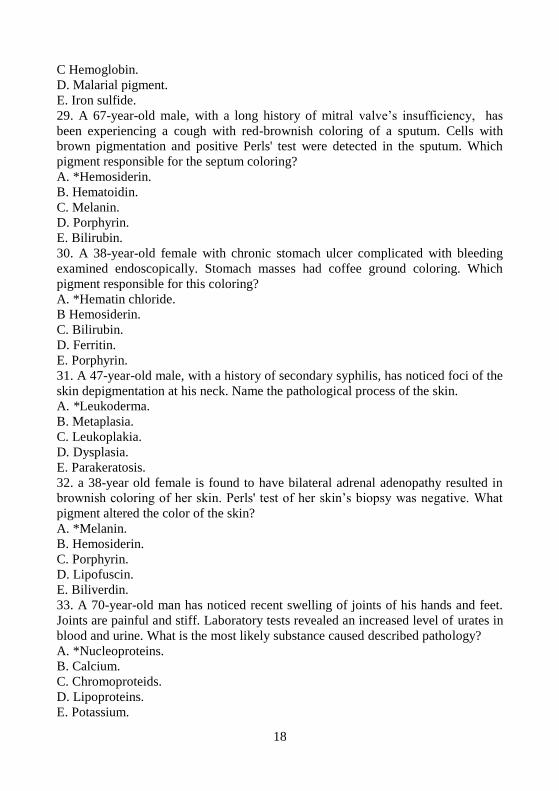
pigment responsible for the septum coloring?
A. *Hemosiderin.
B. Hematoidin.
C. Melanin.
D. Porphyrin.
E. Bilirubin.
30. A 38-year-old female with chronic stomach ulcer complicated with bleeding
examined endoscopically. Stomach masses had coffee ground coloring. Which
pigment responsible for this coloring?
A. *Hematin chloride.
B Hemosiderin.
C. Bilirubin.
D. Ferritin.
E. Porphyrin.
31. A 47-year-old male, with a history of secondary syphilis, has noticed foci of the
skin depigmentation at his neck. Name the pathological process of the skin.
A. *Leukoderma.
B. Metaplasia.
C. Leukoplakia.
D. Dysplasia.
E. Parakeratosis.
32. a 38-year old female is found to have bilateral adrenal adenopathy resulted in
brownish coloring of her skin. Perls' test of her skin‘s biopsy was negative. What
pigment altered the color of the skin?
A. *Melanin.
B. Hemosiderin.
C. Porphyrin.
D. Lipofuscin.
E. Biliverdin.
33. A 70-year-old man has noticed recent swelling of joints of his hands and feet.
Joints are painful and stiff. Laboratory tests revealed an increased level of urates in
blood and urine. What is the most likely substance caused described pathology?
A. *Nucleoproteins.
B. Calcium.
C. Chromoproteids.
D. Lipoproteins.
E. Potassium.

19
34. A 70 year-old male with a history of chronic shigellosis died. At post-mortem
tissue samples were collected for histopoly. Microscopic investigation of
hematoxylyn and eosin slides revealed amorphous violet deposits in stroma of the
heart, kidneys, lungs and stomach mucosa. Koss' reaction was positive. What is the
most likely pathological developed?
A. *Metastatic calcification.
B. Dystrophic calcification.
C. Metabolic calcification.
D. Amyloidosis.
E. Hyalinosis.
35. A woman presented to intensive care department with symptoms of severe he-
molysis due to a snake bite. An autopsy revealed brown coloration of the spleen,
bone marrow, and lymph nodes. Microscopic examination showed the accumula-
tion of brown pigment in cytoplasm of macrophages of tissue sampler. Which of
the following is the pigment that was accumulated in the tissues?
A. *Hemosiderin
B. Hematoidin.
C. Hematin
D. Lipofuscin.
E. Bilirubin.
36. A patient with prolonged esophageal carcinoma died of cachexy. An autopsy
revealed atrophy of his heart and liver, marked reduction of subcutaneous and re-
troperitoneal fatty tissue. Microscopical examination revealed accumulations of the
golden-brown granules and clumps with negative Prussian blue reaction within the
myocardial fibers. Which of the following pigments accumulated in myocardial
fibers?
A. *Lipofuscin.
B. Melanin.
C. Hematoidin.
D. Ferritin.
E. Hemomelanin.
Necrosis. Postmortem changes. 1. A male had a surgery due to ―acute abdomen‖. During the operation it was noted
that the peritoneum was dull and in the lumen of the mesenteric artery superior a
thrombus was detected. About 80 centimeters of the ileac intestine had a black co-
louring. Which process was diagnosed in the intestine? А. *Gangrene B. Decubitus ulcer C. White infarct D. White infarct with a hemorrhagic crown E. Coagulative necrosis

20
2. A patient with diabetes mellitus presents to his physician with an acute pain in
the right foot. At inspection the toe of foot had a black colour, the tissues of foot
were edematous, with bed smell. Which form of necrosis was likely to be diag-
nosed? А. * Moist (wet) gangrene B. Decubitus ulcer C. Sequester D. Dry gangrene E. Infarct 3. A 63-year-old male died of an endemic typhus. During the post-mortem it was
revealed that the muscles of the abdominal wall and legs were dense with whitish-
yellowish colouring. They resemble a candle. Name the pathological process? А. *Waxy necrosis (Zenker‘s necrosis) B. Apoptosis C. Fibrinoid necrosis D. Colliquative necrosis E. Caseous necrosis 4. A 72-year-old-male had an infarct of the dextral hemisphere of the brain. One
year later a computer tomography of the right hemisphere of the brain reveals a
cavity with smooth walls and filled with liquid. Which pathological process is he
most likely to have? А. *Post-infarct cyst B. Hydrocephalus C. Grey softening of a brain D. Infarct of a brain E. Hematoma 5. A post-mortem revealed a thrombus in the left artery mesencephalicae and a
large locus of grey softening in the tissues of the left hemisphere of a brain. Which
pathological process is most likely to be present in the brain? А. * Ischemic infarct B. Coagulative necrosis C. Abscess D. Moist gangrene E. Sequestrum 6. At a post-mortem of the 46-year-old male a large yellow - grey lesion was found
in the left ventricle of the heart. A fresh thrombus was found in the coronary artery.
What disease is he most likely to have? А. *Infarct of the myocardium B. Cardiosclerosis C. Myocarditis D. Amyloidosis E. Cardiomyopathy

21
7. At autopsy a 60-year-old male is found to have ischemic heart disease and athe-
rosclerosis of the coronary arteries of heart. A section of the myocardium showed a
white-yellowish focus, surrounded by the zone of hemorrhages in the apex, anterior
and lateral walls areas of a left ventricle. Which is the most likely diagnosis? А. *Infarct of the myocardium B. Post-infarction cardiosclerosis C. Diffuse cardiosclerosis D. Myocarditis E. Fatty dystrophy of the myocardium 8. After a long staying in the bed a patient with circulatory deficiency got the skin
and soft tissue darening above the sacrums. These tissues became swallowed.
Sloughing off the epidermis in this area resulted in ulceration. Which complication
is most likely? А. *Decubitus ulcer B. Dry gangrene C. Phlegmon D. Infarct E. Abscess 9. Histological investigation of liver‘s biopsy revealed that some cells burn on
small pieces with separate organellas and nuclei fragments surrounded by a mem-
brane. The inflammatory reaction was missing. Select pathological process, the
described changes are characteristic for: А. *Apoptosis B. Necrosis C. Karyorrhexis D. Plasmolysis E. Plasmorrhexis 10. A male who had a long history of the intermittent claudication demonstrates the
tissue of the foot fingers as being dry with a black colour, resembling a mummy.
On small interval from this place the dichromatic line (red colour is next to un-
changed tissues, and white - yellow colour close to a tered tissues). Which type of a
necrosis occurred? А. *Gangrene. B. Infarct. C. Sequester. D. Decubitus ulcer. E. Maceration. 11. A 62-year-old male got a surgery due to the inguinal hernia. Macroscopic ex-
amination reveals that the wall of the intestine was a cyanotic, inflated, swallowed
and coated with threads of a fibrin. Peristalsis was not heard. Which pathological
process occurred in the wall of the intestine? А. * Moist gangrene. B. Dry gangrene.

22
C. Coagulate necrosis D. Colliquative necrosis E. Decubitus ulcer 12. A postmortem of a man, who died from typhoid revealed muscles on the abdo-
minal wall and legs were dense, fragile, whitish-yellowish colour, resembling a
candle. Which term best characterizes the muscles changes? А. *Zenker‘s necrosis B. Fibrinoid necrosis C. Caseous necrosis D. Colliquative necrosis E. Apoptosis 13. A 48-year-old male, who had a history of hypertension for 12 years, present
acute disturbance of the cerebral circulation. He developed a headache and altera-
tion of the motion in the right extremity. Following right-handed hemiplegia re-
sulted in fatal outcome. A postmortem revealed a systemic hyalinosis of the small
arteries, thrombosis in the left arteria cerebri media. In the left parietal-temporal
area a lesion was found, which is called: А. *Ischemic infarct B. Hemorrhage C. Abscess of a brain D. Hemorrhagic infarct E. Edema of the brain 14. A 45-year-old male suddenly died with the following findings revealed during
the postmortem. In the back wall of the left ventricle of the heart a myocardial in-
farction was found. Which of the following microscopic changes in the myocardi-
ocytes can be seen in the locus of an infarct? А. *Karyolysis B. Fatty dystrophy C. Carbohydrate dystrophy D. Calcification E. Protein dystrophy 15. A postmortem of on a previously ill 48-year-old patient found an obturation of
the lumen of the middle cerebral artery due to a thrombus. In the parietal-temporal
area of the left hemisphere of the brain a locus of grey colour tissue with soft tex-
ture is found. Which tern best characterizes the brain tissue changes? А. *Infarct B. Sequester C. Gangrene D. Caseous necrosis E. Fibrinoid necrosis 16. A postmortem on the upper lobe of the right lung reveals the large triangle-like
locus of the dark red dense tissue. Histological examination indicates necrosis of

23
the walls of the alveolus‘s and the lumens filled with erythrocytes. Which is the
most likely associated finding? А. *Hemorrhagic infarct B. Carnification C. Lung‘s gangrene D. Hemorrhage E. Atelectasis 17. A postmortem on an elderly male with atherosclerosis reveals a thrombus in a
branch of the internal carotid artery as well as a grey locus of a moist softening of
the brain‘s tissue. Which pathological process was found in the brain? А. *Ischemic infarct B. Hemorrhagic infiltration C. Hematoma D. Encephalitis E. Tumor of a brain 18. A surgery on a patient, with a history of syphilis revealed a locus of flabby tis-
sue. Grossly, this locus was yellowish, dry, structures and gummy. The most likely
diagnoses is:
А. *Caseous necrosis B. Infarct C. Waxy necrosis D. Fibrinoid a necrosis E. Steatonecrosis 19. The investigation of the liver‘s biopsy revealed that some separately arranged
cells burn on small-sized pieces surrounded by a membrane. In some of them there
were organelles, other had the fragments of dissolved nuclei. The inflammatory
reaction around these cells missed. Name these changes: А. *Apoptosis B. Atrophy C. Necrosis D. Hypoplasia E. Dystrophy 20. An ill elderly patient with a atherosclerosis, develops pain in the left foot.
Grossly was found the foot enlargement, its tissues were black, flabby and mace-
rated. The demarcation zone was not expressed. Which term best characterized the
foot tissues changes? А. * Moist (wet) gangrene. B. Mummification. C. Coagulate necrosis. D. Dry gangrene. E. Sequestrum. 21. A 62-year-old female with atherosclerosis was admitted to the hospitalized. At
surgery gross examination revealed purulent peritonitis. During the operation a

24
thrombus in the mesenteric arterias was found. Which was the most likely cause of
the peritonitis? А. *Hemorrhagic infarct. B. Angiospastic ischemia C. Ischemic infarct D. Stasis E. Compressive ischemia 22. The examination of a child, who had a history of measles, revealed reddish-
black, uneven, swollen, slightly fluctuated lesions of cheaks and perineum area.
Name the complication of measles?
А. * Moist gangrene (noma) B. Dry gangrene C. Gas gangrene D. Decubitus ulcer E. Trophic ulcer 23. A postmortem was performed on a female who died due to the cystadenocarci-
noma metastases. The postmortem revealed large segments of a necrosis of the skin
and soft tissues within cubitus area. Name the form of the necrosis. А. *Decubitus ulcer B. Infarct C. Sequester D. Caseous necrosis E. Zenker‘s necrosis 24. A 58-year-old female with the history of atherosclerosis dies suddenly due to
acute heart failure. Gross inspection of the left ventricle of the heart revealed a whi-
tish-yellowish 6x5 cm, dense lesion with uneven boundaries and hemorrhagic zone
next to it. Which is most likely diagnose? А. *Infarct of the myocardium B. Postinfarction fibrosis C. Healed infarct D. Myocarditis
E. Ischemic cardiomyopathy
25. An ultrastructural examination of a salivary gland revealed within cells pieces
of the nuclei surrounded by a membrane. Also condensate fragments of nuclear
material and separate organelles were found. An inflammatory reaction around
these cells was missing. Which term most correctly defines these alteration? А. *Apoptosis B. Karyorrhexis C. Coagulation necrosis D. Karyopyknosis E. Karyolysis 26. A patient with tuberculosis has a kidney biopsy performed. Histological inves-
tigation revealed the caseous necrosis of the tissue accompanied by disorderly ar-

25
ranged fine grains of a chromatin. Which term most correctly defines describe le-
sion?
А. * Karyorrhexis B. Karyolysis C. Karyopyknosis D. Mitotic activity of nuclei E. Apoptosis 27. A postmortem of a 48-year-old male who had a history of typhoid fever re-
vealed that the rectus abdominis at the wall was dense, a whitish colour, and re-
sembled a candle. Which is the most likely diagnosis? А. * Waxy necrosis B. Fibrinoid necrosis C. Colliquative necrosis D. Caseous necrosis E. Apoptosis 28. A 44 year old ill patient died due to the severe chronic heart failure. Pathologist
diagnosed rheumatic granulomatous myocarditis. Microscopic evaluation of the
myocardium indicated the presence of granulomas which consisted of macrophages
with hyperchromatic nuclei and clear cytoplasm. Also necrosis was seen in the cen-
ter of a lesion. Which is the most likely type of necrosis in the center of the lesion? А. * Fibrinoid necrosis. B. Waxy necrosis. C. Caseous necrosis. D. Colliquative necrosis. E. Fatty dystrophy.
29. A 57-year-old patient has a long history of the type II diabetes mellitus. Physi-
cal examination revealed the alteration of the right foot tissues. They are dense,
black with precise boundaries from normal tissues. Which term most correctly de-
scribe the lesion? А. *Dry gangrene. B. Wet (moist) gangrene. C. Gas gangrene. D. Decubitus. E. Trophic ulcer. 30. A postmortem examination of a dead body revealed a cloudy corneas, dry skin
with yellowish - brown lesion. Which term most correctly identifies describes a
post-mortem alterations? А. *Cadaver desiccation B. Clotting of blood C. Livor mortis D. Rigor mortis E. Algor mortis

26
31. The postmortem of a 48-year-old male reveals in the right temporal lobe of the
brain a large grey lesion with a softening, porridge-like texture. The basal arteries
of the brain had numerous white - yellow thickenings of an intima which signifi-
cantly decreased lumen. Which is the most likely diagnosis? А. *Ischemic infarct B. Abscess of a brain C. Hematoma D. Hemorrhagic infarct E. Edema of the brain 32. At post-mortem, a 60-years-old man, with a history of typhoid fever, is found to
have rectus muscles of the anterior abdominal wall dense, whitish, and look like a
stearic candle. What is the most likely diagnose?
A. *Zenker's necrosis.
B. Fibrinoid necrosis.
C. Colliquative necrosis.
D. Caseous necrosis.
E. Apoptosis.
33. A 65-year-old female, with a long history of diabetes mellitus, presented her
black, edematous and painful thumb of the right foot. Gross inspection revealed a
focal epidermal detachment and malodorous discharge. What is the most likely
clinicopathologic form of necrosis?
A. *Moist (wet) gangrene.
B. Decubitus ulcer.
C. Sequester.
D. Dry gangrene.
E. Infarction.
34. A 5-year-old boy with measles presents to his pediatrician with necrotic
changes of his cheeks. Gross inspection revealed that the cheeks soft tissues were
edematous with reddish black fluctuated indistinctly outlined foci. What is the
most likely complication of a measles?
A. *Moist gangrene.
B. Dry gangrene.
C Gas gangrene.
D. Decubitus ulcer.
E. Trophic ulcer.
35.A physical examination of 67-year old lady, with a history of femoral bone frac-
ture, revealed a sequester formation accompanied with chronic inflammation of a
bone marrow and adjacent tissues. What is the most likely disease caused such le-
sions?
A. *Osteomyelitis.
B. Reticulosarcoma.
C. Multiple myeloma.
D. Osteoclastoma.

27
E. Periostitis.
36. An elderly woman with a history of a stroke one year ago complains of a left
limbs immobility. A computer tomography examination revealed a cavity filled
with liquor, at right hemisphere of her brain. What is the most likely diagnose?
A. *Postinfarction cyst.
B. Hydrocephaly.
C. Grey encephalomalacia.
D. Cerebral infarction.
E. Hematoma.
37. A section of the left lung was found to have an area of dense red tissue. The
area was cone-shaped, stood out distinctly from the healthy tissue, with its base
directed to the pleura. The dissected tissue was granular, dark-red. What is the most
likely diagnosis?
A. *Haemorrhagic infarction
B. Lung gangrene
C. Lung abscess
D. Croupous pneumonia
E. Primary tuberculous affection
38. An autopsy of a dead patient with typhoid fever revealed firm, fragile, whitish-
yellow, and waxy muscles of the anterior abdominal wall and thighs. What is the
most likely pathology of muscles?
A. *Zenker necrosis.
B. Fibrinoid necrosis.
C. Caseous necrosis.
D. Colliquative necrosis.
E. Apoptosis.
39. A 55-year-old patient with 12 years history of hypertensive disease presented
to a hospital with disturbances of cerebral blood circulation resulted in right-side
hemiplegia. He died shortly after arrival. A postmortem revealed systemic hyali-
nosis of small blood vessels and thrombosis of medial cerebral artery. There was
also a focus of a structureless grayish tissue in the temporal lode of left hemis-
phere. The focus was interpreted as:
A. *Ischemic infarction
B. Hemorrhage
C. Abscess
D. Hemorrhagic infarction
E. Edema
40. A 59-year-old patient with transmural myocardial infarction died of cardiac
tamponade due to ruptured myocardial infarct. Which process resulted in a heart‘s
wall rupture?
A. *Autolytic process leading to myocardial softening (myomalacia)
B. Replacement of infracted areas by connective tissue (organization)
C. Increasing of blood pressure in pulmonary circulation

28
D. Myocarditis
E. Chronic aneurism
41. A 58-years-old smoker with a 5-year history of angina pectoris died during the
heart attack. An autopsy revealed atherosclerosis of the coronary arteries with
thrombosis, uneven color of anterior wall of the left ventricle and flabbiness of the
myocardium. What is the most likely diagnosis?
A. Myocardial infarction
B. Angina pectoris
C. Myocarditis
D. Postinfarction cardiosclerosis
E. Myocardial aneurysm
42. A 74-year-old patient with a history of hypertensive syndrome developed col-
lapse and died of increased disturbances of respiration and heart activity. An autop-
sy revealed a dark-red 2x1x5 cm bloody focus in the trunk of the brain. Vessels of
the brain base were considerably thickened, whitish-yellow, with narrowed lumen.
What is the most likely diagnosis?
A. Hemorrhagic infarct
B. Cyst of the brain
C. Gumma of brain
D. Ischemic infarct
E. Meningioma
Blood and lymph circulation disorders.
1. The postmortem of a 48-year-old male, with a long history of a chronic heart
failure, revealed an enlarged liver. Grossly, a liver had a motley pattern. Macros-
copically, the sectional view looked like a nutmeg on incision. Which term most
correctly defines these alteration? А. * General venous plethora B. General arterial plethora C. Anemia D. Hemorrhage E. Bleeding
2. A post-mortem of a 43-year-old female revealed multiple, hemorrhagic infarcts
in lungs. Some lungs‘ vessels had reddish-brown dense masses within lumens,
which were not attached to the vessel walls. Varicose phlebectasia of the legs with
thromboses of some veins were also determined. Which pathological process oc-
curred in this case? А. * Thromboembolism of pulmonary artery. B. Fat embolism of pulmonary artery. C. Tissue embolism of pulmonary artery. D. Foreign bodies embolism of pulmonary artery. E. Gas embolism.

29
3. A male patient died after a abdominal surgery. During the postmortem numerous
thrombuses were found in the veins of the pelvis. Thromboembolic syndrome was
diagnosed. Where it is necessary to search for thromboembolies? А. *Lungs artery B. Portal vein C. Left ventricle of heart D. Brain E. Veins of the lower extremity 4. The histological investigation of a liver detects venous plethora of center lobules,
dystrophy and atrophy of hepatocytes in the venous plethora‘s area, fatty a dystro-
phy of hepatocytes on the periphery of a lobe. The replacement fibrosis in places of
an atrophy of the hepatocytes was also revealed. Which pathological process does
this refer to? А. * ‗Nutmeg‘ liver with precirrhotic phenomena. B. Biliary hepatic cirrhosis. C Fatty hepatosis. D. Hepatitis. E. Toxic dystrophy of a liver. 5. A 35-year-old patient complains of repeated vomiting, diarrhea, decreased arteri-
al pressure and tachycardia. He supposes this condition related to the food poison-
ing. Laboratory tests detected Salmonella‘s infection. Hematological examination
revealed an increased number of erythrocytes per unit volume. Which circulatory
disturbance took place to create this pathology? А. *Clotting of a blood. B. Hemolysis of erythrocytes and compensatory induction of a hemogenesis C. General arterial plethora D. Polycythemia E. Hyperchromatic anemia 6. A male with a history of myocardial infarct died of cardiovascular failure. A
post-mortem revealed a replacement fibrosis, hypertrophy of the myocardium and
dilatation of the cavities, especially the right ventricle. The liver was enlarged. Its
surface was smooth. Grossly, a motley pattern with dark red dots on the grey a
background was revealed. Histologically, central zones of the lobules were hyper-
emic. On the periphery, around of periportal tracts hepatocytes demonstrated fatty
dystrophy. Name these liver changes. А. *‖Nutmeg‖ liver (chronic venous plethora) B. Pseudo ‗nut-meg‘ liver C. Amyloidosis D. Cirrhosis of a liver E. Steatosis of a liver 7. A male patient involved in the traffic accident received a wound in the neck due
to broken glass. The bleeding was small, but a short time after the accident he died

30
of acute dyspnea. A post-mortem of the heart revealed bubbles excretion when pe-
ricardium filled with water. Indicate which pathological process took place: А. *Air embolism B. Gas embolism C. Fat embolism D. Thromboembolism E. Foreign bodies embolism 8. A 52-year-old male with long history of rheumatic heart disease died of chronic
heart failure. A post-mortem revealed brown colour, enlarged dense lungs. Name
the changes in lungs. А. *Brown induration of lungs B. Acute bronchitis C. Horny-comb lungs D. Chronic bronchitis E. Chronic emphysema 9. A patient, who had fast elimination of 10 liters of an ascitic liquid from abdo-
men, suddenly lost his consciousness. What was the cause of this phenomenon? А. *Anemia of the brain B. Thrombosis of the cerebral arteries C. Brain hemorrhage D. Arterial hyperemia E. Thrombosis of the cerebral veins 10. A young male with a history of rheumatic heart disease since childhood, grad-
ually develops mitral stenosis, accompanied with episodes of heart failure. He
presents to a hospital complaining of coughing with a reddish-brown sputum.
Name probable changes at patient‘s lungs. А. *Brown induration of lungs B. Emphysema of lungs C. Atelectasis of lungs D. Pneumosclerosis E. Bronchiectasis
11. At autopsy an elderly female is found to have a blood clot in the femoral artery,
which grossly had a striped pattern. Microscopy revealed a congestions of fibrin
fibers and broken down red and white cells. Which is the most likely type of
thrombus ?
А. *Mixed thrombus B. Postmortem convolutions of a blood C. Thromboemboli D. Hyaline thrombus E. Red thrombus 12. A patient has a purulent otitis and thrombosis of a sigmoid sinus of a dura ma-
ter. Name the complication if thrombus breaks down and fragments circulate to
distal vessels.

31
А. *Thromboembolism of branches of pulmonary artery B. Thromboembolism of vessels of a brain with development of a grey softening of
the brain C. Thromboembolism of vessels of a retina of an eye and development of blindness D. Thromboembolism of coronary arteries E. Local anemia 13. A 43-year-old male, with a history of traffic accident and poly trauma, includ-
ing multiple bone fractures, presented to the hospital. On the second day after the
trauma, he complained of a pain in the right half of the chest, heavy breathing. At
night, he died due to progressive heart and respiratory failure. Microscopic
investigation revealed Sudan-positive orange drops in pulmonary and cerebral ves-
sels that completely occlude the lumens of microcirculatory vessels. What is the
most likely complication led to a patient‘s death?
A. *Fat embolism.
B. Gaseous embolism.
C. Medicamentous embolism.
D. Microbial embolism.
E. Thromboembolism.
14. A 62-year-old patient had a surgery due to acute appendicitis. She had been
placed in a bed for 5 days. After she decided to get out of bed she experienced a
shortage of air, her face became cyanotic and she lost her consciousness. After un-
successful resuscitation the patient died. A post-mortem revealed a thromboembol-
ism of the pulmonary artery. Which of the following is most likely source of
thromboembolism? А. *Thrombosis of veins of the lower extremity B. Thrombosis of a portal vein C. Thrombosis of mesenteric arteries D. Thrombosis in a left ventricle of heart E. Ball-shaped thrombus of an auricle 15. A male with a fracture of the shoulder bone carried on overlapped plaster ban-
dage. Suddenly, an arm and the visible part of the forearm became cyanotic, cold,
and edematous. Which of the following is most likely happened? А. *Local venous plethora B. Local arterial plethora C. Local anemia D. Stasis E. Thrombosis 16. An elderly female develops acute disorder of the cerebral blood circulation,
followed with coma, resulted in fatal outcome. A post-mortem revealed in the right
hemisphere of the brain a large cavity, filled with blood. Which pathological
process took place in the brain? А. *Hematoma B. Hemorrhagic infiltration

32
C. Infarct of the brain D. Diapedesis E. Edema of the brain 17. A male suddenly died after an open fracture of the clavicle. A post-mortem
revealed in the right ventricle of the heart and pulmonary arteries the foamy blood.
Which one was the cause of death? А. *Air embolism B. Bacterial embolism C. Hemorrhage D. Tissue embolism E. Fat embolism 18. A male with a history of the decompression sickness developed symptoms of
acute cerebral circulation disorder and died soon. A post-mortem revealed in the
left hemisphere of the brain the locus of a grey softening of the brain, which was
6х5х3 cm. Which one is most likely to cause the death of the patient? А. *Gas embolism. B. Fat embolism. C. Thrombosis. D. Thromboembolism. E. Atherosclerosis of the vessels. 19. A post-mortem of an elderly man with a long history of the ischemic heart dis-
ease and heart failure revealed a nutmeg liver, brown induration of lungs, cyanotic
induration of kidney and spleen. Indicate, what type of the blood circulation dis-
order is most likely? А. * Chronic general venous plethora. B. Arterial hyperemia. C. Acute general venous plethora. D. Acute anemia. E. Chronic anemia. 20. A 50-year-old male with a myocardial infarction died from the heart failure. A
post-mortem revealed the edema of the lungs and petechial hemorrhages at serous
and mucus membranes. Microscopic examination indicated marked dystrophic alte-
rations of the nephron‘s epithelium at proximal canaliculi of kidneys. Also, the
centrolobular hemorrhage and necrotic zones were found in the liver. Name the
type of the blood circulation disorder. А. * Acute general venous plethora. B. Arterial hyperemia. C. Chronic general venous plethora. D. Acute anemia. E. Chronic anemia. 21. A neonate died of intoxication. A microscopic examination of an umbilical vein
revealed a diffuse inflammatory infiltration of the vessel‘s wall. Also, its lumen
was filled with thrombus, rich with leucocytes and bacterial colonies. Karyorrhexis

33
was detected in many leucocytes. Which is most likely outcome of a thrombus in
that case? А. * Septic autolysis. B. Aseptic autolysis. C. Organization and vascularization of the thrombus. D. Thromboembolism. E. Calcification of a thrombus. 22. A 68-year-old female is admitted to the hospital for treatment of deep vein
thromboses. The next day she suddenly died. At autopsy a large plug of laminated
blood clot is found to occlude the main pulmonary artery. Which is likely to be
found in her lungs?
А. *Thromboembolism B. Thrombosis C. Tissue embolism D. Foreign bodies embolism E. Fat embolism 23. A 77-years-old female with unstable angina presents to physician with slowly
increasing heart failure symptoms. On the night of admission to the hospital she
dies. A post-mortem examination revealed the enlarged liver with dense texture and
the rounded edges. Sectional view had a nutmeg pattern with dark red dots on the
yellowish background. Which pathological process resulted in liver‘s alteration? А. * Chronic venous plethora B. Acute venous plethora C. Arterial plethora D. Arterial anemia E. Chronic hemorrhage 24. A post-mortem of a 53-year-old male with a long history of hypertension re-
vealed the cavity in the occipital lobe of the brain. It measured 2х1 cm, had a
brownish smooth walls and filled with transparent liquid. Which is the most likely
diagnosis? А. * Cyst after hemorrhages B. Softening of the brain C. Abscess of a brain D. Developmental defect of a brain E. Cyst after softening of the brain 25. A 65-years-old patient, who presented with a 10 years history of ischemic heart
disease, died due to heart failure. An autopsy revealed the cyanotic induration of
both spleen and kidneys, brown induration of lungs and ―nutmeg‖ liver. Which is
the most likely type of the blood circulation disorder, resulted in such changes of
internal organs? А. *General chronic venous hyperemia B. General acute venous hyperemia C. General arterial hyperemia after an anemia

34
D. Arterial ischemia as a result of reallocating a blood E. Local chronic venous hyperemia 26. A 63-year-old patient with long history of ischemic heart disease and repeated
myocardial infarction died due to progressive cardiovascular failure. A post-
mortem revealed an enlarged, dense spleen with the dark red colour of the sectional
view. At the microscopic examination of spleen the pulp‘s sclerosis and atrophy of
the follicles were found. Which term most correctly defines theses changes? А. *Cyanotic induration of a spleen B. Sago spleen C. Waxy spleen D. Porphyry spleen E. Septic spleen 27. A young male fractures his pelvis and femur in a motor vehicle accident. On the
third day he dies after respiratory distress and cerebral dysfunction. A histological
examination revealed Sudan-positive orange globules scattered within cerebral cor-
tex, kidneys and lung microcirculatory vessels. Which is most likely diagnoses?
А. * Fat embolism B. Gas embolism C. Tissue embolism D. Microbial embolism E. Thromboembolism 28. A cosmonaut died due to the air-tightless of the space-shuttle cabin. Microscop-
ic examination of the vessels of the internal organs revealed multiple bubbles. Liver
cells developed fatty dystrophy. In the brain and spinal cord multiple small, grey,
soft lesions were discovered. Indicate the most probable cause of such alterations. А. *Gas embolism B. Air embolism C. Fat embolism D. Thromboembolism E. Tissue embolism 29. A patient with a long history of rheumatic disease died of cardiopulmonary
failure. A post-mortem revealed a stenosis of the mitral orifice as well as a brown
induration of lungs. Which term most correctly defines this blood circulation dis-
order? А. *Chronic left ventricular failure. B. Chronic right ventricular failure. C. Acute left ventricular failure. D. Acute right ventricular failure. E. Portal hypertension. 30. A post-mortem of a 59-year-old patient with long history of idiopathic hyper-
tension revealed in brain a cavity with rusty colour walls. Which process preceded
these changes?
А. *Hematoma

35
B. Diapedesis C. Ischemic infarct D. Plasmorrhagia E. Abscess 31. A 65-year-old- patient presented with liver cirrhosis. The removal from his ab-
dominal cavity of 10 liters of ascitic liquid resulted a collapse. Grossly his perito-
neum was hyperemic. Define the type of hyperemia in peritoneum.
А. * Post-anemic hyperemia B. Collateral hyperemia C. Complementary hyperemia D. Inflammatory hyperemia E. Caused by arteriovenous fistula
32. A post-mortem of a 60-year-old female with a history of secondary hyperten-
sion revealed a cavity at the right hemisphere of the brain 4х2,5 cm, filled with red
clots of blood and softened brain tissue. What term best characterized the brain
changes? А. *Hematoma. B. Hemorrhagic impregnation. C. Ischemic infarct. D. Cyst. E. Abscess. 33. An inexperienced scuba diver ascends from a depth of 55 meters to the surface
in about 5 minutes. Shortly after surfacing he complains of severe muscle contrac-
tion and intense abdominal pain. What is most likely diagnose? А. *Gas embolism B. Air embolism C. Fat embolism D. Thromboembolism E. Tissue embolism 34. A 78-year-old male, with a history of stroke 2 years ago, died of pneumonia as
a complication of severe influenza. At post-mortem, gross investigation of his brain
revealed a cerebral cyst with rusty color of its walls. Perls' test was positive. Name
the process reveled within a cystic wall.
A. *Local hemosiderosis.
B. Common hemosiderosis.
C. Local hemomelanosis.
D. Infiltration of bilirubin.
E. Primary hemochromatosis.
35. A 54-years old male with a history of ischemic heart disease presents at hospital
with recurrent myocardial infarction. Few days later, he died due to cardiac failure.
Post-mortem revealed an enlarged solid spleen of dark cherry color on the cut sur-
face. Microscopically, pulp sclerosis and follicles atrophy were found out. What is
the most likely term to define spleen‘s alterations??

36
A. *Cyanotic induration of spleen.
B. Sago spleen.
C. Lardaceous spleen.
D. Porphyry spleen.
E. Septic splenitis.
36. A post-mortem of 77-year-old male, with a long history of cerebral atheroscle-
rosis, revealed an atrophy of his cerebral cortex. What was the most likely cause of
cortex alterations?
A. *Insufficiency of blood supply.
B. Pressure.
C. Physical and chemical factors.
D. Neurotic.
E. Dysfunctional.
37. A 56-year-old male with a history of cerebral atherosclerosis suddenly dies. At
a there is a thrombus within a middle cerebral artery's lumen. Gross inspection re-
vealed a grey flabby focus at the parietotemporal part of the left hemisphere of his
brain .What are the most likely alterations diagnosed in a brain tissues?
A. *Infarction.
B. Sequestrum.
C. Gangrene.
D. Caseous necrosis.
E. Fibrinoid necrosis.
38. An autopsy of a patient who died suddenly at emergency room revealed in his
brain a cavity of irregular shape (5 x 3.5 cm) filled with blood clots and macerated
cerebral tissue. An area of cavitary destruction had a rim of brown discoloration.
This lesion was found within the subcortex nuclei at the right hemisphere of brain.
What is the most likely definition of described pathology?
A. *Hematoma.
B. Hemorrhagic impregnation.
C. Ischemic infarction.
D. Cyst.
E. Abscess.
39. 2 hours after a skeletal extension was performed to a 27 year old patient with
multiple traumas (closed injury of chest, closed fracture of right thigh) his condi-
tion abruptly became worse and the patient died from acute cardiopulmonary de-
compensation. Histological examination of pulmonary and cerebral vessels stained
with Sudan III revealed orange drops occluding the vessel lumen. What complica-
tion of polytrauma was developed?
A. *Fat embolism
B. Thromboembolism
C. Air embolism
D. Microbal embolism
E. Gaseous embolism

37
40. A 65-year-old patient died of chronic heart failure due to rheumatic valve de-
fect. An autopsy revealed the enlarged, dense, brown lungs. What are the most like-
ly changes in the lungs:
A. *Brown induration of the lungs.
B. Acute venous congestion C. Honeycomb lungs.
D. Chronic bronchitis.
E. Chronic emphysema.
41. A 55-year-old patient had a prolonged immobilization due to the surgery opera-
tion. Once, after getting up from a bed he developed sudden onset of dyspnea,
breathing difficulties and cyanosis. Shortly after that he got unconscious state and
then died. A post-mortem revealed a pulmonary thromboembolism. Choose the
most probable source of thromboembolism:
A. *Veins of lower extremities
B. Portal vein
C. Mesenterial arteries
D. Left heart ventricle
E. Left atrium
42. A patient died of a heart failure. An autopsy revealed enlarged and hyperemic
liver, with dark red foci surrounded by paler yellow- brownish zones on its cut sur-
face. Microscopical investigation showed congestion of central veins and fat dy-
strophy of hepatocytes on the periphery of lobules. Specify the most probable pa-
thology of a liver.
A. *Nutmeg liver
B. False nutmeg liver
C. Hepatic amyloidosis
D. Liver cirrhosis
E. Liver steatosis
43. An autopsy of 86-year-old female with a long history of a chronic coronary
disease followed with heart failure revealed ‗nutmeg‘ liver, brown induration of
lungs, cyanotic induration of the kidneys and spleen. Name the kind of a circulatory
disturbance.
A. *Chronic general venous plethora
B. Acute anaemia
C. Chronic anaemia
D. Arterial hyperaemia
E. Acute general venous plethora
Inflammation.
1. A histological investigation of enlarged neck lymphatic node revealed a conges-
tion of epithelial cells, lymphocytes and giant Pirogov-Langhan‘s cells. In the cen-
ter of lesion a caseous necrosis was present. Which pattern of inflammation do the
lymphatic node display?

38
А. * Tuberculosis
B. Rhinoscleroma
C. Sarcoidosis
D. Glanders.
E. Syphilis.
2. A 3-year-old child died of a meningococcal fever. A macroscopic study revealed
swallowed meninges which had yellow-green colouring. Which pattern of inflam-
mation was most likely in meninges?
А. *Fibrinous-purulent inflammation
B. Serous inflammation
C. Hemorrhagic inflammation
D. Catarrhal inflammation
E. Necrotic inflammation
3. A 37-year-old male presents with a fever, dyspnea and pain in the right part of
the chest. The pleurocentesis gave 700 mls of thick yellow-greenish liquid. What
pathological process was diagnosed in a pleural cavity?
А. *Empyema of a pleura
B. Bronchopneumonia
C. Serous pleuritis
D. Hemorrhagic pleuritis
E. Carcinomatosis of a pleura
4. A 25 years old male presents with a 2-day history of itching and redness in the
buccal area that appeared after shaving. Physical examination by family physician
revealed vesicles filled with transparent fluid on the background the hyperemic
buccal area. What is the most likely type of fluid in the vesicles?
A. *Serous exudate.
B. Transsudate.
C. Purulent effluent.
D. Mucous exudate.
E. Hemorrhagic exudate.
5. An autopsy of a 34-years old female, with a history of croupous pneumonia re-
vealed opaque fluid in her pleural cavity. There was also a grey membrane on the
visceral pleura. What is the most likely type of the pleura‘s inflammation?
A. *Fibrinous.
B. Catarrhal.
C. Suppurative.
D. Granulomatous.
E. Hemorrhagic.
6. A 4-year-old girl with 3 days history of diphtheria presents to the emergency
with croup symptoms. Intensive care was unsuccessful and child died at the hospit-
al. A post-mortem revealed thickened, edematous, dull mucous tunic of the larynx,
trachea, and bronchi covered by grayish membrane easily separated from tissues
underneath. What is the most likely type of inflammation?

39
A. *Fibrinous.
B. Serous.
C. Suppurative.
D. Mixed.
E. Catarrhal.
7.At post-mortem a 5-year-old boy is found to have a severe tracheobronchitis
complicated with asphyxia. Grossly, a thick, gray, leathery membrane covered the
mucous tunic of trachea and bronchi. The membrane loosely attached to underlying
tissues and easily removed with forceps. The lumen of the segmental bronchi was
blocked with gray masses of tissue debris, which microscopically consisted with
necrotic tissues, neutrophils, fibrin and bacteria. What is the most likely type of
inflammation?
A. *Croupous.
B. Catarrhal.
C. Diphtheritic.
D. Purulent.
E. Serous
8. An autopsy of a 50-year-old male, who died of dysentery, revealed the hyperem-
ic mucosa of the colon, coated with grey membranes, which can be removed from
tissues underneath with some effort. Which type of an inflammation described in
the intestine?
А. *Diphtheroid inflammation
B. Croupous inflammation
C. Hemorrhagic inflammation
D. Serous inflammation
E. Catarrhal inflammation
9. A 30-year-old male has a neck lymph node removed for examination. Histologi-
cal investigation revealed granulomas, which consisted of epithelioid, lymphoid
and multinuclear Langhans giant cells. Granulomas‘ centers were presented with
necrosis. What causative agent needs to be revealed in the necrosis zone for support
of the diagnosis of tuberculosis?
А. *Mycobacterium tuberculosis
B. Treponema pallidum
C. Staphylococcus
D. Frisch bacillus
E. Salmonella
10. An autopsy of a 53-year-old male, with a history of crupous pneumonia, re-
vealed in his dextral pleural cavity 900 ml of cloudy, grey-yellowish colored liquid.
Pleural membranes were found to be dim and plethoric. Name the clinical -
morphological form of the inflammation in the pleural cavity?
А. * Empyema.
B. Fibrinous inflammation.
C. Phlegmon.

40
D. Chronic abscess.
E. Acute abscess.
11. An autopsy of a 53-year-old male, who died from chronic renal failure, re-
vealed the colon‘s mucosa, covered with grey – yellow membranes, densely cohe-
rent to tissues underneath. The removal of the membranes resulted in ulcers forma-
tion. Name a type of an inflammation?
А. *Diphtheroid
B. Serous
C. Catarrhal
D. Croupous
E. Purulent
12. A 65-year-old female with a long history of chronic glomerulonephritis died
from chronic renal failure. A post-mortem revealed on the surface of the epicar-
dium and pericardium grey-whitish villous membranes. Which pathological process
took place in the pericardium?
А. *Fibrinous inflammation
B. Organization
C. Proliferative inflammation
D. Hemorrhagic inflammation
E. Arterial plethora
13. An elderly man presented to his physician with the complicated nasal breathing.
The histological investigation of a biopsy from his nose septum revealed granulo-
matous inflammation in the mucosa with groups of large vacuolated histiocytes
containing bacteria (Mikulicz‘s cells). What is the most likely diagnosis?
А. *Rhinoscleroma
B. Syphilis
C. Tuberculosis
D. Malleus
E. Lepra
14. An elderly man, with a history of an operation, presented with a sepsis, devel-
oped on a background of a decreased immune reactivity of an organism. A disease
resulted in fatal outcome. Microscopical investigation of the abdominal wall re-
vealed the diffuse segmentonuclear leukocyte‘s infiltration of intermuscular spaces,
edema of a tissue and lysis of muscle fibers. Define the type of inflammation?
А. *Phlegmon.
B. Diphtheroid inflammation.
C. Abscess.
D. Necrosis.
E. Catarrhal inflammation.
15. A 64-years-old male died of rheumatism. An autopsy revealed epicardium cov-
ered with villiferous grey color membranes, which were easily separated from tis-
sues underneath. The separation of membranes presented an edematous, hyperemic
surface of epicardium. What type of an inflammation revealed in pericardium?

41
А. *Fibrinous pericarditis
B. Purulent pericarditis
C. Hemorrhagic pericarditis
D. Proliferative pericarditis
E. Catarrhal pericarditis
16. A 50-year-old male died of a pulmonary and cardiac insufficiency. An autopsy
revealed in a left lung a 4 x 4 сm cavity, filled with pus. The wall of a cavity had a
rough pattern, presented by pulmonary tissue. What is the most likely diagnose?
А. *Acute abscess
B. Chronic abscess
C. Cavernous tuberculosis
D. Hydatidosis
E. Fibrosing alveolitis
17. A post-mortem of a man revealed in his abdominal cavity approximately 2.0
liters of a purulent exudate. A peritoneum was found to be dull, with a grayish tint.
The intestinal serous membranes were covered with grayish membranes, which
were removed easily. Which of the following is the most accurate diagnosis?
А. * Fibro-purulent peritonitis
B. Hemorrhagic peritonitis
C. Serous peritonitis
D. Tuberculous peritonitis
E.
18. A histological examination of the myocardium of a man, who died from heart
failure, revealed a sclerosis of the pericardial connective tissue and diffuse infiltra-
tion by lymphocytes, macrophages, plasmocytes and single neutrophils. What is the
most likely type of inflammation in a heart?
А. * Interstitial productive.
B. Granulomatous.
C. Alterative.
D. Exudative diffuse.
E. Exudative focal.
19. A 28-year-old male presented to the hospital with a fever and weakness, (asthe-
nia). A biopsy was taken from one of his enlarged neck lymph nodes for histologi-
cal examination. Microscopic investigation revealed the foci of necrosis, sur-
rounded by epithelioid cells, giant multinuclear Langhans' cells and also lympho-
cytes. What is the most likely diagnosis?
А. *Tuberculosis
B. Hodgkin‘s disease
C. Lympholeukemia
D. Sarcoidosis
E. Syphilis
20. A microscopic examination of a kidney biopsy revealed foci with granular, eo-
sinophilic masses in their centers. These lesions were surrounded by an infiltrate,

42
consisted of lymphocytes, epithelioid cells and giant Langhans' cells. What is the
most likely pathological process?
А. * Granulomatous inflammation
B. Coagulative necrosis
C. Caseous necrosis
D. Alterative inflammation
E. Proliferation and derivation of macrophages
21. A young male presented to his physician with considerably enlarged, hyperem-
ic, painful tonsils. A gross examination of tonsils revealed dense, dirty-gray mem-
branes on the surface. These membranes have spread to the hard palate and were
intimately attached to tissues underneath. An attempt to remove the membranes
resulted in bleeding. Which pathological process indicates these morphological
changes?
А. *Diphtheroid exudative inflammation.
B. Croupous exudative inflammation.
C. Catarrhal exudative inflammation.
D. Purulent exudative inflammation.
E. Hemorrhagic exudative inflammation.
22. A 38-year-old man, with a history of chronic pyelonephritis, died from the
chronic renal failure. Medical record showed that an auscultation in the hospital
determined a patient‘s ―pericardial frictions rub‖. An autopsy revealed a dim,
rough, and hairy – like epicardium. What type of pericarditis presented in that case?
А. *Croupous
B. Diphtheroid
C. Purulent
D. Ichorous
E. Serous
23. A child presented to a hospital with symptoms of asphyxia. A physical exami-
nation revealed in the larynx whitish membranes, which obstructed lumen and were
easily separated from tissues underneath.
Name the most probable type of an inflammation in the larynx?
А. *Croupous inflammation
B. Hemorrhagic inflammation
C. Diphtheroid inflammation
D. Catarrhal inflammation
E. Purulent inflammation
24. A macroscopic investigation of trachea revealed a dim, hyperemic mucosa,
covered with grey - white membranes. Which one is the most likely form of the
inflammation?
А. *Fibrinous
B. Purulent
C. Serous
D. Proliferative

43
E. Mixed
25. An elderly man presented with a decomposition of some segments of spongy
and cortical layers of an anticnemion bones. The cavities are filled with creamy
greenish-yellow masses. What is the type of inflammation?
А. *Purulent
B. Catarrhal
C. Serous
D. Proliferative
E. Mixed
26. A female patient presented to the hospital with a fever, asphyxia and pain in
the right part of her chest. During a pleurocentesis procedure 700 ml of a viscid
yellow-green liquid was removed. What is the most likely diagnosis?
А. *Empyema of a pleura
B. Carcinomatosis of a pleura
C. Serous pleurisy
D. Fibrinous pleurisy
E. Hemorrhagic pleurisy
27. An autopsy of the 58-year-old man, revealed in his liver a focus of tissue de-
struction, 4 cm in diameter, filled with a yellow-green liquid. What is the most like-
ly diagnose?
А. *Abscess
B. Phlegmon
C. Anthrax
D. Empyema
E. Granuloma
28. A histological examination of biopsy from the nose mucous revealed epithelio-
id cells, plasmocytes, Mikulicz cells, and eosinophilic (acidophilic, Russell's) bo-
dies. What is the most likely diagnosis?
А. * Rhinoscleroma
B. Syphilis
C. Tuberculosis
D. Droplet infection
E. Allergic rhinitis
29. A physical examination of a 5-year-old boy reveals the fauces and the tonsils
enlarged, plethoric, and coated with irremovable whitish membranes. Which one of
the following characterizes the changes in the fauces and tonsils?
А. * Diphtheritic inflammation
B. Caseous necrosis
C. Fibrinous necrosis
D. Croupous inflammation
E. Purulent inflammation
30. A 49-year-old man, with 14 days history of acute lobar staphylococcal pneu-
monia in the left lower lobe of a lung, died of pulmonary and cardiac insufficiency.

44
A post-mortem revealed in the upper lobe of a right lung an oval cavity, approx-
imately 5 cm in diameter, filled with pus and connected with bronchus. Which
complication of acute pneumonia took place in this case?
А. *Abscess of the lung
B. Gangrene of the lung
C. Thromboembolia of a pulmonary trunk
D. Bronchiectasis disease
E. Acute bronchitis
31. An autopsy of an elderly male revealed a cavity with compacted walls filled
with a thick, greenish liquid, which had a stinking odor. What is the most likely
disease?
А. * Abscess of the lung.
B. Gangrene of the lung.
C. Infarct of the lung.
D. Cavern of the lung.
E. Tuberculoma of the lung.
32. A 62-year-old man died of croupous pneumonia. A post-mortem revealed in the
pleural cavity an opaque liquid and a grayish membrane on visceral pleura. What is
the most likely type of inflammation on the visceral pleura?
А. * Fibrinous inflammation
B. Catarrhal inflammation
C. Purulent inflammation
D. Granulomatous inflammation
E. Hemorrhagic inflammation
33. A 5-year-old child has a fever and pain at swallowing. A physical examination
revealed enlarged, dark-red palatine tonsils. They were coated with a grey - yellow
membrane which was intimately attached to the surface of the tonsils. Which kind
of inflammation described in the tonsils?
А. * Diphtheritic inflammation
B. Croupous inflammation
C. Hemorrhagic inflammation
D. Purulent inflammation
E. Catarrhal inflammation
34. An autopsy of a dead body revealed 200 ml of a viscid yellow-green liquid in
the abdominal cavity. What is the most likely form of exudate inflammation?
А. * Purulent inflammation
B. Serous inflammation
C. Fibrinous inflammation.
D. Hemorrhagic inflammation.
E. Ichorous inflammation
35. A post-mortem of a young male revealed congested, thickened, opaque, yello-
wish-green meninges. What type of exudative inflammation these changes are cha-
racteristic for?

45
А. * Purulent inflammation
B. Serous inflammation
C. Hemorrhagic inflammation
D. Fibrinous inflammation
E. Catarrhal inflammation
36. A woman, with a history of her hands‘ skin thermal burn, presented to physi-
cian painful blisters, filled with opaque liquid. What is the most likely type of in-
flammation?
А. * Serous inflammation
B. Productive inflammation
C. Croupous inflammation
D. Granulomatous inflammation
E. Diphtheritic inflammation
37. A 44-year-old male had a skin biopsy procedure. Histological investigation of a
tissue sample showed a caseous necrosis surrounded by infiltrate, consisted from
lymphocytes, some giant cells and the growth of connective tissue. In addition, the
endovasculitis was diagnosed. What is the most likely pathological process?
А. * Productive granulomatous inflammation.
B. Productive interstitial inflammation.
C. Abscess.
D. Catarrhal inflammation.
E. Serous inflammation
38. A 7-year-old child, with a history of angina, presented with enlarged paratra-
cheal, bifurcation and neck lymph nodes. A histological examination of his neck
lymph node revealed the foci of necrosis, surrounded by lymphocytes, epithelioid
cells and Pirogov-Langhans‘ cells. Which one is the most likely pathology?
А. *Tuberculosis
B. Sarcoidosis
C. Rhinoscleroma
D. Malleus
E. Syphilis
39. The histological examination of a lung tissue revealed a lesion presented with a
locus of necrosis, surrounded by regular lines of epithelioid, lymphoid cells, plasma
cells, macrophages and giant Pirogov-Langhans‘ cells.
Define a kind of inflammation?
А. *Tuberculosis inflammation
B. Banal productive inflammation
C. Inflammation at a lepra
D. Exudative inflammation
E. Alterative inflammation
40. A 6-year-old child presented to infectious disease department with a body tem-
perature 38С, punctulated, bright red rash on a skin. The scarlet fever diagnosis is
made. The fauces mucosa was brightly hyperemic, edematous; tonsils were mar-

46
kedly enlarged, with the dim yellowish-grayish centers and sites of black color.
What inflammation underlies changes in fauces?
А. *Purulent-necrotic
B. Fibrinous
C. Hemorrhagic
D. Serous
E. Catarrhal
41. A rectoromanoscopy revealed edematous, reddish color mucosa of rectum and
sigmoid colon, covered with a thick layer of mucus. Name the kind of inflamma-
tion?
A. *Catarrhal
B. Purulent
C. Mixed
D. Hemorrhagic
E. Fibrinous
42 A patient died with symptoms of the brain‘s edema and the dislocation of the
stem. A post-mortem revealed thickened, opaque, grayish-green color pia mater.
Which pathological process took place in pia mater of meninges?
А. *Diffuse purulent leptomeningitis
B. Meningococcal leptomeningitis
C. Tuberculous leptomeningitis
D. Malignant arachnoendothelioma
E. Hydrocephalus
43. A 5-year-old child presented to a hospital with acute fever (up to 380
C), rhini-
tis, cough, lacrimation, and a patchy rash on the skin. His pharyngeal mucosa was
edematous and hyperemic, with whitish maculae on the cheeks. What type of in-
flammation is the background of described changes?
А. *Catarrhal
B. Purulent
C. Fibrinous
D. Hemorrhagic
E. Serous
44. A 3-year-old child presented to a hospital with asphyxia. Examination of the
larynx revealed whitish membranes, which occluded a lumen and easily extracted.
Diphtheria was suspected. Which form of inflammation took place in the larynx?
А. *Croupous inflammation
B. Catarrhal inflammation
C. Diphtheroid inflammation
D. Serous inflammation
E. Purulent inflammation
45. A 47-year-old male presented with a sub-febrile temperature and enlarged
lymph nodes. A biopsy investigation of a lymph node revealed numerous granulo-

47
mas with a caseous necrosis in the centers, surrounded by epithelioid cells, giant
multinuclear Langhans' cells and lymphocytes.
What is the most likely diagnosis?
А. *Tuberculosis
B. Lymphogranulomatosis
C. Lymphosarcoma
D. Lympholeukemia
E. Simple lymphadenitis
46. At a 45-year-old man the biopsy procedure of his nasal cavity‘s mucosa was
performed. The diagnosis of rhinoscleroma made. What cells are typical for the
disease?
А. *Mikulicz‘s cells
B. Pirogovs‘ – Langhans‘ cells
C. Plasma cells
D. Lymphocytes
E. Schaumann‖s bodies
47. An autopsy of a 53-year-old male showed many white, millet sized nodules in
his lungs. A microscopic examination revealed granulomas with a necrosis in their
centers and epithelioid, lymphoid, plasma cells, macrophages and numerous large
multinucleated cells (Langhans' cells) on the periphery of a lesion.
What is the most likely form of granuloma?
А. *Giant - cell granuloma
B. Macrophagal
C. Foreign body granuloma
D. Epithelioid cells
E -
48. A patient presented to the hospital with combustion of his right hand. A physi-
cal examination revealed a desquamation of epidermis with blisters formation. The
blisters were filled with opaque liquid. What most probable inflammation described
in that case?
А. *Serous
B. Purulent
C. Catarrhal
D. Putrefactive
E. Fibrinous
49. A 43-year-old patient complains of hindered nasal breathing. Histological in-
vestigation of a biopsy of his nasal cavity mucosa revealed lymphoid cells, epithe-
lioid cells, plasma cells and Mikulicz‘s cells. What is the most likely diagnosis?
А. *Rinoskleroma
B. Glanders (malleus )
C. Tuberculosis
D. Lepra
E. Syphilis

48
50. A 54-year-old patient complains of complicated nasal breathing. A nasal muco-
sa was thickened. Histological investigation of a biopsy revealed Mikulicz‘s cells,
epithelioid cells, plasma cells, lymphoid cells, and hyaline sphere- like aggregates.
What is the most likely diagnosis?
А. *Skleroma
B. Adenoviral rhinitis
C. Allergic rhinitis
D. Rhinoviral infection
E. Meningococcal nasopharyngitis
51. A sample of cervix uteri was taken for histological examination. A microscopi-
cal investigation revealed a tissue‘s inflammatory infiltration with vascular walls
involvement of small vessels, arteries and veins. In addition, there were also de-
termined a presence of plasma cells, lymphocytes, epithelioid cells in the infiltrate
and foci of sclerosis and hyalinosis of tissue. What is the most likely disease?
А. *Syphilis
B. Tuberculosis
C. Leukoplakia
D. Erosio colli uteri
E. Condyloma
52. A physical examination of tonsils and soft palate mucosa revealed white-grey
color membranes, which are intimately attached to tissues underneath. An attempt
to take out membranes results in formation of a deep tissue defect. Diagnose pa-
thology on a mucosa of tonsils and a soft palate.
А. *Diphtheritic inflammation
B Serous inflammation
C. Croupous inflammation
D. Purulent inflammation
E. Mixed inflammation
53. Histological investigation of a biopsy revealed a granuloma, consisted of lym-
phocytes, plasma cells, macrophages with foamy cytoplasm (Mikulicz‘s cells) and
numerous hyaline‘s sphere- like aggregates. About what disease it is possible to
think?
А. *Rinoskleroma
B. Lepra
C Syphilis
D. Tuberculosis
E. Actinomycosis
54. A 40-year-old woman, with a history of the right palm‘s burn, presented to her
physician an acute pain, reddening and swelling of a palm. In a few minutes there
was a bubble, filled with transparent yellowish liquid. The display of what patho-
logical process the described changes are?
А. *Exudative inflammation
B. Traumatic edema

49
C. Alterative inflammation
D. Proliferative inflammation
E. Vacuolar dystrophy
55. A 44-year-old man presented to the policlinic with a local pain in occipital area
of a head and rise of a body temperature there. Gross investigation of a lesion zone
revealed the cone-shaped cyanotic infiltration with a yellow-greenish apex, which
rose above the surface of skin. What is the most likely diagnosis?
А. *Furuncle
B. Phlegmon
C. Abscess
D. Carbuncle
E. Empyema.
56. A skin sample was taken for histological research. Microscopical investigation
revealed granulomas, consisted of macrophages‘ knots with adding of lymphocytes
and plasma cells. There were also determined large macrophages with fatty va-
cuoles, which contained packed layers of causative agents of a disease (Virchow's
cells). For what disease the given description is characteristic?
А. *Lepra
B. Tuberculosis
C. Syphilis
D. Rinoskleroma
E Glanders (malleus)
57. Histological investigation of a skin biopsy revealed granulomas, which con-
sisted of epithelioid cells, surrounded mainly by T-lymphocytes. In addition, few
giant multinucleated cells (Langhans' cells) were determined among epithelioid
cells. The caseous necrosis was diagnosed within the centers of granulomas. There
was lack of blood vessels. What is the most likely disease?
А. *Tuberculosis
B. Syphilis
C. Lepra
D. Rinoskleroma
E. Glanders (malleus)
58. A histological investigation of a liver biopsy revealed granulomas, consisted of
plasma cells and lymphoid cells. Small vessels were characterized by endo - and
perivasculitis. Some foci of caseous necrosis were also determined. Such granulo-
mas are typical for:
А. *Syphilis
B. Tuberculosis
C. Lepra
D. Rinoskleroma
E. Glanders (malleus)
59. A young man had a removal of mucous membrane new growths in his nasal
cavity. A histological investigation revealed diffuse lymphocytes, plasma cells and

50
macrophages infiltration. Name the type of inflammation.
А. *Inflammation with formation of polyps
B. Inflammation with formation pointed condyloma
C. Interstitial inflammation
D. granulomatous inflammation
E.Exudative inflammation
60. A 4-year-old child presented to his physician with a cold, high temperature,
nausea and vomiting. He was hospitalized and died in two days. An autopsy re-
vealed thickened, dim, edematous, congested, greenish-yellow pia mater of me-
ninges. What variety of exudative inflammation developed in pia maters?
А. *Purulent
B. Catarrhal
C. Hemorrhagic
D. Fibrinous
E. Serous
61. A young man was operated because of phimosis. Histological investigation of a
removed prepuce revealed polymorphic infiltrates, consisted of randomly located
plasmocytes, epithelioid and lymphoid cells. The vasculitis was diagnosed in ves-
sels. What disease such changes are characteristic for?
А. *Syphilis
B. Tuberculosis
C. Actinomycosis
D. Periarteritis nodosa
E. Lepra
62. Microscopic investigation an elderly man‘s lungs revealed the foci of necrosis,
surrounded by the bank of epithelioid cells and lymphocytes. Between lymphocytes
and epithelioid cells the rounded giant cells, with a considerable quantity of nuclei,
located on a periphery of a lesion were determined. Give the name for these
changes:
А. *Tuberculous granuloma
B. Sarcoid granuloma
C. Syphilitic granuloma
D. Lepromatous granuloma
E. Scleromatous granuloma
63. A 6-year-old child presented to department of infectious diseases with acute
pain in a throat, difficulty at swallowing, rise of body temperature up to 390С and
edema of neck. A gross oral investigation revealed enlarged, hyperemic tonsils,
covered with plenty of yellow membranes, which intimately attached to the mucous
membrane. An attempt to take off membrane results in deep, bleeding defect for-
mation. What type of inflammation takes place?
А. *Diphtheritic
B. Purulent
C. Serous

51
D. Croupous
E. Hemorrhagic
64. A fragment of a stomach mucosa was taken for histology during endoscopy
procedure. A microscopic investigation revealed intact mucous membrane, cov-
ered by mucus, thickened, edematous and hyperemic with a numerous tiny hemorr-
hages. Define the type of acute gastritis?
А. *Catarrhal (simple)
B. Erosive
C. Fibrinous
D. Purulent
E. Necrotic
65. An operatively removed appendix was sent to pathology department. Macros-
copical investigation revealed markedly enlarged appendix. His serous membrane
was dim, congested, covered by fibrin‘s membranes. The appendix‘ walls were
thickened. On a cut section pus in the lumen was detected. Microscopical investiga-
tion showed a plethora of vessels, edema of all appendixes‘ layers and diffuses in-
filtration by leucocytes. What is the type of inflammation?
А. *Phlegmonous
B. Apostematous
C. Simple
D. Superficial
E. Gangrenous
66. A 18-year-old girl had a neck lymphatic node removal at the biopsy procedure.
Histological investigation showed nodules, consisted of banks of epithelioid, lym-
phoid cells and large multinucleated cells (Langhans' cells), located between them.
In the centers of nodes a caseous necrosis was determined. What causative agent
needs to be revealed in the necrosis zone for making final diagnosis?
А. *Koch‘s mycobacterium
B. Mycobacterium leprae
C. Treponema pallidum
D. Rickettsia
E. Fungi
67. A young man complains of an itch and a skin reddening, which has appeared
after shaving. Objectively: a cheek skin is hyperemic; the blisters, filled with a
transparent liquid, were detected. What character of a liquid in blisters?
А. *Serous exudate
B. Transudate
C. Purulent exudate
D. Mucosal exudate
E. Hemorrhagic exudate
68. A 4-year-old boy presented to a hospital with pain in a throat at swallowing and
malaise. A physical examination revealed grayish - whitish membranes on a pha-
rynx and tonsils on a background of moderate edema and hyperemia. These mem-

52
branes were intimately attached to tissues underneath. What pathology the de-
scribed changes testify to?
А. *Inflammation
B. Dystrophy
C. Necrosis
D. Metaplasia
E. Reganerative process
69. A 68-year-old man died from meningitis. An autopsy revealed thickened, con-
gested, dim, edematous meninges (pia maters), which were saturated by creamy
greenish-yellow exudate. Define the type of inflammation?
А. *Purulent
B. Serous
C. Fibrinous croupous
D. Fibrinous diphtheritic
E. Hemorrhagic
70. Biopsy of an oral cavity‘s ulcer presented for a histological research. A micro-
scopical investigation revealed areas of caseous necrosis, surrounded by plasmo-
cytes, epithelioid, lymphoid cells and few giant multinuclear Pirogov-Langhans
cells. There were also small vessels with signs of endo- and perivasculitis. Choose
the most likely diagnosis.
А. *Syphilis
B. Tuberculosis
C. Lepra
D. Rinoskleroma
E. Glanders (malleus)
71. An autopsy of 32-year-old male revealed slightly swollen and hyperemic lungs,
covered with numerous millet-like grayish nodules. Histological investigation de-
termined lesions consisted of lymphoid, epithelioid and Langhans giant cells. What
is the most likely diagnosis?
А. *Tuberculosis
B. Croupous pneumonia
C. Bronchopneumonia
D. Interstitial pneumonia
E. Bronchiectatic disease
72. An operatively removed appendix was sent for histological examination. Ma-
croscopical investigation revealed thickened appendix. His serous membrane was
dim, congested, with whitish, loose membranes; the lumen contained turbid, whi-
tish-yellow exudate. Histological study showed the diffuse neutrophil‘s infiltration
of the appendix wall. Choose the most likely type of appendicitis.
А. *Phlegmonous
B. Gangrenous
C Simple
D. Superficial

53
E. Chronic
73. A 17 year old boy fell seriously ill, the body temperature rose up to 38,5°C,
there appeared cough, rhinitis, lacrimation, nasal discharges. What inflammation is
it?
A. *Catarrhal
B. Purulent
C. Fibrinous
D. Hemorrhagic
E. Serous
74. Histological examination of a skin tissue sampling revealed granulomas con-
sisting of macrophagal nodules with lymphocytes and plasmatic cells. There are
also some big macrophages with fatty vacuoles containing causative agents of a
disease packed up in form of spheres (Virchow's cells). Granulation tissue is well
vascularized. What disease is this granuloma typical for?
A. *Lepra
B. Syphilis
C. Rhinoscleroma
D. Tuberculosis
E. Glanders
75. A 5 year-old child is found to have meningococcal meningitis. He died of a
brain edema shortly after admission to the hospital. An autopsy revealed that pia
mater of a brain was thickened, opaque, green-yellowish, and edematous. Name the
kind of inflammation in pia mater?
A. *Purulent
B. Serous
C. Hemorrhagic
D. Catarrhal
E. Putrescent
76. An autopsy of a 34-year-old man with a medical history of rheumatic fever
revealed the epicardium, covered with grayish membranes, producing shaggy pat-
tern. A removal of these membranes displayed edematous plethoric epicardium.
What is the most likely kind of inflammation?
A. *Fibrinous pericarditis
B. Purulent pericarditis
C. Hemorrhagic pericarditis
D. Proliferative pericarditis
E. Catarrhal pericarditis
77. A post-mortem of a 47-years-old patient, who died of cardiopulmonary failure,
revealed a cavity in his left lung. A lesion was 4 cm in diameter, with uneven walls,
composed by lung tissue and filled with pus. What is the most likely diagnosis?
A. *Acute abscess
B. Chronic abscess
C. Cavernous tuberculosis

54
D. Echinococcus
E. Diffuse fibrosing alveolitis
78. A male with purulent otitis died on the 9th day at hospital of a brain oedema.
An autopsy revealed a cavity at the temporal region of his left hemisphere. A lesion
had uneven rough inner walls and was filled with yellow-greenish thick fluid. The
outer walls of the cavity were presented with the cerebral tissues. Name the most
likely pathological process?
A. *Acute abscess
B. Colliquative necrosis
C. Phlegmon
D. Empyema
E. Chronic abscess
Immunopathological processes 1. A 15 year old boy died of the heart failure. He had a history of rheumatic heart
disease in his medical record. Histological investigation of the left auricle of the
heart revealed the rheumatic granuloma (the Aschoff-Talalayev body) with a cen-
tral fibrinous focus associated, with a perimeter plasma cells, macrophages and
giant cells. Which of following best describe the type of immune response?
A. *Cell mediated (Type IV, delayed hyper sensitivity reaction)
B. Anaphylactic (Type I, immediate)
C. Cytolytic (Type II, immediate)
D. Immune complex (Type III, immediate)
E. Reaction of transplant rejection.
2. A 4-yers-old boy presents with multiple malformation of craniofacial bones. He
died soon due to sepsis, resulted from bronchopneumonia. Blood test revealed the
immunoglobulins within normal range. At autopsy the thymical aplasia was identi-
fied. Name the mane cause of death.
A. *Cellular immunodeficiency
B. Combined immunodeficiency
C. Secondary immunodeficiency
D. Acute leukemia
E. Chronic intoxication
3. A 48-year-old women with asthma presents with viral respiratory infection. The
infection caused a status asthmaticus with a fatal outcome. Histological investiga-
tion of lung tissue revealed a contracted bronchus and a luminal plug containing
mucus and cell debris. The submucosa was edematous and contained a mixed in-
flammatory infiltrate, including a lot of eosinophils and degranulated must cells.
What is the mechanism of hypersensitivity presented in this case?
A. *Humoral immunity (Type I, anaphylactic reaction)
B. Inflammatory reaction
C. Autoimmune reaction
D. Immune complex reaction (Type III of reaction)

55
E. Cell-mediated immunity (Type IV of reaction)
4. A young man presents with the enlargement of the regional lymph nodes next to
his inflamed wound. Histological investigation of the lymph node biopsy revealed
the increased number of macrophages, lymphocytes and lymphoid follicules within
cortex zone. Plasma cells number also increased. These alterations are most likely
result of:
A. *Antigen stimulation
B. The acquired insufficiency of lymphoid tissue (secondary immunodeficiency
state)
C. Hereditary immunodeficiency state
D. Neoplastic transformation
E. Hypersensitivity reaction
5. A newborn died after 3 days of life, as a result of a lethal outcome of intrauterine
pneumonia. At autopsy a newborn is sound have a great reduction of his thymic
weight and it‘s sizes. Histological investigation of thymus revealed the unclear
boundary between cortex and medulla zone as a result of lymphocytes amount re-
duction. A few Hassall corpuscles were found. They were focally keratinized and
had concentric aggregated of epithelial cells. The gland was also exhibited lobe
atrophy and sclerosis. What pattern of reaction did the thymus demonstrate?
A. *Accidental thymus transformation (involution)
B. Thymus atrophy
C. Thymus hypotrophy
D. Thymus aplasia (agenesis)
E. Thymus neoplasia
6. A 3-month-old child died because of sepsis. An autopsy revealed thymic hypop-
lasia, both sizes and mass of spleen reduction. Histologically, in spleen there was
lack of the periarterial follicular T-zone and remarkable absence of cells at a red
spleeny pulp. Lymph nodes investigation showed also absence of cells within para-
cortical (T-cells area). B-cells areas in immune system organs looked normal. What
term best characterizes described above changes?
A. Di George‘s syndrome
B. Combined immunodeficiency syndrome
C. Bruton‘s disease (X-linked agammaglobulinemia)
D. Accidental thymus transformation
E. Acquired immunodeficiency syndrome (AIDS) 7. A morphological investigation of a kidney graft revealed a diffuse stromal infil-tration by lymphocytes, plasmocytes, lymphoblasts, plasmablasts, and also a ne-crotic arteriitis. What pathology has developed in a graft? A. *Immunological graft rejection
B. Glomerulonephritis
C. Ischemic kidney damage
D. Tubular necroses
E. Pyelonephritis

56
8. An 8-year-old girl, with a history of acute tonsillitis 3 weeks before the applica-
tion, presented with nephrotic syndrome (proteinuria, haematuria and cylindruria).
These testify the glomerular basal membrane‘s lesion. What is the mechanism of
the basal membrane pathology?
A. * Immune complex mechanism
B. Granulomatous mechanism
C. Antibody-mediated mechanism
D. Reagin-mediated mechanism
E. Cytotoxical mechanism
9. At 46-year-old patient with autoimmune gastritis a biopsy was taken. Histologi-
cally, there was an infiltration by lymphocytes and macrophages of a gastric muc-
ous membrane. What type of hypersensitivity presented in that case?
A. * Type IY (cell-mediated hypersensitivity)
B. Type (reagin type)
C. Type II (antibody-mediated cytotoxicity)
D. Type III (immune complex cytotoxicity)
E. Type V (granulomatous)
10. Histological investigation of a lymph node revealed a considerable quantity of
the enlarged lymphoid follicles with big germinal centers, and plenty of mitotic
figures. What pathology these changes are characteristic for?
A. *Antigen stimulation with follicle hyperplasia
B. Atrophy of lymphoid tissue
C. Lymphosarcoma
D. Lymphogranulomatosis
E. Lympholeukemia
11. A child had a subcutaneous injection of tuberculin for a testing. Twenty-four
hours later a physical examination revealed the expressed hyperemia and induration
of tissues in the place of injection. What was the mechanism of these changes?
A. *Cellular cytotoxicity
B. Reagin cytotoxicity
C. Antibody cytotoxicity
D. Granulomas formation
E. Immune complex
12. A 6-year-old child had the Mantoux test. In 3 days there was an inflammatory
induration and a reddening of a skin 15 mm in diameter. That was considered as the
positive test. What type of hypersensitivity reaction presented in that case?
A. *Hypersensitivity of slow type
B. Immune complex hypersensitivity
C. Complement-mediated cytotoxicity
D. Hypersensitivity immediate type
E.
13. Physical examination of a pregnant woman with an Rh-negative blood revealed
a high level of antierythrocyte antibodies. The cutaneous flap of her Rh-positive

57
husband applied for depression of antibodies. In 2 weeks a flap was rejected. Mi-
croscopical investigation showed a disturbance of blood circulation, edema and
cellular infiltration mainly by lymphocytes, neutrophils and macrophages. What is
the most probable pathology?
A. *Transplantation immunity
B. Hypersensitivity reaction immediate type
C. Hypersensitivity reaction slow type
D. Granulomatous inflammation
E. Interstitial inflammation 14. A 3-year-old child died of an acute destructive staphylococcal pneumonia. Ma-croscopically, thymus mass reduced to 3,0 gr. Microscopical investigation of thy-mus showed reduction of lobules, considerable decline of lymphocytes quantity, stromal collapse, and inversion of layers with cystic enlargement of the Hassall‘s bodies. What is the most likely diagnosis? A. *Accidental thymus involution
B. Thymomegaly
C. Thymus hypoplasia
D. Thymus dysplasia
E. Thymus agnenesia 15. The experiment animal with a history of previous sensibilization received the next subcutaneous dose of antigen. In the place of injection a fibrinous inflamma-tion with alteration of vessels walls, matrix and fiber structures of connective tissue (mucoid and fibrinoid swelling) and fibrinoid necrosis developed. What immuno-logical reaction takes place? A. * Hypersensitivity reaction immediate type
B. Hypersensitivity reaction slow type
C. Reaction of transplantation immunity
D. Normergic reaction
E. Granulomatosis 16. A man with a history of bronchial asthma died of asphyxia. Microscopical in-vestigation of lungs revealed excess of mucus with lots of eosinophils in the bron-chial lumen, sclerosis of interalveolar septas and alveoli dilatation. What mechan-ism of hypersensitivity reaction presented in that case? A. *Reagin reaction
B. Cytotoxic reaction
C. Immune complex reaction
D. Cytolysis, conditioned by lymphocytes
E. Granulomatosis
17. The infiltration anesthesia by ultracain with adrenaline solution has been done
to a young man. Then redness, edema of the skin with itchy blisters suddenly de-
veloped. What type of hypersensitivity took place?
A. * Anaphylaxis
B. Cytotoxicity
C. Immune complex damage

58
D. Hypersensitivity slow type
E. Granulomatosis
18. A 33-year-old patient with a history of acute tonsillitis presented with the uri-
nary syndrome (haematuria, proteinuria and leukocyturia). Kidneys biopsy revealed
an intracapillary proliferative glomerulonephritis. Electronic microscopy deter-
mined large subepithelial deposits. What is the pathogenesis of this disease?
A.*Immune complex mechanism
B. Atopy
C. Cytotoxic, cytolytic action of antibodies
D. Cellularally caused cytolysis
E. Granulomatosis
19. A 10-month-old child has developed pneumonia. Clinical and laboratory tests
revealed absence of mature B-lymphocytes in peripheral blood and in B-zones of
lymph nodes, acute reduction of immunoglobulins in serum. In peripheral blood a
total quantity of lymphocytes was normal. What kind of an immunodeficiency de-
veloped in that case?
A. *Brutone sindrome
B. Neselof syndrome
C. Severe combined immunodeficiency
D. Di-George syndrome
E. Viskotta-Oldrich syndrome
20. A patient with a history of serious radioactive irradiation had bone marrow
transplantation. In 2 months a rush on the skin and diarrhea developed. Clinical and
laboratory tests showed hepatic insufficiency, local necrosis of the skin‘s epithelial
cells, intestinal crypts and liver parenchymal cells. What was the reason for symp-
toms aggravation?
A. * ‖Graft against the owner‖ disease
B. Sepsis development
C. An acute graft rejection
D. A chronic graft rejection
E. An exacerbation of a chronic hepatitis 21. A 7-month-old child died of severe pneumonia complicated by sepsis. A post-mortem revealed an absence of thymus. In lymph nodes there were no lymphoid follicles and cortical substance, in a spleen follicles were markedly reduced, ger-minal centers were absent. What is the most likely diagnosis? A. *Thymus agenesia
B. Thymus aplasia
C. Thymus atrophy
D. Thymus hypoplasia
E. Acidental thymus involution
22. A 1-year-old child died of a relapsing pneumonia. A post-mortem revealed hy-
poplasia of a thymus and peripheral lymphoid tissue, atrophy of cerebellum cortex,

59
angiotelectasis of bulbar conjunctiva. Immunohistochemistry revealed decreased
level of immunoglobulins. What is the most likely disease?
A. * Louis-Bar‘s syndrome
B. Di-George syndrome
C. Nezelof syndrome
D.West‘s syndrome
E. Brutone syndrome
23. A 20-year-old woman died of postnatal sepsis. A post-mortem revealed en-
larged congested spleen with abundant curettage from a cut section. Microscopical
investigation showed hyperplasia and infiltration by plasmocytes of the red pulp
and spleen‘s follicles. The red pulp was also rich with macrophages. What patholo-
gy resulted in spleen‘s alteration?
A. * Antigenic stimulation of an organism
B. Hereditary insufficiency of a peripheric lymphoid tissue
C. Reaction of hypersensitivity of immediate type
D. Reaction of hypersensitivity of the slowed type
E. Autoimmunization
24. Microscopical investigation of arterioles showed plasmatic saturation, mucoid
and fibrinoid swelling, fibrinoid necrosis of walls, diffuse infiltration by lympho-
cytes, plasmocytes and monocytes. In addition, the focal proliferation of hysti-
ocytes, endotheliocytes and pericytes were determined. The final diagnosis was the
―periarteritis nodosa‖. What type of inflammation took place in arterioles?
A. * Acute immune inflammation
B. Acute not immune inflammation
C. Focal exudative inflammation
D. Diffusive exudative inflammation
E. Fibrinous inflammation
25. During operation of kidney transplantation in 15 minutes after vascular anasto-
moses application, transplant became flabby, cyanotic and spotty; the pulsation was
absent. Microscopical investigation of a kidney specimen revealed periendothelial
accumulation of neutrophils in glomeruli vessels. In addition there were also a
thrombosis and necrosis of some glomeruli. Specify the type of graft rejection.
A. *Hyperacute rejection
B. Accelerated rejection
C. Acute intersticial rejection
D. Acute vascular rejection
E. Chronic rejection
26. A 30 year old woman has applied a lipstick with a fluorescent substance for a
long time. Then she got a limited erythema and slight peeling on her lip border,
later there appeared transversal striae and cracks. Special methods of microscopic
examination of the affected area helped to reveal sensibilized lymphocytes and ma-
crophages in the connective tissue; cytolysis. What type of immunological hyper-
sensitivity was developed?

60
A. *IV type (cellular cytotoxicity)
B. I type (reaginic)
C. III type (immune complex cytotoxicity)
D. II type (antibody cytotoxicity)
E. Granulomatosis
27. 48 hours after tuberculine test (Mantoux test) a child had a papule 10 mm in
diameter on the spot of tuberculine injection. What hypersensitivity mechanism
underlies these changes?
A. *Cellular cytotoxicity
B. Granulomatosis
C. Antibody-dependent cytotoxicity
D. Anaphylaxy
E. Immunocomplex cytotoxicity
Adaptation and compensation. 1. A young male complains of diminishing leg muscles in size and volume as a
result of the complicated femoral fracture. The innervations of the muscles was not
lost. Name the type of atrophy?
A. *Disuse atrophy
B. Degeneration atrophy
C. Ischemic atrophy
D. Atrophy due to compression
E. Atrophy due to chemical and physical influence.
2. At autopsy a male is found to have big kidneys with marked dilatation of the
renal pelvis and calyces filled with transparent liquid. A thinning of the renal pa-
renchyma accompanied above lesions. Which is the most likely finding?
A. *Hydronephrosis
B. Glomerulonephritis
C. Amyloidosis
D. Tuberculosis
E. Pyelonephritis
3. An autopsy of an elderly male revealed the atherosclerosis of the brain arteries
accompanied with the thinning of the brains cortex. Name the cause of the atrophy?
A. *Ischemia
B. Compression
C. Chemical and physical factors
D. Loss of innervations
E. Decreased workload
4. A 62-yer-old male died at the hospital after developing heart failure. At autopsy
the weight of the heart was 500gm, with thickened right ventricular wall and di-
lated chamber. Name the alteration at the right ventriculum.
A. *Hypertrophy
B. Hyperplasic alteration due to inflammation

61
C. Metaplasia
D. Atrophy
E. Sclerosis
5. A young male presents with a large and lacerated wound covered with pus. The
peripheral zone of the tissue defect was filled with large amount of granulation tis-
sue. Name the type of healing?
A. *Healing by first intention
B. Healing by second intention
C. Healing under a scab
D. Covering a tissue defect with epithelial cells
E. Organization followed by scarring
6. An autopsy revealed marked enlargement of a kidney. Gross examination of a
kidney‘s section presented with dilation of both renal pelvis and calyces resulted
from renal stones. What from diagnoses is most faithful?
A. *Hydronephrosis
B. Simple cysts
C. Pyelonephritis
D. Benign nephrosclerosis
E. Polycystic kidney disease
7. A physical investigation of an elderly man with history of esophagus cancer with
stenosis but without metastasis revealed an atrophy of skeletal muscles and subcu-
taneous fat tissue. Skin was grayish-brown color, an epidermis was thinned, and
size of the heart decreased. Myocardium and liver also had a brown coloring. What
is the most likely diagnosis?
A. *Alimentary cachexia.
B. Myasthenia.
C. Cancer cachexy
D. Brown atrophy.
E. The Addisona Illness
8. At the man with long history of smoking the bronchial biopsy is taken. Micro-
scopical investigation showed the thickened mucous membrane with chronic in-
flammation and transformation of ciliated epithelium into stratified one. What pa-
thology such changes are characteristic for?
A. *Metaplasia
B. Hyperplasia of epithelium
С. Planocellular cancer
D. Leukoplakia
E. Hypertrophy of epithelium
9. A patient with a history of frequent hemorrhoid bleeding died of acute myocar-
dial infarction. A post-mortem revealed a red, succulent bone marrow of a hip dia-
physis. What pathology developed in a bone marrow?
A. *Compensatory hyperplasia
B. Vicarious hypertrophy

62
C. Hypertrophy excrescences
D. Hormonal hyperplasia
E. Work hypertrophy
10. A 59-year-old patient with a history of prostate glandular hyperplasia operated
in the urology. An operation revealed a thickening of a urinary bladder wall up to 1
cm. It may be caused by:
A. *Work hypertrophy
B. Vicarious hypertrophy
C. Hormonal hypertrophy
D. Hormonal hyperplasia
E. Protective hyperplasia
11. A 7-year-old child presented with a poliomyelitis. His somatic muscles are
weak, their volume is reduced, and skin is dry and pale. Choose the most likely
pathology, which takes place in the soft tissues?
A. *Atrophy.
B. Hypertrophy.
C. Hyperplasia.
D. Metaplasia.
E. Hypoplasia.
12. An elderly man with a long history of smoking presented with a cough accom-
panied by viscid mucous sputum, weakness after the insignificant physical activity
and pale color of the skin. He lost 12,0 kg of weight for the last two months. Mi-
croscopical investigation of endoscopic biopsy showed squamous cell carcinoma.
What pathology preceded the tumor formation?
A. *Metaplasia.
B. Hypoplasia.
C. Hyperplasia.
D. Necrosis.
E. Sclerosis.
13. A 48-year-old patient with a history of a hypertensive disease died of heart fail-
ure. An autopsy revealed an enlarged heart with dilated chambers. A thickness of
the left ventricle wall was 2,5cm. Microscopically myocardial cells were consider-
ably enlarged, with fatty dystrophy and hyperchromic barrel-shaped nuclei. What is
the most likely pathology diagnosed in a heart?
A. *Excentric hypertrophy
B. Myocarditis
C. Concentric hypertrophy
D. Angiogenic cardiosclerosis
E. Cardiomyopathy 14. A 60-year-old man has a history of chronic bronchitis. Histological investiga-tion of bronchus biopsy showed thinned mucous membrane, cystic transformation of mucous glands, and replacement of prismatic epithelium on stratified one. What of the listed pathological processes the most likely?

63
A. * Metaplasia.
B. Hyperplasia.
C. Heterotopia.
D. Heteroplasia.
E. Displasia.
15. Microscopical investigation of tissue from the edge of a chronic gastric ulcer
showed necrosis, granulation tissue, abundant development of connective tissue
and metaplasia of epithelium. What type of pathology takes place in that case?
A. *Pathological regeneration with disorder of phases.
B. Hypertrophy.
C. Physiology regeneration.
D. Reparative regeneration (substitution).
E. Reparative regeneration (restitution).
16. At a patient with a chronic cystitis biopsy of urinary bladder mucosa was taken.
Microscopical investigation revealed a transitional epithelium with foci of stratified
epithelium without keratinization. What process underlies the described changes in
an epithelium?
A. *Metaplasia
B. Dystrophy
C. Hyperplasia
D. Dysplasia
E. Hyperkeratosis
17. After traumatic damage of a liver, subsequently there was complete hepatic
structural and functional restoration. Name the type of regeneration?
A. *Restitution
B. Incomplete regeneration
C. Pathological regeneration
D. Physiological regeneration
E. Substitution
18. At a patient with chronic bronchitis biopsy of bronchus mucous membrane was
taken. Microscopical investigation revealed the areas of stratified epithelium with-
out keratinization. What pathology takes place in the mucous membrane of bron-
chus?
A. *Metaplasia
B. Atrophy
C. Hyperplasia
D. Dysplasia
E.
19. A 42-year-old woman presented to the gynecologist with acyclic, profuse he-
morrhagic discharge. A histological investigation of a curettage material revealed
increased quantity of glands and their cystic dilation. What is the most likely diag-
nosis?
A. * Endometrial hyperplasia

64
B. Endometrial atrophy
C. Endometrial hypertrophy
D. Metaplasia
E. Organization
20. A post-mortem of a 64-year-old woman with a long story of hypertensive dis-
ease revealed considerably diminished, dense kidneys with a fine-grained surface.
What pathology such changes are characteristic for?
A. * Blood insufficiency atrophy
B. Atrophy from pressure
C. Senile atrophy
D. Dysfunctional atrophy
E. Hypoplasia
21. An elderly man died of cardiac insufficiency. Twenty years ago he had the right
lung pulmonectomy because of a cyst. A post-mortem revealed an enlarged left
lung. Diagnose pathology in the left lung.
A. *Vicarious hypertrophy
B. Neurohumoral hypertrophy
C. Dysfunctional atrophy
D. Dyscirculatory atrophy
E. Neurotic atrophy
22. A 42-year-old man had a lower extremity amputation. After a while painful
knots appeared in a stump. Microscopical investigation revealed amputation neu-
romas. What pathology such changes are characteristic for?
A. *Pathological regeneration
B. Complete reparative regeneration
C. Inflammation
D. Incomplete reparative regeneration
E. Metaplasia
23. A 38-year-old woman presented with complaints about the frequent uterine
bleeding. The diagnostic curettage was performed. A histological investigation of a
curettage material revealed increased quantity of coiled glands, some of them were
with cystic dilation. What pathology these changes are characteristic for?
A. *Glandular-cystic hyperplasia.
B. Atrophy
C. Metaplasia
D. Dysplasia
E. Hypertrophic vegetations
24. A man had the right lung pulmonectomy because of a tumor 7 years ago. Then
the volume of the left lung increased on 40%. What process developed in the left
lung?
A. *Vicarious hypertrophy
B. Neurohumoral hypertrophy
C. False hypertrophy

65
D. Work hypertrophy
E. Hypertrophic vegetation
25. A histological investigation of an endometrium revealed coiled extended glands
with ‗saw-‘ and a ‗spin-like‘ pattern. A stromal proliferation with hyperplasia of its
cells was also determined. What is the most likely diagnosis?
A. * Glandular hyperplasia of endometrium
B. Acute endometritis
C. Leiomyoma
D. Hydatidiform mole
E. Placental polyp
26. A patient presented with small knots on a thin peduncle in his rectum. Micro-
scopical investigation of the biopsy material revealed a growth of connective tissue
and glands. What is the most likely pathology?
A. *Hyperplastic polyp.
B. Hypertrophy.
C. Metaplasia.
D. Atrophy.
E. Sclerosis.
27. The biopsy of central bronchus mucosa of miner is taken. Microscopically,
there is a transformation of a cylindrical epithelium to the mature stratified one.
What pathology such changes are characteristic for?
А.* Metaplasia
В. Hyperplasia
C. Leucoplakia
D. Dysphasia
E. Reparative regeneration
28. The biopsy of a vaginal portion of uterus cervix is taken. There is an augmenta-
tion of quantity of cells, rising of mitotic activity and alteration of polarity of cells
in basal part of an epithelial layer. What pathology such changes are characteristic
for?
A.* Dysphasia
B. Metaplasia
C. Atrophy
D. Acantosis
E. Hyperplasia
29. A 40-year-old man has a keloid scar after a treatment of the left arm burn. This
process may be an example of:
A. *Pathologic regeneration
B. Hyperplasia
C. Metaplasia
D. Reparative regeneration
E. Vicarious hypertrophy
30. A 70-years-old patient admitted to a hospital with acute lung abscess. He sud-

66
denly died of progressing cardiopulmonary failure shortly after admission. An au-
topsy revealed an enlargement of mediastinal lymph nodes. They were pink-reddish
and elastic. A spleen was enlarged and flabby with plentiful scrape of a pulp. What
was the cause of lymph node and spleen‘s alterations?
A. *Protective hyperplasia
B. Compensatory hypertrophy
C. Hormonal hyperplasia
D. Venous congestion
E. Purulent inflammation
31. A 70-year-old patient had a surgery due to severe prostatic hyperplasia. A sur-
gery revealed a thickening of urinary bladder‘s wall up to one cm. What is the most
likely cause of this thickening?
A. *Workload hypertrophy
B. Vicarious hypertrophy
C. Hormonal hypertrophy
D. Hormonal hyperplasia
E. Compensatory hypertrophy
32. A patient with a long history of the chronic cystitis had a cystoscopy and biopsy
was performed. Histological examination of biopsy material revealed chronic cysti-
tis and foci of transitional epithelium replaced by squamous - cell non - keratinized
epithelium. These foci were the manifestations of:
A. *Metaplasia
B. Degeneration
C. Hyperplasia
D. Dysplasia
E. Hyperkeratosis
Epithelium neoplasms
1. A teenager male presents with slowly enlarging, painless nodule on his right
hand‘s skin. Microscopic examination of removed lesion revealed an increased
number of epithelial layers, with a stroma underneath, with developed a papillo-
matous pattern. Identify most likely type of atypia?
A. * Tissue
B. Cellular.
C. Metabolic.
D. Functional.
E. Hystochemical
2. A 35 year-old female with a family history of colon cancer inquires about
screening. Colonoscopy revealed a tumor and a tissue sample was taken for histol-
ogy. Microscopic investigation has shown cells were arranged in glandular-like
pattern. They exhibit cell pleomorphism, atypia, invasive growth and pathologic
mitosis‘s. What is the most likely diagnose?
A. *Adenocarcinoma

67
B. Basal-cell carcinoma
C. Solid carcinoma
D. Mucous carcinoma
E. Undifferentiated carcinoma
3. A 50-year-old female is found to have a urinary bladder neoplasm. Biopsy inves-
tigation revealed that tumor consists of thin, branching out papillae, covered with
few layers of transitional cell epithelium. What is the most likely diagnose?
A. *Papilloma
B. Basal cell
C. Transitional cell carcinoma
D. Squamous cell carcinoma
E. Fibroadenoma
4. A 60-year-old postmenopausal woman has been feel unhealthy and weak for
about 3 month. A gross investigation of her cervix uteri revealed a lesion and a
biopsy from this area was obtained. Microscopically, a lesion composed of atypical
squamous cell, many of them displayed pathological mitoses. Keratin pearl forma-
tion was also observed histologically. What is the most likely diagnose?
A. * Squamous cells carcinoma with keratinisation
B. Transitional cell carcinoma
C. Squamous cells without keratinisation
D. Adenocarcinoma
E. Anaplastic carcinoma
5. A 48-year-old woman complains to her doctor of uterine bleeding. Endometrial
biopsy investigation revealed a presence of gland-forming cells with enlarged
hyperchromic nuclei, cell atypia, abnormal mitoses. These cells were also deter-
mined at myometrium. What term most correctly identifies this pathological
process?
A. * Adenocarcinoma of uterus.
B. Adenomatous endometrial hyperplasia.
C. Acute endometritis.
D. Placental polyp.
E. Chorionepitelioma of uterus.
6. A 39-year-old woman presented in oncology with a stomach cancer. Physical
investigation revealed metastatic tumors in ovaries (Krukenberg tumors). What is
most likely pattern of spread in that case?
A. *Lymphatic retrograde
B. Lymphatic orthograde
C. Gematogenous
D. Implantation
E. Along epithelium-lined surfaces
7. A 23-year-old female decided to have a surgeon‘s removal of a small nodule at
her leg‘s skin. An operation material delivered to pathology department. A
histological examination of a new growth has shown that parenchyma has been

68
formed of integumentary epithelium with an increased amount of layers. Stroma
together with epithelial proliferation forms papillae. What is the most likely form of
atypism?
A. *Tissue.
B. Cellular.
C. Histochemical.
D. Functional.
E. Metabolic.
8. A 45-year old male with a family history of a gastric cancer inquires about
screening. A gastroscopic examination of a patient revealed a pedunculated tumor
mass 1.5 cm in diameter in the area of the lesser curvature of stomach. What kind
of growth does the tumor have?
A. *Exophytic.
B. Expansive.
C. Infiltrating.
D. Appositional.
E. Endophytic.
9. A 70 year-old male is found to have a nodule at his right bronchus mucous
membrane. A microscopic examination of the biopsy material from the tumor re-
vealed the cell and tissue atypism, keratin pearls formation. What is the most likely
pathologic process at biopsy material?
A. *Malignant tumor.
B. Benign tumor.
C. Hyperplasia.
D. Metaplasia.
E. Hypoplasia.
10. A 57-year-old postmenopausal woman is found to have a chronic nonhealing
lesion at her portio vaginalis uteri.A biopsy was taken form this area. A
histological examination of the biopsy material revealed a cellular atypism within
epithelial layer, but basic membrane was unchanged. What is the most likely diag-
nosis?
A. *Carcinoma in situ.
B. Erosion.
C. Adenocarcinoma.
D. Papilloma.
E. Endometriosis.
11. A 15-year-old boy is found to have a small, dense, nodule at his right forearm
skin. Grossly it had a papillary surface, which looked like a cauliflower. Micro-
scopically, the tumor consists of many papillae. Parenchyma formed of integumen-
tary epithelium with an increased amount of layers. The polarity of epithelial cells,
their stratification and membrane wholeness are preserved. A connective tissue
forms a stroma within a center of papillae. What is the most likely diagnosis?
A. *Papilloma.

69
B. Fibroma.
C. Adenoma.
D. Fibroadenoma.
E. Cystadenoma.
12. A 66-year old male lost his appetite and has been loosing his weight for about 5
months. X-ray study revealed a stomach neoplasm. Histological examination of a
tumor‘s biopsy showed a great amount of signet-ring cells. Name the histological
variant of cancer.
A. *Adenocarcinoma.
B. Solid carcinoma.
C. Sarcoma.
D. Mucinous carcinoma.
E. Carcinoid.
13. A 45-year-old woman has a small tumor of her left breast. Histological exami-
nation of a breast biopsy revealed poorly differentiated atypical epithelial cells.
They formed trabecules separated from one another by connective tissue. The cells
and stroma proportion was approximately 1:1.What is the most likely histological
variant of the cancer.
A. * Solid carcinoma.
B. Adenocarcinoma.
C. Epidermoid carcinoma.
D. Sccirrhous fibrocarcinoma.
E. Small cell carcinoma.
14. Histological investigation of a node in the removed mammary gland revealed
complexes of atypical polymorphic epithelial cells, which had various sizes and
forms. There were clear spaces at the centers of complexes. The cells had large
nuclei, with presence of atypical mitoses. Diagnose the pathology.
A. *Adenocarcinoma
B. Squamous cell nonkeratinous carcinoma
C. Solid carcinoma
D. Fibroadenoma of breast
E. Nondifferentiated polymorphocellular carcinoma
15. A microscopical investigation of a tumour revealed signet-ring cells. Name a
histological variant of a cancer.
A. *Mucinous carcinoma
B. Adenocarcinoma
C. Sarcoma
D. Solid carcinoma
E. Carcinoid
16. A 34-year-old woman presented with a hoarseness of a voice. A laryngoscopy
revealed a tumour of a larynx. The neoplasm had a grey-white color and papillary
surface. Microscopical investigation showed a new growth of a connecting tissue,

70
covered by a stratified epithelium with the marked keratinization. Cellular atypia
was absent. Most likely the tumor is:
A. *Papilloma
B. Fibroma
C. Polyp
D. Angioma
E. Angiofibroma
17. A microscopical investigation of a 50-year-old woman‘s endometrial curettage
material with the clinical diagnosis « ovarian – menstrual cycle‘s disorder » re-
vealed growth of glandular structures. These glands consisted of polymorphic cells
with hyperchromic nuclei and mitoses figures. For what pathology the revealed
histological changes are characteristic?
A. *Adenocarcinoma of uterus
B. Placental polyps
C. Acute endometritis
D. Glandular hyperplasia of endometrium
E. Chorioepithelioma of uterus
18. A 52-year-old female patient with a history of the chronic bronchitis and pneu-
mosclerosis presented to the hospital for biopsy diagnostics. A microscopical inves-
tigation of a left bronchial‘s mucous from the suspicious site revealed cellular and
tissue atypia. There were also found some structures in the form of‖ cancer pearls‖.
What is the most likely pathology presented in that case?
A. * Squamous cell keratinous carcinoma of bronchus
B. Chronic polypous bronchitis
C. Bronchiectasis
D. Sharp bronchitis
E. Squamous cell metaplasia of mucous of bronchus
19. Histological investigation of a bronchial biopsy revealed a tumour which is
constructed from nests of stratified epithelium‘s atypical cells with some characte-
ristic "pearls". What is the most likely diagnosis?
A. *Squamous cell keratinous carcinoma
B. Squamous cell nonkeratinous carcinoma
C. Solid carcinoma
D. Mucous carcinoma E. Scirrhous carcinoma 20. A microscopical investigation of a breast tumour revealed that the neoplasm
was constructed of undifferentiated atypical epithelial cells. These cells formed
trabecules, separated by layers of a connective tissue. A parity of cells and stroma
was approximately 1:1. Name a histological variant of a cancer.
A. *Solid carcinoma
B. Adenocarcinoma
C. Squamous cell carcinoma
D. Fibrocarcinoma

71
E. Small cell carcinoma
21. Histological investigation of the removed breast node revealed different sizes
and the form complexes of atypical polymorphic epithelial cells among abundant
stroma. The complexes had a clear space center. Cells were characterized by large
nuclei, the increased number of nucleoli, nucleoli organizers and presence of atypi-
cal mitoses. What is the most likely diagnosis?
A. *Adenocarcinoma
B. Fibroadenoma of breast
C. Solid carcinoma
D. Squamous cell nonkeratinous carcinoma
E. Nondifferentiated polymorphic cells carcinoma
22. A post-mortem of a 59-year-old man, who died of a lung cancer, revealed plural
metastases. What kind from the listed below metastases it is possible to regard as
implantation (contact) one according to a mechanism of development?
A. *Multiple tumorous nodules of pleura
B. Metastasis in prebronchial, paratracheal lymphatic knots
C. Metastasis in a brain
D. Metastases in an adrenal gland
E. Invasion of tumor from bronchus in an esophagus
23. A 55-year-old man presented to a hospital with the atelectasis of a right lung‘s
middle lobe, resulted from the obturation of a midlobar bronchus by a node of soft
tissues. A bronchoscopy revealed a new growth within an obturation zone. A mi-
croscopical investigation of a biopsy sample showed the growths of an atypical
glandular epithelium with pathological mitoses, which spread in tissues underneath
and a cartilage. What is the most likely disease?
A. *Bronchogenic carcinoma of lungs
B. Dysplasia of epithelium of bronchus
C. Inflammatory polyps
D. Deforming bronchitis
E. Sarcoma of bronchus
24. A 65-year-old woman presented to the hospital with the menopausal bleeding.
At histological investigation revealed in the curettage material from the mucosa of
her cervix uteri revealed a new growth of atypical epithelium with formation so-
called " cancer pearls ". What is the most likely diagnosis?
A. *Squamous cell keratinous carcinoma
B. Adenocarcinoma
C Squamous cell nonkeratinous carcinoma
D. Mucous cancer
E. Nondifferentiated carcinoma
25. A post-mortem of a 48-year-old woman with a history of an operated stomach
tumour in the past revealed markedly enlarged, dense, whitish color ovary. Histo-
logical investigation of the ovarian tissue showedutterly atypical epithelial cells,

72
placed among layers and cords of a connective tissue. What if the most likely dis-
ease?
А. *Krukenberg's carcinoma of ovary
B. Serous cystadenocarcinoma
C. Pseudomucinous cystcarcinoma
D. Malignant thecoma
E. Malignant granular cell tumor
26. A histological investigation of a lung biopsy revealed atypical cells which form
plural acinar structures and produce mucus. What histological form of a cancer of
lungs takes place at the patient?
A. *High differentiated adenocarcinoma
B. Low differentiated adenocarcinoma
C. Nondifferentiated carcinoma
D. Differentiated
E. Glandular squamous cell carcinoma
27. A gastroscopy of a 37-year-old man revealed a tumorous formation 1,5 cm in
diameter in a small curvature of his stomach. What character of a tumor growth?
A. *Exophytic
B. Expansive
C. Invasive
D. Infiltrating
E. Endophytic
28. Histological investigation of a biopsy from a tumor of the right bronchus‘ muc-
ous membrane revealed a cellular and tissue atypia, appearance of structures in the
form of ' cancer pearls '. Define the pathological process.
A. *Malignant tumor
B. Benign tumor
C. Hyperplasia
D. Metaplasia
E. Hypoplasia
29. A histological investigation of the biopsy from cervix uteri of a 45-year-old
woman revealed signs of cellular atypia with intact basal membrane. What is the
most likely diagnosis?
A. *Carcinoma in situ
B. Erosion
C. Adenocarcinoma
D. Papilloma
E. Endometriosis
30. A physical examination of a 42-year-old patient revealed enlarged supraclavi-
cular lymph nodes. A histological investigation of a lymph node biopsy showed the
metastasis of a signet-ring cancer. Choose the most probable localization of a pri-
mary tumour.
A. *Carcinoma of stomach

73
B. Cancer of esophagus
C. Cancer of thyroid gland
D. Carcinoma of lungs
E. Carcinoma of uterine cervix
31. A female patient in a climacteric period presented with relapsing uterine bleed-
ings. The diagnostic curettage of a uterus is executed. Microscopical investigation
showed among blood glandular elements of different size and forms created by
atypical cells with hyperchromatic nuclei numerous mitoses (including pathologi-
cal). What is the most likely pathology?
A. *Adenocarcinoma
B. Glandular hyperplasia of endometrium
C. Choriocarcinoma
D. Adenomatous polypus
E. Signs of the interrupted pregnancy
32. A bronchoscopy of a 65-year-old patient revealed a polyp-like formation, 1,0
cm in diameter, in the proximal part of the upper lobe bronchus of his right lung.
A histological research revealed the tumour which consisted from fine lymphocyte-
like cells with hyperchromic nuclei. The cells grew like layers and cords. Specify,
what of the listed below kinds of tumors is the most authentic?
A. *Nondifferentiated small cell carcinoma
B. Nondifferentiated large cell carcinoma
C. Squamous cell carcinoma
D. Adenocarcinoma
E. Glandular squamous cell carcinoma 33. A gastroscopic study of a 50-year-old patient revealed a crater-like lesion on small curvature in pre-pyloric zone of stomach. From a regional site of formation a biopsy is taken. биопсия. A histological investigation showed a tumor with glands-like structures of the various form and the sizes, growing into surrounding tissue. The cells had marked signs pf atypia. Name a histological variant of the presented tumor. A. *Adenocarcinoma
B. Squamous cell carcinoma
C. Scirrhous carcinoma of stomach
D. Mucous carcinoma of stomach
E. Solid carcinoma of stomach
34. A patient presented to her physician with gradually developed plaque on a skin
of a cheek. The formation had necrosis and an ulcer in the center. A histological
investigation of a skin biopsy revealed a growth of atypical epithelial cells with a
lot of pathological mitoses. What is the most likely diagnosis?
A. *Carcinoma of skin
B. Sarcoma
C. Papilloma
D. Trophic ulcer
E. Fibroma

74
35. A 48- year-old man with a history of a chronic bronchitis has died of a cachexy.
A post-mortem revealed in a lumen of the right bronchus an endophytic growth of a
light- grey softish tissue. Microscopic investigation showed a development of an
atypical stratified epithelium with a presence of an « epithelial [epidermic] pearl,
pearly body ». What is the most likely diagnose.
A. *Squamous cell keratinous carcinoma
B. Squamous cell nonkeratinous carcinoma
C. Adenocarcinoma
D. Nondifferentiated carcinoma
E. Apudoma
36. An autopsy of a 50-year-old man, who died of cancer intoxication, revealed the
thickening of a stomach‘s wall to 1, 2 cm. The mucosa was fixed, with ill-defined
gastric folds. On a cut view a tissue was homogeneous, whitish with chondroid
density. For what macroscopical form of a tumour the described changes are cha-
racteristic?
A. *Infiltrate
B. Node
C. Ulcer
D. Ulcerous-infiltration
E. Cyst
37. A histological investigation of a breast tumor‘s biopsy revealed solid layers of
fine epithelial cells with polymorphic nuclei and a considerable quantity of patho-
logical mitoses. In addition, the tumor had very little stroma and a lymphocytic
infiltration. Name the kind of presented tumor.
A. *Medullary carcinoma
B. Scirrhous carcinoma
C. Paget‘s disease
D. Adenoma
E. Adenofibroma
38. A mucus membrane biopsy is taken from a bronchus of a 52-year-old patient. A
histological investigation revealed cords of atypical epithelial cells, which grew
into the tissues underneath. In the cord‘s center a concentric pink color formations
were determined (―cancer pearls‖, ―epithelial [epidermic] pearl, pearly body‖).
Name a kind of a tumor?
А. *Squamous cell keratinous carcinoma
В. Differentiated adenocarcinoma
C. Squamous cell nonkeratinous carcinoma
D. Melanoma
E. Transitional cell carcinoma
Tumours derived from a mixture of tissues
1. A 55-year-old female is found to have asymptomatic microscopic hematuria. A
plain radiograph of the abdomen revealed a neoplasm at her right kidney. A gross

75
investigation of removed kidney revealed a node 8cm in diameter at its upper pole.
The cut surface of a tumor presented with multiple hemorrhages and necroses.
Histologically, it consists of light cells that form alveolar and papillary structures.
The invasive growth of the tumor is moderate. Many cells have hyperchromic nuc-
lei and atypical mitoses figues. What is the most likely diagnosis?
A. *Renal cell carcinoma.
B. Clear cell adenoma.
C. Adenocarcinoma.
D. Nephroblastoma.
E. Acidophilic adenoma with malignancy.
2. A 60-year-old man noticed worsening urinary tract symptoms and thus inquires
about screening tests for kidney pathology. Investigation revealed a node 8 cm in
diameter at right kidney‘s apex. After surgery, grossly tumor presented a patchy
pattern with hemorrhages and zones of necrosis. Histological study showed large
anaplastic tumor cells with abundant foamy cytoplasm and with small central
densely staining nucleus. Abnormal mitoses were also found. What is the most
likely diagnose?
A. * Clear -cell carcinoma
B. Clear -cell adenoma
C. Adenocarcinoma
D. Nephroblastoma
E. Acidophilic adenoma with malignization
3. A 45-yer-old female examined by her gynecologist due to uterine bleeding. A
neoplasm was diagnosed and then removed at surgery department. The uterine le-
sion presented with huge necrotic and hemorrhagic spongy masses. Microscopic
investigation revealed large clear atypical epithelial cells and variety of abnormal
dark cells, arranged around material blood spaces. Stroma was not defined. Atypi-
cal cells resembled cytotrophoblast and syncytiotrophoblast cells. What is the most
likely diagnose?
A. *Choriocarcinoma
B. Invasive hydatidiform mole
C. Adenocarcinoma
D. Cavernous haemangioma.
E. Medullary carcinoma
4. A 32-year-old woman with a history of abortion 8 months earlier presented to the
emergency department complaining of uterine bleeding. Examination done by gy-
necologist revealed a neoplasm of the uterus. Grossly, tumor had a spongy structure
with multiple hemorrhages. Microscopically, atypical clear epithelial Langhan‘s
cells and abnormal syncytiotrophoblast cells arranged around maternal blood spac-
es were detected. What is the most likely diagnosis?
A. *Choriocarcinoma
B. Squamous cell carcinoma without keratinisation
C. Adenocarcinoma

76
D. Fibromyoma
E. Hydatidiform mole
5. A 45-year-old woman presented to oncology with a breast tumor. A biopsy re-
vealed a tissue atypia with stromal predominance over neoplastic parenchyma.
Breast ducts and ductules were variable, they had one or two layered epithelium
without atypical mitotic activity. The intralobular stroma was dence. What is the
most likely diagnosis?
A. *Fibroadenoma
B. Papilloma
C. Non-invasive carcinoma
D. Invasive carcinoma
E. Mastitis
6. The patient on a face skin had a tumorous formation plaque-like form with an
ulcer. What is the most likely diagnosis?
A. * Basal cell epithelioma
B. Carcinoid
C. Thecoma
D. Pinealoma
C. Thymoma
7. A physical examination of a 22-year-old woman, with few years‘ history of
myasthenia, revealed a big tumor at anterior mediastinum. Histologically a tumor
consisted of the oblong (spindle-shaped) cells with oval dark nuclei, which form
bands and nests and has little Hassall‘s bodies. After a surgical removal of a tumor
symptoms of a myasthenia have started to disappear progressively. Diagnose a tu-
mor which has etiological and pathogenic connection with a myasthenia.
A. * Thymoma
B. Adenoma of thyroid gland
C. Adenoma of parathyroid gland
D. Paraganglioma
E. Medulloblastoma
8. A 48-year-old man has presented to his physician a plaque-like formation on a
neck. Histological investigation of a skin biopsy revealed tumorous cells located as
nests, having round and oval form with narrow rim of basophilic cytoplasm. They
reminded cells of a skin basal layer. Specify the tumor name.
A. * Basal cell epithelioma
B. Epidermal cancer
C. Hidroadenoma
D. Trichoepithelioma
E. Syringadenoma
9. A physical investigation of a 25-year-old-woman revealed in her breast a dense
node 1,0 cm in diameter. A biopsy research showed encapsulated growth of a con-
nective tissue round a basal membrane of a mammary gland ducts. Glandular ele-

77
ments had different diameter, did not form lobes. What is the most likely diagno-
sis?
A. *Fibroadenama
B. Fibroma
C. Metastasis of a cancer
D. Adenoma
E. Fibrous cancer
10. A physical examination of a 39-year-old woman revealed a soaking area by her
breast nipple, a superficial ulcer with inflammatory hyperemia and skin edema. A
histological research of a biopsy from this area revealed in basal layer of thickened
epidermis atypical big cells with light and optically empty cytoplasm, with absence
of intercellular bridges. Such cells are found and in the ostium of the big ducts of a
gland. What is the most likely diagnosis?
A. *Paget‘s desease
B. Intraductal cancer
C. Basal cell cancer
D. Epidermoid carcinoma
E. Melanoma
11. A 45 year old man consulted a doctor about a plaque-like formation on his
neck. Histological examination of a skin bioptate revealed clusters of round and
oval tumour cells with a narrow border of basophilic cytoplasm resembling of cells
of basal epidermal layer. What tumour is it?
A. *Basal cell carcinoma
B. Hydroadenoma
C. Trichoepithelioma
D. Epidermal cancer
E. Syringoadenoma
12. 6 months after labour a woman had uterine hemorrhage. Gynecological exami-
nation of uterine cavity revealed a dark-red tissue with multiple cavities resembling
of a "sponge". Microscopic examination of a tumour revealed in blood lacunas
atypical light epithelial Langhans cells and giant cells of syncytiotrophoblast. What
tumour is it?
A. *Chorioepithelioma
B. Cystic mole
C. Fibromyoma
D. Adenocarcinoma
E. Squamous cell nonkeratinous carcinoma
Mesenchymal (connective tissue) tumors
1. A 30-year-old man has a node on his left leg the skin. Physical investigation re-
vealed dense, mobile tumor, circumscribed by a connective tissue capsule. Grossly
it cut section was presented with dense witish fibres. Microscopically the tumor
composed of mature fibroblasts and a collagenous stroma. What is the most likely

78
diagnosis?
A. *Fibroma
B. Myoma
C. Histiocytoma
D. Dermatofibroma
E. Desmoid.
2. A 14x6x5 cm neoplasm excised from a retroperitoneum of a 66-year-old woman
at surgery department. Microscopic investigation revealed atypical anaplastic cells,
which contained round cytoplasmic vacuoles of lipid that scallop the nucleus. The
majority of cells were pleomorphic, some of them were round with chromosomal
abnormalities. What is the most likely diagnosis?
A. *Liposarcoma
B. Lipoma
C. Myosarcoma
D. Fibrosarcoma
E. Mesothelioma
3. A physical examination of 47-year-old woman of gynecology department re-
vealed that her uterus contained dicrete, firm, white nodules. Histological examina-
tion excised lesion demonstrated a tissue atypia of a sample. It presented well-
defferentiated mature cells of smooth muscules. What is the most likely diagnosis?
A. *Leiomyoma.
B. Carcinoma of the uterus.
C. Fibromyoma.
D. Chorionepithelioma.
E. Leiomyosarcoma
4. A 20-year-old man has had a slowly growing reddish nodule on his upper lip. He
finally decides to have a surgeon remove it. Microscopically the nodule is com-
posed of benign varying sized tiny blood vessels. What is the most likely diagno-
sis?
A. *Capillary hemangioma.
B. Venous hemangioma.
C. Cavernous hemangioma.
D. Hemangiopericytoma.
E. Glomus-angioma.
5. The patient, a previously dealthy man of 25 years, presented with a painless
neoplasm in a soft tissues of his left thigh. Grossly, a tumor had uneven boundaries
and on cut section it looked like a fish flesh. Microscopic investigation revealed
immature connective tissue cells with pleomorphism, numerous mitotic figures and
lymphocyte infiltration at the edge of the tumor. What is the most likely diagnosis?
A. *Fibrosarcoma
B. Myosarcoma
C. Fibroma
D. Carcinoma

79
E. Myoma
6. A 40-year-old woman presented with a very slowly enlarging subcutaneous mass
at the right side of the chest wall. Physical examination revealed a soft lobulated
fluctuant swelling, not attached to the skin or underlying muscle. Histologically a
neoplasm was well-encapsulated and consisted of mature cells with clear cytoplasm
that varied considerably in size. What is the most likely diagnosis?
A. *Lipoma.
B. Fibroma.
C. Hygroma.
D. Papilloma.
E. Hemangioma.
7. A macroscopical investigation of operatively removed uterus revealed a tumour
with a soft consistence, hemorrhages and necroses. The tumor cut surface reminds '
the fish meat '. Histological research has found an expressed cellular and tissue
atypia; there were cells with pathological mitoses figures. What is the most likely
diagnosis?
A. Sarcoma.
B. Adenocarcinoma.
C. Angioma.
D. Fibroma.
E. Lipoma.
8. At the young man in a skin depth the dense, mobile tumour, is defined. A micro-
scopical research revealed chaotically located fascicles of collagen fibers with a
small amount of spindle-shaped cells. What tumour is removed?
A. *Dense fibroma.
B. Leiomyoma.
C. Melanoma.
D. Lipoma.
E. Glomus-angioma.
9.A 4-year-old child presented with a flat red color knot on his neck skin, which
turns pale at pressing by glass the knot. What is the most probable diagnosis? А. Hemangioma B. Pigmented nevus C. Melanoma D. Leiomyoma E. Lymphangioma 10. A 28-year-old man with a history of an elbow bruise 3 years ago presented with
a tumorous growth in the area of an epiphysis of a humeral bone. The formation did
not have accurate borders. A histological investigation of biopsy material revealed
a considerable quantity of polymorphic cells of osteoblastic type with numerous
pathological mitoses. Make the presumable diagnosis.
A. *Osteosarcoma
B. Chondrosarcoma
C. Osteoid-osteoma

80
D. Fibrosarcoma
E. Sinovial sarcoma
11. A 48-year-old man presented to physician with mobile 1, 0 x 0, 7 cm formation
under a skin of mandible. It had precise borders, dough-like consistence and slow
growth. A histologic research of formation revealed fat tissue cells (lipocytes),
which created lobules of different forms and the sizes, divided by thin layers of a
connective tissue with vessels. What is the most likely diagnosis?
A. *Lipoma
B. Fibroma
C. Angioma
D. Liposarcoma
E. Fibrosarcoma
12. A 33-year-old woman presented with a tumor-like formation on a white line of
her abdomen, which during pregnancy has started to increase in sizes. A histologi-
cal research revealed that a tumor is constructed of the differentiated connective
tissue, in which collagen fibers prevail of cells. What tumor presented in this case?
A. * Desmoid
B. Dense fibroma
C. Fibrosarcoma
D. Soft fibroma
E. Dermatofibroma
13. A 15-year-old young man presented with a tumorous formation in the central
site of his wrist bone. The node grew slowly within last 3 years. A histological re-
search of a removed neoplasm revealed mature chondrocytes without mitoses,
which randomly located in chondral lacunas. Cartilages of a capsule had different
form and the sizes due to variable quantity of chondral cells, between which there
were basic substance with liquid layers of a connective tissue. What is the most
likely diagnosis?
A. *Chondroma
B. Chondroblastoma
C. Chondrosarcoma
D. Teratoma
E.
14. A 50-year-old patient presented to the doctor with a ball-shaped, dense, motion-
less neoplasm, 2 cm in diameter, under a skin in the right parietal site of his head.
A histological research of a removed neoplasm revealed a chaotic osteal beams
pattern with a connective tissue between. What is the most likely diagnosis?
A. *Cancellous osteoma (Osteoma spongiosum)
B. Compact osteoma (Osteoma durum)
C. Osteoporosis
D. Osteomalacia
E. Osteosarcoma

81
15. At the young woman in the area of her distal extremity of a femur the tumor,
which quickly grew, is removed. Grossly, it had a motley pattern – from white-
sulphur to brown-red color and a quaggy consistence. Microscopical investigation
revealed the basic tissue component of a tumor presented with osteal and the ossi-
form structures covered by atypical osteoblasts, with numerous thin-walled vessels
and atypical mitoses figures. Make a diagnosis.
A. *Osteocarcoma
B. Chondroma
C. Osteoma
D. Саркома Юинга
E. Angiosarcoma
16. A 16-year-old child presented with a painful softish node in his femur diaphy-
sis. The formation grows quickly with destruction of a spongy layer of a bone. Mi-
croscopical investigation revealed monomorphic round cells little bit bigger than
mature lymphocytes, with jejunely light cytoplasm which contains glycogen. In
some zones these cells form pseudo- rosettes with few mitoses. Between cells there
are fibrinous membranes. What is the most likely diagnosis?
A. *Ewing‘s sarcoma
B. Limphoma
C. Rhabdomyosarcoma
D. Neuroblastoma
E. Hemangioma
17. Examination of a 55 year old woman revealed under the skin of submandibular
area a movable slowly growing pasty formation with distinct borders 1,0x0,7 cm
large. Histological examination revealed lipocytes that form segments of different
forms and sizes separated from each other by thin layers of connective tissue with
vessels. What is the most probable diagnosis?
A. *Lipoma
B. Fibroma
C. Liposarcoma
D. Fibrosarcoma
E. Angioma
18. Examination of the anterior abdominal wall of a pregnant woman revealed a
tumour-like formation that arose on the spot of a tumour that was removed two
years ago. The neoplasm was well-defined, dense, 2x1 cm large. Histological ex-
amination revealed that the tumour was composed of differentiated connective tis-
sue with prevailing collagen fibres. What tumour might be suspected?
A. *Desmoid
B. Hibernoma
C. Leiomyoma
D. Fibrosarcoma
E. Lipoma
82
Neoplasms of the nervous system (including meninges) and
melanin producing tissues 1. An eye of 53-year-old patient, excised at surgery due to neoplasm, presented in
pathology department. Gross investigation reveled a 1×0,4 cm black lesion in the
retina. Microscopic appearance of a tumor was characterized by nests of immature
cell with eccentric nuclei, prominent macronucleoli and cytoplasm brown pigment.
What is the most likely diagnosis?
A. *Melanoma
B. Neurinoma
C. Angiosarcoma
D. Neuroblastoma
E. Ganglioneuroblastoma
2. At autopsy a 8-year-old child is found to have a poorly circumscribed tumor of
cerebellum. Histologically investigation reveled crowds of small immature cells
with hyperchromatic, round-oval nuclei and scant cytoplasm. A few rosettes were
also found by pathologist at slide examination. What is the most likely diagnosis in
that case?
A. *Medulloblastoma
B. Astrocytoma
C. metastasis of cancer
D. metastasis of sarcoma
E. Glioblastoma
3. An elderly man with a 2-year history of right leg amputation presents with 2 cm
encapsulated tumor at the area of a past surgical trauma of soft tissue. Histology
investigation revealed disordered orientation of mature nerve fiber bundles inter
mixed with connective tissue. Higher magnification showed bundles of axons,
Schwann cells, fibroblasts and perineurial cells within tumor mass. What is most
likely diagnosis?
A. Neurinoma
B. Neurofibroma
C. Malignant neurinoma
D. Soft fibroma
E. Fibrosarcoma
4. A 66-year-old woman present with right eye bad vision, ophtalmoscopy revealed
a neoplasm of retina which was soon excised, at surgery together with an eye ball.
Grossly, a neoplasm was soft, irregular in contour 1×1 cm in size and had a brown
coloring. Under microscope a lesion demonstrated nodular aggregates of infiltrat-
ing cells. There cells contained large brown pigment nuclei with chromatin
clumped at the periphery of nuclear membrane and prominent nucleoli. Atypical
mitoses figures were also revealed. What is most likely diagnosis?
A. *Melanoma
B. Schwannoma
C. Glomus tumor

83
D. Paraganglioma
E. Angiosarcoma
5. A 38-year-old woman has a seizure while shopping and is taken to the hospital.
A scan of a brain demonstrated a poorly circumscribed 5 cm tumor at right parietal
lobe. A biopsy of this area contains of increased number of glial cell nuclei and an
interening feltwork of file cell processes that give the background a fibrillary ap-
pearance. Abnormal mitoses and atypical central nervous system cells were absent
within biopsy sample. What is the most likely diagnosis?
A. Astrocytoma
B. Oligodendroglioma
C. Ganglioneuroma
D. Ependimoma
E. Chorioid papilloma
6. A 50-year-old woman presented with a pigmented painful skin lesion above the
ankle. It had been present for many years but in recent months it had enlarged quite
rapidly, its outline got irregularity. Microscopic investigation of a lesion biopsy
revealed nests of atypical cells and single cells with eccentric nuclei, prominent
macronucleoli and cytoplasmic brown pigment. What is the most likely diagnosis?
A. *Melanoma
B. Basal cell carcinoma
C. Hemangioma.
D. Haematoma.
E. Carcinoid.
7. The man of 45 years has completely lost hearing on the right ear. Physical inves-
tigation has not revealed any pathological changes in system of the right acoustic
analyzer. The tomography of a brain has revealed a neoplasm, 7 cm in diameter,
homogeneous, without precise contours in a site of cerebellopontine angle. Name a
neoplasm.
A. *Neurilemmoma B.Gangliocytoma C. Astrocytoma
D. Chorioidal papilloma
E. Neuroblastoma
8. The tumor of a brain is diagnosed for the patient with neurologic disturbances.
During operation the tumor, which looks like the dense node bound to a firm cere-
bral membrane, is removed. Histological investigation revealed the neoplasm con-
structed from endothelium-like cells closely adjoining to each other. What is the
most likely diagnosis?
A. *Meningioma
B. Glioblastoma
C. Anaplastic meningeoma
D. Astrocytoma
E. Neuroblastoma

84
9. The tumour of a brain is diagnosed for the patient with quickly increasing intra-
cranial hypertension. The removed tumor of a parietal -temporal part of brain had a
soft consistence and a motley pattern of a cut surface. Histological investigation
revealed neoplastic tissue constructed of polymorphic cells with sites of necroses
and a hemorrhage. What is the most likely diagnosis?
A. * Glioblastoma
B. Oligodendroglioma
C. Neuroblastoma
D. Astrocytoma
E. Meningioma
10. The woman on her face skin had a pigmental formation in the form of a nodule
which quickly grew. The biopsy is made. Microscopical investigation of biopsy
sample revealed fields of spindle-shaped and polymorphic cells which contain a
brown pigment. In addition, there were diagnosed numerous mitoses. What is the
most likely diagnosis?
A. *Melanoma
B. Nevus
C. Cancer
D. Papilloma
E. Dermatofibroma
11. A physical investigation of a 60-year-old woman with one year history of a
formation on a face revealed a brown skin plaque with irregular form and black
impregnations. A histological research of a skin biopsy showed in epidermis and
through all derma polymorphic big cells with pathological mitoses, large nucleoli
and yellowy-brown pigment in a cytoplasm of many cells. The specified cells grow
in a kind of fine groups and also alone. Make a diagnosis.
A. * Melanoma
B. Nevus
C. Papilloma
D. Xeroderma
E. Melanosis
12. Examination of a young woman revealed a node-like, soft and elastic homo-
genous tumour of pinkish-white colour along the acoustic nerve. The tumour con-
tains cell bundles with oval nuclei. Cellular fibrous bundles form rhythmic struc-
tures made up by parallel rows of regularly oriented cells arranged in form of a
palisade with cell-free homogenous zone (Verocay bodies) between them. What
tumour is it?
A. *Neurinoma
B. Ganglioneuroblastoma
C. Malignant neurinoma"
D. Neuroblastoma
E. Ganglioneurinoma

85
13. Examination of a young woman revealed a node-like, soft and elastic homo-
genous tumour of pinkish-white colour along the acoustic nerve. The tumour con-
tains cell bundles with oval nuclei. Cellular fibrous bundles form rhythmic struc-
tures made up by parallel rows of regularly oriented cells arranged in form of a
palisade with cell-free homogenous zone (Verocay bodies) between them. What
tumour is it?
A. *Neurinoma
B. Ganglioneuroblastoma
C. Malignant neurinoma
D. Neuroblastoma
E. Ganglioneurinoma
Diseases of haemopoetic and allied systems
1. A 34- year-old male is found to have weakness confusion, other neurologic ma-
nifestation and polyuria X-ray examination revealed multifocal destructive bone
lesions throughout the skeletal system. Electrophoretic analysis revealed increased
level of Ig in the blood and Bence Jones protein in the urine. What is the most like-
ly diagnosis?
A.*Myeloma
B. Acute monocytic leukemia
C. Chronic myeloleukemia
D. Lymphogranulomatosis
E. Histiocytosis
2. A 42-year-old female is admitted to the hospital for treatment a blood disorder.
A disease complicated with pneumonia led to lethal outcome. An autopsy revealed
hyperplasia of a bone marrow with ―pus‖ – like appearance, splenomegaly (5 kg
weight), hepatomegaly (6 kg weight), systemic lymph nodes enlargement. What is
the most likely diagnosis?
A* Chronic myeloleukemia
B. Chronic lymphatic leukemia
C. Myeloma
D. Polycytemia vera (erythremia, Osler's disease)
E. Lymphogranulomatosis
3. An elderly male is admitted to the hospital for treatment of humeral bone fruc-
ture. His fracture‘s area X-ray showed a new growth and lytic zone within lesion.
Histological examination of a biopsy revealed abnormal plasma cells. What is the
most likely diagnosis?
A. * Myeloma
B. Chronic osteomyelitis
C. Chondrosarcoma.
D. Bone fibrous dysplasia
E. Metastasis of adenocarcinoma
4. At autopsy an elderly female is found to have enlarged groups of a neck, axillary

86
and mediastinal lymph nodes matted together. They were firm and rubbery. The
cut surface was gray-white, producing a ―fish-flesh‖ appearance. Microscopy re-
vealed heterogeneous cellular infiltrate wich contained lymphocytes, classic and
mononuclear Reed-Berezovsky-Sternberg cells. What is the most likely diagnosis?
A. * Lymphogranulomatosis
B. Chronic lymphatic leukemia
C. Lymphosarcoma
D. Retikulosarcoma
E. Mycosis fungoides
5. A 67-year-old female presented with painless enlargement of lymph nodes. His-
tological examination of a biopsy sample revealed that the whole lymph node pat-
tern was unclear, with heterogeneous cellular infiltrate. It included abnormal, im-
mature cells admixed with lymphocytes, eosinophils, plasma cells and macrophag-
es. Binucleated Berezovsky-Reed-Sternberg cells, where surrounded by multiple
cell types. What is the most likely diagnosis?
A. * Lymphogranulomatosis
B. Acute myeloleukemia
C. Chronic myeloleukemia
D. Mycosis fungoides
E. Tuberculosis
6. A 25-year-old patient presented with peripheral adenopathy, involving a single
cervical lymph node. A biopsy sample investigation revealed an unclear node pat-
tern with mixed cellular infiltration. Histologycal study under higher magnification
showed numerous variants of Berezovsky-Reed-Stenberg cells, lymphocytes, ne-
crotic area and mild diffuse fibrosis. What is the most likely diagnosis?.
A. * Lymphogranulomatosis
B. Nodular lymphoma
C. Burkitt's lymphoma
D. Lymphocytic lymphoma
E. Chronic lymphatic leukemia
7. A 65-year-old female is noted to be anemic. He serum protein electrophoresis
demonstration a large monoclonal Ig G kappa protein. In her bone marrow are in-
creased numbers of atypical plasma cells. Her skull X-ray show multiple lytic
areas. What is the most likely diagnosis?
A.* Myeloma
B. Polycytemia vera (erythremia, Osler's disease)
C. Lymphocytic lymphoma
D. Chondrosarcoma
E. Bone fibrous dysplasia
8. A thoracotomy of a 58-year-old woman revealed in her anterior mediastinum the
enlarged and soldered together lymph nodes. Microscopical investigation revealed
atypical cells with predominance of Hodgkin cells and giant Reed—Berezovsky -
Sternberg cells. A sclerosis was absent. What is the most likely diagnosis?

87
A. *Lymphogranulomatosis with low-spirited development of lymphoid tissue
B. Lymphogranulomatosis with predominance of nodularis sclerosis
C. Lymphogranulomatosis with predominance of lymphoid tissue
D. Lymphosarcoma
E. Mixed-cellular variant of lymphogranulomatosis
9. A tomography revealed enlarged lymphatic nodes. A histological investigation
of lymph node‘s biopsy showed a circular growths of a connective tissue, which
surrounded a granuloma - like formation, made from lymphocytes, plasmocytes
and giant double- nuclear cells. What is the most likely diagnosis?
A. *Lymphogranulomatosis
B. Lymphosarcoma
C. Tuberculosis
D. Sarcoidosis
E. Lymphatic leukemia
10. The young man presented to his physician with enlarged neck lymph nodes. A
microscopical investigation of a lymph node biopsy sample revealed the lymphoid
tissue proliferation with presence of a giant Reed—Berezovsky - Sternberg cells,
eosinocytes, zones of necrosis and sclerosis. What is the most likely diagnosis?
A. *Lymphogranulomatosis
B. Chronic lymphatic leukemia
C. Multiple myeloma
D. Lymphosarcoma
E. Histiocytosis
11. A biopsy of the enlarged lymph node was taken. A histological investigation
revealed a diffuse growth of lymphoid cells with adding of eosinocytes, atypical
histiocytes, solitary giant Reed—Berezovsky - Sternberg cells with two and more
nuclei, cell‘s necrosis and sclerosis. What is the most likely diagnosis?
A. * Lymphogranulomatosis
B. Lymphatic leukemia
C. Burkett‘s lymphoma
D. Sarcoidosis
E. Myeloleukemia
12. An autopsy of a 67-year-old man revealed the systemic enlargement of lymph
nodes with formation of tumorous conglomerates. The spleen was also enlarged
with a motley pattern of a cut surface. There were plural, tiny, yellowish-white
spots on a red background of a spleen‘s pulp. What is the most likely diagnosis?
A. * Lymphogranulomatosis
B. Sarcoidosis
C. Lymphosarcoma
D. Carcinoma of lung
E. Lymphatic leukemia 13. Clinical investigation of a patient revealed the enlarged lymph nodes, spleen
and liver. A microscopical study of the enlarged cervical (neck) lymph node

88
showed the blurring of its structures and absence of lymphatic follicles. All micro-
scopical views were presented by cells with round nuclei and narrow ring of a ba-
sophilic cytoplasm. What is the most likely diagnosis?
A. *Lymphatic leucosis
B. Lymphogranulomatosis
C. Lymphosarcoma
D. Myeloleukemia
E. Multiple myeloma
14. An autopsy of a 35-year-old woman revealed the enlarged spleen, (weight
800gm), liver (weight 4000 gm) and lymph nodes. A bone marrow of a femur dia-
physis was juicy (succulent), crimson-red color. Microscopical study of a liver de-
fined dense infiltrates within portal tracts, consisted of immature blood cells. These
cells had a round nuclei and narrow ring of a cytoplasm. What is the most likely
diagnosis?
A. *Chronic lymphatic leukemia
B. Chronic myeloid leucosis
C. Generalizated form of lymphogranulomatosis
D. Acute myeloblastic leucosis
E. Acute lymphoblastic leucosis
15. A microscopic investigation of the enlarged neck lymph node biopsy revealed
the blurring of its structures, plenty of proliferating lymphocytes with adding of
solitary giant Reed—Berezovsky - Sternberg cells. What is the most likely diagno-
sis?
A. *Lymphogranulomatosis, with predominance of lymphatic tissue
B. Lymphogranulomatosis, with exhaustion of lymphatic tissue
C. Mixed cell variant of lymphogranulomatosis
D. Lymphosarcoma
E. Nodular sclerotic variant of lymphogranulomatosis
16. At young men the increased cervical lymph node is removed. Microscopic in-
vestigation revealed the altered node‘s structure, an absence of lymphoid follicles,
sites of a sclerosis and necrosis. The cellular infiltrate is polymorphic with a pres-
ence of lymphocytes, eosinocytes, and atypical one-nuclear cells and multinuclear
giant cells (Reed—Berezovsky - Sternberg cells). What is the most likely diagno-
sis?
A. * Lymphogranulomatosis
B. Acute lymphatic leukemia
C. Chronic lymphatic leukemia
D. Burkett‘s lymphoma
E. Mycosis fungoides
17. A liver biopsy was taken from a 66-year-old man, with a history of increased
quantity of lymphocytes and pro-lymphocytes in his blood. A histological investi-
gation of a liver sample revealed plural accumulations of the mentioned above

89
cells, mainly between hepatic segments. For what disease above listed changes are
characteristic?
A. *Chronic lymphatic leukemia
B. Acute lymphatic leukemia
C. Lymphogranulomatosis
D. Chronic persistence hepatitis
E. Hepatocellular carcinoma of liver
18. A 4-year-old girl died due to a post-hemorrhagic anemia, resulted from a ga-
stro-intestinal profuse bleeding. An autopsy revealed an anemia of her organs, the
enlargement of the different groups of lymph nodes, thymomegaly, mild hepato-
megaly, splenomegaly and bright red bone marrow. Microscopical study showed
hyper cellularity of a bone marrow with monomorphic blast cells infiltrate, diffuse
inflammatory tumor-like infiltrates in a liver, a spleen, lymph nodes, meninges and
substance of a brain. What is the most likely diagnosis?
A. * Acute lymphoblastic leucosis
B. Acute myeloblastic leucosis
C. Acute nondifferentiated leucosis
D. Acute monoblastic leucosis
E. Acute plazmoblastic leucosis
19. A 14-year-old boy presented to a hospital with enlarged submaxillary and cer-
vical lymph nodes. A biopsy procedure was performed. Microscopical investigation
revealed the disorder of a lymph node typical structure, a heterogeneous cellular
population with a presence of giant multinuclear cells and plural one-nuclear big
cells. There were also eosinocytes, neutrophils and lymphocytes in the cell infil-
trate. In addition, sites of necrosis and sclerosis were found. What is the most likely
diagnosis?
A. *Lymphogranulomatosis
B. Hyperplasia of lymphatic nodes
C. Granulomatous lymphadenitis
D. Purulent lymphadenitis
E. Non-Hodgkin‘s lymphoma
20. A radiological investigation of a man‘s head revealed in his maxillary and
mandibular bones numerous round defects with smooth walls. A histological study
showed osteolysis and osteoporosis accompanied with insufficient bone repair. The
laboratory test of the urine detected the Bence Jones protein. What is the most like-
ly diagnosis?
A. *Multiple myeloma
B. Chronic myeloleukemia
C. Chronic erythromyelosis
D. Acute myeloleukemia
E. Acute nondifferentiated leucosis
21. A physical examination of a young men‘s oral cavity revealed the atrophy of
mucous membrane and red spots on his tongue (atrophic; Hunter's; Moeller's glos-
90
sitis). Sclera had a yellow coloring. A blood test showed the color index above one.
For what anemia these changes are characteristic?
A. * Nutritional anemia due to vitamine B12 deficiency
B. Asiderotic anemia
C. Acute posthemorrhagic
D. Chronic posthemorrhagic
E. Hemolytic anemia
22. A physical examination of a 42-year-old man revealed enlarged lymph nodes.
A histological investigation of a lymph node showed lymphocytes, histiocytes, reti-
cular cells, small and big Hodgkin‘s cells, multinuclear Berezovsky -Reed-
Sternberg cells (Sternberg-Reed cells) infiltration with solitary necrotic areas. What
disease such changes characteristic for?
A. * Lymphogranulomatosis
B. Lymphosarcoma
C. Chronic leucosis
D. Acute leucosis
E. Metastasis of carcinoma of lungs
23. A gross examination of a dead body revealed the skin‘s pallor and a yellowness
of a sclera. Livores mortis were not defined. The volume of blood in a heart and
large vessels was reduced. A blood looked aqueous. In a skin, mucosa and serous
membranes there were petechial hemorrhages. The internal organs, especially a
spleen, a liver and kidneys had a rusty color on a cut. A bone marrow of flat bones
was a crimson-red and succulent. In cortical [tubular, cylindrical] bones it looked
like a crimson jelly. Name the disease, connected with a deficiency of vitamin В12.
A. *Pernicious anemia
B. Drepancytic (sickle-cell) anemia
C. Panmyelophthisis
D. Toxic anemia
E. Acute posthemorrhagic anemia
24. A post-mortem of a 56-year-old woman revealed the Hunter's (atrophic; Moel-
ler's) glossitis, atrophy of mucous membrane of a stomach and liver‘s hemosidero-
sis. A bone marrow in all investigated bones was red. A microscopical study
showed hyper cellular infiltration in a lamina propria of a stomach with a presence
of lymphatic follicles. In a spinal cord there was a funicular myelosis and also
haemopoiesis foci detected in a spleen. What is the most likely diagnosis?
A. *Addison-Biermer anemia
B. Fanconi's (congenital aplastic) anemia
C. Hypoplastic anemia
D. Chronic gastritis (type A)
E. Chronic gastritis (type B)
25. A post-mortem of a 4-year-old girl revealed plural petechial hemorrhages on
her skin, serous and mucous membranes, large focal hemorrhage in a brain and
necrotic tonsillitis. Microscopical study showed multiple cell infiltrates with pre-

91
vailing lymphocytes in a bone marrow, a liver, a spleen, a thymus, lymph nodes,
tonsils and a skin. What is the most likely diagnosis?
A. *Acute lymphatic leukemia
B. Chronic lymphatic leukemia
C. Hodgkin‘s lymphoma
D. Follicular non-Hodgkin‘s lymphoma
E. Mycosis fungoides
26. A post-mortem of a 15-year-old girl revealed enlarged neck, mediastinal and
mesenteric lymph nodes, which were integrated in conglomerates. On a cut section,
the tissue pattern of the nodes was non-uniform, with foci of necrosis. Microscopi-
cal investigation showed the uneven structure of lymph nodes, foci of sclerosis and
necrosis. The cell population was also heterogeneous and included uninuclear atyp-
ical cells, giant multinuclear atypical cells, a significant amount of eosinocytes and
neutrophils, and sparse lymphocytes. What is the most likely form of Hodgkin
lymphoma (lymphogranulomatosis)?
A. * Lymphogranulomatosis, mixed cell variant
B. Lymphogranulomatosis, lymphohistiocytic variant
C. Lymphogranulomatosis, nodular sclerosis
D. Lymphogranulomatosis, variant with low-spirited development of lymphoid
tissue
E. Hodgkin‘s sarcoma
27. A 63-year-old man, with 20 years history of working as the engineer for the
service of electronic microscopes, died of a sepsis. An autopsy revealed plural he-
morrhages in serous and mucous membranes, a general hemosiderosis, a fatty dy-
strophy of a myocardium, liver and kidneys, ulcerative –necrotic and purulent
processes in a gastro –intestinal system. The red bone marrow was replaced by a
fatty. What is the most likely diagnosis?
A. *Hypoplastic anemia
B. Megaloblastic anemia
C. Posthemorrhagic anemia
D. Iron deficiency anemia
28. A 38-year-old man, with a history of an ulcer, resulted in a stomach‘s resection,
in his blood test had a normal quantity of erythrocytes, but reduced hemoglobin‘s
concentration and decreased color index. An autopsy revealed pale skin and visible
mucous membranes; bone marrow of long tubular bones was brightly red. Erythro-
cytes in a smear had a normal form and the sizes. They look very pale because of
bad staining by dyes. What pathological process took place in this case?
A. * Hypochromic iron deficiency anemia
B. В12-folic acid deficiency anemia
C. Acute lymphoblast leukemia
D. Sicklemia sickle cell anemia
E. Aplastic anemia

92
29. A 44-year-old man presented to a gastroenterologist with pains in his epiga-
strium. A physical examination revealed an icteritiousness of his skin and scleras,
an alteration of a tongue‘s mucous membrane. A tongue grossly looked shining,
smooth, with red spots. In peripheral blood‘s smear there were found enlarged
erythrocytes (megaloblasts). A histological study of a gastrobiopsy from a body of
a stomach showed a thinning of mucosa, a reduction of glands quantity, super-
fluous growth of a connective tissue. Specify, what of diagnoses is the most proba-
ble in this case:
A. * В12-folic acid deficiency anemia
B. Chronic posthemorrhagic anemia
C. Hemolytic anemia .
D. Chronic myeloid leukemia.
E. Aplastic anemia
30. A man, with a history of getting a high doze of ionizing radiation, presented to
his physician with marked stomatorrhagia (gingival hemorrhage), spontaneous skin
and mucosas‘ haemorrhages. A blood test showed a normochromal anemia and
pancytopenia. The concentration of iron in blood‘s serum was normal. A histologi-
cal investigation of a bone marrow puncture sample revealed a replacement of a
hemopoietic tissue by the fatty tissue. What is the most likely diagnosis?
A. * Aplastic anemia
B. В12-folic acid deficiency anemia
C. Hemolytic anemia
D. Myelodysplastic syndrome
E. Immune cytopenia
31. A patient presented with an infiltrative, plaque-like polymorphic skin rash,
which had various contours, sizes and a congested -cyanotic color. The lesions
tended to peripheral growth and fusion. A microscopical investigation of a skin
biopsy revealed massive lymphocytes‘ proliferation, which occupied the entire
derma and a hypodermic fatty layer. What is the most likely diagnosis?
A. *Limphoma of skin
B. Systemic lupus erythematosus
C. Mycosis fungoi‘des
D. Intradermal nevus
E.
32. A patient has a cluster of matted together dense lymph nodes on his neck. His-
tological examination of a removed lymph node revealed proliferation of reticular
cells, presense of Reed-Sternberg cells. What disease is meant?
A. *Lymphogranulomatosis
B. Myeloblastic leukosis
C. Lymphoblastic leukosis
D. Myelocytic leukosis
E. Lymphocytic leukosis

93
33. Microscopical examination of an enlarged cervical lymph node revealed blur-
ring of its structure, absence of lymphoid follicles; all the microscopic fields
showed cells with roundish nuclei and thin limbus of basophil cytoplasm. It is
known from the clinical data that other groups of lymph nodes are also enlarged as
well as spleen and liver. What disease might be suspected?
A. *Lymphoid leukosis
B. Myeloid leukosis
C. Lymphosarcoma
D. Multiple myeloma
E. Lymphogranulomatosis