MicroArray Technology possibile application in the diagnostic virology Dott. ssa Maria Concetta...
57
MicroArray Technology possibile application in the diagnostic virology Dott. ssa Maria Concetta Bellocchi Prof Carlo Federico Perno UNIVERSITA' DEGLI STUDI DI ROMA "TOR VERGATA"DIPARTIMENTO DI MEDICINA SPERIMENTALE E SCIENZE BIOCHIMICHE VIA MONTPELLIER 1 00133 ROMA TEL. 06 72596552 FAX 06 72596039
-
Upload
alvaro-dynes -
Category
Documents
-
view
220 -
download
0
Transcript of MicroArray Technology possibile application in the diagnostic virology Dott. ssa Maria Concetta...

MicroArray Technology possibile application in the diagnostic virology
Dott.ssa Maria Concetta Bellocchi
Prof Carlo Federico PernoUNIVERSITA' DEGLI STUDI DI ROMA "TOR VERGATA"DIPARTIMENTO DI MEDICINA SPERIMENTALE E SCIENZE BIOCHIMICHE
VIA MONTPELLIER 1 00133 ROMA TEL. 06 72596552 FAX 06 72596039

Uses for MicroArrays
Identification of sequence (gene / gene mutation) Sequencing Arrays - tests for nucleotide sequence in a fragment of DNA (sequencing by hybridization - ideal for detection of single nucleotide polymorphisms[snps]).
Mutation Analysis
Determination of expression level (abundance) of genes. Expression Arrays: tests for mRNA expressed in a tissue or cells
Expression Analysis
Identification of presence viral contamination
Diagnostic Analysis

HYBRIDIZATION
Base-pairing or hybridization is the underlining principle of DNA
microarray. (i.e., A-T and G-C for DNA; A-U
and G-C for RNA)

Traditional methods in molecular biology generally work on a "one gene in one experiment" basis, which means that the throughput is very limited and the "whole picture" of gene function is hard to obtain.
This technology promises to monitor the whole genome on a single chip so that researchers can have a better picture of the interactions among thousands of genes simultaneously.

An array is an orderly arrangement of samples. It provides a medium for matching known and “unknown” DNA samples based on base-pairing rules and automating the process of identifying the unknowns.
An array experiment can make use of common assay systems such as microplates or standard blotting membranes, and can be created by hand or make use of robotics to deposit the sample.
Terminologies that have been used in the literature to describe this technology include, but not limited to: biochip, DNA chip, DNA microarray and gene array

Overview of Array Technology
MicroArrays
Macroarrays
In general, arrays are described as macroarrays or microarrays, the difference being the size of the sample spots.

Macroarrays contain sample spot sizes of about 300 microns
or larger and can be easily imaged by existing gel and blot
scanners.
Microarrays require specialized robotics and imaging
equipment. The sample spot sizes in microarray are typically
less than 200 microns in diameter and these arrays
usually contains thousands of spots.
• RT-PCR
• Ribonuclease Protection Assays
• Northern blot
Compared to:

Microarray Technology
There are two variants of the DNA, in terms of the property of
arrayed DNA
sequence with known identity:

Format I : probe cDNA (500~5,000 bases long) is immobilized to a solid
surface such as glass using robot spotting and exposed to a set of targets either separately or in a mixture. This method, "traditionally“ is called DNA
microarray is widely considered as developed at Stanford
University

Format II : an array of oligonucleotide (20~80-mer oligos) or peptide nucleic acid
(PNA) probes is synthesized either in situ (on-chip) or by conventional
synthesis followed by on-chip immobilization. The array is exposed to
labeled sample DNA, hybridized, and the identity/abundance of
complementary sequences are determined. This method, "historically"
is called DNA chips

It was developed at Affymetrix, Inc. , which sells its photolithographically fabricated
products under the GeneChip® trademark. Many companies are manufacturing oligonucleotide based chips using
alternative in-situ synthesis or depositioning technologies.

"DNA microarray(s)" and "DNA chip(s)" are used interchangeably. But viewers should aware this technical difference.

cDNA microarrays
• mechanical placement of pre-assembled genes on a glass slide
• suited for gene expression analysis and novel gene discovery
• allow two mRNA populations to be compared on the same chip, a technique known as ratiometric gene expression analysis.

DNA chips • synthesis of oligonucleotide sequences on silicon chips by light activation
• well suited for high-efficiency analysis for gene expression and mutation
• not suited for the discovery of novel genes

What Are MicroArrays
Arrangements of DNA on matrix supports
Cloned DNAs, PCR products, oligos
Usually glass slides or silicon chips
DNA is spotted in regular arrays
Typically ≤ 1nl of ~1µg/µl DNA
Spots can be 80-200µm dia
100µM spots every 130µm = ~70,000 on
slide

Typical Arrays
All ~4400 coding regions of E. coli
All ~6300 coding regions of yeast
~30,000 human genes & eSTs
~20,000 mouse or rat genes & eSTs
Sets for other mainstream organisms
Custom sets of eg ~400 “cytokine”
human genes

What to Spot
• cDNAs– cDNA libraries already exist for many species– Amplified inserts available for eg humans, mouse– Significant contamination problems– Cross talk between clones for multigene families
• Long oligos– Does not require manipulation of libraries– Can be gene or splice variant specific– Require extensive high quality sequence information– Probably to be 50-70mers for consistent TMs

Arrayers
• Spotters
• Growers

Spotters Pins transfer DNA from reservoir to matrix Spot size very consistent ± 10%
Growers
Affymetrix synthesise oligos on matrix (photolithography) Size limited - ~25 nucleotides (median 18mer) Densely packed ~10µm dia

Detection - Array Readers
• Radioactive Detection
• Fluorescence Detection
• Electrical Detection

Fluorescence Detection
Label DNA/RNA with fluor
Hybridise to DNA spots on matrix
Detect bound DNA by scanning

LABELLING
RNA/DNA labelling with fluor-nucleotides
eg Cy3-dNTP and Cy5-dNTP
poor, uneven incorporation
Amino-allyl Labelling
incorporate amino-allyl nucleotides
chemically couple to eg Cy3 and
Cy5
Hybridise control & test sample on same slide

Scanner Process
Dye Photons Electrons Signal
Laser PMT A/DConvertor
excitation amplification FilteringTime-spaceaveraging
Laser Detection•Laser passes over the slide•Excites fluor which releases photon•Photon hits PMT and converted to signal•Digital signal summed over eg 10µm2 area•Image built up from these “pixel” values

Image Quantification
• Alignment
• Quantification
– Intensity
– Background
– Area
– Variance of pixel intensity

Alignment

Cy3
Cy5

Registration
composite

Quantified Data
Thousands of cDNA Thousands of cDNA sequences spotted on sequences spotted on a glass arraya glass array
Hybridise and scan Hybridise and scan arrayarray
Image Analysis
• Tests for the presence of a nucleic acid sequence by hybridizing a probe bound to a matrix to the target sequence.
• Many different probes can be bound to the same matrix.
• Therefore, a single sample can be evaluated for many different target sequences simultaneously.

No virus + Virus
Annealing
cDNA synthesis
Degratation of mRNA template
Purification of cDNA
Coupling cDNA with CyDye NHS ester
Purification of CyDye labelled cDNA

APPLICATION MICROARRAY IN VIROLOGY

Performing a Microarray Study
NormalInfected /tumor/blood
Extract RNA
Extract RNA
Make cDNAAmplify by PCR
PCR ProductLabeled withGreen Dye
PCR ProductLabeled withRed DyeMix
Hybridize on Micro Array
Green SignalRNA Expressedin Normal Tissue
Red Signal RNA Expressedin Infected /Tumor Tissue or Blood


HIVHIV DIAGNOSTIC:DIAGNOSTIC:
Analysis of viral resistance by Analysis of viral resistance by microarraysmicroarrays

HIV-1 genomeHIV-1 genome

DRUGSDRUGS
NRTI (n = 11):– AZT,ddI, ddC, d4T, 3TC, Abacavir (1592U89),
Lodenosine (FddA), FTC, Adefovir (PMEA), bisPOC PMPA, BCH10652
NNRTI (n = 6):– Atevirdine, Delavirdine, Loviride, Nevirapine,
Efavirenz (DMP266), MKC442 PI (n = 8):
– Ritonavir, Indinavir, Saquinavir, Nelfinavir, Amprenavir, ABT378, PNU140690, PD178390
Foscarnet
The therapeutic failure is often related to the appearance of virus strains resistant to antivirals. Thus, the development of methods able to predict the efficacy of drugs can help in the selection of antivirals to be used in HIV-infected patients.
Viral resistanceViral resistance

Methods for genotyping HIV
Direct sequencing Selective detection of point mutations Wide spectrum analysis of viral RNA by microarrays

Mutations in proteaseMutations in protease

Mutations in RT:Mutations in RT:
NRTI
NNRTI
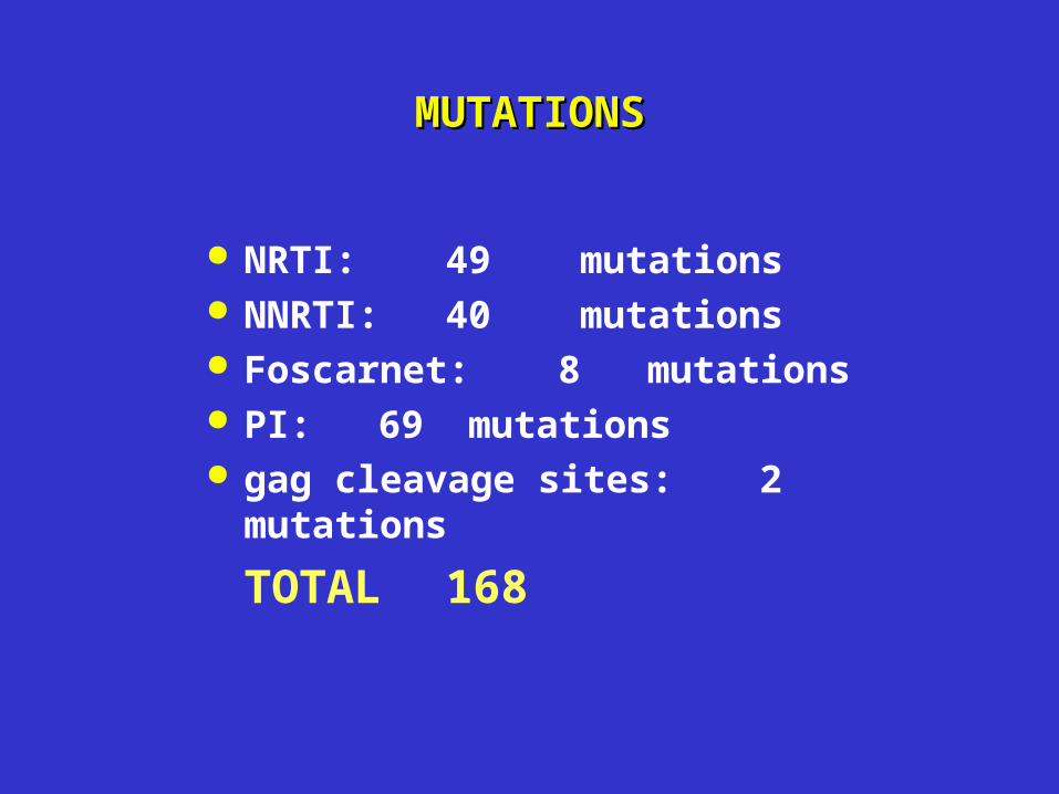
MUTATIONSMUTATIONS
NRTI: 49 mutations NNRTI: 40 mutations Foscarnet: 8 mutations PI: 69 mutations gag cleavage sites: 2 mutations
TOTAL 168

HIV ChipHIV Chip
Prot
RT
gag
1 Amplicon Includes 2 Gag cleavage sites: p7/p1 (2086) and p1/p6 (2134)
I ncludes the cleavage site at start of protease (2252) and between protease and RT (2546)
Includes all known mutations in protease
3 AmpliconsIncludes all known mutations in RT
Includes the cleavage site between RT
and RNAseH (3866)
2 AmpliconsIncludes the cleavage site between
p17 and p24 (1186)
Includes 2 cleavage sites: p24/p2
(1879) and p2/p7 (1921)

HIV PRT Mutant AnalysisHIV PRT Mutant Analysis
T215T215
T215YT215Y
T215FT215F
F214L / T215FF214L / T215F


The next step in microarrays
The next step in microarrays would then be the introduction of a more global analysis of the proteome, where at least 5,000 to 10,000 proteins could be studied using this principle.
The probes used for such approach would have to be based on recombinant antibodies and phage display libraries with several billions of antibody members.
As the antibodies cannot be synthesized on the surface of the chips, as is possible for DNA arrays, the probes have to be spotted on the chips in an array format and coupled to a downstream high-throughput detection system.
Some obvious choices for detection include: fluorescent tags, nano-electrodes, and in the case of smaller arrays, MS.

Protein chips architecture
Protein chips use a “probe” on the surface of a silicon chip to be able to catch native and post-translationally modified proteins.
One of the more obvious choices for such probes is antibodies because of the exquisite specificity of the their molecular design.
In fact, antibodies have already been used in a limited analysis of cellular changes occurring in cultured human cells, using a very small array and detected by SELDI (surface enhanced laser desorption ionization) MS.

Analysis of the proteome

Reversing the problem
In addition to developments in the use of microarrays of capture antibodies to measure protein levels, protein chips have been printed with antigens that are used for the detection of circulating antibodies in clinical specimens.

“Catcher” molecules
In contrast to DNA arrays, in which binding molecules may be defined by sequence and synthesized onto the surface of the array, protein-based catcher molecules with defined and predetermined specificities cannot be produced in such a way.
Instead, protein-based “catcher” molecules need to be developed for each ligand.
Arrays can be seen as miniaturized variants of assay formats having existed for many years.
Typically, such formats include enzyme-linked immunosorbent assay (ELISA) and other types of immunometric assays utilizing antibodies as catcher molecules.

Until now, the few protein-based arrays that have been presented have utilized monoclonal and full-length antibodies produced by conventional hybridoma technology and obtained from commercial or in-house sources.
These arrays have consisted of antibodies with specificities against known antigens, e.g., cytokines, cell signal, or cell matrix proteins, but also other proteins, and have counted as many as 250 different antibody specificities.
These antibodies may be developed through immunization of animals yielding monoclonal or polyclonal formats or may be developed using recombinant technologies using libraries of randomly recombined antibody fragments.


Disadvantages of Microarray Testing
• Use of Complex 'Research' Procedures
• Labor Intensive
• High Cost

•Multiple arrays.
•Multiple spots containing the same DNA oligo sequence/ protein on the same microarray.
•Multiple spots containing different oligo sequences/protein that assay the same gene RNA on the same microarray.
•Multiple spots containing different oligo sequences that assay different RNA products from the same gene on the same microarray.
•Replication of total experiment •Replication of just the hybridization step•Replication of only some other set of steps.
Levels of Replication