Metallo-Beta-Lactamase Producing Pseudomonas … cross sectional study included 1583 clinical...
12
Polish Journal of Microbiology 2017, Vol. 66, No 3, 297–308 ORIGINAL PAPER * Corresponding author: A.F. Abaza, Microbiology Department, High Institute of Public Health, Alexandria University, Egypt; e-mail: [email protected] Introduction Pseudomonas aeruginosa is considered one of the most leading causes of healthcare associated infections (HCAIs) worldwide (Varaiya et al., 2008). It is consid- ered the fourth most commonly isolated nosocomial pathogen accounting for 10% of all HCAIs. P. aeruginosa infections can range from superficial skin infections to fulminant sepsis. Even colonization of such strains in critical systems can be fatal (Sivaraj et al., 2012). World Health Organization (2015) has identified antimicrobial resistance as one of the three most impor- tant problems for human health. P. aeruginosa represents a phenomenon of resistance since all known mecha- nisms of antimicrobial resistance can be encountered; nevertheless enzyme production is the major mecha- nism of acquired resistance in these strains especially with β-lactam antibiotics, which are considerd a major line of treatment for P. areuginosa. Of these enzymes are the β-lactamases (Strateva and Yordanov, 2009). ere are four classes of β-lactamases: A, C and D which act through a serine based mechanism and metallo-beta- lactamases (MBL); a class B type of β-lactamases that is the most worrisome and require bivalent metal ions, usually zinc as a cofactor for their activity (Bush and Jacopy, 2010). is group can be suppressed by biva- lent ionic chelators as ethylene diamine tetra acetic acid (EDTA), but not inhibited by commercial β-lactamase inhibitors as clavulanic acid and tazobactam. ey can hydrolyze β-lactams from all classes except the mono- bactams (Aoki et al., 2010). MBL producing P. aeruginosa isolates were first reported in Japan in 1991, and since then there has been a substantial increase in the reporting of MBLs among carbapenem-resistant P. aeruginosa isolates worldwide (Pitout et al., 2005). ese isolates increas- ingly have been responsible for several nosocomial out- breaks in tertiary centers in different parts of the world (Walsh, 2008). Also the association between infections caused by MBL producing P. aeruginosa and longer hospital stay with high mortality rates has been reported (Zavascki et al., 2006). Metallo-Beta-Lactamase Producing Pseudomonas aeruginosa in a Healthcare Setting in Alexandria, Egypt AMANI F. ABAZA 1 , SORAYA A. EL SHAZLY 1 , HEBA S.A. SELIM 1 and GEHAN S.A. ALY 2 1 Microbiology Department, High Institute of Public Health, Alexandria University, Alexandria, Egypt 2 Alexandria University Students’ Hospital, Alexandria, Egypt Submitted 7 December 2016, revised 25 April 2017, accepted 5 May 2017 Abstract Pseudomonas aeruginosa has emerged as a major healthcare associated pathogen that creates a serious public health disaster in both develop- ing and developed countries. In this work we aimed at studying the occurrence of metallo-beta-lactamase (MBL) producing P. aeruginosa in a healthcare setting in Alexandria, Egypt. is cross sectional study included 1583 clinical samples that were collected from patients admit- ted to Alexandria University Students’ Hospital. P. aeruginosa isolates were identified using standard microbiological methods and were tested for their antimicrobial susceptibility patterns using single disc diffusion method according to the Clinical and Laboratory Standards Institute recommendations. irty P. aeruginosa isolates were randomly selected and tested for their MBL production by both phenotypic and genotypic methods. Diagnostic Epsilometer test was done to detect metallo-beta-lactamase enzyme producers and polymerase chain reaction test was done to detect imipenemase (IMP), Verona integron-encoded (VIM) and Sao Paulo metallo-beta-lactamase (IMP) encod- ing genes. Of the 1583 clinical samples, 175 (11.3%) P. aeruginosa isolates were identified. All the 30 (100%) selected P. aeruginosa isolates that were tested for MBL production by Epsilometer test were found to be positive; where 19 (63.3%) revealed bla SPM gene and 11 (36.7%) had bla IMP gene. bla VIM gene was not detected in any of the tested isolates. Isolates of MBL producing P. aeruginosa were highly susceptible to polymyxin B 26 (86.7%) and highly resistant to amikacin 26 (86.7%). MBL producers were detected phenotypically by Epsilometer test in both carbapenem susceptible and resistant P. aeruginosa isolates. bla SPM was the most commonly detected MBL gene in P. aeruginosa isolates. Key words: Pseudomonas aeruginosa, Epsilometer test, metallo-beta-lactamases, MBL encoding genes
-
Upload
truongnhan -
Category
Documents
-
view
215 -
download
1
Transcript of Metallo-Beta-Lactamase Producing Pseudomonas … cross sectional study included 1583 clinical...

Polish Journal of Microbiology2017, Vol. 66, No 3, 297–308
ORIGINAL PAPER
* Corresponding author: A.F. Abaza, Microbiology Department, High Institute of Public Health, Alexandria University, Egypt; e-mail: [email protected]
Introduction
Pseudomonas aeruginosa is considered one of the most leading causes of healthcare associated infections (HCAIs) worldwide (Varaiya et al., 2008). It is consid-ered the fourth most commonly isolated nosocomial pathogen accounting for 10% of all HCAIs. P. aeruginosa infections can range from superficial skin infections to fulminant sepsis. Even colonization of such strains in critical systems can be fatal (Sivaraj et al., 2012).
World Health Organization (2015) has identified antimicrobial resistance as one of the three most impor-tant problems for human health. P. aeruginosa represents a phenomenon of resistance since all known mecha-nisms of antimicrobial resistance can be encountered; nevertheless enzyme production is the major mecha-nism of acquired resistance in these strains especially with β-lactam antibiotics, which are considerd a major line of treatment for P. areuginosa. Of these enzymes are the β-lactamases (Strateva and Yordanov, 2009). There are four classes of β-lactamases: A, C and D which act
through a serine based mechanism and metallo-beta-lactamases (MBL); a class B type of β-lactamases that is the most worrisome and require bivalent metal ions, usually zinc as a cofactor for their activity (Bush and Jacopy, 2010). This group can be suppressed by biva-lent ionic chelators as ethylene diamine tetra acetic acid (EDTA), but not inhibited by commercial β-lactamase inhibitors as clavulanic acid and tazobactam. They can hydrolyze β-lactams from all classes except the mono-bactams (Aoki et al., 2010).
MBL producing P. aeruginosa isolates were first reported in Japan in 1991, and since then there has been a substantial increase in the reporting of MBLs among carbapenem-resistant P. aeruginosa isolates worldwide (Pitout et al., 2005). These isolates increas-ingly have been responsible for several nosocomial out-breaks in tertiary centers in different parts of the world (Walsh, 2008). Also the association between infections caused by MBL producing P. aeruginosa and longer hospital stay with high mortality rates has been reported (Zavascki et al., 2006).
Metallo-Beta-Lactamase Producing Pseudomonas aeruginosain a Healthcare Setting in Alexandria, Egypt
AMANI F. ABAZA1, SORAYA A. EL SHAZLY1, HEBA S.A. SELIM1 and GEHAN S.A. ALY2
1 Microbiology Department, High Institute of Public Health, Alexandria University, Alexandria, Egypt2 Alexandria University Students’ Hospital, Alexandria, Egypt
Submitted 7 December 2016, revised 25 April 2017, accepted 5 May 2017
A b s t r a c t
Pseudomonas aeruginosa has emerged as a major healthcare associated pathogen that creates a serious public health disaster in both develop-ing and developed countries. In this work we aimed at studying the occurrence of metallo-beta-lactamase (MBL) producing P. aeruginosa in a healthcare setting in Alexandria, Egypt. This cross sectional study included 1583 clinical samples that were collected from patients admit-ted to Alexandria University Students’ Hospital. P. aeruginosa isolates were identified using standard microbiological methods and were tested for their antimicrobial susceptibility patterns using single disc diffusion method according to the Clinical and Laboratory Standards Institute recommendations. Thirty P. aeruginosa isolates were randomly selected and tested for their MBL production by both phenotypic and genotypic methods. Diagnostic Epsilometer test was done to detect metallo-beta-lactamase enzyme producers and polymerase chain reaction test was done to detect imipenemase (IMP), Verona integron-encoded (VIM) and Sao Paulo metallo-beta-lactamase (IMP) encod-ing genes. Of the 1583 clinical samples, 175 (11.3%) P. aeruginosa isolates were identified. All the 30 (100%) selected P. aeruginosa isolates that were tested for MBL production by Epsilometer test were found to be positive; where 19 (63.3%) revealed blaSPM gene and 11 (36.7%) had blaIMP gene. blaVIM gene was not detected in any of the tested isolates. Isolates of MBL producing P. aeruginosa were highly susceptible to polymyxin B 26 (86.7%) and highly resistant to amikacin 26 (86.7%). MBL producers were detected phenotypically by Epsilometer test in both carbapenem susceptible and resistant P. aeruginosa isolates. blaSPM was the most commonly detected MBL gene in P. aeruginosa isolates.
K e y w o r d s: Pseudomonas aeruginosa, Epsilometer test, metallo-beta-lactamases, MBL encoding genes
Abaza A.F. et al. 3298
The problem is aggravated by the fact that most of the MBL encoding genes reside on integrons and plas-mids which in turn allows for widespread dissemina-tion of these genetic elements, hence poses a threat for spread resistance patterns among the Gram-negative bacteria (Mohamed and Raafat, 2011).
As regards molecular structure: five MBL types have been widely recognized; imipenemase (IMP), Verona integron-encoded (VIM), Sao Paulo (SPM), German imipenemase (GIM) and Seoul imipenemase (SIM). Several types of MBL enzymes have been identified in P. aeruginosa among which the VIM-type enzymes appear to be the most prevalent. IMP is also considered one of the most important types of MBLs. Camagaro et al., (2011) reported that after being restricted for more than ten years to Brazilian hospitals; SPM seems to become a global challenge, warning for the role of human traffic in spreading MBL genes (Salabi et al., 2010).
It is well known that poor outcome occurs when patients with serious infections due to MBL produc-ing organisms are treated with antibiotics to which the organism is completely resistant. Therefore early and proper detection of MBL producing Gram-negative bacilli especially P. aeruginosa is crucial; for optimal treatment of particularly critically ill and hospitalized patients and to permit rapid initiation of strict infection control procedures to prevent nosocomial spread and control the dissemination of resistance (Cuzon et al., 2012). This work aimed at studying the occurrence of MBL producing P. aeruginosa in a healthcare setting in Alexandria, Egypt.
Experimental
Material and Methods
This cross sectional study was carried out during an 18-month period from January 2013 to June 2014. It included different clinical samples that were collected from patients admitted to the Alexandria University Students’ Hospital (AUSH). Collected clinical samples were processed in AUSH laboratory and the Microbi-ology laboratory at the High Institute of Public Health (HIPH). The study was approved by the Ethics Com-mittee of the HIPH. Informed consents of all enrolled patients were obtained before collection of samples and after explanation of the purpose and benefits of the research.
Sampling
Data collection. A questionnaire sheet including all the relevant information (name, age, sex, date of admis-sion, medical history, diagnosis, antibiotic administra-
tion etc.) was filled in for every patient enrolled in the present study.
Samples collection and processing. A total of 1583 different clinical samples were collected during the study period from patients showing signs and symptoms sug-gestive of infection and were delivered to the laboratory. The samples were distributed as 660 respiratory sam-ples (500 bronchoalveolar lavage (BAL) and 160 sputum samples), 446 urine samples, 209 blood samples, 142 pus and exudate samples, 58 peritoneal fluid samples, 35 ear discharge, and 33 conjunctival secretions. Col-lected samples were subjected to macro scopical and microscopical examination. The samples were cultured on blood (Oxoid 9191118 UK) and MacConkey’s agar (Oxoid 567362 UK) plates. Plates were incubated aero-bically at 37°C for 24 hours (Tille et al., 2014).
Identification procedures of P. aeruginosa. After proper incubation of inoculated blood and MacCon-key’s agar plates, isolates that appeared as medium sized, grayish, opaque, large flat pigmented colonies, with feathered edges, producing a sweet or grape like odour either hemolytic or non hemolytic on blood agar plates, and were pale, non lactose fermenting on MacConkey’s agar plates, and microscopically appeared as Gram-negative bacilli were further differentiated and identified according to standard microbiological methods (Tille et al., 2014).
Antimicrobial susceptibility testing (AST). All 175 confirmed P. aeruginosa isolates were tested for their antibiotic susceptibility patterns using single disc dif-fusion method according to the Clinical and Labora-tory Standards Institute (CLSI) recommendations (Patel et al., 2014). The test was done on Mueller Hinton (MH) agar plates (Difco 00252-01), using the selected antibio-tic discs. All antimicrobial discs used in this study were supplied by oxoid laboratories. After 24 hours aerobic incubation at 37°C, each plate was examined and inhibi-tion zones were measured, recorded, and interpreted as susceptible (S), intermediate (I) or resistant (R) accord-ing to the interpretive criteria of CLSI (Patel et al., 2014).
Isolates that were confirmed to be P. aeruginosa and tested for their antimicrobial susceptibility pat-terns were then subcultured on blood agar plates and incubated aerobically at 37°C for 24 hrs. Isolated colo-nies were inoculated on soft agar deeps and incubated aerobically at 37°C for 24 hrs.
Identification of metallo beta lactamase produc-tion by Epsilometer test (E-test). Thirty confirmed P. aeruginosa isolates were selected to be tested for their MBL enzyme production by E test. MBL diag-nostic E-test strip consists of a double sided seven dilu-tion range of imipenem (IP) (4 to 256 microgram/ml) and IP overlaid with EDTA (1 to 64 microgram/ml) (Pitout et al., 2005). MBL E-test was performed accord-ing to the manufacturer’s instructions (AB BioMerieux,

MBL producing Pseudomonas aeruginosa3 299
Solna, Sweden). Isolates stored in agar deeps were subcultured on blood agar plates and were incubated aerobically at 37°C for 24 hrs. Individual colonies were picked from overnight agar plates and suspended in a 0.85% saline and were adjusted to a turbidity of 0.5 McFarland standard.
E-test MBL strips were applied to MH agar plates inoculated with adjusted suspensions. Seeded MH agar plates were incubated aerobically for 24 hrs at 37°C.The minimum inhibitory concentration (MIC) end points were read where the inhibition ellipses intersected the strip. A reduction of imipenem MIC in the presence of EDTA that is greater than or equal to eight-fold (IP/IPI > 8 mm) was interpreted as indicating MBL activity. The presence of a phantom zone or a defor-mation of the imipenem ellipse was also considered a positive result (Pitout et al., 2005).
Detection of blaIMP, blaVIM, and blaSPM MBL genes by PCR. Thirty P. aeruginosa isolates that were con-firmed as MBL producers by MBL diagnostic E-test, were tested for the presence of blaIMP, blaVIM, blaSPM genes.
DNA extraction
Procedure. DNA for PCR was extracted by the boil-ing method. Two or three colonies were taken from fresh culture of the confirmed MBL P. aeruginosa iso-lates and suspended in 500 μl saline, then vortexed to get a uniform suspension. The cells were lysed by heat-ing them at 100°C for 10 minutes, and then centrifuged at 12,000 rpm for 10 min. The supernatant was used directly as a template DNA in the PCR mixture.
DNA amplification. The extracted DNA was sub jec-ted to PCR amplification reaction using three pairs of primers specific for MBL genes (blaIMP, blaVIM, and blaSPM). The DNA amplification was done using Dream Taq Green PCR Master mix (Thermo Scientific, Waltham, United States). The primers were purchased lyophi-lized; (Biosearch Tech, Petaluma, California, United States). They were reconstituted by the addition of sterile nuclease free water to a final concentration of 100 pico mol/μl, distributed in aliquots and stored at –20°C.
Primers sequence of MBL genes (Sader et al., 2005)
b-PCR amplification protocol. (I) Reaction mix-tures were prepared using sterile nuclease free water. To each tube a total volume of 50 ml was reached by
adding Master mix (25 μl), sense primer (1 μl), anti-sense primer (1 μl), DNA template (sample) (10 μl), nuclease free water (13 μl). A negative control was pre-pared by the addition of the same contents to the tube with 10 μl nuclease free water instead of the sample.
(II) The tubes were transferred to the thermal cycler (BioCycler TC-S, Boeco-Germany) for amplification. The thermocycler program conditions for blaIMP and blaVIM genes included: 30 cycles of amplification under the following conditions: denaturation at 95°C for 30 seconds, annealing for 1 minute at specific tempera-tures (blaIMP at 45°C and blaVIM–66°C), and extension at 72°C for 1 minutes/kb product (Khosravi et al., 2011). The cycling parameters of PCR to amplify blaSPM gene were: initial denaturation at 95°C for 5 min, followed by 30 cycles of denaturation at 95°C for 1 min, annealing at 50°C for 1 minute and extension at 68°C for 1 minute. The cycle was followed by a final extension at 72°C for 10 minutes (Gaspareto et al., 2007).
DNA detection by gel electrophoresis. PCR pro-ducts were loaded on 2% agarose in tris borate EDTA (TBE) containing 0.5 μl of ethidium bromide per ml. After electrophoresis, the gel was visualized under ultraviolet light.
The DNA bands were visualized on a 320 nm UV transilluminator and photographed. The gel was exam-ined for specific bands; positive results of PCR were confirmed by detection of 432 bp band for blaIMP gene, 500 bp band for blaVIM gene and 650 bp band for blaSPM gene as determined by the molecular weight markers run at the same time. (Khosravi et al., 2011; Gaspareto et al., 2007).
Statistical analysis of the data. Data were fed to the computer and analyzed using IBM SPSS software package version 20.0 (Kirkpatrick and Feeney, 2013). Qualitative data were described using number and percent. Quantitative data were described using Range (minimum and maximum), mean, standard deviation and median. Comparison between different groups regarding categorical variables was tested using Chi-square test, Fisher’s exact test, Monte Carlo correction, independent t-test and Mann Whitney test. Signifi-cance of the obtained results was judged at the 5% level.
Results
The present study included 175 P. aeruginosa iso-lates that were recovered from a total of 1583 clinical samples. These samples were collected randomly from patients admitted to the AUSH (984 from ICUs and 599 from wards).
The highest percentage of P. aeruginosa isolates 75 (42.9%) were recovered from respiratory samples, fol-lowed by urine samples 57 (32.6%) and pus and exudate
IMP 1 Sense: 5’dCTACCGCAGCAGAGTCTTTGC3’ Antisense: 5’dGAACAACCAGTTTTGCCTTACC3’VIM 2 Sense: 5’dATGTTCAAACTTTTGAGTAGTAAG3’ Antisense: 5’dCTACTCAACGACTGAGCG3’SPM 1 Sense: 5dCCTACAATCTAACGGCGACC3’ Antisense: 5’dTCGCCGTGTCCAGGTATAAC3’
Primer Sequence (5’ to 3’)

Abaza A.F. et al. 3300
samples 33 (18.9%). From blood samples, four P. aeruginosa isolates were recovered (2.3%), while each of con-junctival secretion, ear discharge, and peritoneal fluid samples yielded two P. aeruginosa isolates (1.1% each).
In the current work, the highest percentage of resist-ance among 175 detected P. aeruginosa isolates was for aztreonam 150 (85.7%), followed by ceftazidime 140 (80%), cefipime 139 (79.4%), imipenem 137 (78.3%), ciprofloxacin 134 (76.6%), and meropenem 129 (73.7%).
On the other hand, the highest susceptibility percen-tages were recorded for polymyxin B 152 (86.9%), piper-acillin tazobactam 52 (29.7%), 48 ofloxacin (27.4%), piperacillin 46 (26.3 %), gentamycin 39 (22.3%) and cefepime 28 (16%).
Of the 175 confirmed P. aeruginosa isolates, 30 iso-lates were randomly selected to be tested phenotypi-cally for their MBL enzyme production by E test. Thirty selected P. aeruginosa isolates were as follows:
•Twenty P. aeruginosa isolates were resistant to three classes of β-lactams including: cephems, carba penems and monobactams (aztreonam).
•Three P. aeruginosa isolates were resistant to three classes of β-lactams (penicillins, cephalospor-ins and carbapenems) and were susceptible to aztreonam.
• Seven P. aeruginosa isolates were resistant to three classes of antibiotics (cephems, aminoglycosi-des, and carbapenems), but were susceptible to imipenem, and 4 of them were also susceptible to meropenem.
This study showed that the highest percentage of MBL producing P. aeruginosa isolates were from respir-atory samples 11/30 (36.7%), followed by urine, and pus and exudate samples 9/30 (30%) each. Only one isolate was recovered from an ear sample 1/30 (3.3%) (Fig. 1).
Thirty selected isolates were all positive for MBL production by MBL E test, and were tested for blaIMP, blaVIM and blaSPM genes using conventional PCR (results as in Fig. 2). Of the 30 tested isolates; 19 (63.3%)
Fig. 1. Distribution of 30 MBL producing P. aeruginosa isolatesaccording to type of samples.
Fig. 2C blaVIM
Lane 1: Marker 100 bp Ladder Lane 2: Negative test strain for blaVIM (500 bp)Lane 3: Negative test strain for blaVIM (500 bp)Lane 4: Negative control Lane 5: Negative test strain for blaVIM (500 bp)Lane 6: Negative test strain for blaVIM (500 bp)Lane 7: Negative test strain for blaVIM (500 bp)Lane 8: Negative test strain for blaVIM (500 bp)
Fig. 2. A, B, C. Electrophoresis results for P. aeruginosa blaIMP,blaSPM and blaVIM genes.
Fig. 2A blaIMP
Lane 1: Marker 100 bp Ladder Lane 2: Positive test strain for blaIMP (432 bp)Lane 3: Positive test strain for blaIMP (432 bp)Lane 4: Positive test strain for blaIMP (432 bp)Lane 5: Positive test strain for blaIMP (432 bp)Lane 6: Negative controlLane 7: Positive test strain for blaIMP (432 bp)
Fig. 2B blaSPM
Lane 1: Marker 100 bp Ladder Lane 2: Negative controlLane 3: Positive test strain for blaSPM (650 bp)Lane 4: Positive test strain for blaSPM (650 bp)Lane 5: Positive test strain for blaSPM (650 bp)Lane 6: Positive test strain for blaSPM (650 bp)Lane 7: Positive test strain for blaSPM (650 bp)Lane 8: Positive test strain for blaSPM (650 bp)Lane 9: Positive test strain for blaSPM (650 bp)

MBL producing Pseudomonas aeruginosa3 301
revealed blaSPM gene and 11(36.7%) had blaIMP gene, blaVIM gene was not detected in any of the tested isolates.
Of the 30 patients with MBL producing P. aeruginosa isolates, 14(46.7%) were of age group 20 ≤ 30 years, 9 (30.0%) belonged to the age group 30–50 years, and seven patients (23.3%) were above 50 years (Table I). Moreover, our data showed that the highest percent-age of patients who revealed P. aeruginosa isolates with positive blaSPM gene were aged 20 ≤ 30 years old (47.4%), followed by those aged > 50 years old (31.6%).
In this study the majority of patients with MBL producing P. aeruginosa isolates 21 (70%) were admit-ted to the ICU and 9 (30%) were admitted in inpatient
wards. In addition, the highest percentage (76.7%) was recovered from those who had duration of hospital stay of more than 7 days. Regarding antibiotic intake, the majority of patients 26/30 (86.7%) had taken antibiot-ics within 2 weeks from the study period, and this was found to be statistically significant (p ≤ 0.001). On the other hand, 23/30 (76.7%) patients were readmitted to the hospital within 3 months. This was statistically significant (p ≤ 0.006) (Table II).
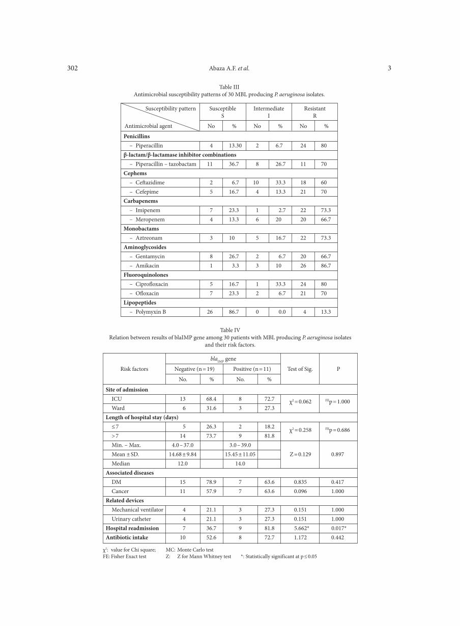
Our work revealed that the 30 MBL producing P. aeruginosa isolates were highly susceptible to poly-mxin B 26 (86.7%), followed by piperacillin-tazobactam 11 (36.7%), then gentamycin 8 (26.7%). Seven (23.3%) isolates were susceptible to each of imipenem and oflo-xa cin. The highest percentage of isolates were resistant to Amikacin 26/30 (86.7%), followed by piperacillin and ciprofloxacin 24 /30(80% each) (Table III).
More than half of the patients who had P. aeruginosa isolates with blaIMP genes 6/11 (54.5%) were aged between 20 ≤ 30 years old, this was followed by those of 30–50 years old 3/11 (27.3%). The lowest percen- tage of P. aeruginosa isolates with blaIMP gene 2 (18.2%) was recovered from patients above 50 years. On the other hand, the highest percentage of patients who revealed positive blaSPM gene were aged 20 ≤ 30 years old 9/19 (47.4%), followed by those aged > 50 years old 6/19 (31.6%).
Table IDistribution of the 30 patients with MBL producing P. aeruginosa
isolates according to their gender and age.
2: Chi square test; MC: Monte Carlo test
Gender
Agein years
Male(n = 12)
Female(n = 18)
Total(n = 30)
No. %No. %No. %
20– < 30 7 58.3 7 38.9 14 46.730–50 1 8.3 8 44.4 9 30.0> 50 4 33.3 3 16.7 7 23.3χ2 (MCp) 4.570 (0.104)
Site of admissionICU (n = 126) 21 70.0 χ2 = 0.072 0.789Ward (n = 49) 9 30.0Length of hospital stay (days)≤ 7 (n = 54) 7 23.3 χ2 = 0.961 0.327> 7 (n = 121) 23 76.7 Min. – Max. 3.0–39.0Mean ±SD. 14.97 ± 10.12 Z = 0.519 0.604Median 12.50 Associated diseasesDM 22 73.3 χ2 = 18.114* < 0.001*Cancer 18 60.0 χ2 = 9.052* 0.003*Related devicesMechanical ventilator 7 23.3 χ2 = 0.784 0.376Urinary catheter 7 23.3 χ2 = 8.823* FEp = 0.024*Readmission (n = 94) 23 76.7 χ2 = 7.672* 0.006*Antibiotic intake (n = 97) 26 86.7 χ2 = 14.301* < 0.001*
Table IIDistribution of 30 patients with positive MBL producing P. aeruginosa isolates according to risk factors.
χ2: Chi square test; Z: Z for Mann Whitney test; *: Statistically significant at p ≤ 0.05
Frequency of isolation
Risk factors
Patients with MBL producingP. aeruginosa isolates
(n = 30) Test of sig. P
No. %
Abaza A.F. et al. 3302
Penicillins – Piperacillin 4 13.30 2 6.7 24 80β-lactam/β-lactamase inhibitor combinations – Piperacillin – tazobactam 11 36.7 8 26.7 11 70Cephems – Ceftazidime 2 6.7 10 33.3 18 60 – Cefepime 5 16.7 4 13.3 21 70Carbapenems – Imipenem 7 23.3 1 2.7 22 73.3 – Meropenem 4 13.3 6 20 20 66.7Monobactams – Aztreonam 3 10 5 16.7 22 73.3Aminoglycosides – Gentamycin 8 26.7 2 6.7 20 66.7 – Amikacin 1 3.3 3 10 26 86.7Fluoroquinolones – Ciprofloxacin 5 16.7 1 33.3 24 80 – Ofloxacin 7 23.3 2 6.7 21 70Lipopeptides – Polymyxin B 26 86.7 0 0.0 4 13.3
Table IIIAntimicrobial susceptibility patterns of 30 MBL producing P. aeruginosa isolates.
Susceptibility pattern
Antimicrobial agent
SusceptibleS
IntermediateI
ResistantR
No % No % No %
Site of admission ICU 13 68.4 8 72.7 χ2 = 0.062 FEp = 1.000 Ward 6 31.6 3 27.3Length of hospital stay (days) ≤ 7 5 26.3 2 18.2 χ2 = 0.258 FEp = 0.686 > 7 14 73.7 9 81.8 Min. – Max. 4.0 – 37.0 3.0 – 39.0 Mean ± SD. 14.68 ± 9.84 15.45 ± 11.05 Z = 0.129 0.897 Median 12.0 14.0Associated diseases DM 15 78.9 7 63.6 0.835 0.417 Cancer 11 57.9 7 63.6 0.096 1.000Related devices Mechanical ventilator 4 21.1 3 27.3 0.151 1.000 Urinary catheter 4 21.1 3 27.3 0.151 1.000Hospital readmission 7 36.7 9 81.8 5.662* 0.017*Antibiotic intake 10 52.6 8 72.7 1.172 0.442
Table IVRelation between results of blaIMP gene among 30 patients with MBL producing P. aeruginosa isolates
and their risk factors.
χ2: value for Chi square; MC: Monte Carlo testFE: Fisher Exact test Z: Z for Mann Whitney test *: Statistically significant at p ≤ 0.05
Risk factors
blaIMP gene
PTest of Sig.Negative (n = 19) Positive (n = 11)
No. % No. %
MBL producing Pseudomonas aeruginosa3 303
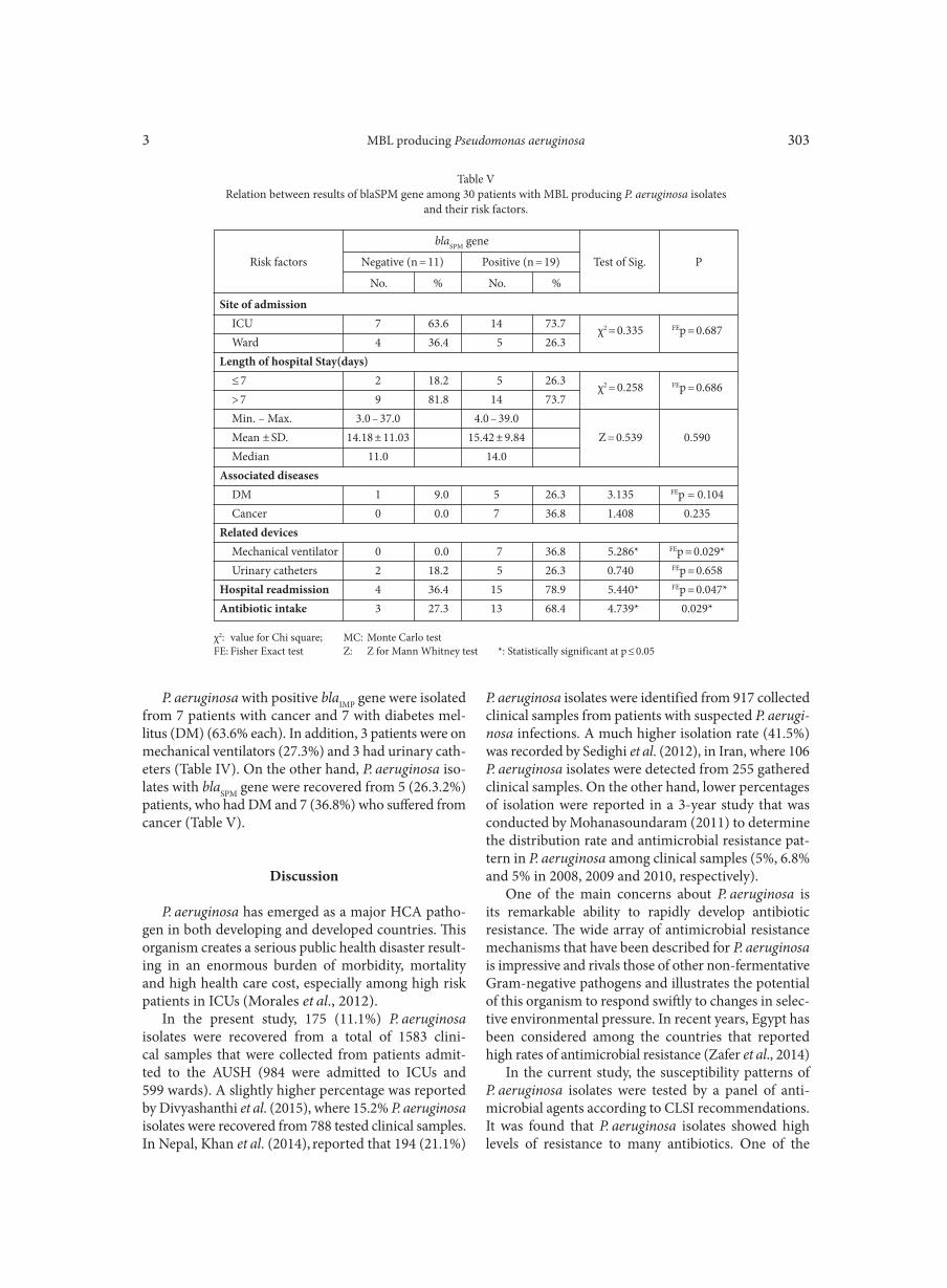
P. aeruginosa with positive blaIMP gene were isolated from 7 patients with cancer and 7 with diabetes mel-litus (DM) (63.6% each). In addition, 3 patients were on mechanical ventilators (27.3%) and 3 had urinary cath-eters (Table IV). On the other hand, P. aeruginosa iso-lates with blaSPM gene were recovered from 5 (26.3.2%) patients, who had DM and 7 (36.8%) who suffered from cancer (Table V).
Discussion
P. aeruginosa has emerged as a major HCA patho-gen in both developing and developed countries. This organism creates a serious public health disaster result-ing in an enormous burden of morbidity, mortality and high health care cost, especially among high risk patients in ICUs (Morales et al., 2012).
In the present study, 175 (11.1%) P. aeruginosa isolates were recovered from a total of 1583 clini-cal samples that were collected from patients admit-ted to the AUSH (984 were admitted to ICUs and 599 wards). A slightly higher percentage was reported by Divyashanthi et al. (2015), where 15.2% P. aeruginosa isolates were recovered from 788 tested clinical samples. In Nepal, Khan et al. (2014), reported that 194 (21.1%)
P. aeruginosa isolates were identified from 917 collected clinical samples from patients with suspected P. aeruginosa infections. A much higher isolation rate (41.5%) was recorded by Sedighi et al. (2012), in Iran, where 106 P. aeruginosa isolates were detected from 255 gathered clinical samples. On the other hand, lower percentages of isolation were reported in a 3-year study that was conducted by Mohanasoundaram (2011) to determine the distribution rate and antimicrobial resistance pat-tern in P. aeruginosa among clinical samples (5%, 6.8% and 5% in 2008, 2009 and 2010, respectively).
One of the main concerns about P. aeruginosa is its remarkable ability to rapidly develop antibiotic resistance. The wide array of antimicrobial resistance mechanisms that have been described for P. aeruginosa is impressive and rivals those of other non-fermentative Gram-negative pathogens and illustrates the potential of this organism to respond swiftly to changes in selec-tive environmental pressure. In recent years, Egypt has been considered among the countries that reported high rates of antimicrobial resistance (Zafer et al., 2014)
In the current study, the susceptibility patterns of P. aeruginosa isolates were tested by a panel of anti- microbial agents according to CLSI recommendations. It was found that P. aeruginosa isolates showed high levels of resistance to many antibiotics. One of the
Site of admission ICU 7 63.6 14 73.7 χ2 = 0.335 FEp = 0.687 Ward 4 36.4 5 26.3Length of hospital Stay(days) ≤ 7 2 18.2 5 26.3 χ2 = 0.258 FEp = 0.686 > 7 9 81.8 14 73.7 Min. – Max. 3.0 – 37.0 4.0 – 39.0 Mean ± SD. 14.18 ± 11.03 15.42 ± 9.84 Z = 0.539 0.590 Median 11.0 14.0Associated diseases DM 1 9.0 5 26.3 3.135 FEp = 0.104 Cancer 0 0.0 7 36.8 1.408 0.235Related devices Mechanical ventilator 0 0.0 7 36.8 5.286* FEp = 0.029* Urinary catheters 2 18.2 5 26.3 0.740 FEp = 0.658Hospital readmission 4 36.4 15 78.9 5.440* FEp = 0.047*Antibiotic intake 3 27.3 13 68.4 4.739* 0.029*
Table VRelation between results of blaSPM gene among 30 patients with MBL producing P. aeruginosa isolates
and their risk factors.
χ2: value for Chi square; MC: Monte Carlo testFE: Fisher Exact test Z: Z for Mann Whitney test *: Statistically significant at p ≤ 0.05
Risk factors
blaSPM gene
PTest of Sig.Negative (n = 11) Positive (n = 19)
No. % No. %
Abaza A.F. et al. 3304
alarming results is the high resistance against carbapen-ems; where 78.3% of isolates were resistant to imipenem and 73.7% were resistant to meropenem. In agreement with our results, Diab et al. (2013) revealed a high rate (72%) of imipenem resistance among P. aeruginosa isolates. In the Middle East the occurrence of imipenem resistant P. aeruginosa was also recognized. In Saudi Arabia, of a total of 350 P. aeruginosa isolates, 135 (38.57%) were found to be resistant to imipenem (Mohamed et al., 2011). Hashemi et al., (2016) reported that all their MBL-producing P. aeruginosa isolates were resistant to meropenem and imipenem.
Among 33 European countries participating in the European Antimicrobial Resistance Surveillance Sys-tem (EARSS) in 2007, six countries reported carbap-enem resistance rates of more than 25% among P. aeruginosa isolates (Souli et al., 2008); the highest rate was reported from Greece (51%). Reasons for increased antimicrobial resistance in Greece are numerous as explained by Miyakis et al. (2011). As Greece has the highest antibiotic consumption rate in Europe, both in total and in out-patients, population mobility can intro-duce resistant strains and infrastructure and resources for infection control are insufficient. This, along with reduced awareness for detection, increases the likeli-hood of in-hospital spread of multi drug resistant organisms (MDROs). Many of these reasons exist in our country and can explain the high antimicrobial resistance encountered among P. aeruginosa isolates in the current study.
During the last decade, emergence and dissemina-tion of the most prevalent MBL genes such as; blaIMP, blaVIM, blaGIM, blaSIM, and the newly identified blaAIM and blaNDM have been extensively documented around the world. Because of the efficient carbapenemase activ-ity of the MBL enzymes, they account for up to 40% of worldwide imipenem resistant P. aeruginosa. Xavier et al. (2010) stated that MBL enzymes increase antimi-crobial MICs more effectively than does either efflux pump over-expression or porin down-regulation alone. Unfortunately, screening only carbapenem-resistant organisms for these enzymes, as most often performed, is suboptimal. With increasing reports of MBLs in carbapenem-susceptible isolates, it becomes crucial to improve laboratory methods used for detection of what is called: hidden MBLs (Ntokou et al., 2008).
In the present study determination of MBL fre-quency in both carbapenem resistant and susceptible P. aeruginosa isolates was done by phenotypic and geno-typic methods. Accordingly, 30 identified P. aeruginosa isolates were selected to cover: the commonly known definition of MBL producing P. aeruginosa isolates i.e. those that are resistant to all beta lactams but sensitive to monobactams (aztreonam), and isolates that showed resistance to carbapenems and aztreonam, together
with isolates that were carbapenem (imipenem) sensi-tive to exclude hidden MBLs.
The most widely accepted standardized MBL screen-ing test is the MBL E-test. However, due to the high cost and relative unavailability of E-test strips, many clini-cal microbiology laboratories use alternative screening methods, such as double-disk synergy test (DDST) and combined disk test (CDT). Although the DDST and the CDT assays are simple to perform and cheaper than the MBL E-test, they have shown discordant results, depending on the employed methodology, β-lactam substrates, MBL inhibitors, and bacterial genus tested (Picão et al., 2008; Ranjan et al., 2015). In this piece of work, all of the 30 (100%) selected P. aeruginosa iso-lates were found to be MBL producing strains using E-test. Similarly Bashir et al. (2011) reported that all their thirty isolates were positive for MBL producing P. aeruginosa by E-test.
In the current work, of the 30 identified MBL iso-lates; the highest percentage was recovered from res-piratory samples 11 (36.7%), followed by 9 (30%) from each of urine and pus and exudate samples, and only one isolate (3.3%) was from ear discharge. This is nearly in agreement with Zavascki et al. (2006), who found that the lung was the most frequent site of nosocomial infection (50.3%), followed by urinary tract (20.5%), and skin and soft tissue (15.8%).
Many factors may contribute to the acquisition of MBL resistant enzymes. Patients in critical care units are likely to have higher probability for these isolates. It was found from this study that the highest percent-age of MBL producing P. aeruginosa isolates was among ICU patients (70%). This was in line with Zavascki et al. (2006) and Lucena et al. (2014) who found that ICUs admission increased the risk for MBL producing P. aeru ginosa infections.
One of the most important parameters for MBL acquisition is associated diseases. Of the 30 patients, who revealed MBL P. aeruginosa isolates, 22 (73.3%) had DM and 18 (60%) suffered from cancer. These results were statistically significant (p < 0.001 and p = 0.003, respectively). In concordance, Varaiya et al. (2008) reported that out of 33 (14.3%) MBL produc-ing isolates, 24 (72.7%) were diabetic patients, and 6 (18.1%) were cancer patients. They attributed their findings to the associated immune deficiency among diabetic patients, and recurrent foot infections being good poly-microbial media for a high incidence of multidrug-resistant P. aeruginosa. In addition, Vaishali et al. (2013) reported that the presence of underlying diseases as DM is a significant risk factor in acquisition of MBL P. aeruginosa infection.
Other risk factors that were significantly associated with the presence of MBL producing P. aeruginosa infection in this study were the use of medical devices
MBL producing Pseudomonas aeruginosa3 305
as mechanical ventilators and urinary catheters (23.3%) each, readmission to hospitals (76.7%), and history of previous antibiotic intake (86.7%). On the other hand, the length of hospital stay for more than 7 days rep-resented 76.7%, but was not found to be statistically significant. Kumar et al. (2011), found that all MBL-positive patients were exposed to different risk fac-tors as prolonged hospital stay for more than 8 days, cathe terization, previous antibiotic use, and mechanical ventilation. In the study done by Vaishali et al. (2013) the duration of hospital stay of more than 10 days had 1.7 times risk of acquisition of MBL P. aeruginosa infec-tion more than duration of hospital stay of less than or equal to10 days.
The occurrence MBL production is not restricted to carbapenem resistant strains, but some reports have argued about their presence in carbapenem susceptible organisms. They might be unrecognized as the MBL detection has not been routinely performed in most clinical microbiology laboratories (Picão et al., 2012). Such organisms often carry hidden MBL genes. As a consequence, these isolates will be able to participate in horizontal MBL gene transfer with other Gram-negative pathogens and may contribute significantly to MBL related outbreaks. In the present study, of the 30 identified MBL producing P. aeruginosa isolates, 7 (23.3%) were imipenem sensitive and 4 (13.3%) were sensitive to meropenem. Diab et al. (2013) documented that among the studied imipenem susceptible isolates, 58.3% of which were proved to be MBL producers. A much lower percentage was published by Anoar et al. (2014) in Iraq, where of the 46 detected MBL produc-ing isolates, 7 (3.95%) were meropenem sensitive. They explained their findings by the possibility that there might be hidden MBL genes among isolated strains which cannot be diagnosed by phenotypic tests, lead-ing to the dissemination of these genes in the hospital silently among patients even within normal HCWs who can act as carriers for MBL genes in future.
In this piece of work, multi drug resistance was noticed. MBL producing P. aeruginosa had the high-est percentage of resistance against amikacin 86.7%, followed by piperacillin and ciprofloxacin 80% each. In addition, carbapenems showed high percentages of resistance; imipenem and meropenem (73.3%, and 66.7%, respectively). Bhongle et al. (2012) found only one isolate sensitive to imipenem, but it was found to be positive for MBL, thus indicating that MBL producers could show susceptibility to imipenem. These isolates can appear to be susceptible to carbapenems though they carry carbapenemases, such organisms thus carry hidden MBL genes, whereby the microbiologists may remain unaware of their presence.
MBL producers are commonly known as those organisms that potently hydrolyze all beta-lactam anti-
biotics except aztreonam. In the current study, high resistance to monobactams (aztreonam) was detected (73.3%), and only 3 (10%) of the isolates were suscep-tible to aztreonam and conformed to such definition. A lower percentage of aztreonam resistance (45.1%) was reported by Zafer et al. (2014). In this study, 86.7% of MBL producing P. aeruginosa isolates were suscep-tible to polymyxin B. This supports the evidence that polymyxin B has increasingly become the last viable therapeutic option for MDR Pseudomonas infections. In accordance with the current results Bashir et al. (2011) recorded that MBL producers showed very high resistance to all antimicrobials except polymyxin B. However, resistance to amikacin (73%) and cipro-floxacin (55%) was lower when compared to the results in the present study (86.7% and 80%, respectively). Piperacillin tazobactam was the second most effective antibiotic after polymyxin B, where 36.7% of our iso-lates were sensitive to it.
Although phenotypic methods are considered to be useful and reliable in detecting MBL producers, results should be validated and confirmed by genotypic meth-ods. In the present study, all the 30 (100%) identified MBL producing P. aeruginosa isolates by E test were further screened for the presence of MBL encoding genes (blaIMP, blaSPM, and blaVIM) using conventional PCR. blaIMP gene was the most prevalent MBL among the isolates accounting for 63.3%, while 36.7% of the isolates revealed blaIMP gene and none of the isolates had blaVIM genes.
In agreement with the results of this study, Gaspa-reto et al. (2007) stated that blaSPM gene was the most common gene among MBL P. aeruginosa isolates (73%), and that no blaVIM gene was detected. Camargo et al. (2011) reported that blaSPM gene accounted for 71% of positive MBL strains, while blaIMP was detected in 29%.
On the contrary, blaVIM gene was detected in many Egyptian studies. Zafer et al. (2014) demonstrated that 58.3% were positive for blaVIM gene, with blaIMP gene detected in 2.1% and no blaSPM gene was iden- tified. Another study by Diab et al. (2013) revealed that 70% were positive for blaVIM, while blaIMP gene was not detected.
In the current work, all the 30 P. aeruginosa isolates that were identified as MBL producers by the phenotypic method E-test, were found to be MBL producers by the molecular method PCR; where 19 isolates had MBLSPM gene and 11 had blaIMP gene. A lower percentage was reported by Doosti et al. (2013); where 36/41 (87.8%) of isolates were phenotypically MBL positive, but PCR results confirmed presence of MBL genes in only 33/41 (80%) of isolates. blaIMP producers have been detected worldwide: in Europe (Docquier et al., 2003) and in the U.S. (Hanson et al., 2006). In the current study, posi-tive MBL IMP gene producers were more commonly

Abaza A.F. et al. 3306
isolated from respiratory samples (36.4%), followed by urine and pus and exudate samples (27.3% each). It was detected in only one ear discharge sample (9.1%).
In the current work, hospital readmission was found to be a significant risk factor for acquiring blaIMP gene, where 81.8% of patients were readmitted to the hospital within 3 months of the study (p = 0.017).
Among MBL genes, blaSPM has been widely detected in Brazil and Switzerland, the dissemination of this gene in various regions seems to be caused by a sin-gle epidemic P. aeruginosa clone (Salabi et al., 2010) In the present study, blaSPM gene producers were mostly recovered from respiratory samples (47.4%), followed by urine (26.3%) and pus and exudate samples (21.1%). In line to our data, Zavascki et al. (2006) in southern Brazil, described the first nosocomial outbreak of P. aeru ginosa producing MBL SPM gene, and noted that IMP isolates were highly revealed from respiratory samples (43.0%), followed by urine and surgical wound samples (33.3% and 5.9%, respectively). While Matos et al. (2016) documented that their study was the first report describing the detection of the blaSPM-1-like gene in northern Brazil.
In the present study, the majority of patients with positive blaSPM gene P. aeruginosa isolates were admitted to the ICU (73.7%) and 14 (73.7%) had a hospital stay duration of more than 7 days. In addition 5 (26.3%) had DM and 7 (36.8%) suffered from cancer. It was also found that the use of mechanical ventilators was signifi-cantly associated with positive blaSPM genes (p = 0.029).Other risk factors that were significantly associated with higher rates of positive blaSPM gene were history of hospital readmission and antibiotic intake (78.9% and 68.4%, respectively). In agreement with the current findings, a study to evaluate risk factors for coloniza-tion or infection due to MDR P. aeruginosa carrying blaSPM gene recorded that 50% of patients with positive blaSPM gene had Foley’s catheters, 21% were on mechan-ical ventilation, 78% had been previously hospitalized within the preceding year and all patients (100%) had taken antibiotics which was the main significant risk factor detected (Nouer et al., 2005). Matos et al. (2016) reported that 20% (4/20) of their P. aeruginosa isolates were positive for the blaSPM-1-like gene, and that MDR occurred most frequently among isolates from adults who had been hospitalized for an average of 87.1 days, where the use of mechanical ventilation and urinary catheters were risk factors for infection.
In contrast with other studies (Zafer et al., 2014; Mohd et al., 2015) which suggested successful global dissemination of blaVIM resistant gene and considered it to be of great concern, no blaVIM genes were detected in the present work. Zafer et al., in 2014 reported that blaVIM gene was detected in 85% of MBL producing P. aeruginosa isolates, while Gonçalves et al., (2017)
reported that among the 157 analyzed P. aeruginosa strains, 5.3% were positive for blaVIM gene.
Ignorance of rational antibiotics prescribing princi-ples and prolonged clinical use of carbapenems for the treatment of MDR P. aeruginosa infections have been recognized as the main reasons behind MBL carba-penamase producing strains. Determination of MBL genes in MDR P. aeruginosa gives useful data about their epidemiology and risk factors associated with this group. Hence, early recognition of MBL pro ducers is indispensable and necessitates rigorous infection con-trol measures (Cantas et al., 2013).
Conclusions
– MBL producing P. aeruginosa isolates were more prevalent among patients admitted to the ICUs.
– Polymyxin B was the most effective antimicrobial agent against MBL producing P. aeruginosa isolates, while amikacin was the least effective one.
– Monobactam (aztreonam) susceptible P. aeruginosa isolates were also found to be MBL producers as aztreonam resistant ones.
– MBL producers were detected phenotypically by E test in both carbapenem susceptible and resistant P. aeruginosa isolates.
– blaSPM was the most commonly detected MBL gene in P. aeruginosa isolates.
– blaSPM and blaIMP MBL encoding genes were detected in both carbapenem susceptible and resistant P. aeruginosa isolates.
– Associated diseases (DM, cancer), indwelling urinary catheters, hospital readmission, and antibiotic intake were considered as significant risk factors for MBL producing P. aeruginosa infections.
Recommendations
1. Accurate laboratory methods including culture and antimicrobial susceptibility testing with routine screening for MBL production should be the base for proper diagnosis and management of P. aeruginosa infections.
2. Early and reliable detection of MBL production in P. aeruginosa isolates including monobactam and car-bapenem susceptible and resistant strains; to permit timely institution of effective antimicrobial therapy and control dissemination of resistance in hospitals.
Literature
Anoar K.A., F.A. Ali and S.A. Omar. 2014. Detection of metallo-beta-lactamase enzyme in some Gram-negative bacterial isolated from burn patients in Sulaimani City, Iraq. Euro. Sci. J. 10(3): 1857–1881.

MBL producing Pseudomonas aeruginosa3 307
Aoki N., Y. Ishii, T. Saga, K. Tateda, S.Y. Kimura, T. Kikuchi, Y. Kobayashi, H.Tanabe, F. Tsukada, Gejyo and others. 2010. Effi-cacy of calcium-EDTA as an inhibitor for metallo-β-lactamase in a mouse model of Pseudomonas aeruginosa pneumonia. Antimicrob. Agents Chemother. 54(11): 4582–4588.Bashir D., M.A. Thokar, B.A. Fomda, G. Bashir, D. Zahoor, S. Ahmed and A.S. Toboli. 2011. Detection of metallo-beta-lacta-mase (MBL) producing Pseudomonas aeruginosa at a tertiary care hospital in Kashmir. Afric. J. Microb. Res. 5(2): 164–172.Bhongle N.N., N.V. Nagdeo and V.R. Thombare. 2012. The preva-lence of metallo-β-lactamases in the clinical isolates of Pseudomonas aeruginosa in a tertiary care hospital: an alarming threat. J. Clin. Diag. Res. 6: 1200–1202.Bush K. and G.A. Jacopy. 2010. Updated functional classification of β-lactamases. Antimicrob. Agents Chemother. 54(3): 969–976.Camagaro C.H., A. Nascimento-Brudr, A.L. Mondelli, A.C. Montelli and T. Sadatsune. 2011. Detection of IMP and IMP metallo-β-lactamases in clinical specimens of Pseudomonas aeruginosa from a Brazilian public tertiary hospital. Braz. J. Infect. Dis. 15(5): 478–481.Cantas L., S.Q. Shah, L.M. Cavaco, C. Manaia, F. Walsh, M. Popowska, H. Garelick, H. Bürgmann and H. Sørum. 2013. A brief multi-disciplinary review on antimicrobial resistance in medicine and its linkage to the global environmental microbiota. Front. Microbiol. 4: 1–14.Cuzon G., T. Naas, P. Bogaerts, Y. Glupczynski and P. Nord-mann. 2012. Evaluation of a DNA microarray for the rapid detec-tion of extended-spectrum β-lactamases (TEM, SHV and CTX-M), plasmid-mediated cephalosporinases (CMY-2-like, DHA, FOX, ACC-1, ACT/MIR and CMY-1-like/MOX) and carbapenemases (KPC, OXA-48, VIM, IMP and NDM). J. Antimicrob. Chemother. 67: 1865–1869.Diab M., N. Fam, M. El-Said, E. El-Dabaa, I. El-Defrawy and M. Saber. 2013. Occurrence of VIM-2 metallo-β-lactamases in imi-penem resistant and susceptible Pseudomonas aeruginosa clinical isolates from Egypt. Afr. J. Microbiol. Res. 7(35): 4465–4472.Divyashanthi C.M., S. Adithiyakumar and N. Bharathi. 2015. Study of prevalence and antimicrobial susceptibility pattern of bacterial isolates in a tertiary care hospital. Int. J. Pharm. Sci. 7(1): 185–190. Docquier J.D., M.L. Riccio, C. Mugnaioli, F. Luzzaro, A. Endi-miani, A. Toniolo, G. Amicosante and G.M. Rossolini. 2003. IMP-12, a new plasmid-encoded metallo-β-lactamase from a Pseudomonas putida clinical isolate. Antimicrob. Agents Chemother. 47: 1522–1528.Doosti M., A. Ramazani and M. Garshasb. 2013. Identification and characterization of metallo-β-lactamases producing Pseudomonas aeruginosa clinical isolates in university hospital from Zanjan Pro-vince, Iran. Iran Biomed. J. 17(3): 129–133.Gaspareto P.B., A.F. Martins, A.P. Zavascki and A.L. Barth. 2007 Occurrence of blaIMP-1 genes of metallo-β-lactamase in clinical iso-lates of Pseudomnas aeruginosa from three university hospitals in the city of Porto Alegre, Brazil. Braz. J. Microbiol. 38: 108–109.Gonçalves I., R.C. Dantas, M.L. Ferreira, D.W.D.F. Batistão, P.P. Gontijo-Filho and R.M. Ribas. 2017. Carbapenem-resistant Pseudomonas aeruginosa: association with virulence genes and bio-film formation. Braz. J. Microbiol. 48(2): 211–217. Hanson N.D., A. Hossain, L. Buck, E.S. Moland and S.K. Thom-son. 2006. First occurrence of a Pseudomonas aeruginosa isolate in the United States producing an IMP metallo-β-lactamase, IMP-18. Antimicrob. Agents Chemother. 50: 2272–2273.Hashemi A., F. Fallah, S. Erfanimanesh, A.S. Chirani and M. Dadashi. 2016. Detection of antibiotic resistance genes among Pseudomonas aeruginosa strains isolated from burn patients in Iran. British. Microbiol. Res. J. 12(4): 1–6.
Khan S., P. Singh, R. Rashmi, A. Asthana and K. Khanal. 2014. Recent trend of acquisition of multi-drug resistance in Pseudomonas aeruginosa. Asian Pacific J. Microbiol. Res. 2(1): 1–5.Khosravi Y., K.M. Vellasamy, S.T. Tay and J. Vadivelu. 2011. Molecular detection and characterization of metallo-beta-lactamase (MBL) genes and integrons of imipenem-resistant P. aeruginosa in Malaysia. J. Med. Microbiol. 60 (Pt 7): 988–994.Kirkpatrick L.A. and B.C. Feeney. 2013. A simple guide to IBM SPSS statistics, p. 128. Version 20.0. for student ed. Wadsworth, Cengage Learning, Belmont, California.Kumar V., M.R. Sen, S. Anupurba, P. Prakash and R. Gupta. 2011. An observational study of metallo-beta-lactamase production in clinical isolates of Pseudomonas aeruginosa: an experience at tertiary care hospital in north India. Indian J. Prev. Soc. Med. 42: 173–176. Lucena A., L.M. Dalla Costa, K.S. Nogueira, A.P. Matos, A.C. Gales, M.C. Paganini, M.E. Castro and S.M. Raboni. 2014. Nosocomial infections with metallo-beta-lactamase producing P. aeruginosa: molecular epidemiology, risk factors/clinical features and outcomes. J. Hosp. Infec. 87: 234–240.Matos E.C.O.D, H.J.D. Matos, M.L. Conceição, Y.C. Rodrigues, I.C.D.S Caneiro and K.V.B. Lima. 2016. Clinical and microbio-logical features of infections caused by Pseudomonas aeruginosa in patients hospitalized in intensive care units. Rev. Soc. Bras. Med. Trop. 49(3): 305–311.Miyakis S., G.M. Eliopoulos, A. Pefanis and A. Tsakris. 2011. The challenges of antimicrobial drug resistance in Greece. Clin. Infect. Dis. 53(2): 177–184.Mohamed A.A., A.M. Shibl, S.A. Zaki and A.F. Tawfik. 2011. Anti-microbial resistance pattern and prevalence of metallo-β-lactamases in Pseudomonas aeruginosa from Saudi Arabia. Afr. J. Microbiol. Res. 5(30): 5528–5533.Mohamed M.N. and D. Raafat. 2011. Phenotypic and genotypic detection of metallo-beta-lactamase resistant Acinetobacter baumanii isolated from a tertiary hospital in Alexanrdia, Egypt. Res. J. Microbiol. 6: 750–760.Mohanasoundaram K.M. 2011. The antimicrobial resistance pat-tern in the clinical isolates of Pseudomonas aeruginosa in a tertiary care hospital; 2008–2010 (A 3 Year Study). J. Clin. Diag. Res. 5(3): 491–494.Mohd N.M., M.H. Nurnajwa, J. Lay, J.C. Teoh, A.N. Syafinaz and M.T. Niazlin. 2015. Risk factors for multidrug-resistant Pseudomonas aeruginosa among hospitalized patients at a Malaysian hospital. Sains Malaysiana 44(2): 257–260. Morales E., F. Cots, M. Sala, M. Comas, F. Belvis, M. Riu, M. Salvado, S. Grau, J.P. Horcajada, M.M. Montero and others. 2012. Hospital costs of nosocomial multi-drug resistant Pseudomonas aeruginosa acquisition. BMC Health Serv. Res. 12: 122.Nouer S.A., M. Nucci, M.P. de-Oliveira, F.L. Pellegrino and B.M. Moreira. 2005. Risk factors for acquisition of multidrug-resis-tant Pseudomonas aeruginosa producing IMPmetallo-beta-lactamase. Antimicrob. Agents Chemother. 49(9): 3663–3667.Ntokou I.A., A.N. Maniatis, A. Tsakris and S. Pournaras. 2008. Hidden VIM-1 metallo-β-lactamase phenotypes among acineto-bacter baumannii clinical isolates. J. Clin. Microbiol. 46(1): 346–349.Patel J.B., F.R. Cockerill, J. Alder, P.A. Bradford, G.M. Eliopoulos, D.J. Hardy, J.A. Hindler, S.G. Jenkins, J.S. Lewis, L.A. Miller and others. 2014. Performance standards for antimicrobial susceptibility testing; twenty fourth informational supplement. M100-S24. CLSI 34(1): 1–219.Peleg A.Y., C. Franklin, J. Bell and D.W. Spelman. 2004. Emer-gence of IMP-4 metallo-β-lactamase in a clinical isolate from Aus-tralia. J. Antimicrob. Chemother. 54: 699–700. Picão R.C., F.E. Carrara-Marroni, A.C. Gales, E.J. Venâncio, D.E. Xavier, M.C. Tognim and J.S. Pelayo. 2012. Metallo-β-lactamase production in Meropenem susceptible Pseudomonas
Abaza A.F. et al. 3308
aeruginosa isolates: risk for silent spread. Mem. Inst. Oswaldo Cruz 107(6): 747–751. Picão R.C., S.S. Andrade, A.G. Nicoletti, E.H. Campana, G.C. Moraes, R.E. Mendes and A.C. Gales. 2008. Metallo-β-lactamase detection: comparative evaluation of double-disk synergy versus combined disk tests for IMP-, GIM-, SIM- IMP-, or VIM-producing isolates. J. Clin. Microbiol. 46: 2028–2037.Pitout J.D., D.B. Gregson, L. Poirel, J. McClure, P. Le and D.L. Church. 2005. Detection of Pseudomonas aeruginosa produc-ing metallo-β-lactamases in a large centralized laboratory. J. Clin. Microbiol. 43(7): 3129–3135.Ranjan S., G.S. Banashankari and P.R.S. Babu. 2015. Evaluation of phenotypic tests and screening markers for detection of metallo-β-lactamases in clinical isolates of Pseudomonas aeruginosa: a prospec-tive study. Med. JDY Patil Univ. 8: 599–605.Sader H.S., A.O. Reis, S. Silbert and A.C. Gales. 2005. IMPs, VIMs and IMPs: the diversity of metallo-β-lactamases produced by car-bapenem-resistant P. aeruginosa in a Brazilian hospital. Clin. Microbiol. Infect. 11(1): 73–76.Salabi A.E., M.A. Toleman, J. Weeks, T. Bruderer, R. Frei and TR Walsh. 2010. First report of metallo-beta-lactamase IMP-1 in Europe. Antimicrob. Agents Chemother. 54(1): 582.Sedighi M., S. Safiri, S. Pirouzi, H. Jayasinghe, M. Sepidarkish and H. Fouladseresht. 2015. Detection and determination of the antibiotic resistance patterns in Pseudomonas aeruginosa strains isolated from clinical specimens in hospitals of Isfahan, Iran, 2012. Scimetr 3(1): e21133. Sivaraj S., P. Murugesan, S. Muthurelu, S. Purusothaman and A. Silambarasan. 2012. Comparative study of Pseudomonas aeruginosa isolate recovered from clinical and environmental samples against antibiotics. Inter. J. Pharm. Sci. 4(3): 103–107.Souli, M., I. Galani and H. Giamarellou. 2008. Emergence of extensively drug-resistant and pandrug-resistant Gram-negative bacilli in Europe. Euro Surveill 13(47): 1–11.
Strateva T. and D. Yordanov. 2009. Pseudomonas aeruginosa is a phenomenon of bacterial resistance. J. Med. Microbiol. 58(9): 1133–1148.Tille P., B.A. Forbes, D. Sahm and A. Weissfeld. 2014. Overview of bacterial identification methods and strategies, pp. 193–232. Bailey and Scott’s diagnostic microbiology, 13th ed. Elsevier, St. Louis, Missouri.Vaishali G., B. Renu and D. Vaishali. 2013. Study the prevalence and risk factors of MBL producing P. aeruginosa from tertiary care centre. Int. J. Sci. Res. 4: 438.Varaiya A., M. Kulkarni, P. Bhalekar and J. Dogra. 2008. Incidence of metllo-beta-lactamase-producing Pseudomonas aeruginosa in dia-betes and cancer patients. Indian J. Pathol. Microbiol. 51(2): 200–203.Walsh T.R. 2008. Clinically significant carbapenamases: an update. Curr. Opin. Infect. Dis. 21: 367–731.World Health of Organization (WHO). 2015. Antibacterial resistance, p. 256. Fact sheet N°194. WHO, Geneva.Xavier D.E., R.C. Picão, R. Girardello, L.C. Fehlberg, A.C. Gales. 2010. Efflux pumps expression and its association with porin down-regulation and β-lactamase production among Pseudomonas aeruginosa causing bloodstream infections in Brazil. BMC Microbiol. 10: 217.Zafer M.M., H.A. El-Mahallawy, M.A. Amin, M.S. Ashour and M.H. El-Agam. 2014. Characterization of metallo-β-lactamase pro-ducing Pseudomonas aeruginosa in Egypt. Egy. J. Med. Microbiol. 23(1): 69. Zafer M.M., M.H. Al-Agamy, H.A. El-Mahallawy, M.A. Amin and M.S. Ashour. 2014. Antimicrobial resistance pattern and their beta-lactamase encoding genes among Pseudomonas aeruginosa strains isolated from cancer patients. Biomed. Res. Int. 2014: 101635.Zavascki A.P., A.L. Barth, A.L. Goncalves, A.L. Moro, J.F. Fer-nandes and A.F. Martins. 2006. The influence of metallo-beta-lactamase production on mortality in nosocomial Pseudomonas aeruginosa infections. J. Antimicrob. Chemother. 58: 387–392.