Metabolic dysregulation in the Atp7b−/− Wilson’s disease ...
8
Metabolic dysregulation in the Atp7b -/- Wilson’s disease mouse model Clavia Ruth Wooton-Kee a,1 , Matthew Robertson b , Ying Zhou a,c , Bingning Dong a , Zhen Sun a , Kang Ho Kim a , Hailan Liu d , Yong Xu a,d , Nagireddy Putluri a , Pradip Saha a , Cristian Coarfa b , David D. Moore a,c,1 , and Alli M. Nuotio-Antar d,1 a Department of Molecular and Cellular Biology, Baylor College of Medicine, Houston, TX 77030; b Dan L. Duncan Cancer Center, Baylor College of Medicine, Houston, TX 77030; c Integrative Molecular and Biomedical Sciences Graduate Program, Baylor College of Medicine, Houston, TX 77030; and d Department of Pediatrics, Baylor College of Medicine, Houston, TX 77030 Contributed by David D. Moore, December 8, 2019 (sent for review August 18, 2019; reviewed by Ronald M. Evans and Mitchell A. Lazar) Inactivating mutations in the copper transporter Atp7b result in Wilson’s disease. The Atp7b -/- mouse develops hallmarks of Wilson’s disease. The activity of several nuclear receptors de- creased in Atp7b -/- mice, and nuclear receptors are critical for maintaining metabolic homeostasis. Therefore, we anticipated that Atp7b -/- mice would exhibit altered progression of diet-induced obesity, fatty liver, and insulin resistance. Following 10 wk on a chow or Western-type diet (40% kcal fat), parameters of glucose and lipid homeostasis were measured. Hepatic metabolites were measured by liquid chromatography–mass spectrometry and corre- lated with transcriptomic data. Atp7b -/- mice fed a chow diet pre- sented with blunted body-weight gain over time, had lower fat mass, and were more glucose tolerant than wild type (WT) lit- termate controls. On the Western diet, Atp7b -/- mice exhibited reduced body weight, adiposity, and hepatic steatosis compared with WT controls. Atp7b -/- mice fed either diet were more in- sulin sensitive than WT controls; however, fasted Atp7b -/- mice exhibited hypoglycemia after administration of insulin due to an impaired glucose counterregulatory response, as evidenced by reduced hepatic glucose production. Coupling gene expression with metabolomic analyses, we observed striking changes in he- patic metabolic profiles in Atp7b -/- mice, including increases in glycolytic intermediates and components of the tricarboxylic acid cycle. In addition, the active phosphorylated form of AMP kinase was significantly increased in Atp7b -/- mice relative to WT controls. Alterations in hepatic metabolic profiles and nuclear receptor signaling were associated with improved glucose tolerance and insulin sensitivity as well as with impaired fasting glucose pro- duction in Atp7b -/- mice. nuclear receptors | liver | copper W ilson’s disease is an autosomal recessive disorder with equal prevalence in males and females caused by mutations in the copper transporter Atp7b. The result is free copper con- centrations that exceed the functional capacity of copper-binding proteins, causing oxidative stress, hepatic inflammation, steatosis, fibrosis, and cirrhosis (1). The variable clinical presentation of metabolic disorders in Wilson’s disease patients has been at- tributed to differing environmental and genetic factors (2). Recent metabolomic analysis of serum from Wilson’s disease patients identified unique signatures including enrichment in the tricarboxylic acid cycle, amino acid metabolism, choline metabo- lism, and oxidative stress pathways (3). In agreement with these findings, a Wilson’s disease rat model of chronic copper feeding had alterations in serum metabolite profiles, including decreased serum glucose (4). Nuclear receptors play a central role in the regulation of hepatic metabolism. Defects in nuclear receptor signaling and lipid me- tabolism have been identified in patients with Wilson’s disease and in Atp7b -/- mice (5–7). In accord with this, we found that activity of the nuclear receptors FXR, RXRα, HNF4α, and LRH-1 de- creased in livers of Atp7b -/- mice (8). Additional results in humans [PPARα (9, 10)] and mice [FXR (7, 8), LXR (5)] link metabolic and nuclear receptor dysfunction to Wilson’s disease. Whole- genome sequencing revealed differentially methylated regions for genes involved in lipid metabolism, including HNF4α and RXR α targets, in Wilson’s disease patients (11). We sought to further define metabolic consequences of Wilson’s disease, hypothesizing that Atp7b -/- mice would exhibit an altered metabolic phenotype linked to changes in nuclear receptor sig- naling. Here, we describe an unexpected increase in whole-body glucose tolerance and insulin sensitivity coupled with decreased fasting glucose production in Atp7b -/- mice. Metabolomic and transcriptomic profiles link these findings with alterations in nu- clear receptor target gene expression. Results Atp7b -/- Mice Are Protected from Diet-Induced Obesity. In accord with expectations from Wilson’s disease and previous reports (8), Atp7b -/- mice on a C57BL/6J background showed elevated he- patic copper, but not iron, levels, which were associated with in- duction of metallothionein 2 (Mt2) expression (SI Appendix, Fig. S1 A and B). At the end of the study, Atp7b -/- mice had elevated serum ALT, ALP, and bilirubin (total, direct, and indirect), and lower albumin; however, globulin level and the albumin/glob- ulin ratio were unchanged, suggesting that liver function was not significantly impaired (SI Appendix, Fig. S1 C–H). Similar Significance Wilson’s disease is an autosomal recessive disorder that results in accumulation of toxic levels of copper in the liver, which can lead to cirrhosis and liver failure. Emerging evidence suggests that hepatic metabolic processes are altered in Wilson’s disease pa- tients and Wilson’s disease animal models. Our studies revealed an unexpected improvement in glucose tolerance and insulin sensitivity in a Wilson’s disease mouse model (Atp7b -/- ) that correlate with activation of AMPK. These findings have the po- tential to uncover therapeutic options for patients with Wilson’s disease, as well as for patients with metabolic disorders such as type 2 diabetes and fatty liver disease. Author contributions: C.R.W.-K., D.D.M., and A.M.N.-A. designed research; C.R.W.-K., Y.Z., B.D., Z.S., K.H.K., H.L., P.S., and A.M.N.-A. performed research; C.R.W.-K., M.R., Y.X., N.P., P.S., C.C., D.D.M., and A.M.N.-A. analyzed data; and C.R.W.-K., D.D.M., and A.M.N.-A. wrote the paper. Reviewers: R.M.E., Salk Institute for Biological Studies; and M.A.L., University of Pennsylvania. The authors declare no competing interest. Published under the PNAS license. Data deposition: The data reported in this paper have been deposited in the Gene Ex- pression Omnibus database, https://www.ncbi.nlm.nih.gov/geo/ (accession no. GSE125637). 1 To whom correspondence may be addressed. Email: [email protected], moore@bcm. edu, or [email protected]. This article contains supporting information online at https://www.pnas.org/lookup/suppl/ doi:10.1073/pnas.1914267117/-/DCSupplemental. First published January 10, 2020. 2076–2083 | PNAS | January 28, 2020 | vol. 117 | no. 4 www.pnas.org/cgi/doi/10.1073/pnas.1914267117 Downloaded by guest on February 22, 2022
Transcript of Metabolic dysregulation in the Atp7b−/− Wilson’s disease ...

Metabolic dysregulation in the Atp7b−/− Wilson’sdisease mouse modelClavia Ruth Wooton-Keea,1, Matthew Robertsonb, Ying Zhoua,c, Bingning Donga, Zhen Suna, Kang Ho Kima, Hailan Liud,Yong Xua,d, Nagireddy Putluria, Pradip Sahaa, Cristian Coarfab, David D. Moorea,c,1, and Alli M. Nuotio-Antard,1
aDepartment of Molecular and Cellular Biology, Baylor College of Medicine, Houston, TX 77030; bDan L. Duncan Cancer Center, Baylor College of Medicine,Houston, TX 77030; cIntegrative Molecular and Biomedical Sciences Graduate Program, Baylor College of Medicine, Houston, TX 77030; and dDepartment ofPediatrics, Baylor College of Medicine, Houston, TX 77030
Contributed by David D. Moore, December 8, 2019 (sent for review August 18, 2019; reviewed by Ronald M. Evans and Mitchell A. Lazar)
Inactivating mutations in the copper transporter Atp7b resultin Wilson’s disease. The Atp7b−/− mouse develops hallmarks ofWilson’s disease. The activity of several nuclear receptors de-creased in Atp7b−/− mice, and nuclear receptors are critical formaintaining metabolic homeostasis. Therefore, we anticipated thatAtp7b−/− mice would exhibit altered progression of diet-inducedobesity, fatty liver, and insulin resistance. Following 10 wk on achow or Western-type diet (40% kcal fat), parameters of glucoseand lipid homeostasis were measured. Hepatic metabolites weremeasured by liquid chromatography–mass spectrometry and corre-lated with transcriptomic data. Atp7b−/− mice fed a chow diet pre-sented with blunted body-weight gain over time, had lower fatmass, and were more glucose tolerant than wild type (WT) lit-termate controls. On the Western diet, Atp7b−/− mice exhibitedreduced body weight, adiposity, and hepatic steatosis comparedwith WT controls. Atp7b−/− mice fed either diet were more in-sulin sensitive than WT controls; however, fasted Atp7b−/− miceexhibited hypoglycemia after administration of insulin due to animpaired glucose counterregulatory response, as evidenced byreduced hepatic glucose production. Coupling gene expressionwith metabolomic analyses, we observed striking changes in he-patic metabolic profiles in Atp7b−/− mice, including increases inglycolytic intermediates and components of the tricarboxylicacid cycle. In addition, the active phosphorylated form of AMPkinase was significantly increased in Atp7b−/− mice relative toWT controls. Alterations in hepatic metabolic profiles and nuclearreceptor signaling were associated with improved glucose toleranceand insulin sensitivity as well as with impaired fasting glucose pro-duction in Atp7b−/− mice.
nuclear receptors | liver | copper
Wilson’s disease is an autosomal recessive disorder withequal prevalence in males and females caused by mutations
in the copper transporter Atp7b. The result is free copper con-centrations that exceed the functional capacity of copper-bindingproteins, causing oxidative stress, hepatic inflammation, steatosis,fibrosis, and cirrhosis (1). The variable clinical presentation ofmetabolic disorders in Wilson’s disease patients has been at-tributed to differing environmental and genetic factors (2).Recent metabolomic analysis of serum from Wilson’s diseasepatients identified unique signatures including enrichment in thetricarboxylic acid cycle, amino acid metabolism, choline metabo-lism, and oxidative stress pathways (3). In agreement with thesefindings, a Wilson’s disease rat model of chronic copper feedinghad alterations in serum metabolite profiles, including decreasedserum glucose (4).Nuclear receptors play a central role in the regulation of hepatic
metabolism. Defects in nuclear receptor signaling and lipid me-tabolism have been identified in patients with Wilson’s disease andin Atp7b−/− mice (5–7). In accord with this, we found that activityof the nuclear receptors FXR, RXRα, HNF4α, and LRH-1 de-creased in livers of Atp7b−/− mice (8). Additional results in humans[PPARα (9, 10)] and mice [FXR (7, 8), LXR (5)] link metabolic
and nuclear receptor dysfunction to Wilson’s disease. Whole-genome sequencing revealed differentially methylated regions forgenes involved in lipid metabolism, including HNF4α and RXR αtargets, in Wilson’s disease patients (11).We sought to further define metabolic consequences of Wilson’s
disease, hypothesizing that Atp7b−/− mice would exhibit an alteredmetabolic phenotype linked to changes in nuclear receptor sig-naling. Here, we describe an unexpected increase in whole-bodyglucose tolerance and insulin sensitivity coupled with decreasedfasting glucose production in Atp7b−/− mice. Metabolomic andtranscriptomic profiles link these findings with alterations in nu-clear receptor target gene expression.
ResultsAtp7b−/− Mice Are Protected from Diet-Induced Obesity. In accordwith expectations fromWilson’s disease and previous reports (8),Atp7b−/− mice on a C57BL/6J background showed elevated he-patic copper, but not iron, levels, which were associated with in-duction of metallothionein 2 (Mt2) expression (SI Appendix, Fig.S1 A and B). At the end of the study, Atp7b−/− mice had elevatedserum ALT, ALP, and bilirubin (total, direct, and indirect), andlower albumin; however, globulin level and the albumin/glob-ulin ratio were unchanged, suggesting that liver function wasnot significantly impaired (SI Appendix, Fig. S1 C–H). Similar
Significance
Wilson’s disease is an autosomal recessive disorder that results inaccumulation of toxic levels of copper in the liver, which can leadto cirrhosis and liver failure. Emerging evidence suggests thathepatic metabolic processes are altered in Wilson’s disease pa-tients and Wilson’s disease animal models. Our studies revealedan unexpected improvement in glucose tolerance and insulinsensitivity in a Wilson’s disease mouse model (Atp7b-/-) thatcorrelate with activation of AMPK. These findings have the po-tential to uncover therapeutic options for patients with Wilson’sdisease, as well as for patients with metabolic disorders such astype 2 diabetes and fatty liver disease.
Author contributions: C.R.W.-K., D.D.M., and A.M.N.-A. designed research; C.R.W.-K., Y.Z.,B.D., Z.S., K.H.K., H.L., P.S., and A.M.N.-A. performed research; C.R.W.-K., M.R., Y.X., N.P.,P.S., C.C., D.D.M., and A.M.N.-A. analyzed data; and C.R.W.-K., D.D.M., and A.M.N.-A.wrote the paper.
Reviewers: R.M.E., Salk Institute for Biological Studies; and M.A.L., University ofPennsylvania.
The authors declare no competing interest.
Published under the PNAS license.
Data deposition: The data reported in this paper have been deposited in the Gene Ex-pression Omnibus database, https://www.ncbi.nlm.nih.gov/geo/ (accession no.GSE125637).1To whom correspondence may be addressed. Email: [email protected], [email protected], or [email protected].
This article contains supporting information online at https://www.pnas.org/lookup/suppl/doi:10.1073/pnas.1914267117/-/DCSupplemental.
First published January 10, 2020.
2076–2083 | PNAS | January 28, 2020 | vol. 117 | no. 4 www.pnas.org/cgi/doi/10.1073/pnas.1914267117
Dow
nloa
ded
by g
uest
on
Feb
ruar
y 22
, 202
2
to Atp7b−/− mice on a mixed genetic background, Atp7b−/− micehad pleiomorphic nuclei, ballooned hepatocytes, and micro-steatosis; however, significant inflammation was not apparent (SIAppendix, Fig. S2) (12).There are conflicting reports of metabolic status in Wilson’s
disease patients (13, 14). We assessed differences in body weightand composition, as well as the response of Atp7b−/− mice to aWestern-type diet challenge. For metabolic phenotyping experi-ments, we chose to study male mice since female C57BL/6J micehave been reported to be protected from obesity and insulin re-sistance in comparison to males (15). Starting at 6 wk of age,Atp7b−/− and wild-type (WT) mice were provided a normal chowor a high-fat, high-sucrose Western-type diet and weighed for 10wk prior to metabolic phenotyping experiments. Body-weight gainover time, total fat mass, and epididymal white adipose tissue(eWAT) weight were moderately decreased in chow-fedAtp7b−/− mice in comparison with WT littermate controls(Fig. 1A). On a Western diet (WD), body-weight gain was moreblunted and endpoint total fat mass and liver and eWAT weightswere reduced in Atp7b−/− mice compared with WT mice (Fig. 1A).Lean body mass and kidney weight, representative of lean mass,did not differ between genotypes on either chow or WD (SIAppendix, Fig. S3 A and B).The decreased adiposity exhibited by Atp7b−/− mice suggested
differences in energy balance between Atp7b−/− and WT mice.Therefore, indirect calorimetry was performed simultaneously
with measurement of activity and food intake at a time pointprior to significant differences in body weight. Total daily foodconsumption decreased for chow-diet–fed Atp7b−/− mice com-pared with littermate WT controls due to decreased food con-sumption during the dark (active) cycle (SI Appendix, Fig. S4 Aand B). Although respiratory exchange ratio (RER) did notdiffer during the light (inactive) cycle, RER was increased dur-ing the dark cycle, indicating enhanced carbohydrate oxidationfor Atp7b−/− mice vs. WT controls (SI Appendix, Fig. S4C). Bothambulatory x-axis (walking) and z-axis (rearing or jumping) ac-tivities increased, whereas total X-activity (walking and stereo-typy) did not differ for Atp7b−/− mice compared with WTcontrols (SI Appendix, Figs. S4 D–F and S5). Although locomotoractivity profiles differed, VO2 and energy expenditure (heat) didnot differ between Atp7b−/− mice and WT littermate controls (SIAppendix, Fig. S4 G and H). These results link the decreasedbody-weight gain of Atp7b−/− mice primarily to decreased foodintake, with an additional potential contribution from increasedactivity.
Decreased Hepatic Steatosis in Atp7b−/− Mice. In accordance withprevious reports (5, 6), parameters of lipid metabolism were al-tered in Atp7b−/− mice. WD-fed WT animals had increased serumand hepatic cholesterol (CHOL), triglycerides (TG), and free fattyacids (FFA) (Fig. 1B). However, WD-fed Atp7b−/− mice had de-creased hepatic CHOL, TG, and FFA in comparison with WT
A
B
Fig. 1. Blunted weight gain and adiposity in Atp7b−/−mice. (A) Body and liver weights, fat mass, and eWATweights inAtp7b−/− andWTmice on either chow orWD.(B) Plasma and liver tissue CHOL, TG, and FFA were measured. #P < 0.05 vs. WT chow diet; *P < 0.05 vs. WTWD; two-way ANOVA, and Sidak’s post hoc test. n = 4 to11/group.
Wooton-Kee et al. PNAS | January 28, 2020 | vol. 117 | no. 4 | 2077
MED
ICALSC
IENCE
S
Dow
nloa
ded
by g
uest
on
Feb
ruar
y 22
, 202
2

mice (Fig. 1B). Consistent with quantitative assessment of hepaticlipid content, Oil Red O staining showed decreased neutral-lipidstaining for both the chow-fed and the WD-fed Atp7b−/− micerelative to WT controls (SI Appendix, Fig. S6A). Oil Red Ostaining was also notable in nuclei of Atp7b−/− mice on both diets(SI Appendix, Fig. S6). Cd36 expression increased, suggesting thatAtp7b-deficient hepatocytes take up more fatty acids (SI Appendix,Fig. S6B). However, in keeping with previous studies (7), bothAcadl and Mgll expression decreased, suggesting decreased betaoxidation, although Cpt1a expression was not different. Ucp2 ex-pression increased in Atp7b−/− vs. WT control livers.
Atp7b−/− Mice Are More Glucose Tolerant and Insulin Sensitive.Glucose tolerance tests (GTT) revealed increased glucose toler-ance in Atp7b−/− mice fed chow diet in comparison with WT mice(Fig. 2A). Although the GTT curves did not differ between WD-fed Atp7b−/− and WT mice, insulin levels in response to glucoseinjection decreased at all time points for WD-fed Atp7b−/− mice incomparison to WT mice, suggesting enhanced insulin sensitivity inboth chow and WD-fed Atp7b−/− mice (Fig. 2 B and C). Insulintolerance tests (ITT) and homeostatic model assessment of insulinresistance (HOMA-IR) levels supported increased insulin sensi-tivity in both chow and WD-fed Atp7b−/− mice (Fig. 2 D–F). Inaccord with this, hyperinsulinemic euglycemic clamp studiesshowed increased glucose infusion rate and rate of disappearanceof glucose (Rd) in Atp7b−/− mice (Fig. 2 G and H). This was ac-companied by an increased metabolic index of glucose (Rg) (amarker of glucose uptake) in gastrocnemius muscle and eWAT inAtp7b−/− mice (Fig. 2 I and J). The endogenous rate of appearanceof glucose (Ra), an index of endogenous glucose production, wasnot different between groups prior to or post insulin infusion (Fig.2K). Consistent with enhanced glucose uptake, increased AMPKactivation was observed in gastrocnemius muscle of chow-diet–fedAtp7b−/− mice in comparison with WT controls (Fig. 2L).Both chow- and WD-fed Atp7b−/− mice exhibited striking
hypoglycemia following insulin administration after fasting, withblood glucose levels dipping below 80 mg/dL by 15 and 45 min,respectively, suggesting an impaired glucose counterregulatoryresponse (Fig. 2D and E) (16). Pyruvate tolerance tests (PTT) alsoindicated that fasted-state gluconeogenesis in Atp7b−/− mice fedeither chow or WDmay be blunted relative to WT controls (Fig. 3A and B). Expression of glycogen synthase was significantly de-creased in Atp7b−/− relative to WT mice (SI Appendix, Fig. S7A).Although initial UDP-glucose measurements did not differ be-tween groups, quantitative (colorimetric enzyme assays) andqualitative [periodic acid–Schiff (PAS) staining] methods demon-strated decreased hepatic glycogen content in ad-libitum-fedAtp7b−/− vs. WT mice, reflecting a potential defect in synthesisand/or storage of glucose (SI Appendix, Fig. S7 B–D). In contrastwith normal hepatocytes, hepatocytes with abnormal morphology(ballooning with pleiomorphic nuclei containing inclusions) wereunstained by PAS in Atp7b−/− mice (SI Appendix, Fig. S7C). Theseresults reveal that hepatic glucose production in the fasted stateand glycogen stores may be decreased in Atp7b−/− mice.
Hepatic AMPK Activation in Atp7b−/− Mice. AMPK is activated inresponse to diverse cellular stresses, including increased reactiveoxygen species (ROS) production. The well-established con-nection between excess intracellular copper and redox stress inWD suggested that hepatic AMPK activation could play a role inthe metabolic phenotypes observed in Atp7b−/− mice. Therefore,we measured total and phospho-AMPK in the liver. Chow-fedAtp7b−/− mice did indeed have increased hepatic p-AMPKα, butnot total AMPK, as shown by Western blot (Fig. 3C). In agree-ment with the expected impact of hepatic AMPK activation todecrease gluconeogenesis and lipogenesis, Pepck1, Pcx, Fbp1,FASN, ACC1, and Srebp-1c expression levels were reduced inAtp7b−/− mice in comparison with WT controls (Fig. 3 D and E).
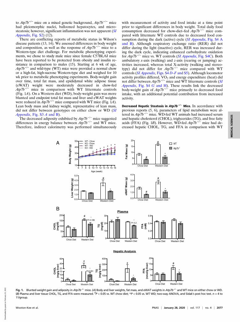
Hepatic Metabolite Composition in Atp7b−/− Mice. Analysis of theTCA cycle and glycolytic metabolic pathways revealed dramaticchanges in hepatic metabolites. Atp7b−/− mice had decreasedhepatic glucose, pyruvate, and lactate levels; however, severalglycolytic intermediates increased (G6P/F6P, 2PG/3PG, andPEP) (Fig. 4A). Similarly, there was an accumulation of TCAintermediates (oxaloacetate, citrate, fumarate, and malate),suggesting metabolic adaptation in the livers of Atp7b−/− mice.Cancer metabolism and integrated stress-response studies havedescribed a role for Pepck2 (mitochondrial Pepck) in the pro-motion of cataplerotic mechanisms that promote “pyruvate recy-cling” as a mechanism for cell adaptation in conditions of stress(17, 18). Despite decreased Pepck1, Pepck2 expression signifi-cantly increased in Atp7b−/− mice (Fig. 4B). While themechanism(s) for increased Pepck2 in Atp7b−/− mice are pres-ently unclear, it is possible that copper-induced cellular stress isdriving activation of endoplasmic reticulum-stress–mediatedpathways previously shown to increase Pepck2 (18).In contrast, acetyl-CoA and α-KG decreased in Atp7b−/− mice
(Fig. 4A), which was accompanied by an increase in pyruvatedehydrogenase kinase 4 (Pdk4) and a decrease in pyruvate dehy-drogenase a1 (Pdha1) expression (Fig. 4C). Pyruvate dehydroge-nase inactivation is reversed via dephosphorylation by the pyruvatedehydrogenase phosphatase enzyme (19). Together with increasedPdk4 expression, the decreased Pdpr expression in Atp7b−/− mice(Fig. 4C) may limit pyruvate flux into the TCA cycle. Furthermore,the changes in TCA cycle intermediates coincided with decreasedMdh1 and succinate dehydrogenase complex (Sdhb) and increasedIdh2 gene expression (Fig. 4D).
Hepatic Transcriptomic Analysis and Predicted Transcription FactorAlterations in Atp7b−/− Mice. Microarray transcriptomic analysiswas performed with 3-mo-old WT and Atp7b−/− mice (SI Appendix,Figs. S8 and S9), an age at which nuclear receptor dysfunction wasdetectable but livers lacked significant pathology (8, 12, 20). KyotoEncyclopedia of Genes and Genomes (KEGG) pathway analysisshowed that up-regulated pathways included proteasome, cellcycle, protein catabolism, oxidative phosphorylation, detoxification(glutathione metabolism), and TCA cycle. Pathways involved inimmunity, calcium signaling, and synthesis of unsaturated fattyacids were down-regulated (SI Appendix, Fig. S9A). These findingsare in keeping with transcriptomic studies in younger Atp7b−/−miceon a mixed background and in Wilson’s disease patients (5, 6).Furthermore, microarray data revealed significantly increased
hepatic expression of the anorectic hepatokine Gdf15 (SI Ap-pendix, Table S1). Real-time PCR confirmed a fourfold increasein hepatic Gdf15 expression for chow-diet–fed Atp7b−/− mice atboth 3 and 6 mo of age (SI Appendix, Fig. S10A). Serum GDF15was also increased fourfold for chow-fed Atp7b−/− mice at both 3and 6 mo of age. Although Western diet feeding significantly increasedserum GDF15 in WT mice, circulating GDF15 levels remainedsignificantly greater in WD-fed Atp7b−/− mice in comparison withWT controls (SI Appendix, Fig. S10B).Transcription-factor–binding sites enriched within the regula-
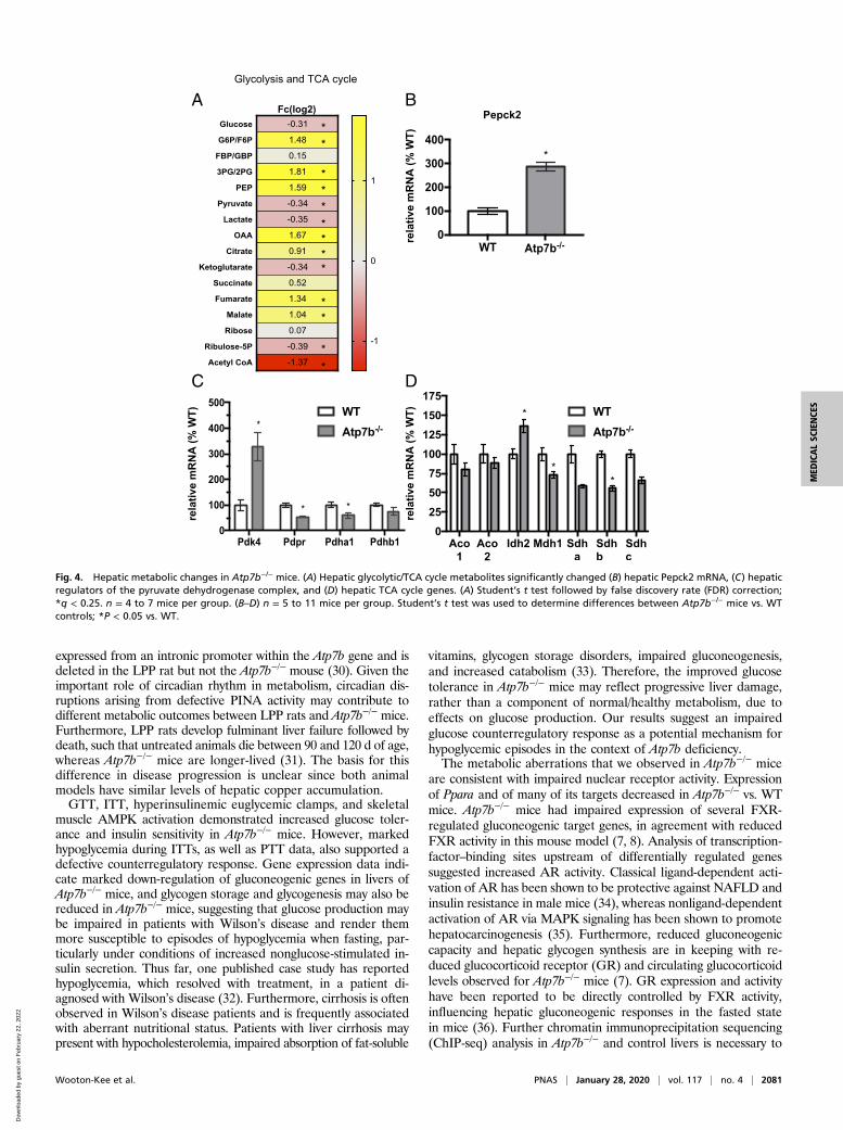
tory regions of significantly changed gene sets were identifiedusing EnrichR Analysis. As expected, nuclear-receptor–bindingelements were strongly associated with down-regulated genes. Inagreement with previous reports (7, 20), RXRα- and LXRα-binding sites were the most highly enriched in down-regulatedgenes, likely reflecting dysfunctional nuclear receptor activity(Fig. 5). LXR-binding elements were also enriched to a lesserextent in the up-regulated gene dataset. PPARα elements, alongwith PXR (a xenobiotic detoxification nuclear receptor), wererepresented in the down-regulated gene data set. Transcriptomicanalysis of decreased genes containing PXR-binding elementsincluded Cyp7b1 and Elov2, revealing a link between aberrantxenobiotic and lipid metabolism and lipogenesis (SI Appendix,Table S1).
2078 | www.pnas.org/cgi/doi/10.1073/pnas.1914267117 Wooton-Kee et al.
Dow
nloa
ded
by g
uest
on
Feb
ruar
y 22
, 202
2

A B C
D E F
G H I
J K L
Fig. 2. Glucose tolerance and insulin sensitivity in Atp7b−/− mice. (A and B) GTT. (C) Insulin released during intraperitoneal (IP)-GTT time course. (D and E) ITTwas performed on 6-mo-old Atp7b−/− and WT mice fed either (A and D) chow or (B and E) WD. (F) HOMA-IR. (G–K) Hyperinsulinemic euglycemic clamp wasperformed with 4- to 7-mo-old chow-fed Atp7b−/− and WT mice. (L) Western blot analysis of gastrocnemius muscle AMPK and p-AMPK levels in chow-fed WTand Atp7b−/− mice. (A–F) *P < 0.05, WT vs. Atp7b−/−, chow; #P < 0.05 WT vs. Atp7b−/−, WD; two-way ANOVA, and Sidak’s post hoc test. n = 4 to 11/group. (G–K) *P < 0.05, Student’s t test for Atp7b−/− mice vs. WT; n = 5 to 6 mice/group.
Wooton-Kee et al. PNAS | January 28, 2020 | vol. 117 | no. 4 | 2079
MED
ICALSC
IENCE
S
Dow
nloa
ded
by g
uest
on
Feb
ruar
y 22
, 202
2

The most abundant transcription-factor–binding elements withinthe up-regulated genes were binding sites for FoxM1, E2F4, andandrogen receptor (AR), which is consistent with cell-cycle pathwayenrichment in the KEGG pathway analysis of the transcriptomicdata (SI Appendix, Fig. S9B).
DiscussionCopper-mediated impairment of liver function is well character-ized in Wilson’s disease patients and in Atp7b−/− animal models.The most commonly described dysfunctions in Wilson’s diseasepatients are hepatic steatosis and cirrhosis. Surprisingly little isknown regarding glucose and lipid metabolism in these patients,and the limited available case reports disagree regarding glucosetolerance in Wilson’s disease patients (13, 14). Wilson’s disease is arare autosomal recessive disorder (1:30,000 occurrence), and thislow incidence together with environmental and genetic variabilitylikely contribute to the lack of a clear conclusion regarding glucosehomeostasis in Wilson’s disease patients (2). Atp7b−/− mice pro-vide a robust model for studying metabolism because environ-mental and genetic factors are more precisely controlled.We measured multiple metabolic parameters in Atp7b−/− mice
and WT littermate controls maintained on either normal chow orWestern-type diets. Chow-fed Atp7b−/− mice exhibited modestlyreduced adiposity and body weight by 14 wk of age. On WD,Atp7b−/− mice exhibited more pronounced reductions in bodyweight, adiposity, and hepatic steatosis in comparison with WT.The reduction in hepatic steatosis may be secondary to reducedobesity observed for WD-fed Atp7b−/− mice in comparison withWT littermate controls. Atp7b−/− mice ate less food and weremore active, which may implicate a central component in theobserved metabolic differences. Indeed, our microarray and real-time PCR data revealed a fourfold increase in hepatic Gdf15expression for chow-diet–fed Atp7b−/- mice in comparison withWT controls at 3 mo of age, prior to observed weight differences,and continuing through 6 mo of age. GDF15 has recentlyemerged as an inhibitor of food intake (21), suggesting that thisanorectic hepatokine may contribute to decreased food intakeobserved in Atp7b−/− mice. Serum GDF15 levels were similarlyelevated for chow-fed Atp7b−/− mice of both age groups. Western
diet feeding increased serum GDF15 levels in WT mice, witheven higher levels in Atp7b−/− mice (SI Appendix, Fig. S10).Our findings support previous reports of decreased hepatic
and serum lipids in mixed-background Atp7b−/− mice (5, 6). How-ever, significantly decreased hepatic and fasting plasma lipid levelsrelative to WT were evident only for Atp7b−/− mice when they werefed WD. This may reflect altered genetic susceptibility to lipidsynthesis, oxidation, and accumulation among mouse strains sinceAtp7b−/− mice on WD did have decreased serum and hepatic cho-lesterol and triglyceride levels in comparison with WT mice.In previous studies, white adipose tissue (WAT) in mixed-
background Atp7b−/− mice showed a correlation between copperlevels and lipolysis in explant WAT (22), and copper levels wereinversely correlated with steatosis in nonalcoholic fatty liverdisease (NAFLD) patients (23, 24) and rodent NAFLD models(25, 26). While we did not observe an effect on fasting-plasmafree fatty acids, it remains to be determined whether the decreasedadiposity observed in Atp7b−/− mice on the C57BL/6 background isin part due to a primary alteration in adipocyte function or to asecondary effect of copper accumulation in the liver or elsewhere.Interestingly, hepatocyte-specific knockout of Atp7b (Atp7bΔHep)resulted in increased adiposity and weight gain despite limitedhepatic inflammation, possibly due to alterations in cell signalingin nonparenchymal cells and to increased metallothionein ex-pression (copper chelation) in Atp7bΔHep mice (27). Furthermore,Atp7b−/−, but not Atp7bΔHep, mice have abnormal enterocytemorphology, accumulation of enteric lipid droplets, and disruptedchylomicron assembly (28), which may account for differencesin metabolic phenotype between Atp7b−/− and Atp7bΔHep mice.Recent findings in an Atp7b−/− rat model (cross breed between
the Long–Evans–Cinnamon rat and the Piebald Virol Glaxo rat,defective in pineal night-specific ATPase [PINA] and Atp7b [LPPrat]) demonstrated increased visceral fat, hepatic triglyceride con-tent, liver weight, and macrosteatosis in response to a high-caloriediet (45% kcal from fat and 722 kcal/L fructose-supplementeddrinking water), which correlated with increased hepatic mito-chondrial damage (29). In contrast with Atp7b−/− mice, LPP ratsdid not have aberrations in visceral fat or other parameters of lipidmetabolism on a normal diet (29). There are potential genetic andphysiologic reasons underlying these differences. The PINA gene is
0 15 30 45 60 75 90 1051200
50100150200250300
Time (min.)
* ** ***
0 15 30 45 60 75 90 1051200
50100150200250300
Time (min.)
** *
***
WTKO
FASN ACC1 DGAT2 Elovl3 SCD1 Chrebp SREBP-1c
0
100
200
300
* **
* *
** ***
#
*
*
Pepck1 Pcx Fbp1 G6pc0
50
100
150
** * * #
# #
WT: WDAtp7b-/- : WD
WT: ChowAtp7b-/- : Chow
A B C
D E
Fig. 3. Fasted-state gluconeogenesis and AMPK activation. (A and B) PTT on Atp7b−/− and WT mice. (B) Western blot analysis of hepatic AMPK and p-AMPKlevels in chow-fed 6-mo-old WT and Atp7b−/− mice. (C) Hepatic gluconeogenic and (D) lipogenic gene mRNA expression measured by real-time PCR analysis.(A and B) *P < 0.05, Atp7b−/− vs. WT diet-matched control, two-way ANOVA, and Sidak’s post hoc test. n = 4 to 11/group. (D and E) *P < 0.05 vs. WT chow; #P <0.05 vs. WT WD; Student’s t test. n = 5 mice/group.
2080 | www.pnas.org/cgi/doi/10.1073/pnas.1914267117 Wooton-Kee et al.
Dow
nloa
ded
by g
uest
on
Feb
ruar
y 22
, 202
2
expressed from an intronic promoter within the Atp7b gene and isdeleted in the LPP rat but not the Atp7b−/− mouse (30). Given theimportant role of circadian rhythm in metabolism, circadian dis-ruptions arising from defective PINA activity may contribute todifferent metabolic outcomes between LPP rats and Atp7b−/− mice.Furthermore, LPP rats develop fulminant liver failure followed bydeath, such that untreated animals die between 90 and 120 d of age,whereas Atp7b−/− mice are longer-lived (31). The basis for thisdifference in disease progression is unclear since both animalmodels have similar levels of hepatic copper accumulation.GTT, ITT, hyperinsulinemic euglycemic clamps, and skeletal
muscle AMPK activation demonstrated increased glucose toler-ance and insulin sensitivity in Atp7b−/− mice. However, markedhypoglycemia during ITTs, as well as PTT data, also supported adefective counterregulatory response. Gene expression data indi-cate marked down-regulation of gluconeogenic genes in livers ofAtp7b−/− mice, and glycogen storage and glycogenesis may also bereduced in Atp7b−/− mice, suggesting that glucose production maybe impaired in patients with Wilson’s disease and render themmore susceptible to episodes of hypoglycemia when fasting, par-ticularly under conditions of increased nonglucose-stimulated in-sulin secretion. Thus far, one published case study has reportedhypoglycemia, which resolved with treatment, in a patient di-agnosed with Wilson’s disease (32). Furthermore, cirrhosis is oftenobserved in Wilson’s disease patients and is frequently associatedwith aberrant nutritional status. Patients with liver cirrhosis maypresent with hypocholesterolemia, impaired absorption of fat-soluble
vitamins, glycogen storage disorders, impaired gluconeogenesis,and increased catabolism (33). Therefore, the improved glucosetolerance in Atp7b−/− mice may reflect progressive liver damage,rather than a component of normal/healthy metabolism, due toeffects on glucose production. Our results suggest an impairedglucose counterregulatory response as a potential mechanism forhypoglycemic episodes in the context of Atp7b deficiency.The metabolic aberrations that we observed in Atp7b−/− mice
are consistent with impaired nuclear receptor activity. Expressionof Ppara and of many of its targets decreased in Atp7b−/− vs. WTmice. Atp7b−/− mice had impaired expression of several FXR-regulated gluconeogenic target genes, in agreement with reducedFXR activity in this mouse model (7, 8). Analysis of transcription-factor–binding sites upstream of differentially regulated genessuggested increased AR activity. Classical ligand-dependent acti-vation of AR has been shown to be protective against NAFLD andinsulin resistance in male mice (34), whereas nonligand-dependentactivation of AR via MAPK signaling has been shown to promotehepatocarcinogenesis (35). Furthermore, reduced gluconeogeniccapacity and hepatic glycogen synthesis are in keeping with re-duced glucocorticoid receptor (GR) and circulating glucocorticoidlevels observed for Atp7b−/− mice (7). GR expression and activityhave been reported to be directly controlled by FXR activity,influencing hepatic gluconeogenic responses in the fasted statein mice (36). Further chromatin immunoprecipitation sequencing(ChIP-seq) analysis in Atp7b−/− and control livers is necessary to
A B
C D
Fig. 4. Hepatic metabolic changes in Atp7b−/− mice. (A) Hepatic glycolytic/TCA cycle metabolites significantly changed (B) hepatic Pepck2 mRNA, (C) hepaticregulators of the pyruvate dehydrogenase complex, and (D) hepatic TCA cycle genes. (A) Student’s t test followed by false discovery rate (FDR) correction;*q < 0.25. n = 4 to 7 mice per group. (B–D) n = 5 to 11 mice per group. Student’s t test was used to determine differences between Atp7b−/− mice vs. WTcontrols; *P < 0.05 vs. WT.
Wooton-Kee et al. PNAS | January 28, 2020 | vol. 117 | no. 4 | 2081
MED
ICALSC
IENCE
S
Dow
nloa
ded
by g
uest
on
Feb
ruar
y 22
, 202
2
determine the broad patterns of nuclear-receptor binding in re-sponse to increased copper load.Increased hepatic AMPK activation may be responsible for
reduced gluconeogenic and lipogenic gene expression. AMPKserves as a sensor of energy status, as well as cellular stress (suchas ROS production) (37–40). Copper-induced production ofROS production is well characterized in Wilson’s disease patientsand animal models (2) and may provide a stimulus for AMPKactivation in Atp7b−/- mice. Alternatively, AMPK is activated inresponse to UCP2-mediated depletion of cellular ATP (41, 42).Ucp2 has been identified as a transcriptional target of bothFOXM1 (43) and E2F4 (44, 45), the transcription factors with thetwo most highly up-regulated binding sites identified in our study.FOXM1 expression has been reported to be up-regulated byGDF15, activated by oxidative stress, and to negatively regulateexpression of the copper transporter Ctr1, suggesting that FoxM1may play a larger role in mediating the cellular response to excesscopper (46, 47). Future studies will investigate the underlyingmechanisms for AMPK activation and the impact on metabolismin Atp7b−/− mice in relation to ROS generation.Decreased gluconeogenesis, hepatic glucose, pyruvate, and lactate
concentrations were accompanied by a significant increase in gly-colytic intermediates, as well as unexpected increases in many TCAcycle intermediates despite decreased Ac-CoA. In support of de-creased TCA cycle metabolic flux, we found increased Pdk4 mRNAexpression. Future studies measuring TCA cycle flux are necessaryto determine the mechanism for increased TCA cycle intermediates.Here we describe metabolic changes in Atp7b−/− mice that are
likely a consequence of impaired hepatic nuclear receptor func-tion. We observed dramatic alterations in glucose homeostasis,metabolome, and AMPK activation in Atp7b−/− mice. Futurestudies to elucidate the mechanisms for decreased adiposity andaltered whole-body glucose homeostasis have the potential touncover therapeutic options for patients with Wilson’s disease,as well as for patients with metabolic disorders such as type 2diabetes and fatty liver disease.
MethodsAnimal Care. Atp7b−/− (C57BL×129S6/SvEv) mice (48) were backcrossed for 10generations with C57BL/6j mice to generate Atp7b+/− mice on a C57BL/6Jbackground strain. Male Atp7b−/− and WT cagemate/littermate controls forthe study were obtained from heterozygous Atp7b+/− mating cages. Micewere fed either a standard chow diet (Harlan Laboratories 2920X) or a 40%kcal from fat, 35% sucrose Western-type diet (Research Diets). Coppercontent of the chow and Western-type diet was 17.23 and 10 mg/kg, re-spectively. Diets and water were provided ad libitum, and mice weremaintained on a 12-h light–dark cycle and euthanized at Zeitgeber Time 2–3.All experiments were performed under the guidelines detailed in the Guidefor the Care and Use of Laboratory Animals (49), and animal experimentswere approved by the Institutional Animal Care and Use Committee atBaylor College of Medicine (BCM).
Inductively Coupled Plasma–Optical Emission Spectrometry. Liver sampleswere processed as previously described (8).
Body Composition. Total fat and lean mass were measured on ad-libitum-fedmice at the end of the diet study using an EchoMRI Whole Body CompositionAnalyzer (Echo Medical Systems) located in the Mouse Metabolic ResearchUnit (MMRU) at the US Department of Agriculture/Agricultural ResearchService (USDA/ARS) Children’s Nutrition Research Center (CNRC) at BCM.
Plasma and Hepatic Lipids. Mice were fasted for 6 h prior to blood collectionbetween 1:00 and 2:00 PM. Livers were dissected frommice in the ad-libitum-fed state. Plasma and liver triglycerides (Pointe Scientific), cholesterol (PointeScientific), and free fatty acids (BioVision) were measured following manu-facturer protocols. Serum chemistry profiles (alanine transaminase, alkalinephosphatase, bilirubin, albumin, globulin) were performed in the Center forComparative Medicine at BCM.
Histology. Liver samples were fixed in 10% formalin (for hematoxylin andeosin, PAS, and PAS-diastase staining) or frozen in Optimal Cutting Tem-perature compound for Oil Red O staining, and staining was performed atthe Digestive Disease Core at BCM.
Indirect Calorimetry, Food Intake, and Activity. Indirect calorimetry, food in-take, and activity measurements were performed in the MMRU at the USDA/ARS CNRC at BCM as previously described (50). To minimize the confoundingeffect of different body weights on energy expenditure (51), experiments wereperformed on chow-diet–fed mice, when mice were 12 wk old, prior to sig-nificant differences in body weight between genotypes. Age- and weight-matched littermates were singly housed and acclimated for 6 d in metabolicchambers. Subsequently, indirect calorimetry, food intake, and activity weremeasured over a 24-h period using an Oxymax/Comprehensive Lab AnimalMonitoring System (Oxymax/CLAMS, Columbus Instruments). Data were ac-quired using the CLAMS data eXamination Tool (CLAX, Columbus Instruments).RER and energy expenditure were calculated as previously described (50).
Whole-Body Glucose Homeostasis. Prior to intraperitoneal injection, micewere fasted for 6 h for GTT and ITT, or overnight for PTT. For GTT, 1.5 g/kgdextrose (Sigma); for ITT, 0.75 U/kg human insulin (Eli Lilly and Company); orfor PTT, 1.0 g/kg sodium pyruvate (Sigma) was injected. Immediately priorand subsequent to injections, blood glucose was measured via glucometer(OneTouch) at time points shown. For GTT, blood was collected, and plasmainsulin was measured using a Rat/Mouse Insulin ELISA kit (EMD Millipore).Hyperinsulinemic euglycemic clamp studies were performed at the MouseMetabolomic and Phenotyping Core (MMPC) at BCM.
Protein Analysis.Western blot analysis on liver and gastrocnemius dissected inthe ad-libitum-fed state was performed with 1:1,000 α-AMPK (23A3, CellSignaling), phospho(T172)-α-AMPK (40H9, Cell Signaling), α-tubulin (ab-4074,Abcam), 36B4 (RPLP0) (Cell Signaling), and 1:50,000 secondary antibody.
Real Time-PCR. RNA was harvested from 100 mg of liver samples using TRIzolReagent (Invitrogen), and synthesized cDNA (Invitrogen) was used as tem-plate for real-time PCR with SYBR Green reagent (Roche) on a 384-wellLightCycler (Roche). Primers were designed with the Roche UniversalPrimer website. Reactions were normalized to 36B4 mRNA expression.
MetaboliteMeasurements. Six-month-old glycolytic and TCA cycle metaboliteswere measured in livers taken from ad-libitum-fed mice in the BCMMetabolomics Core. The extended protocol is provided in SI Appendix.
0 5 10 15 20 25 30 35 40 45
GATA1
PXR
PPAR
CEBP
STAT5
LXR
RXR
Combined Score
Enric
hR A
naly
sis
* 13.4
* 13.4
* 3.7
* 2.3
* 3.8
* 1.9
* 1.4
* -log10(p-value)
Fig. 5. Transcriptomic analysis in Atp7b−/− mice livers. Pathway analysis oftranscription factor binding was performed with the significantly changedmicroarray analysis genes (Fc > 1.5, FDR < 0.05) using the web-basedEnrichR platform (27141961). Binding sites identified from ChIP-Seq datawithin the following down-regulated genes: RXR(NR2B1)_22158963_ChIP-Seq_LIVER_Mouse; LXR(NR1H3)__22158963_ChIP-Seq_LIVER_Mouse; STAT5_23275557_ChIP-Seq_MAMMARY-EPITHELIUM_Mouse; CEBPa_23403033_ChIPSeq_LIVER_Mouse; PPARa(NR1C1)_22158963_ChIP-Seq_LIVER_Mouse;PXR(NR1I2)_20693526_ChIP-Seq_LIVER_Mouse; GATA1_22025678_ChIP-Seq_K562_Human. *-log10(P value).
2082 | www.pnas.org/cgi/doi/10.1073/pnas.1914267117 Wooton-Kee et al.
Dow
nloa
ded
by g
uest
on
Feb
ruar
y 22
, 202
2
Microarray Analysis. RNA was harvested from male and female 3-mo-oldAtp7b−/− mice and WT controls with TRIzol Reagent (Invitrogen), followedwith RNeasy kit (Qiagen). Microarray processing using a GeneChip Mouse430 2.0 (Affymetrix) was performed in the Genomic and RNA Profiling Coreat BCM. Data are available at the Gene Expression Omnibus database un-der accession no. GSE125637 (52). Datasets were analyzed as previouslydescribed (31).
Serum Growth/Differentiation Factor 15 Measurement. Serum from ad-libitum-fed mice was collected and analyzed using the Mouse/Rat GDF-15 QuantikineELISA Kit (R&D Systems) per manufacturer protocol.
Statistical Analysis. Statistical analysis was performed with GraphPad Prismsoftware (v7.0). Data are presented as mean ± the SEM, and P < 0.05 wasconsidered statistically significant. Student’s t test was used to comparedifferences between two groups. For multiple-group comparison, two-way ANOVA tests followed by Sidak’s post hoc test were performed.
ACKNOWLEDGMENTS. We thank Drs. Curtis Hughey and Svetlana Lutsenkofor helpful discussion and edits; Mr. Jordan Mar for technical assistance withthe mouse diet study; Mr. Firoz Vohra and Dr. Marta Fiorotto, both of theCNRC MMRU Core; Dr. Michael Grusak for mineral composition studiesperformed at the CNRC; and Dr. Milton Finegold and Dr. Michael Ittmannfor assistance with analysis of liver histology. This work was supported byNational Institute of Diabetes and Digestive and Kidney Disease (NIDDK)Grants K01DK111716-03 and P30 DK056338 (to C.R.W.-K.); USDA-ARS Grant3092-51000-060-03S and American Heart Association Grant 18CDA34110137 (toA.M.N.-A.); R. P. Doherty, Jr., Welch Chair in Science Grant Q-0022 and CancerPrevention & Research Institute of Texas (CPRIT) Grant RP150587 (to D.D.M.);CPRIT Proteomics and Metabolomics Core Facility RP170005 (to N.P. and C.C.);Mouse Metabolic and Phenotyping Core (MMPC) at Baylor College of Medicineand NIH Grants R01DK114356 and UM1HG006348 (to P.S.). We acknowledgethe joint participation of the Diana Helis Henry Medical Research Foundationthrough its direct engagement in the continuous active conduct of medicalresearch in conjunction with BCM. The MMRU at the USDA/ARS CNRC, BaylorCollege of Medicine, is supported by funds from the USDA/ARS (https://www.bcm.edu/research/advanced-technology-core-labs/lab-listing/mouse-metabolic-research-unit).
1. S. Lutsenko, N. L. Barnes, M. Y. Bartee, O. Y. Dmitriev, Function and regulation ofhuman copper-transporting ATPases. Physiol. Rev. 87, 1011–1046 (2007).
2. S. Lutsenko, E. S. LeShane, U. Shinde, Biochemical basis of regulation of humancopper-transporting ATPases. Arch. Biochem. Biophys. 463, 134–148 (2007).
3. G. V. Sarode et al., Metabolomics profiles of patients with Wilson disease reveal adistinct metabolic signature. Metabolomics 15, 43 (2019).
4. J. Xu et al., 1H NMR-based metabolomics investigation of copper-laden rat: A modelof Wilson’s disease. PLoS One 10, e0119654 (2015).
5. J. P. Hamilton et al., Activation of liver X receptor/retinoid X receptor pathway amelio-rates liver disease in Atp7B(-/-) (Wilson disease) mice. Hepatology 63, 1828–1841 (2016).
6. D. Huster et al., High copper selectively alters lipid metabolism and cell cycle ma-chinery in the mouse model of Wilson disease. J. Biol. Chem. 282, 8343–8355 (2007).
7. P. A. Wilmarth et al., A systems approach implicates nuclear receptor targeting in theAtp7b(-/-) mouse model of Wilson’s disease. Metallomics 4, 660–668 (2012).
8. C. R. Wooton-Kee et al., Elevated copper impairs hepatic nuclear receptor function inWilson’s disease. J. Clin. Invest. 125, 3449–3460 (2015).
9. H. Nagasaka et al., Relationship between oxidative stress and antioxidant systems inthe liver of patients with Wilson disease: Hepatic manifestation in Wilson disease as aconsequence of augmented oxidative stress. Pediatr. Res. 60, 472–477 (2006).
10. H. Nagasaka et al., Fatty liver and anti-oxidant enzyme activities along with peroxi-some proliferator-activated receptors γ and α expressions in the liver of Wilson’sdisease. Mol. Genet. Metab. 107, 542–547 (2012).
11. C. E. Mordaunt et al., Epigenomic signatures in liver and blood of Wilson disease patientsinclude hypermethylation of liver-specific enhancers. Epigenetics Chromatin 12, 10 (2019).
12. D. Huster et al., Consequences of copper accumulation in the livers of the Atp7b-/-(Wilson disease gene) knockout mice. Am. J. Pathol. 168, 423–434 (2006).
13. K. Johansen, G. Gregersen, Glucose intolerance in Wilson’s disease. Normalizationafter treatment with penicillamine. Arch. Intern. Med. 129, 587–590 (1972).
14. R. Krysiak, G. Handzlik-Orlik, B. Okopien, Endocrine symptoms as the initial mani-festation of Wilson’s disease. Yale J. Biol. Med. 85, 249–254 (2012).
15. K. Bwanahali, M. Mbuyi, B. Kapita, Osteoarthrosis, gout and arthritis rheumatoid in in-ternal medicine in Kinshasa [in French]. Rev. Rhum. Mal. Osteoartic. 58, 105–111 (1991).
16. J. E. Ayala et al.; NIH Mouse Metabolic Phenotyping Center Consortium, Standardoperating procedures for describing and performing metabolic tests of glucose ho-meostasis in mice. Dis. Model. Mech. 3, 525–534 (2010).
17. A. Méndez-Lucas et al., PEPCK-M expression in mouse liver potentiates, not replaces,PEPCK-C mediated gluconeogenesis. J. Hepatol. 59, 105–113 (2013).
18. A. Méndez-Lucas, P. Hyroššová, L. Novellasdemunt, F. Viñals, J. C. Perales, Mito-chondrial phosphoenolpyruvate carboxykinase (PEPCK-M) is a pro-survival, endo-plasmic reticulum (ER) stress response gene involved in tumor cell adaptation tonutrient availability. J. Biol. Chem. 289, 22090–22102 (2014).
19. T. E. Roche, Y. Hiromasa, Pyruvate dehydrogenase kinase regulatory mechanisms and in-hibition in treating diabetes, heart ischemia, and cancer. Cell. Mol. Life Sci. 64, 830–849 (2007).
20. J. P. Hamilton et al., Activation of LXR/RXR pathway ameliorates liver disease in atp7b(Wilson disease) mice. Hepatology (2015).
21. V. W. W. Tsai, Y. Husaini, A. Sainsbury, D. A. Brown, S. N. Breit, The MIC-1/GDF15-GFRAL pathway in energy homeostasis: Implications for obesity, cachexia, and otherassociated diseases. Cell Metab. 28, 353–368 (2018).
22. L. Krishnamoorthy et al., Copper regulates cyclic-AMP-dependent lipolysis. Nat. Chem.Biol. 12, 586–592 (2016).
23. E. Aigner et al., A role for low hepatic copper concentrations in nonalcoholic fattyliver disease. Am. J. Gastroenterol. 105, 1978–1985 (2010).
24. V. Nobili et al., Levels of serum ceruloplasmin associate with pediatric nonalcoholicfatty liver disease. J. Pediatr. Gastroenterol. Nutr. 56, 370–375 (2013).
25. M. Song et al., High fructose feeding induces copper deficiency in Sprague-Dawleyrats: A novel mechanism for obesity related fatty liver. J. Hepatol. 56, 433–440 (2012).
26. M. Song et al., Modest fructose beverage intake causes liver injury and fat accumu-lation in marginal copper deficient rats. Obesity (Silver Spring) 21, 1669–1675 (2013).
27. A. Muchenditsi et al., Targeted inactivation of copper transporter Atp7b in hepato-cytes causes liver steatosis and obesity in mice. Am. J. Physiol. Gastrointest. LiverPhysiol. 313, G39–G49 (2017).
28. H. Pierson et al., The function of ATPase copper transporter ATP7B in intestine.Gastroenterology 154, 168–180.e5 (2018).
29. C. Einer et al., A high-calorie diet aggravates mitochondrial dysfunction and triggers severeliver damage in Wilson disease rats. Cell. Mol. Gastroenterol. Hepatol. 7, 571–596 (2019).
30. S. Ahmed, J. Deng, J. Borjigin, A new strain of rat for functional analysis of PINA. BrainRes. Mol. Brain Res. 137, 63–69 (2005).
31. H. Zischka et al., Liver mitochondrial membrane crosslinking and destruction in a ratmodel of Wilson disease. J. Clin. Invest. 121, 1508–1518 (2011).
32. R. Krysiak, B. Okopie�n, Whipple’s triad as a clinical manifestation of hepatolenticulardegeneration. Pol. Arch. Med. Wewn. 117, 53–55 (2007).
33. Y. Arakawa, M. Moriyama, Y. Arakawa, Liver cirrhosis and metabolism (sugar, pro-tein, fat and trace elements). Hepatol. Res. 30S, 46–58 (2004).
34. H. Y. Lin et al., Increased hepatic steatosis and insulin resistance in mice lacking he-patic androgen receptor. Hepatology 47, 1924–1935 (2008).
35. T. Kanda, O. Yokosuka, The androgen receptor as an emerging target in hepatocel-lular carcinoma. J. Hepatocell. Carcinoma 2, 91–99 (2015).
36. B. Renga et al., Glucocorticoid receptor mediates the gluconeogenic activity of thefarnesoid X receptor in the fasting condition. FASEB J. 26, 3021–3031 (2012).
37. D. G. Hardie, S. A. Hawley, J. W. Scott, AMP-activated protein kinase: Development ofthe energy sensor concept. J. Physiol. 574, 7–15 (2006).
38. S. L. Choi et al., The regulation of AMP-activated protein kinase by H(2)O(2). Biochem.Biophys. Res. Commun. 287, 92–97 (2001).
39. M. Lee et al., Critical roles of AMP-activated protein kinase in the carcinogenic metal-induced expression of VEGF and HIF-1 proteins in DU145 prostate carcinoma. Biochem.Pharmacol. 72, 91–103 (2006).
40. Y. Wu, M. Viana, S. Thirumangalathu, M. R. Loeken, AMP-activated protein kinasemediates effects of oxidative stress on embryo gene expression in a mouse model ofdiabetic embryopathy. Diabetologia 55, 245–254 (2012).
41. Y. Shang et al., Targeted expression of uncoupling protein 2 to mouse liver increasesthe susceptibility to lipopolysaccharide/galactosamine-induced acute liver injury.Hepatology 50, 1204–1216 (2009).
42. K. D. Chavin et al., Obesity induces expression of uncoupling protein-2 in hepatocytesand promotes liver ATP depletion. J. Biol. Chem. 274, 5692–5700 (1999).
43. A. Lachmann et al., ChEA: Transcription factor regulation inferred from integratinggenome-wide ChIP-X experiments. Bioinformatics 26, 2438–2444 (2010).
44. ENCODE Project Consortium, The ENCODE (ENCyclopedia of DNA elements) project.Science 306, 636–640 (2004).
45. ENCODE Project Consortium, A user’s guide to the encyclopedia of DNA elements(ENCODE). PLoS Biol. 9, e1001046 (2011).
46. B. F. Peake, S. M. Eze, L. Yang, R. C. Castellino, R. Nahta, Growth differentiation factor15 mediates epithelial mesenchymal transition and invasion of breast cancers throughIGF-1R-FoxM1 signaling. Oncotarget 8, 94393–94406 (2017).
47. H. Ye et al., Hepatocyte nuclear factor 3/fork head homolog 11 is expressed in pro-liferating epithelial and mesenchymal cells of embryonic and adult tissues. Mol. Cell.Biol. 17, 1626–1641 (1997).
48. O. I. Buiakova et al., Null mutation of the murine ATP7B (Wilson disease) gene resultsin intracellular copper accumulation and late-onset hepatic nodular transformation.Hum. Mol. Genet. 8, 1665–1671 (1999).
49. National Research Council, Guide for the Care and Use of Laboratory Animals (Na-tional Academies Press, Washington, DC, ed. 8, 2011).
50. A. M. Nuotio-Antar, D. L. Hachey, A. H. Hasty, Carbenoxolone treatment attenuatessymptoms of metabolic syndrome and atherogenesis in obese, hyperlipidemic mice.Am. J. Physiol. Endocrinol. Metab. 293, E1517–E1528 (2007).
51. A. A. Butler, L. P. Kozak, A recurring problem with the analysis of energy expenditurein genetic models expressing lean and obese phenotypes. Diabetes 59, 323–329 (2010).
52. C. R. Wooton-Kee, C. Coarfa, M. Robertson, Gene expression in wild type and Atp7b−/−
mice. Gene Expression Omnibus. https://www.ncbi.nlm.nih.gov/geo/query/acc.cgi?&acc=GSE125637. Deposited 25 January 2019.
Wooton-Kee et al. PNAS | January 28, 2020 | vol. 117 | no. 4 | 2083
MED
ICALSC
IENCE
S
Dow
nloa
ded
by g
uest
on
Feb
ruar
y 22
, 202
2
https://www.bcm.edu/research/advanced-technology-core-labs/lab-listing/mouse-metabolic-research-unit
https://www.bcm.edu/research/advanced-technology-core-labs/lab-listing/mouse-metabolic-research-unit