
Combined Transbronchial Needle Aspiration And PET/CT For Mediastinal Staging Of Lung Cancer
Upload
scu-hospitalCategory
view
81download
2
MS
Dsetctbg
taltlstfi1aasTrotapa
atlclwwyTd
D
A
1
ediastinal Staging Prior to Surgical Resectionteven J. Mentzer, MD
cj(
secpatbdpTdaAir
fcplmaf
fs(scn
SMtadtpc
ocumenting the anatomic extent of lung cancer be-fore surgical resection has value in therapeutic deci-
ion-making. In the past, documenting mediastinal dis-ase spared the patient the morbidity of an exploratoryhoracotomy.1 Currently, the presence of stage IIIA lungancer is an indication for neoadjuvant chemoradiationherapy.2 In the future, it is likely that tissue biopsies wille used for biomarkers staging,3 tissue typing for onco-enic mutations,4 and/or drug sensitivity testing.5
The pathophysiologic assumption in cancer staging ishat the progressive growth of the primary tumor eventu-lly leads to the embolic spread of metastatic cells to theymphatic system and subsequently to the blood circula-ion. This predictable sequence of tumor progression hased to staging systems designed to provide consistent de-criptions of the anatomic extent of cancers at specificimes in their clinical progression. Denoix and colleaguesrst advocated the “TNM” classification in the 1940s.6 In986, the American Joint Committee on Cancer (AJCC)nd the International Union Against Cancer (UICC)dopted a common TNM staging system.7 In 1997, thistaging system was reconciled with the 1983 Americanhoracic Society statement on clinical staging.8 The cur-ent staging system is based on the relative size and extentf the primary tumor (T), the absence, presence, and ex-ent of regional lymph node involvement (N), and thebsence or presence of distance metastases (M). Theresent TNM staging system has been universally adopteds the basis for therapeutic comparisons worldwide.
In potentially operable lung cancer, a description of thenatomic extent of the cancer depends on the size of theumor (T) and the involvement of regional or mediastinalymph nodes (N). Clinical T stage is typically assessed byhest CT scans. T2 tumors are greater than 3 cm, involve aarge bronchus (�2 cm from the carina), or are associatedith atelectasis.9 The survival of clinically staged patientsith T2N0 disease is only 75% at 1 year and 40% at 5ears.8 The survival of pathologically staged patients with2N0 disease is 90% at 1 year and 60% at 5 years.8 Theisappointing survival of these early-stage (stage IB) lung
ivision of Thoracic Surgery, Brigham and Womens Hospital, Harvard Med-ical School, Boston, MA.
ddress reprint requests to Steven J. Mentzer, MD, Brigham and WomensHospital, Division of Thoracic Surgery, Room 259, 75 Francis Street,
mBoston, MA 02115-6110. E-mail: [email protected]
52 1522-2942/05/$-see front matter © 2005 Elsevier Inc. All rights reserved.doi:10.1053/j.optechstcvs.2005.06.001
ancer patients suggests two conclusions: (1) we do a poorob of characterizing the extent of malignant disease, and2) we do a particularly poor job of clinical staging.
Recent attempts to improve the sensitivity of all cancertaging have focused on lymphatic mapping. Clinical andxperimental evidence of sentinel node mapping in breastancer and melanoma suggest that (1) lymph drainageatterns are predictable; (2) lymph node drainage followssequential pattern; and (3) lymph nodes filter and entrap
umor cells. In lung cancer, the lymphatic map developedy Naruke and coworkers and adopted by the AJCC hasemonstrated an even more predictable pattern of lym-hatic drainage than is seen in trunkal malignancies.10,11
he success of the lymph node map likely reflects theefined hilar and mediastinal lymphatic anatomy as wells the unidirectional lymph flow along the thoracic duct.ttempts to replace tissue biopsies with noninvasive stag-
ng have been plagued with a consistent 10% error rateegardless of the technique employed.
The mapping of mediastinal lymph nodes can be per-ormed by using a mediastinoscopic or thoracoscopic pro-edure. Cervical mediastinoscopy provides access to thearatracheal (levels 2R, 2L, 4R, and 4L) and subcarinal
ymph nodes (level 7). The lymph nodes in the aortopul-onary window (levels 5 and 6) are accessible through an
nterior mediastinotomy—a procedure commonly re-erred to as an anterior or parasternal mediastinoscopy.
Biopsies of mediastinal lymph nodes can also be per-ormed by thoracoscopy. In the right hemithorax, thoraco-copic staging provides access to the right paratracheal nodeslevel 4R), inferior pulmonary ligament nodes (level 9), andubcarinal nodes (level 7). In the left hemithorax, thoracos-opy provides access to the aortopulmonary (AP) windowodes (level 5 and 6) and posterior hilar nodes.
urgical Procedureediastinoscopy is performed by using a general anes-
hetic. At our institution, mediastinoscopy is performed asn ambulatory procedure in patients with suspected me-iastinal disease. Patients suspected of having no medias-inal malignancy are typically staged by a combination ofreoperative PET/CT scanning and intraoperative thora-oscopic staging to confirm the absence of mediastinal
alignancy.
Mediastinal staging prior to surgical resection 153
Figure 1 The patient is positioned for mediastinoscopy in the supine position so that the head extends just beyond theend of the operating table. A roll is placed under the shoulders to facilitate extension of the neck. The head is in themidline position and is stabilized by using a circular cushion. Intravenous tubing and monitoring lines are placed to oneside to facilitate surgical access to the patient’s head and neck. The suprasternal notch is palpated to identify apotentially ectatic innominate artery. The supraclavicular regions are palpated because previously unrecognized lymphnodes may be identified in this position.
The patient is prepped from the chin to the upper abdomen so that any inadvertent arterial injury can be addressedthrough a median sternotomy. The surgical drapes are only suspended by the intravenous tubing and monitoring lines
on one side of the patient’s head.
154 S.J. Mentzer
Figure 2 The cervical mediastinoscopy incision is made in the suprasternal notch 1 fingerbreadth above the manu-brium. The horizontal incision is approximately 1 fingerbreadth in length and is placed in Langer’s lines to minimizescarring. The horizontal dissection is continued through the platysma. The dissection is then continued verticallybetween the strap muscles. Because of the significant mobility of the strap muscles and veins in the neck, there is littleneed to ligate or cauterize vessels in this dissection. If vessels are cauterized during this dissection, adequate hemostasiscan be assessed by an end inspiratory hold maneuver that approximates the venous pressure during postoperativecoughing or valsalva maneuvers.
The most important step in the cervical dissection is the definition of the pretracheal fascia. The pretracheal space,an avascular potential space between the pretracheal fascia and the anterior trachea, is the conduit for the safe passageof the mediastinoscope. A relatively small hole in the pretracheal fascia can be expanded sufficiently to admit a fingerby mobilizing the pretracheal fascia laterally as well as inferiorly within the neck. Attempts to blindly mobilize thepretracheal fascia in the mediastinum—particularly the blind spreading of the Metzenbaum scissors within the medi-astinum—risks injury to an ectatic or atherosclerotic innominate artery. Repair of these posterior wall injuries of theinnominate artery typically require a median sternotomy. Similarly, an excessively large hole in the pretracheal fascia
may complicate the insertion of the mediastinoscope and may even lose access to the pretracheal space.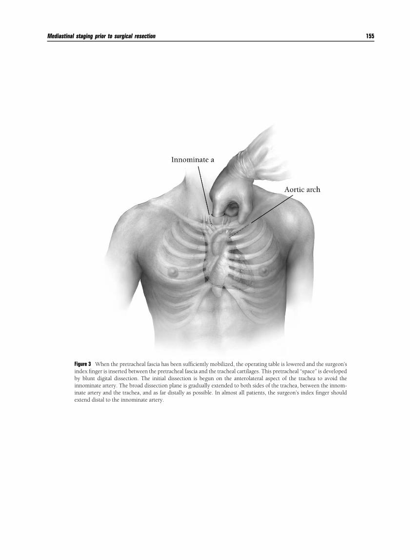
Mediastinal staging prior to surgical resection 155
Figure 3 When the pretracheal fascia has been sufficiently mobilized, the operating table is lowered and the surgeon’sindex finger is inserted between the pretracheal fascia and the tracheal cartilages. This pretracheal “space” is developedby blunt digital dissection. The initial dissection is begun on the anterolateral aspect of the trachea to avoid theinnominate artery. The broad dissection plane is gradually extended to both sides of the trachea, between the innom-inate artery and the trachea, and as far distally as possible. In almost all patients, the surgeon’s index finger should
extend distal to the innominate artery.
156 S.J. Mentzer
Figure 4 The lower right paratracheal lymph nodes (level 4R) are located at the distal extent of the surgeon’s finger andexternal to the pretracheal fascia. The surgeon’s finger is used to penetrate the pretracheal fascia and facilitate exposureof the lower paratracheal lymph nodes during subsequent mediastinoscopic examination. Digital palpation of the lowerright paratracheal lymph nodes is a critical step in the procedure. Pathologic nodes are often better appreciated by
palpation than by visual inspection.
Mediastinal staging prior to surgical resection 157
Figure 5 The surgeon’s finger is withdrawn and the mediastinoscope is introduced into the pretracheal space. Somesurgeons raise the operating table and stand when performing the mediastinoscopy, while other surgeons sit on a stoolfor the procedure. An advantage of sitting, in addition to comfort, is that the surgeon can readily reassess themediastinum by palpation without changing the operating table position. The beveled end of the mediastinoscope isused to retract the soft tissue of the neck and pretracheal fascia. Rotation of the mediastinoscope during insertion oftenfacilitates visualization of the pretracheal fascia and the anterior wall of the trachea. The mediastinoscope is inserted
along the axis of the trachea: approximately a 30 degree angle relative to the sternum.Figure 6 The initial view of the mediastinum is the innominate artery apposed to the anterior wall of the trachea. Theimportance of an intact pretracheal fascia is apparent in this step: the pretracheal fascia provides a potential space forpassing the mediastinoscope distal to the artery. Although rarely involved with metastatic cancer, the upper paratra-
cheal lymph nodes (2R and 2L) are accessible at this level of the dissection.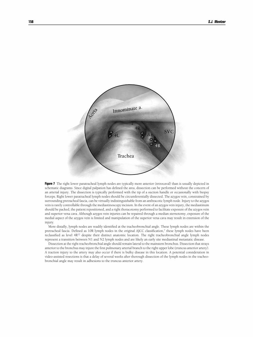
158 S.J. Mentzer
Figure 7 The right lower paratracheal lymph nodes are typically more anterior (retrocaval) than is usually depicted inschematic diagrams. Since digital palpation has defined the area, dissection can be performed without the concern ofan arterial injury. The dissection is typically performed with the tip of a suction handle or occasionally with biopsyforceps. Right lower paratracheal lymph nodes should be circumferentially dissected. The azygos vein, constrained bysurrounding pretracheal fascia, can be virtually indistinguishable from an anthracotic lymph node. Injury to the azygosvein is rarely controllable through the mediastinoscopy incision. In the event of an azygos vein injury, the mediastinumshould be packed, the patient repositioned, and a right thoracotomy performed to facilitate exposure of the azygos veinand superior vena cava. Although azygos vein injuries can be repaired through a median sternotomy, exposure of themedial aspect of the azygos vein is limited and manipulation of the superior vena cava may result in extension of theinjury.
More distally, lymph nodes are readily identified at the tracheobronchial angle. These lymph nodes are within thepretracheal fascia. Defined as 10R lymph nodes in the original AJCC classification,7 these lymph nodes have beenreclassified as level 4R12 despite their distinct anatomic location. The right tracheobronchial angle lymph nodesrepresent a transition between N1 and N2 lymph nodes and are likely an early site mediastinal metastatic disease.
Dissection at the right tracheobronchial angle should remain lateral to the mainstem bronchus. Dissection that straysanterior to the bronchus may injure the first pulmonary arterial branch to the right upper lobe (truncus anterior artery).A traction injury to the artery may also occur if there is bulky disease in this location. A potential consideration invideo-assisted resections is that a delay of several weeks after thorough dissection of the lymph nodes in the tracheo-
bronchial angle may result in adhesions to the truncus anterior artery.
Mediastinal staging prior to surgical resection 159
Figure 8 An alternative approach to sampling the lower right paratracheal lymph nodes is video thoracoscopy. Thora-coscopic staging requires single lung ventilation. The procedure can be performed through two access ports; typicallythese can be placed in the 3rd and 7th interspace in the anterior axillary line. The thoracoscope is inserted through the7th interspace access port and the lung is reflected caudad exposing the supra-azygos mediastinal pleura. The superiorvena cava is reflected anteriorly exposing the retrocaval lymph nodes. A rounded instrument, such as a ring forceps, canbe useful to extract the nodal tissue in the retrocaval location. On occasion, a lower lobe tumor will have an isolated
inferior pulmonary ligament lymph node. These level 9 lymph nodes are uniquely accessible by thoracoscopy.
160 S.J. Mentzer
Figure 9 An early site of embolic spread from middle and lower lobe malignancies is the subcarinal space (level 7).13 Thesubcarinal space is difficult to evaluate by conventional CT scanning and its retrocardiac location compromisesevaluation by PET scanning.
Dissection that is carried down to the level of the subcarinal space frequently reveals anterior tracheal lymph nodes.A note of caution is that these lymph nodes may be associated with an anterior bronchial artery in 5% of the patients.14
The true subcarinal space, however, lies below the subcarinal fascia. Dissection of this fascial plane is required to exposepathologic lymph nodes. Particularly with substantial adenopathy, lymph nodes may be extruded from the subcarinalspace facilitating biopsies, limiting the necessity of extensive dissection.
In contrast to other lymph node levels, circumferential dissection of the subcarinal (level 7) lymph nodes is notadvisable in the subcarinal space because of the potential for arterial bleeding. Excessive dissection in the subcarinalspace may result in bleeding that appears to originate deep within the subcarinal space. Direct pressure may be requiredfor 10 minutes or more to obtain hemostasis. This can be accomplished by using vaginal packing gauze and a biopsyforceps to apply direct pressure. The biopsy forceps is used in preference to a standard packing forceps to facilitatevisualization and direct pressure on the bleeding site. In general, bleeding is less severe in patients with significant aorticatherosclerotic disease and more severe in patients with chronic infections such as bronchiectasis.
The left paratracheal lymph nodes are dissected to identify contralateral (stage IIIB) nodal involvement from a rightlung cancer or ipsilateral (stage IIIA) nodal involvement from a left lung cancer. On the left side of the trachea, thepretracheal fascia inserts into the lateral trachea. The insertion of the pretracheal fascia is progressively more anteriornear the carina. This fascia must be dissected to expose the underlying left lower paratracheal (level 4L) lymph nodes.The left lower paratracheal lymph nodes are often a pair of nodes straddling the left recurrent laryngeal nerve.
The left recurrent nerve courses cephalad over the left mainstem bronchus and along the tracheoesophageal groove.Although the mechanism of postoperative recurrent nerve paralysis is often unclear, excessive dissection should beavoided to minimize ischemic injury and electrocautery should not be used near the recurrent nerve to avoid thermal
injury.
Mediastinal staging prior to surgical resection 161
Figure 10 The distal left lower paratracheal 4L lymph nodes (formerly10L) are found on the lateral aspect of the leftmainstem bronchus just distal to the left tracheobronchial angle. As the left pulmonary artery lies anterior to the leftmainstem bronchus, the lateral dissection can be extended several centimeters along the lateral border of the bronchus.This dissection is only limited by the aortic arch. If the lymph nodes at the left tracheobronchial angle are visible by
chest CT scanning, these lymph nodes are typically positive on biopsy (Fig 10, arrow).
162 S.J. Mentzer
Figure 11 Tumors in the left upper lobe preferentially metastasize to the aortopulmonary lymph nodes. These lymphnodes can be sampled by anterior mediastinoscopy or video thoracoscopy. Contraindications to anterior mediastinos-copy include a significant mediastinal shift or cardiac revascularization procedures. In contrast to thoracoscopic stagingof the AP window, anterior mediastinoscopy is performed within the mediastinal pleura.
The patient is positioned in the supine position identical to the cervical mediastinoscopy position. The operatingtable may be placed in slight reverse Trendelenburg. The head is in the midline position and is stabilized by using acircular cushion. A 1-fingerbreadth-wide incision is made over the second intercostal space or the second intercostalcartilage. Removal of the costal cartilage may lessen the risk of postoperative costochondritis and leaves a cosmeticallyacceptable incision. At this level, the internal thoracic (mammary) artery lies within 1 cm of the lateral sternal border.The internal thoracic fascia medial to the internal thoracic artery is dissected to facilitate access to the retrosternal space.An index finger can be used to sweep the artery laterally to provide access to the anterior mediastinum. Should injury
to the artery occur during this maneuver, bleeding is readily controlled with digital pressure.
Mediastinal staging prior to surgical resection 163
Figure 12 The aortic arch is readily identified within the extrapleural mediastinum. The surgeon’s index finger isadvanced along the lateral border of the aortic arch to the base of the innominate artery. At this level, the phrenic andvagus nerves define a parallel course over the lateral aspect of the aortic arch. These nerves are palpated as a ridge or“bowstring” on the aortic arch. The preaortic (level 6) lymph nodes are generally found straddling the nerves near thebase of the innominate artery. Because of the limited visualization from mediastinal fat, pathologic lymph nodes areinitially palpated and the mediastinoscope is introduced subsequently to facilitate biopsy of the lymph nodes.
Aortopulmonary lymph nodes are palpated by sliding the index finger caudad toward the pulmonary artery. Becauseof the relative compliance and variable orientation of the pulmonary artery, dissection with the suction handle withouta palpable target is inadvisable. Large, soft, and sessile lymph nodes are often found intimately opposed to the main
pulmonary artery. The normal level 5 lymph nodes is often large; size alone is not an indication of malignancy.
164 S.J. Mentzer
Figure 13 An alternative approach to sampling the AP window is video thoracoscopy. As noted previously, thoracoscopyrequires general anesthesia and single lung ventilation. The approach is similar to the right side: 3rd and 7th interspace accessports. The lung is reflected posteriorly using an endoscopic Kitner inserted next to the thoracoscope. Posterior rotation of theoperating table also facilitates exposure of the hilum. The phrenic nerve is traced to the level 6 lymph nodes at the base of theinnominate artery. Thoracoscopy has the advantage of clear visualization of the level 5 lymph node as well as the lymph nodesin the posterior hilum juxtaposed to the mainstem bronchus.
In cases requiring both paratracheal and AP window staging, cervical and anterior mediastinoscopy are performed.Bimanual palpation permits a thorough appreciation of the aortic arch and the thin fascial plane that separates the cervicalfrom preaortic dissections. It is this fascial plane, at the base of the “V” formed by the innominate and carotid arteries, that isdissected in an extended cervical mediastinoscopy.15 Because of the traction of the mediastinoscope on the aorta, this
procedure is contraindicated in patients with atherosclerotic aortic disease.
SIsbasrnrstmvtccptlath
R
1
1
1
1
1
1
1
Mediastinal staging prior to surgical resection 165
ummaryn our current conception of cancer, lymph nodes repre-ent a pivotal transition between a primary tumor treatedy surgical therapy alone and metastatic disease treated byn evolving combination of multimodality therapy. Inva-ive mediastinal staging provides an opportunity for pre-esectional histologic examination of these pivotal lymphodes. The disadvantages of mediastinoscopy is that itequires general anesthesia and, in many cases, a delay inurgical resection. The advantages of mediastinoscopy arehat it is safe and effective.16 In patients with suspectedediastinal lung cancer (stage III), mediastinoscopy pro-
ides lymph node staging and histologic confirmation ofumor type. In these selected patients, we perform suffi-iently extensive mediastinal sampling that it is impracti-al to examine the entire specimen by frozen section. Therice of a thorough examination of the lymph nodes is thathe therapeutic resection may be delayed a week; nonethe-ess, the mediastinoscopy is low risk and can be performeds an outpatient procedure. In appropriately selected pa-ients, invasive mediastinal staging provides importantistologic information with minimal morbidity.
eferences1. Pearson FG: Use of mediastinoscopy in selection of patients for lung
cancer operations. Ann Thorac Surg 30:205-207, 19802. Le Chevalier T, Lynch T: Adjuvant treatment of lung cancer: current
status and potential applications of new regimens. Lung Cancer 46:
S33-S39, 2004 (suppl 2)3. Franklin WA, Carbone DP: Molecular staging and pharmacogenomics.Clinical implications: from lab to patients and back. Lung Cancer 41:S147-S154, 2003 (suppl 1)
4. Oyama T, Osaki T, Baba T, et al: Molecular genetic tumor markers innon-small cell lung cancer. Anticancer Res 25(2B):1193-1196, 2005
5. Perea S, Hidalgo M: Predictors of sensitivity and resistance to epidermalgrowth factor receptor inhibitors. Clin Lung Cancer 6 (suppl 1):S30-S34,2004
6. Denoix PF: Sur l’organisation d’une statistique permanente du cancer.Bull Inst Natl Hyg (Paris) 1:67-74, 1944
7. Mountain CF: A new international staging system for lung cancer.Chest 89:225S-233S, 1986
8. Mountain CF: Revisions in the International System for Staging LungCancer. Chest 111(6):1710-1717, 1997
9. Lung, in American Joint Committee on Cancer: AJCC Cancer StagingManual. New York, NY, Springer, 2002, pp 167-181
0. Naruke T, Suemasu K, Ishikawa S: Lymph node mapping and curabil-ity at various levels of metastasis in resected lung cancer. J ThoracCardiovasc Surg, 76(6):832-839, 1978
1. Liptay MJ: In vivo sentinel lymph node mapping in lung cancer. AnnSurg Oncol 12(2):102-103, 2005
2. Mountain CF, Dresler CM: Regional lymph node classification for lungcancer staging. Chest 111(6):1718-1723, 1997
3. Nohl-Oser HC: An investigation of the anatomy of the lymphatic drain-age of the lungs as shown by the lymphatic spread of bronchial carci-noma. Ann R Coll Surg Engl 51(3):157-176, 1972
4. Liebow AA: Patterns of origin and distribution of the major bronchialarteries in man. Am J Anat 117:19-32, 1965
5. Ginsberg RJ, Rice TW, Goldberg M, et al: Extended cervical mediasti-noscopy. A single staging procedure for bronchogenic carcinoma of theleft upper lobe. J. Thorac Cardiovasc Surg 94:673-678, 1987
6. Ginsberg RJ: Evaluation of the mediastinum by invasive techniques.
Surg Clin North Am 67(5):1025-1035, 1987


![Typical and atypical radiologic manifestations of ......of radiographic staging [1,2,6]. Middle mediastinal nodes (at the left paratracheal level, subcarinal level, and level of the](https://static.fdocuments.us/doc/165x107/5f7d2ff2ec543436a327439a/typical-and-atypical-radiologic-manifestations-of-of-radiographic-staging.jpg)