Invasive Mediastinal Staging of Lung Cancer: ACCP … staging.pdf · ACCP Evidence-Based Clinical...
22
DOI 10.1378/chest.07-1362 2007;132;202-220 Chest Vansteenkiste and Gerard A. Silvestri Frank C. Detterbeck, Michael A. Jantz, Michael Wallace, Johan Guidelines (2nd Edition) ACCP Evidence-Based Clinical Practice Invasive Mediastinal Staging of Lung Cancer: http://chestjournal.org/cgi/content/abstract/132/3_suppl/202S and services can be found online on the World Wide Web at: The online version of this article, along with updated information ). ISSN: 0012-3692. http://www.chestjournal.org/misc/reprints.shtml ( of the copyright holder may be reproduced or distributed without the prior written permission Northbrook IL 60062. All rights reserved. No part of this article or PDF by the American College of Chest Physicians, 3300 Dundee Road, 2007 Physicians. It has been published monthly since 1935. Copyright CHEST is the official journal of the American College of Chest Copyright © 2007 by American College of Chest Physicians on January 8, 2008 chestjournal.org Downloaded from
Transcript of Invasive Mediastinal Staging of Lung Cancer: ACCP … staging.pdf · ACCP Evidence-Based Clinical...
DOI 10.1378/chest.07-1362 2007;132;202-220 Chest
Vansteenkiste and Gerard A. Silvestri Frank C. Detterbeck, Michael A. Jantz, Michael Wallace, Johan
Guidelines (2nd Edition)ACCP Evidence-Based Clinical Practice Invasive Mediastinal Staging of Lung Cancer:
http://chestjournal.org/cgi/content/abstract/132/3_suppl/202Sand services can be found online on the World Wide Web at: The online version of this article, along with updated information
). ISSN: 0012-3692. http://www.chestjournal.org/misc/reprints.shtml(of the copyright holder may be reproduced or distributed without the prior written permission Northbrook IL 60062. All rights reserved. No part of this article or PDFby the American College of Chest Physicians, 3300 Dundee Road,
2007Physicians. It has been published monthly since 1935. Copyright CHEST is the official journal of the American College of Chest
Copyright © 2007 by American College of Chest Physicians on January 8, 2008 chestjournal.orgDownloaded from
Invasive Mediastinal Staging of LungCancer*ACCP Evidence-Based Clinical Practice Guidelines(2nd Edition)
Frank C. Detterbeck, MD, FCCP; Michael A. Jantz, MD, FCCP;Michael Wallace, MD, FCCP; Johan Vansteenkiste, MD, PhD; andGerard A. Silvestri, MD, FCCP
Background: The treatment of non-small cell lung cancer (NSCLC) is determined by accuratedefinition of the stage. If there are no distant metastases, the status of the mediastinal lymphnodes is critical. Although imaging studies can provide some guidance, in many situations invasivestaging is necessary. Many different complementary techniques are available.Methods: The current guidelines and medical literature that are applicable to this issue wereidentified by computerized search and were evaluated using standardized methods. Recommen-dations were framed using the approach described by the Health and Science Policy Committeeof the American College of Chest Physicians.Results: Performance characteristics of invasive staging interventions are defined. However, adirect comparison of these results is not warranted because the patients selected for theseprocedures have been different. It is crucial to define patient groups, and to define the need foran invasive test and selection of the best test based on this.Conclusions: In patients with extensive mediastinal infiltration, invasive staging is not needed. Inpatients with discrete node enlargement, staging by CT or positron emission tomography (PET)scanning is not sufficiently accurate. The sensitivity of various techniques is similar in this setting,although the false-negative (FN) rate of needle techniques is higher than that for mediastinos-copy. In patients with a stage II or a central tumor, invasive staging of the mediastinal nodes isnecessary. Mediastinoscopy is generally preferable because of the higher FN rates of needletechniques in the setting of normal-sized lymph nodes. Patients with a peripheral clinical stage INSCLC do not usually need invasive confirmation of mediastinal nodes unless a PET scan findingis positive in the nodes. The staging of patients with left upper lobe tumors should include anassessment of the aortopulmonary window lymph nodes. (CHEST 2007; 132:202S–220S)
Key words: anterior mediastinotomy; bronchoscopy; Chamberlain procedure; clinical staging; endobronchial ultra-sound; esophageal ultrasound; mediastinal lymph nodes; mediastinoscopy; N2; N3; pathologic staging; staging;transbronchial needle aspiration; transthoracic needle aspiration; video-assisted thoracic surgery
Abbreviations: APW � aortopulmonary window; EBUS � endobronchial ultrasound; EUS � endoscopic ultrasound;FN � false negative; FP � false positive; LUL � left upper lobe; NA � needle aspiration; NSCLC � non-small celllung cancer; PET � positron emission tomography; SCLC � small cell lung cancer; TBNA � transbronchial needleaspiration; TTNA � transthoracic needle aspiration; VATS � video-assisted thoracic surgery
T his chapter addresses invasive procedures for con-firmatory staging of the mediastinum in patients
with lung cancer. The focus is on patients in whomthere is a strong suspicion of lung cancer. Such apresumptive clinical diagnosis is generally possible byan experienced clinician after an assessment of riskfactors, and a review of the clinical presentation and the
radiographic appearance on a CT scan. If the presenceof distant metastatic disease has been ruled out, thestatus of the mediastinum becomes the crucial factor inselecting the optimal treatment strategy. The initialclinical evaluation (ie, clinical presentation and CT scanfindings) already yields a presumptive clinical stagewith respect to the mediastinum, which may have been
SupplementDIAGNOSIS AND MANAGEMENT OF LUNG CANCER: ACCP GUIDELINES
202S Diagnosis and Management of Lung Cancer: ACCP Guidelines
Copyright © 2007 by American College of Chest Physicians on January 8, 2008 chestjournal.orgDownloaded from
supplemented by a positron emission tomography(PET) scan as well. However, noninvasive imaging testscan provide only a suspicion that involvement of themediastinal nodes is present or absent, and in manyclinical situations confirmation of the status of thesenodes by an invasive test is necessary. The reliability ofnoninvasive tests is discussed in chapter 12 in thissupplement. This chapter discusses the performancecharacteristics of the various invasive staging tests forthe mediastinum, how to select a test, and how tointerpret the results.
Several invasive tests are available to stage themediastinum (Table 1). These include mediasti-noscopy, the Chamberlain procedure (also knownas an anterior mediastinotomy), transthoracic nee-dle aspiration (TTNA) of the mediastinum, trans-bronchial needle aspiration (TBNA), endobron-chial ultrasound (EBUS) with needle aspiration(NA), esophageal endoscopic ultrasound (EUS)with NA, and video-assisted thoracic surgery (VATS),which is also known as thoracoscopy. Invasive testsare also sometimes needed to confirm or excludedistant metastases, but these are not discussed in thischapter.
The invasive procedures listed in Table 1 are oftenneeded to more accurately confirm the presumptivemediastinal stage, but they are also sometimes usedsimply to confirm the diagnosis of malignancy. Thisdistinction is important because these are two en-
tirely different situations, involving patients withvery different tumor characteristics, with differenttest parameters that are of great importance, andtherefore with differences in which test should beselected. For example, an invasive test in a patientwith massive mediastinal infiltration by a malignancyis performed primarily for the purpose of diagnosis.In this case, the test to confirm the diagnosis isusually selected based on what can be accomplishedmore easily (both technically and for the patient),and the choice is driven primarily by patient-specificissues rather than the test-specific performancecharacteristics. On the other hand, in many patientsinvasive tests are needed to confirm the mediastinalstage. In this case, the choice of procedure is gov-erned by how reliably the test will define the absenceor presence of nodal involvement (ie, the test per-formance characteristics, and specifically the false-negative (FN) and false-positive (FP) rates for re-sults of the test).
Obviously, in many situations an invasive test canprovide both confirmation of the diagnosis and con-firmation of the stage at the same time. This factunderlies the importance of not immediately pursu-ing a diagnostic test in patients but rather thinkingthrough the presumptive diagnosis, the presumptivestage, and the need for further confirmatory stagingtests first.
In general, patients with lung cancer can beseparated into four groups (Table 2) with respect tointrathoracic radiographic characteristics (includingboth the primary tumor and the mediastinum), aswas discussed in chapter 12 on noninvasive staging.
*From the Division of Thoracic Surgery (Dr. Detterbeck), YaleUniversity, New Haven, CT; Division of Pulmonary and CriticalCare Medicine (Dr. Jantz), University of Florida, Gainesville, FL;Mayo Clinic (Dr. Wallace), Jacksonville, FL; Division of PulmonaryMedicine (Dr. Vansteenkiste), Catholic University, Leuven Bel-gium; and Division of Pulmonary and Critical Care Medicine (Dr.Silvestri), Medical University of South Carolina, Charleston, SC.The authors have reported to the ACCP that no significantconflicts of interest exist with any companies/organizations whoseproducts or services may be discussed in this article.Manuscript received May 30, 2007; revision accepted June 5, 2007.Reproduction of this article is prohibited without written permissionfrom the American College of Chest Physicians (www.chestjournal.org/misc/reprints.shtml).Correspondence to: Frank C. Detterbeck, MD, FCCP, Divisionof Thoracic Surgery, Department of Surgery, Yale University, 330Cedar St, FMB 128, New Haven, CT 06520-8062; e-mail:[email protected]: 10.1378/chest.07-1362
Table 1—Techniques of Invasive Mediastinal Staging
MediastinoscopyEUS-NATBNAEBUS-NATTNAVATS stagingChamberlain procedureExtended cervical mediastinoscopy
Table 2—Definition of Radiographic Groups WithRespect to Intrathoracic Radiographic Characteristics
Group Description Definition
A Mediastinalinfiltration
Tumor mass within themediastinum such that discretelymph nodes cannot bedistinguished or measured*
B Enlarged discretemediastinalnodes
Discrete mediastinal nodes � 1 cmin short-axis diameter on atransverse CT scan image
C Clinical stage II orcentral stage Itumor
Normal mediastinal nodes (� 1 cm)but enlarged N1 nodes (� 1 cm)or a central tumor (withinproximal one third of thehemithorax)
D Peripheral clinicalstage I tumor
Normal mediastinal and N1 nodes(� 1 cm) and a peripheraltumor (within outer two thirdsof hemithorax)
*This does not include a tumor mass within the lung that is abuttingthe mediastinum and tangentially involving the mediastinal pleuraor fat (this situation pertains to the T stage of the primary tumor andnot the N stage of the mediastinum).
www.chestjournal.org CHEST / 132 / 3 / SEPTEMBER, 2007 SUPPLEMENT 203S
Copyright © 2007 by American College of Chest Physicians on January 8, 2008 chestjournal.orgDownloaded from

Briefly, the groups consist of patients with extensivemediastinal infiltration (radiographic group A), pa-tients with enlargement of discrete mediastinalnodes the size of which can be measured (radio-graphic group B), patients with normal mediastinalnodes determined by CT scan but with a centraltumor or suspected N1 disease (radiographic groupC), and patients with normal mediastinal nodes anda peripheral clinical stage I tumor (radiographicgroup D).
The definition of the four radiographic groups isuseful for several reasons. As described in chapter12, it is helpful in determining the chance of findingdistant metastases despite a negative clinical evalua-tion, as well as the FP and FN rates of the CT andPET scan predictions of mediastinal node involve-ment. In addition, the separation into radiographicgroups helps to guide the choice of an invasive testand the performance characteristics of these tests.The radiographic groups are defined by the anatomiccharacteristics found on a CT scan for several rea-sons. First, a CT scan is relatively inexpensive and isessentially always performed as a preliminary step inorder to define the nature of a pulmonary abnormal-ity and to arrive at a clinical diagnosis of suspectedlung cancer. Second, the technical reasons for choos-ing one invasive approach over another are governedprimarily by anatomic factors (ie, the location andsize of the nodes) rather than by metabolic factors(ie, PET scan uptake).
The interpretation and application of the results ofinvasive staging procedures are difficult because thepublished data are defined by patients who haveundergone a particular test, rather than by radio-graphic or clinical criteria that could be used pro-spectively to select patients for a particular approach.The patients who have undergone a particular pro-cedure are a mix of the different radiographic groupsjust discussed, and often include patients in whomthe primary issue was confirmation of the diagnosis,those in whom it was confirmation of nodal involve-ment, and those in whom it was confirmation of thelack of nodal involvement. Furthermore, the locationof suspected nodal involvement influences which testis performed because some nodal stations are easilyaccessible by one test and not by another. Therefore,the patient cohorts included in series of particularinvasive procedures are likely not the same. Thismakes a comparison of the sensitivity and specificityof the different tests inappropriate. However, we haveattempted to make a loose comparison for patients inparticular radiographic subgroups, with recognitionthat this assessment must be taken with a largegrain of salt. In addition, the amount of experienceis very likely to affect the performance character-istics of a procedure and must also be taken into
account in choosing an invasive staging procedurein a specific practice setting. At any rate, it is bestto view the different invasive staging tests ascomplementary and not competitive.
The approach taken in this chapter is to summa-rize the performance characteristics of each invasivetest first, with the recognition that the patientsincluded in studies of a particular test are generallypoorly defined, and that direct comparisons betweentests are inappropriate. This is followed by a some-what speculative discussion about which types ofpatients were included and an analysis of the testresults for particular subgroups, whenever this ispossible. Finally, the last section uses the availabledata and the nuances of patient subgroups to attemptto define an integrated approach for use in invasivestaging tests of the mediastinum.
It must be emphasized that all of the tests dis-cussed in this chapter are used to refine the clinicalstage as defined by the American Joint Committeeon Cancer. The clinical stage is the stage that isdetermined using all information available prior toany treatment, and thus is the most useful stagingclassification in actual practice. The informationavailable may be limited (ie, involving only a chestCT scan) or extensive (ie, involving invasive proce-dures). An invasive staging procedure is still consid-ered to be part of clinical staging, even though it mayinvolve a surgical procedure (ie, mediastinoscopy)and evaluation by a pathologist. The pathologic stageis applicable only to patients who have undergonesurgical resection, including an accurate assessmentof potential areas of spread (such as lymph nodes) bythe surgeon and the pathologist. In general, thepathologic stage is viewed as the closest approxi-mation to the true stage, but is useful only forpostoperative prognostication, and is not applica-ble during patient evaluation and selection of atreatment strategy.
Materials and Methods
The data presented here are based on a systematic search andevaluation of the published literature from January 1980 throughJune 2006. Articles published prior to July 2001 were identifiedaccording to the criteria laid out in the previous version of theAmerican College of Chest Physicians lung cancer guidelines.1Subsequent literature was identified by the authors using thesame search strategy and selection criteria (briefly, studies pub-lished in the English language, peer-reviewed, nonoverlapping,having at least 20 patients, containing an adequate assessment ofthe true nodal status, and with the ability to calculate perfor-mance characteristics).1
The data abstraction was performed for patients suspected ofhaving lung cancer (eg, non-small cell lung cancer [NSCLC] andsmall cell lung cancer [SCLC]). Patients suspected of a diagnosisother than lung cancer were excluded from the study, wherepossible. A definite diagnosis of any lung cancer in the medias-
204S Diagnosis and Management of Lung Cancer: ACCP Guidelines
Copyright © 2007 by American College of Chest Physicians on January 8, 2008 chestjournal.orgDownloaded from
tinal tissues was considered to be positive, while other diagnoses(eg, benign disease or lymphoma) were coded as negative for lungcancer. Equivocal test results were considered to be negative.Biopsies that were aborted or yielded insufficient tissue areincluded as negative findings and are counted as such in thestatistics. The reported feasibility of the test is also reported (ie,the proportion of patients undergoing the test in whom anadequate biopsy was able to be obtained) in order to have anassessment of the technical success rate. The calculation of thesubtotal or total summary performance characteristics was ac-complished by the calculation of an average of the values (eg, ofsensitivity and specificity) from each study; in other words, noweighting according to study size was performed. This waschosen for simplicity, and because a comparison of the resultsusing both methods revealed minimal differences (ie, 1 to 2percentage points).
Various parameters can be used to assess the reliability of atest, including sensitivity, specificity, and FN and FP rates(typically expressed as a percentage). The latter two measures aresometimes expressed in a less intuitive manner as the converse,known as the negative predictive value (1 – FN rate) or thepositive predictive value (1 – FP rate). Sensitivity and specificityare derived from patient populations in whom the true diseasestatus is already known, who either all have or do not have thecondition in question. These parameters provide data about howoften the test results will be positive or negative for theserespective populations. Thus, these measures provide informa-tion about the test, because the disease status has already beendetermined in the patients. In theory, these measures can beused to compare different tests, provided the patient populationsin which the tests are used are the same. Unfortunately, partic-ularly with regard to invasive staging tests, the patients selectedfor different tests are not the same, limiting the value of themeasures of sensitivity and specificity. Furthermore, the FN andFP rates are of much greater practical use to the clinician, whomust interpret the reliability of a test result (positive or negative)in an individual patient. The clinician does not know the truedisease status of the patient, only that the patient falls within thegroup of those with a negative or positive test result. It isimportant to point out that the FN rate or FP rate of the testcannot be estimated from the sensitivity or specificity, becausethese are each derived from different formulas. This is a commonmisconception that frequently creates confusion and inappropri-ate interpretation of the test results. The only exception to thisfact is in the case of “perfect” test performance (ie, a sensitivity of100% does, in fact, imply an FN rate of 0%, and a specificity of100% implies an FP rate of 0%).
This chapter focuses on the clinician’s viewpoint and thereforeplaces an emphasis on the FN and FP rates. The clinician iscaring for individual patients. From this perspective, a test isuseful if one is comfortable basing treatment decisions on theresult, because it is sufficiently predictive of the true diseasestatus in that patient.
Techniques of Invasive MediastinalStaging
Mediastinoscopy
Mediastinoscopy is performed in the operatingroom, usually under general anesthesia, and in mostUnited States centers patients are discharged fromthe hospital the same day.2–4 The procedure involvesan incision just above the suprasternal notch, inser-tion of a mediastinoscope alongside the trachea, and
biopsy of the mediastinal nodes. Rates of morbidityand mortality as a result of this procedure are low(2% and 0.08%, respectively).5 Right and left highand low paratracheal nodes (stations 2R, 2L, 4R, and4L), pretracheal nodes (stations 1 and 3), and ante-rior subcarinal nodes (station 7) are accessible viathis approach. Node groups that cannot be biopsiedwith this technique include posterior subcarinalnodes (station 7), inferior mediastinal nodes (sta-tions 8 and 9), aortopulmonary window (APW)nodes (station 5), and anterior mediastinal nodes(station 6). The availability of a videomediastino-scope allows better visualization, more extensivesampling (including posterior station 7), and evenperformance of a complete lymph node dissectionthrough this approach.6,7
The average sensitivity of mediastinoscopy todetect mediastinal node involvement from canceris approximately 80%, and the average FN rate isapproximately 10% (Table 3).6,8,12,13,15,16,77– 88 Sev-eral authors8 –13 have shown that approximatelyhalf (range, 42 to 57%) of the FN cases were dueto nodes that were not accessible by the medias-tinoscope. The FN rate at mediastinoscopy isprobably also affected by the diligence with whichnodes are dissected and sampled at mediastinos-copy. Ideally, five nodal stations (stations 2R, 4R,7, 4L, and 2L) should routinely be examined, withat least one node sampled from each station unlessnone are present after actual dissection in theregion of a particular node station. Videomedias-tinoscopy appears to yield some improvement insensitivity (90%) and FN rates (7%).6,13,14 Thespecificity and the FP rates of mediastinoscopy arereported to be 100% and 0%, respectively. Strictlyspeaking, these values cannot really be assessedbecause patients with a positive biopsy findingwere not subjected to any further procedures(such as thoracotomy) to confirm the results.Nevertheless, it seems reasonable to assume thatthe FP rate is low. Few studies have reportedfeasibility, but in general it appears to be quitehigh.
The patients included in these series have hadpotentially operable, nonmetastatic lung cancer withvery few exceptions. The majority of these patientswere in the radiographic groups B, C, and D. Only afew studies have reported on specific subgroups ofpatients. In patients with peripheral clinical stage Itumors, the sensitivity was found to be approximately45%, the FN rate 8%, and the prevalence 15%.15,16
Thus, mediastinoscopy appears to be very good inruling out mediastinal node involvement in patientswith normal-sized nodes (because of the low FNrate). An explanation for the lower sensitivity in thispopulation is not readily apparent, but underscores
www.chestjournal.org CHEST / 132 / 3 / SEPTEMBER, 2007 SUPPLEMENT 205S
Copyright © 2007 by American College of Chest Physicians on January 8, 2008 chestjournal.orgDownloaded from
the need for caution in extrapolating the perfor-mance characteristics of a test derived from onepatient population to another population.
EUS-NA
EUS-NA of mediastinal lymph nodes through thewall of the esophagus has been performed with anegligible risk of infection or bleeding. Only onecomplication (transient fever) has been reportedamong 6 studies involving 369 patients.17–22 Nomortality has been reported. This technique is par-ticularly useful for inferior pulmonary ligament,esophageal, subcarinal, and APW nodes (stations 9,8, 7, and 5). Nodes that are anterolateral to thetrachea (stations 2R, 2L, 4R, and 4L) are difficult tosample reliably (but are more commonly involvedwith lung cancer). This procedure requires a skilledendoscopist with specific experience and the neces-sary equipment, which is becoming more commonlyavailable at many tertiary referral centers.
Sixteen studies17–24,26–29,31,34,89,90 met the inclu-sion criteria and assessed the use of EUS-NA in themediastinal staging of 973 evaluable lung cancerpatients (Table 4). There are no data regarding thefeasibility of EUS-NA, but it is assumed to be high
for well-selected patients at experienced centers. Forthe detection of malignant mediastinal (ie, N2 or N3)lymph nodes, the overall sensitivity was 84%, andthe overall FN rate was 19% (range, 0 to 61%).The overall specificity was 99.5%, and the overallFP rate was 0.4%, but only one study23 trulyallowed the evaluation of these performance char-acteristics because it is the only study in which apositive result was investigated further. In thisstudy, a surgical excision of lymph nodes that werepositive, as determined by EUS-NA, was per-formed; a specificity of 97% and an FP rate of 7%were found.23 Interestingly, this is the same as theaverage FP rate for TBNA in those studies thathave assessed this.
The patients included in these studies hadNSCLC without evidence of distant metastases.Most of the patients had enlarged lymph nodes,which is further corroborated by an overall preva-lence of disease of 61% (exactly what is predicted bya CT scan FP rate of 40%). Furthermore, it must beremembered that patients undergoing EUS weregenerally selected because they had suspected nodalinvolvement in locations amenable to EUS-NA.Thus, the population undergoing EUS has been
Table 3—Cervical Mediastinoscopy in Lung Cancer Patients*
Study/YearPatients,
No.PatientType
Feasibility,%
Sensitivity,%
Specificity,%
FP,%
FN,%
Prevalence,% Notes
Hammoud et al12/1999 1,369 cI–III 100 85 100 0 8 36 ?% SCLCCoughlin et al8/1985 1,259 cI–III 92 100 0 3 29 4% SCLCLuke et al77/1986 1,000 cI–III 85 100 0 9 39 12% SCLCDe Leyn et al78/1996 500 cI–III 76 100 0 13 39 NSCLC onlyLardinois13/2003 181 cI–III 87 100 0 8 34 VMSBrion et al79/1985 153 cI–III 67 100 0 15 35 5% SCLCJolly et al80/1991 136 cI–III 92 100 0 9 54 7% SCLCRatto et al81/1990 123 cI–III 88 100 0 6 33 NSCLC onlyEbner et al82/1999 116 cI–III 96 81 100 0 18 50 11% SCLCGdeedo et al83/1997 100 cI–III 78 100 0 9 32 NSCLC onlyDeneffe et al84/1983 124 cI–III 100 68 100 0 12 31 NSCLC onlyAaby et al85/1995 57 cI–III 84 100 0 11 44 NSCLC only
Subtotal 5,118 cI–III 82 100 0 10 38
Page et al86/1987 345 cII–III† 73 100 0 20 48 18% SCLCDillemans et al87/1994 331 cII,III† 72 100 0 16 41 NSCLC onlyKimura14/2003 125 cII–III 85 100 0 8 36 VMSRıordain et al88/1991 74 cII–III† 81 100 0 16 50 3% SCLCVennisac6/2003 154 cIII 100 97 100 0 6 71 VMS
Subtotal 1,029 cII–III 82 100 0 13 49
Choi et al15/2003 291 cI 44 100 0 9 15 NSCLCGurses16/2002 67 cN0 40 100 0 7 15
Subtotal 358 cI 42 100 0 8 15
Total 6,505 78 100 0 11 39
*VMS � videomediastinoscopy; ? � not defined.†Excluded peripheral cI; included central, cII, and cIII.
206S Diagnosis and Management of Lung Cancer: ACCP Guidelines
Copyright © 2007 by American College of Chest Physicians on January 8, 2008 chestjournal.orgDownloaded from
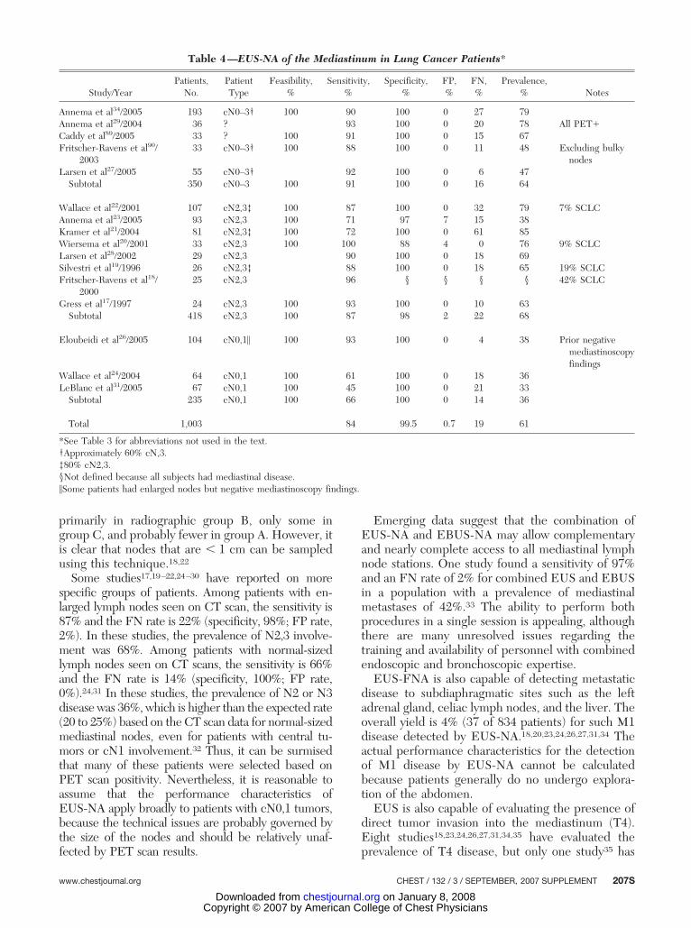
primarily in radiographic group B, only some ingroup C, and probably fewer in group A. However, itis clear that nodes that are � 1 cm can be sampledusing this technique.18,22
Some studies17,19–22,24–30 have reported on morespecific groups of patients. Among patients with en-larged lymph nodes seen on CT scan, the sensitivity is87% and the FN rate is 22% (specificity, 98%; FP rate,2%). In these studies, the prevalence of N2,3 involve-ment was 68%. Among patients with normal-sizedlymph nodes seen on CT scans, the sensitivity is 66%and the FN rate is 14% (specificity, 100%; FP rate,0%).24,31 In these studies, the prevalence of N2 or N3disease was 36%, which is higher than the expected rate(20 to 25%) based on the CT scan data for normal-sizedmediastinal nodes, even for patients with central tu-mors or cN1 involvement.32 Thus, it can be surmisedthat many of these patients were selected based onPET scan positivity. Nevertheless, it is reasonable toassume that the performance characteristics ofEUS-NA apply broadly to patients with cN0,1 tumors,because the technical issues are probably governed bythe size of the nodes and should be relatively unaf-fected by PET scan results.
Emerging data suggest that the combination ofEUS-NA and EBUS-NA may allow complementaryand nearly complete access to all mediastinal lymphnode stations. One study found a sensitivity of 97%and an FN rate of 2% for combined EUS and EBUSin a population with a prevalence of mediastinalmetastases of 42%.33 The ability to perform bothprocedures in a single session is appealing, althoughthere are many unresolved issues regarding thetraining and availability of personnel with combinedendoscopic and bronchoscopic expertise.
EUS-FNA is also capable of detecting metastaticdisease to subdiaphragmatic sites such as the leftadrenal gland, celiac lymph nodes, and the liver. Theoverall yield is 4% (37 of 834 patients) for such M1disease detected by EUS-NA.18,20,23,24,26,27,31,34 Theactual performance characteristics for the detectionof M1 disease by EUS-NA cannot be calculatedbecause patients generally do no undergo explora-tion of the abdomen.
EUS is also capable of evaluating the presence ofdirect tumor invasion into the mediastinum (T4).Eight studies18,23,24,26,27,31,34,35 have evaluated theprevalence of T4 disease, but only one study35 has
Table 4—EUS-NA of the Mediastinum in Lung Cancer Patients*
Study/YearPatients,
No.PatientType
Feasibility,%
Sensitivity,%
Specificity,%
FP,%
FN,%
Prevalence,% Notes
Annema et al34/2005 193 cN0–3† 100 90 100 0 27 79Annema et al29/2004 36 ? 93 100 0 20 78 All PET�Caddy et al89/2005 33 ? 100 91 100 0 15 67Fritscher-Ravens et al90/
200333 cN0–3† 100 88 100 0 11 48 Excluding bulky
nodesLarsen et al27/2005 55 cN0–3† 92 100 0 6 47
Subtotal 350 cN0–3 100 91 100 0 16 64
Wallace et al22/2001 107 cN2,3‡ 100 87 100 0 32 79 7% SCLCAnnema et al23/2005 93 cN2,3 100 71 97 7 15 38Kramer et al21/2004 81 cN2,3‡ 100 72 100 0 61 85Wiersema et al20/2001 33 cN2,3 100 100 88 4 0 76 9% SCLCLarsen et al28/2002 29 cN2,3 90 100 0 18 69Silvestri et al19/1996 26 cN2,3‡ 88 100 0 18 65 19% SCLCFritscher-Ravens et al18/
200025 cN2,3 96 § § § § 42% SCLC
Gress et al17/1997 24 cN2,3 100 93 100 0 10 63Subtotal 418 cN2,3 100 87 98 2 22 68
Eloubeidi et al26/2005 104 cN0,1� 100 93 100 0 4 38 Prior negativemediastinoscopyfindings
Wallace et al24/2004 64 cN0,1 100 61 100 0 18 36LeBlanc et al31/2005 67 cN0,1 100 45 100 0 21 33
Subtotal 235 cN0,1 100 66 100 0 14 36
Total 1,003 84 99.5 0.7 19 61
*See Table 3 for abbreviations not used in the text.†Approximately 60% cN,3.‡80% cN2,3.§Not defined because all subjects had mediastinal disease.�Some patients had enlarged nodes but negative mediastinoscopy findings.
www.chestjournal.org CHEST / 132 / 3 / SEPTEMBER, 2007 SUPPLEMENT 207S
Copyright © 2007 by American College of Chest Physicians on January 8, 2008 chestjournal.orgDownloaded from
specifically evaluated the reliability of EUS for Tstaging. This study found a sensitivity of 88%, aspecificity of 98%, an FN rate of 1%, and an FP rateof 30%. Overstaging appeared to occur when atumor was seen only to invade the mediastinal softtissues. The FP rate was 0% if a tumor was seenwithin a blood vessel or the esophagus. Thus, al-though EUS can be helpful in determining the Tstage, the high FP rate, in general, limits the basingof treatment decisions on this test.
The cost of EUS is less than surgical staging proce-dures, probably due to the ability to perform EUSwithout general anesthesia in an ambulatory setting.Two studies36,37 have suggested that EUS may be morecost-effective compared to mediastinoscopy, althoughthese studies assumed that mediastinoscopy frequentlyrequired inpatient hospital admission.
TBNA
TBNA, also known as a Wang NA, can be per-formed safely with no significant morbidity. It can be
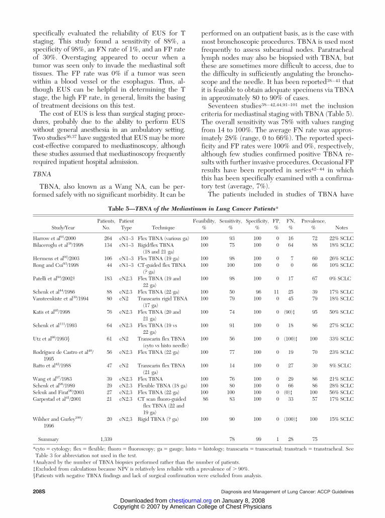
performed on an outpatient basis, as is the case withmost bronchoscopic procedures. TBNA is used mostfrequently to assess subcarinal nodes. Paratracheallymph nodes may also be biopsied with TBNA, butthese are sometimes more difficult to access, due tothe difficulty in sufficiently angulating the broncho-scope and the needle. It has been reported38–41 thatit is feasible to obtain adequate specimens via TBNAin approximately 80 to 90% of cases.
Seventeen studies38–42,44,91–101 met the inclusioncriteria for mediastinal staging with TBNA (Table 5).The overall sensitivity was 78% with values rangingfrom 14 to 100%. The average FN rate was approx-imately 28% (range, 0 to 66%). The reported speci-ficity and FP rates were 100% and 0%, respectively,although few studies confirmed positive TBNA re-sults with further invasive procedures. Occasional FPresults have been reported in series42–44 in whichthis has been specifically examined with a confirma-tory test (average, 7%).
The patients included in studies of TBNA have
Table 5—TBNA of the Mediastinum in Lung Cancer Patients*
Study/YearPatients,
No.PatientType Technique
Feasibility,%
Sensitivity,%
Specificity,%
FP,%
FN,%
Prevalence,% Notes
Harrow et al91/2000 264 cN1–3 Flex TBNA (various ga) 100 93 100 0 16 72 22% SCLCBilaceroglu et al38/1998 134 cN1–3 Rigid/flex TBNA
(18 and 21 ga)100 75 100 0 64 88 18% SCLC
Hermens et al92/2003 106 cN1–3 Flex TBNA (19 ga) 100 98 100 0 7 60 26% SCLCRong and Cui93/1998 44 cN1–3 CT-guided flex TBNA
(? ga)100 100 100 0 0 66 10% SCLC
Patelli et al94/2002† 183 cN2.3 Flex TBNA (19 and22 ga)
100 98 100 0 17 67 0% SCLC
Schenk et al44/1986 88 cN2.3 Flex TBNA (22 ga) 100 50 96 11 25 39 17% SCLCVansteenkiste et al39/1994 80 cN2 Transcarin rigid TBNA
(17 ga)100 79 100 0 45 79 18% SCLC
Katis et al95/1998 76 cN2.3 Flex TBNA (20 and21 ga)
100 74 100 0 (90)‡ 95 50% SCLC
Schenk et al111/1993 64 cN2.3 Flex TBNA (19 vs22 ga)
100 91 100 0 18 86 27% SCLC
Utz et al96/1993§ 61 cN2 Transcarin flex TBNA(cyto vs histo needle)
100 56 100 0 (100)‡ 100 33% SCLC
Rodrıguez de Castro et al40/1995
56 cN2.3 Flex TBNA (22 ga) 100 77 100 0 19 70 23% SCLC
Ratto et al42/1988 47 cN2 Transcarin flex TBNA(21 ga)
100 14 100 0 27 30 8% SCLC
Wang et al97/1983 39 cN2.3 Flex TBNA 100 76 100 0 29 86 21% SCLCSchenk et al98/1989 29 cN2.3 Flexible TBNA (18 ga) 100 80 100 0 66 86 28% SCLCSelcuk and Firat99/2003 27 cN2,3 Flex TBNA (22 ga) 100 100 100 0 (0)‡ 100 56% SCLCGarpestad et al41/2001 21 cN2.3 CT scan fluoro-guided
flex TBNA (22 and19 ga)
86 83 100 0 33 57 17% SCLC
Wilsher and Gurley100/1996
20 cN2,3 Rigid TBNA (? ga) 100 90 100 0 (100)‡ 100 15% SCLC
Summary 1,339 78 99 1 28 75
*cyto � cytology; flex � flexible; fluoro � fluoroscopy; ga � gauge; histo � histology; transcarin � transcarinal; transtrach � transtracheal. SeeTable 3 for abbreviation not used in the text.
†Analyzed by the number of TBNA biopsies performed rather than the number of patients.‡Excluded from calculations because NPV is relatively less reliable with a prevalence of � 90%.§Patients with negative TBNA findings and lack of surgical confirmation were excluded from analysis.
208S Diagnosis and Management of Lung Cancer: ACCP Guidelines
Copyright © 2007 by American College of Chest Physicians on January 8, 2008 chestjournal.orgDownloaded from
generally had a very high prevalence of N2,3 involve-ment, and the general implication is that the medi-astinal nodes have been markedly enlarged, althoughthe specifics about node size are generally vague.The results should not be applied to patients withoutextensive mediastinal involvement. Furthermore, thehigh FN rate makes this test less useful for staging ofthe mediastinum in patients with normal-sizednodes. Positive TBNA results fairly reliably demon-strate mediastinal node involvement. Negative TBNAresults, however, cannot sufficiently exclude medias-tinal nodal involvement, and additional staging pro-cedures should be performed.
EBUS-NA
EBUS-NA is a relatively new technique for medi-astinal staging. Initially, EBUS was accomplished byintroducing a catheter with an ultrasound transducerat the tip of the catheter through the workingchannel of the bronchoscope. The lymph node waslocalized with the probe, and the catheter was thenwithdrawn. The lymph node would then be sampledwith TBNA without real-time guidance. More re-cently, a bronchoscope with a convex ultrasoundprobe has been developed that allows for real-timeultrasound-guided TBNA.45 EBUS-NA can be usedto sample the highest mediastinal, upper and lowerparatracheal, and subcarinal lymph nodes, as well ashilar lymph nodes.
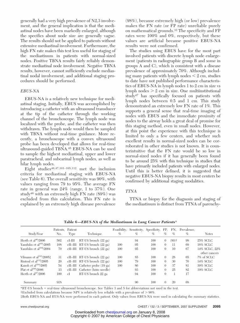
Eight studies46,47,101–105,112 met the inclusioncriteria for mediastinal staging with EBUS-NA(see Table 6). The overall sensitivity was 90%, withvalues ranging from 79 to 95%. The average FNrate in general was 24% (range, 1 to 37%). Onestudy46 with an extremely high FN rate (89%) wasexcluded from this calculation. This FN rate isexplained by an extremely high disease prevalence
(98%), because extremely high (or low) prevalencemakes the FN rate (or FP rate) unreliable purelyon mathematical grounds.32 The specificity and FPrates were 100% and 0%, respectively, but thesevalues are artificial because positive EBUS-NAresults were not confirmed.
The studies using EBUS have for the most partinvolved patients with discrete lymph node enlarge-ment (patients in radiographic group B and some ingroups A and C), which is consistent with a diseaseprevalence of approximately 70%. Although includ-ing many patients with lymph nodes � 2 cm, studiesto date have not published performance characteris-tics of EBUS-NA in lymph nodes 1 to 2 cm in size vslymph nodes � 2 cm in size. One multiinstitutionalstudy47 has specifically focused on patients withlymph nodes between 0.5 and 1 cm. This studydemonstrated an extremely low FN rate of 1%. Thissupports a general sense that real-time imaging ofnodes with EBUS and the immediate proximity ofnodes to the airway holds a great deal of promise forthis staging method, even in small nodes. However,at this point the experience with this technique islimited to only a few centers, and whether suchexcellent results in normal-sized nodes can be cor-roborated in other studies is not known. It is coun-terintuitive that the FN rate would be so low innormal-sized nodes if it has generally been foundto be around 25% with this technique in studies thathave primarily included patients with enlarged nodes.Until this is better defined, it is suggested thatnegative EBUS-NA biopsy results in most centers beconfirmed by additional staging modalities.
TTNA
TTNA or biopsy for the diagnosis and staging ofthe mediastinum is distinct from TTNA of parenchy-
Table 6—EBUS-NA of the Mediastinum in Lung Cancer Patients*
Study/YearPatients,
No.PatientType Technique
Feasibility,%
Sensitivity,%
Specificity,%
FP,%
FN,%
Prevalence,% Notes
Herth et al46/2006 502 cI-III RT-US bronch (22 ga) 94 100 0 (89)† 98 25% SCLCYasufuku et al101/2005 108 cII–III RT-US bronch (22 ga) 100 95 100 0 11 69 16% SCLCYasufuku et al102/2004 70 cII–III RT-US bronch (22 ga) 100 95 100 0 10 67 14% SCLC, 22%
other cancersVilmann et al103/2005‡ 31 cII–III RT-US bronch (22 ga) 100 85 100 0 28 65 ?% of SCLCRintoul et al112/2005 20 cII–III RT-US bronch (22 ga) 100 79 100 0 30 70 14% SCLCKanoh et al104/2005 54 cII–III Catheter probe (19 ga) 100 86 100 0 37 81 30% SCLCPlat et al105/2006 33 cII–III Catheter (histo needle) 93 100 0 25 82 19% SCLCHerth et al47/2006 100 cI RT-US bronch 22 ga 94 100 0 1 17
Summary 918 90 100 0 20 68
*RT-US bronch � real-time ultrasound bronchoscope. See Tables 3 and 5 for abbreviations not used in the text.†Excluded from calculations because NPV is relatively less reliable with a prevalence of � 90%.‡Both EBUS-NA and EUS-NA were performed in each patient. Only values from EBUS-NA were used in calculating the summary statistics.
www.chestjournal.org CHEST / 132 / 3 / SEPTEMBER, 2007 SUPPLEMENT 209S
Copyright © 2007 by American College of Chest Physicians on January 8, 2008 chestjournal.orgDownloaded from
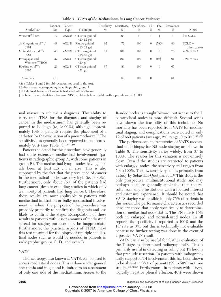
mal masses to achieve a diagnosis. The ability tocarry out TTNA for the diagnosis and staging ofcancer in the mediastinum has generally been re-ported to be high (ie, � 90%), although approxi-mately 10% of patients require the placement of acatheter for the evacuation of a pneumothorax.32 Thesensitivity has generally been reported to be approx-imately 90% (see Table 7).106–110
Patients selected for this procedure have generallyhad quite extensive mediastinal involvement (pa-tients in radiographic group A, with some patients ingroup B). The mediastinal lymph nodes have gener-ally been at least 1.5 cm in size. This is alsosupported by the fact that the prevalence of cancerin the mediastinal nodes was very high (ie, � 80%).Furthermore, only about 75% of the patients hadlung cancer (despite excluding studies in which onlya minority of patients had lung cancer). Therefore,these results are most applicable to patients withmediastinal infiltration or bulky mediastinal involve-ment, in whom the purpose of the procedure wasprobably primarily to confirm the diagnosis and lesslikely to confirm the stage. Extrapolation of theseresults to patients with lesser amounts of mediastinalspread for staging purposes may be inappropriate.Furthermore, the practical aspects of TTNA makethis test unsuited for the biopsy of multiple medias-tinal nodes such as would be needed in patients inradiographic groups C, D, and even B.
VATS
Thoracoscopy, also known as VATS, can be used toaccess mediastinal nodes. This is done under generalanesthesia and in general is limited to an assessmentof only one side of the mediastinum. Access to the
R-sided nodes is straightforward, but access to the Lparatracheal nodes is more difficult. Several serieshave shown the feasibility of this technique. Nomortality has been reported from VATS for medias-tinal staging, and complications were noted in only12 of 669 patients (average, 2%; range, 0 to 9%).48–55
The performance characteristics of VATS medias-tinal node biopsy for N2 node staging are shown inTable 8. The sensitivity varies widely, from 37 to100%. The reason for this variation is not entirelyclear. Even if the studies are restricted to patientswith enlarged nodes, the sensitivity still ranges from50 to 100%. The low sensitivity comes primarily froma study by Sebastian-Quetglas et al49 This study is theonly prospective, multiinstitutional study, and mayperhaps be more generally applicable than the re-sults from single institutions with a focused interestand extensive experience. It should be noted thatVATS staging was feasible in only 75% of patients inthis series. The performance characteristics recordedhere are those that apply specifically to determina-tion of mediastinal node status. The FN rate is 15%both in enlarged and normal-sized nodes. In allreports, the specificity is reported as 100% and theFP rate as 0%, but this is technically not evaluablebecause no further testing was done in the event ofa positive VATS result.
VATS can also be useful for further evaluation ofthe T stage as determined radiographically. This isprimarily useful in detecting or ruling out T4 lesionsthat preclude resection. In patients with radiograph-ically suspected T4 involvement this has been shownto be absent in 38% of patients (29 to 50%) in threestudies.49,50,55 Furthermore, in patients with a cyto-logically negative pleural effusion, 40% were shown
Table 7—TTNA of the Mediastinum in Lung Cancer Patients*
Study/YearPatients,
No.PatientType Technique
Feasibility,%
Sensitivity,%
Specificity,%
FP,%
FN,%
Prevalence,% Notes
Westcott106/1981 72 cN2,3† CT scan-guided(20–22 ga)
94 ‡ ‡ ‡ ‡ ?% SCLC
de Gregorio et al107/1991
48 cN2,3† Fluoro-guided(18–22 ga)
92 72 100 0 (58)§ 90 SCLC �other cancer
Moinuddin et al108/1984
40 cN2,3† CT scan-guided(18–20 ga)
91 100 100 0 0 78 48% SCLC
Protopapas andWestcott109/1996
32 cN2,3 CT scan-guided(20 ga)
100 100 0 (0)§ 91 16% SCLC
Böcking et al110/1995
23 cN2,3 CT scan-guided(22 ga)
87 80 100 0 0 65
Summary 215 89 100 0 81
*See Tables 3 and 5 for abbreviation not used in the text.†Bulky masses, corresponding to radiographic group A.‡Not defined because all subjects had mediastinal disease.§Excluded from calculations because NPV is relatively less reliable with a prevalence of � 90%.
210S Diagnosis and Management of Lung Cancer: ACCP Guidelines
Copyright © 2007 by American College of Chest Physicians on January 8, 2008 chestjournal.orgDownloaded from
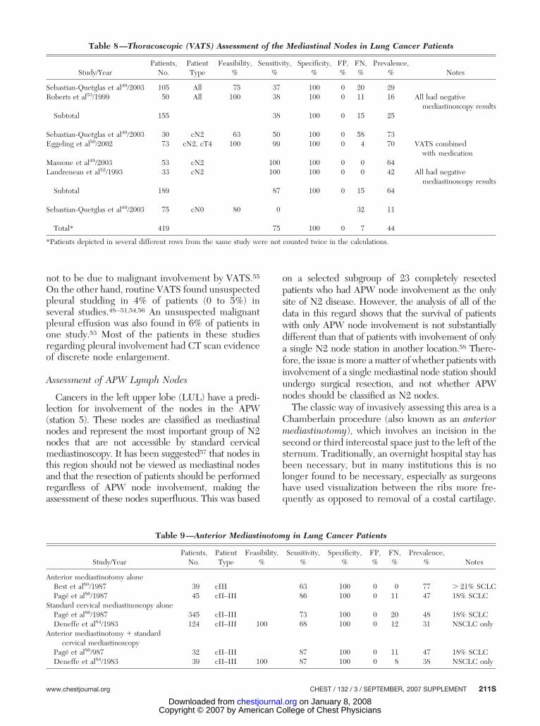
not to be due to malignant involvement by VATS.55
On the other hand, routine VATS found unsuspectedpleural studding in 4% of patients (0 to 5%) inseveral studies.48–51,54,56 An unsuspected malignantpleural effusion was also found in 6% of patients inone study.53 Most of the patients in these studiesregarding pleural involvement had CT scan evidenceof discrete node enlargement.
Assessment of APW Lymph Nodes
Cancers in the left upper lobe (LUL) have a predi-lection for involvement of the nodes in the APW(station 5). These nodes are classified as mediastinalnodes and represent the most important group of N2nodes that are not accessible by standard cervicalmediastinoscopy. It has been suggested57 that nodes inthis region should not be viewed as mediastinal nodesand that the resection of patients should be performedregardless of APW node involvement, making theassessment of these nodes superfluous. This was based
on a selected subgroup of 23 completely resectedpatients who had APW node involvement as the onlysite of N2 disease. However, the analysis of all of thedata in this regard shows that the survival of patientswith only APW node involvement is not substantiallydifferent than that of patients with involvement of onlya single N2 node station in another location.58 There-fore, the issue is more a matter of whether patients withinvolvement of a single mediastinal node station shouldundergo surgical resection, and not whether APWnodes should be classified as N2 nodes.
The classic way of invasively assessing this area is aChamberlain procedure (also known as an anteriormediastinotomy), which involves an incision in thesecond or third intercostal space just to the left of thesternum. Traditionally, an overnight hospital stay hasbeen necessary, but in many institutions this is nolonger found to be necessary, especially as surgeonshave used visualization between the ribs more fre-quently as opposed to removal of a costal cartilage.
Table 9—Anterior Mediastinotomy in Lung Cancer Patients
Study/YearPatients,
No.PatientType
Feasibility,%
Sensitivity,%
Specificity,%
FP,%
FN,%
Prevalence,% Notes
Anterior mediastinotomy aloneBest et al60/1987 39 cIII 63 100 0 0 77 � 21% SCLCPagé et al86/1987 45 cII–III 86 100 0 11 47 18% SCLC
Standard cervical mediastinoscopy alonePagé et al86/1987 345 cII–III 73 100 0 20 48 18% SCLCDeneffe et al84/1983 124 cII–III 100 68 100 0 12 31 NSCLC only
Anterior mediastinotomy � standardcervical mediastinoscopy
Pagé et al86/987 32 cII–III 87 100 0 11 47 18% SCLCDeneffe et al84/1983 39 cII–III 100 87 100 0 8 38 NSCLC only
Table 8—Thoracoscopic (VATS) Assessment of the Mediastinal Nodes in Lung Cancer Patients
Study/YearPatients,
No.PatientType
Feasibility,%
Sensitivity,%
Specificity,%
FP,%
FN,%
Prevalence,% Notes
Sebastian-Quetglas et al49/2003 105 All 75 37 100 0 20 29Roberts et al53/1999 50 All 100 38 100 0 11 16 All had negative
mediastinoscopy resultsSubtotal 155 38 100 0 15 25
Sebastian-Quetglas et al49/2003 30 cN2 63 50 100 0 58 73Eggeling et al50/2002 73 cN2, cT4 100 99 100 0 4 70 VATS combined
with medicationMassone et al48/2003 53 cN2 100 100 0 0 64Landreneau et al52/1993 33 cN2 100 100 0 0 42 All had negative
mediastinoscopy resultsSubtotal 189 87 100 0 15 64
Sebastian-Quetglas et al49/2003 75 cN0 80 0 32 11
Total* 419 75 100 0 7 44
*Patients depicted in several different rows from the same study were not counted twice in the calculations.
www.chestjournal.org CHEST / 132 / 3 / SEPTEMBER, 2007 SUPPLEMENT 211S
Copyright © 2007 by American College of Chest Physicians on January 8, 2008 chestjournal.orgDownloaded from
The reliability of this procedure has not been exten-sively documented, despite its common use. Thesensitivity of a Chamberlain procedure in addition tostandard cervical mediastinoscopy in patients withLUL tumors is approximately 87%, and the FN rateis approximately 10% (Table 9). Two additionalstudies59,60 regarding this procedure have not reallyaddressed the reliability of the procedure for thestaging of NSCLC. In one study,59 no actual biopsieswere performed in most patients, and the procedurewas used to assess resectability (in this series, resect-able patients included those with bulky APW nodalinvolvement). The other study60 used anterior medi-astinotomy primarily for diagnosis (not staging), andincluded pulmonary biopsies and evaluation of pa-tients with mediastinal masses. In fact, only a minor-ity of patients included in this study had lung cancer.
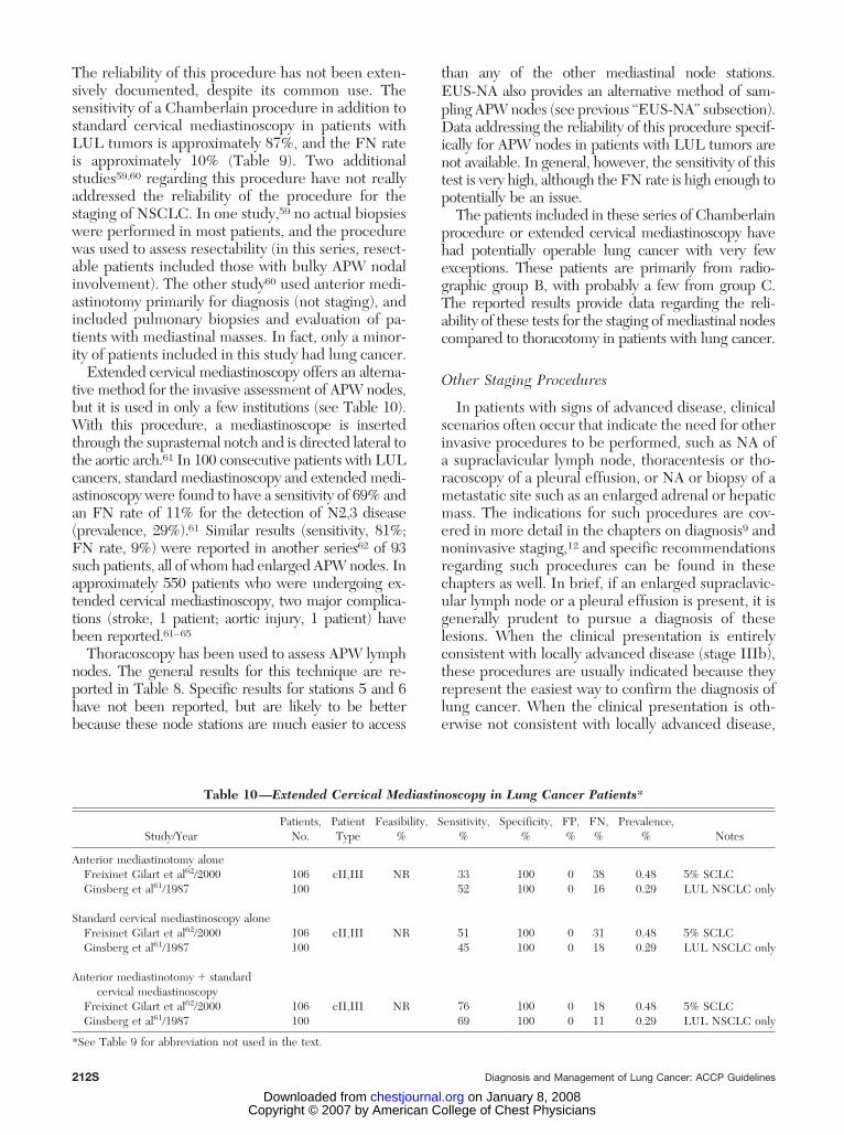
Extended cervical mediastinoscopy offers an alterna-tive method for the invasive assessment of APW nodes,but it is used in only a few institutions (see Table 10).With this procedure, a mediastinoscope is insertedthrough the suprasternal notch and is directed lateral tothe aortic arch.61 In 100 consecutive patients with LULcancers, standard mediastinoscopy and extended medi-astinoscopy were found to have a sensitivity of 69% andan FN rate of 11% for the detection of N2,3 disease(prevalence, 29%).61 Similar results (sensitivity, 81%;FN rate, 9%) were reported in another series62 of 93such patients, all of whom had enlarged APW nodes. Inapproximately 550 patients who were undergoing ex-tended cervical mediastinoscopy, two major complica-tions (stroke, 1 patient; aortic injury, 1 patient) havebeen reported.61–65
Thoracoscopy has been used to assess APW lymphnodes. The general results for this technique are re-ported in Table 8. Specific results for stations 5 and 6have not been reported, but are likely to be betterbecause these node stations are much easier to access
than any of the other mediastinal node stations.EUS-NA also provides an alternative method of sam-pling APW nodes (see previous “EUS-NA” subsection).Data addressing the reliability of this procedure specif-ically for APW nodes in patients with LUL tumors arenot available. In general, however, the sensitivity of thistest is very high, although the FN rate is high enough topotentially be an issue.
The patients included in these series of Chamberlainprocedure or extended cervical mediastinoscopy havehad potentially operable lung cancer with very fewexceptions. These patients are primarily from radio-graphic group B, with probably a few from group C.The reported results provide data regarding the reli-ability of these tests for the staging of mediastinal nodescompared to thoracotomy in patients with lung cancer.
Other Staging Procedures
In patients with signs of advanced disease, clinicalscenarios often occur that indicate the need for otherinvasive procedures to be performed, such as NA ofa supraclavicular lymph node, thoracentesis or tho-racoscopy of a pleural effusion, or NA or biopsy of ametastatic site such as an enlarged adrenal or hepaticmass. The indications for such procedures are cov-ered in more detail in the chapters on diagnosis9 andnoninvasive staging,12 and specific recommendationsregarding such procedures can be found in thesechapters as well. In brief, if an enlarged supraclavic-ular lymph node or a pleural effusion is present, it isgenerally prudent to pursue a diagnosis of theselesions. When the clinical presentation is entirelyconsistent with locally advanced disease (stage IIIb),these procedures are usually indicated because theyrepresent the easiest way to confirm the diagnosis oflung cancer. When the clinical presentation is oth-erwise not consistent with locally advanced disease,
Table 10—Extended Cervical Mediastinoscopy in Lung Cancer Patients*
Study/YearPatients,
No.PatientType
Feasibility,%
Sensitivity,%
Specificity,%
FP,%
FN,%
Prevalence,% Notes
Anterior mediastinotomy aloneFreixinet Gilart et al62/2000 106 cII,III NR 33 100 0 38 0.48 5% SCLCGinsberg et al61/1987 100 52 100 0 16 0.29 LUL NSCLC only
Standard cervical mediastinoscopy aloneFreixinet Gilart et al62/2000 106 cII,III NR 51 100 0 31 0.48 5% SCLCGinsberg et al61/1987 100 45 100 0 18 0.29 LUL NSCLC only
Anterior mediastinotomy � standardcervical mediastinoscopy
Freixinet Gilart et al62/2000 106 cII,III NR 76 100 0 18 0.48 5% SCLCGinsberg et al61/1987 100 69 100 0 11 0.29 LUL NSCLC only
*See Table 9 for abbreviation not used in the text.
212S Diagnosis and Management of Lung Cancer: ACCP Guidelines
Copyright © 2007 by American College of Chest Physicians on January 8, 2008 chestjournal.orgDownloaded from
the etiology of these lesions must be established inorder to accurately define the stage. However, theprocedures used to diagnose an enlarged supracla-vicular node (ie, NA or surgical biopsy) are the sameregardless of whether the issue is to confirm thediagnosis or to define the stage. Similarly, in patientswith a clinical presentation that is consistent withadvanced disease (stage IV), an invasive proceduremay be indicated as the easiest way to confirm thediagnosis and establish the cell type of the lungcancer. In patients with a solitary site that is suspi-cious for a distant metastasis or in patients with aclinical presentation that seems inconsistent withadvanced disease, an invasive procedure is indicatedto accurately define the stage. The procedures usedto assess possible distant sites are the same regard-less of the clinical presentation, and are dictatedprimarily by technical and anatomic factors that arespecific to the particular patient.
No data are available to assess the sensitivity,specificity, and FN and FP rates of NA of a supra-clavicular node. General experience indicates thatthis procedure is usually successful; in addition,surgical biopsy of such a node is easily accomplishedif a NA procedure is not diagnostic. The reliability ofprocedures to diagnose a pleural effusion is coveredin the chapter on diagnosis. Thoracentesis has asensitivity of approximately 60%; thoracoscopy has asensitivity of � 95%. Procedures to diagnose sus-pected distant metastatic sites are too varied todiscuss in detail; furthermore, no data are availablethat expressly assesses the reliability of these tests inpatients with lung cancer.
Approach to Patients
Mediastinal Infiltration
In patients with extensive mediastinal infiltration,the radiographic evidence of mediastinal involve-ment is quite universally considered adequate. Thereare no data to prove this, because invasive confirma-tion is not done. However, even though staging is notan issue, tissue is needed to confirm the diagnosisand to establish what type of cancer is present (eg,NSCLC vs SCLC). In this case, it does not matterwhether tissue is obtained from the primary tumor orfrom a mediastinal site.
In patients in whom the diagnosis is the primaryissue, tissues should be obtained by whatevermethod is easiest to perform. In other words, thechoice of procedure will be governed primarily bypatient-specific factors (ie, anatomic, convenience,and comorbidity factors) instead of the performancecharacteristics of a test. For example, it is still likelythat a test of relatively low sensitivity such as sputum
cytology or cytology of a pleural effusion will bechosen first simply because it is easiest to perform. Itis rare that such a patient will undergo TBNA,EUS-NA, or mediastinoscopy. Details of the perfor-mance characteristics of diagnostics tests of theprimary tumor are summarized in chapter 9, andperformance characteristics of the invasive medias-tinal tests are summarized in the tables here. How-ever, as noted above, the determining factor con-cerning which test to choose will be governed primarilyby patient-specific issues.
Recommendation
1. For patients with extensive mediastinalinfiltration of tumor and no distant metastases,radiographic (CT scan) assessment of the medi-astinal stage is usually sufficient without inva-sive confirmation. Grade of recommendation, 2C
Discrete Mediastinal Lymph Node Enlargement
Many patients present with a CT scan demonstrat-ing the enlargement of discrete mediastinal (N2,3)lymph nodes. An extensive literature32 demonstratesthat enlargement seen on CT scan alone carries anFP rate of approximately 40% (see chapter 12). ThePET scan literature has only recently become de-tailed enough to begin to define FN and FP rates insubgroups of patients such as those with discretenodal enlargement seen on a CT scan. The FP ratefor PET scanning in the mediastinum has beenwidely shown to be around 15 to 20%, although thishas not been defined for this particular subgroup ofpatients. Two metaanalyses66,67 have estimated thePET FN rate to be 13 to 25% in patients with nodalenlargement detected by CT scan, although theseestimates are not based on direct data or clearlydefined patients. Direct data from studies68,69 inpatients with mediastinal or hilar nodal enlargement(radiographic groups B and C combined) have founda PET FN rate of 20 to 28% for N2,3 involvement.Thus, it appears that in patients with enlargedmediastinal nodes detected by CT scanning, the CTscan alone cannot be relied on, and invasive biopsy isneeded whether a PET scan finding is positive ornegative.
In choosing an invasive staging test, several issuesmust be considered. First is the availability of differ-ent procedures. All of the invasive tests require somespecialized experience and skill, and people whoperform these procedures only occasionally may notbe able to achieve the performance characteristicspublished in studies performed at high-volume insti-tutions. Second, the location of the suspicious nodesis important, because nodes in one location may be
www.chestjournal.org CHEST / 132 / 3 / SEPTEMBER, 2007 SUPPLEMENT 213S
Copyright © 2007 by American College of Chest Physicians on January 8, 2008 chestjournal.orgDownloaded from
accessible only by a particular approach. There maybe factors related to patient comorbidity that mayargue against certain approaches, such as mediasti-noscopy, which usually requires general anesthesia.However, patients who are unable to tolerate ageneral anesthetic for such a small procedure asmediastinoscopy are likely not to be well enough totolerate definitive treatment for lung cancer anyhow.The morbidity and mortality of invasive proceduresmay be a consideration, although all of the availableprocedures generally have an excellent safety profile.Finally, cost may be a consideration.
The sensitivity of various invasive mediastinal stag-ing tests in cN2,3 patients appears to be similar. Astrict comparison is not justified because the patientsundergoing these procedures are not comparabledue to differences in how they are selected for aparticular procedure (eg, the location of the nodes).The primary issue is the variability in FN rates. If aNA technique is chosen, it must be remembered thata negative result is not very reliable. A NA proceduremay well be a good first choice because theseprocedures are less invasive than mediastinoscopy.However, a negative needle biopsy finding should befollowed up in general with mediastinoscopy.
An option for the treatment of patients with stageIII NSCLC is induction therapy followed by surgery(see chapter 17). If this approach is chosen, the roleof mediastinal restaging after induction therapy isvery unclear. However, some people have arguedthat the approach should include surgery only inthose patients who have a response in the mediasti-num to induction therapy. It has been shown repeat-edly70 that CT scan evidence of tumor shrinkage isnotoriously misleading. PET scanning for mediasti-nal restaging has also been shown to have high FPand FN rates.70 A repeat mediastinoscopy is gener-ally safe and feasible (82 to 100%) but has mediocreresults (sensitivity, 70 to 82%; FN rate, 15 to25%),13,71–73 and most surgeons are uncomfortablewith this procedure. Because a first-time mediasti-noscopy is probably the best way to accomplish medi-astinal restaging, an argument can be made to use a NAtechnique initially to document N2,3 involvement andto save mediastinoscopy for the restaging procedureafter induction therapy. All of this only applies if theadopted treatment policy is one of induction therapy,with subsequent therapy to be determined by theresults of mediastinal restaging (despite the lack of datadefining the role of surgery and restaging).
Recommendations
2. For patients with discrete mediastinal lymphnode enlargement (and no distant metastases),
invasive confirmation of the radiographic stage isrecommended (regardless of whether a PET scanfinding is positive or negative in the mediastinalnodes). Grade of recommendation, 1B
3. For patients with discrete mediastinal lymphnode enlargement (and no distant metastases),many invasive techniques for the confirmationof the N2,3 node status are suggested as reason-able approaches (eg, mediastinoscopy, EUS-NA,TBNA, EBUS-NA, or TTNA), given the appropri-ate experience and skill (regardless of whether aPET scan finding is positive or negative in themediastinal nodes). Grade of recommendation, 1B
4. For patients with discrete mediastinal lymphnode enlargement (and no distant metastases), anonmalignant result from a needle technique (eg,EUS-NA, TBNA, EBUS-NA, or TTNA) should befurther confirmed by mediastinoscopy (regard-less of whether a PET scan finding is positive ornegative in the mediastinal nodes). Grade of rec-ommendation, 1C
Central and Clinical N1 Tumors
Patients with no evidence of mediastinal nodeenlargement but with a central tumor or N1 nodeinvolvement represent another distinct group (groupC). It is reasonable to consider patients with centraltumors together with those with N1 node enlarge-ment, because it is usually difficult to assess the N1nodes in the case of a central tumor. Extensive dataindicate that the FN rate of a CT scan with respectto the mediastinal nodes is 20 to 25% (see chapter 12on noninvasive staging).32 More limited data demon-strate that the FN rate for PET scanning in themediastinal nodes in this situation is similarly high(24 to 83%).68,69,74,75 Thus, invasive staging is re-quired in these patients despite the negative CT scanresult and even a negative PET scan result.
In patients with normal-sized mediastinal lymphnodes in whom invasive staging is needed, mediasti-noscopy remains the “gold standard.” The generalexperience with mediastinoscopy suggests that theFN rate (approximately 10%) is low in these patients,and those studies15,16 that have specifically reportedon these patients substantiate this. Although it can-not be directly compared to mediastinoscopy, theFN rate (20%) of EUS-NA demonstrates that asignificant number of patients with negativeEUS-NA finding may still harbor metastases. Sub-group analysis in the study by Wallace et al24 hassuggested that approximately one half of these FNcases were due to malignant lymph nodes in theanterior mediastinum, which may be more accessibleby mediastinoscopy or, theoretically, EBUS-NA, al-though to date this area has not been carefully
214S Diagnosis and Management of Lung Cancer: ACCP Guidelines
Copyright © 2007 by American College of Chest Physicians on January 8, 2008 chestjournal.orgDownloaded from
studied. The other half of FN cases were often dueto very small deposits that may be more subject tosampling error of the needle or small biopsy meth-ods.
Other methods of mediastinal staging have gen-erally not been used much in this patient popula-tion (ie, TTNA, TBNA, and EBUS-NA). However,the performance characteristics of these tests,especially the FN rates, in patients with enlargedmediastinal nodes would suggest that TTNA,TBNA, and EBUS-NA are likely not to perform aswell as mediastinoscopy in patients with normal-sized nodes. This is particularly true with regard tothe FN rates. Because the goal of invasive stagingin this situation is to confirm the absence ofmediastinal disease, the FN rate is the parameterof greatest importance. It does not appear that theNA techniques can confirm a negative mediasti-num finding with sufficient reliability. EBUS-NAmay turn out to be sufficiently reliable to rule outmediastinal node involvement in small nodes, butthe data are too preliminary to justify a firmrecommendation.47
Recommendations
5. For patients with a radiographically nor-mal mediastinum (by CT scan) and a centraltumor or N1 lymph node enlargement (and nodistant metastases), invasive confirmation of theradiographic stage is recommended (regardlessof whether a PET scan finding is positive ornegative in the mediastinal nodes). Grade ofrecommendation, 1C
6. For patients with a central tumor or N1 lymphnode enlargement (and no distant metastases), in-vasive staging is recommended. In general, medias-tinoscopy is suggested, but EUS-NA or EBUS-NAmay be a reasonable alternative if nondiagnosticresults are followed by mediastinoscopy. Grade ofrecommendation, 2C
Peripheral Clinical Stage I Tumors
Patients with peripheral tumors in whom thereis no enlargement of N1 or N2,3 nodes seen on CTscans, the FN rate of this radiographic assessmentin the mediastinum is approximately 10%.32 Theincidence is lower in patients with T1 tumors (9%)than in those with T2 tumors (13%).32 Whetherthis incidence is viewed as being high enough tojustify performing mediastinoscopy or PET scan-ning is a matter of judgment. A negative PET scanfinding in the mediastinum carries a FN rate ofapproximately 5% (range, 3 to 6%) in this group ofpatients.68,74 –76 Thus, invasive staging is probably
not needed in this patient group if the findings ofa PET scan of the mediastinum are negative. APET scan is generally not needed in the case of acT1N0M0 tumor (see chapter 12). Invasive stagingof the mediastinum is also generally not indicatedin these cases.
If invasive staging is deemed to be necessary, itappears that mediastinoscopy is the best choicebecause of a low FN rate compared to techniquesinvolving NA. The arguments raised concerning theinvasive staging of normal mediastinal nodes in cN1or central tumors (group C) applies to this group(group D) as well, since these arguments are afunction primarily of the size of the mediastinalnodes.
Recommendations
7. For patients with a peripheral clinicalstage I tumor in whom a PET scan shows uptakein mediastinal nodes (and not distant metasta-ses), invasive staging is recommended. In gen-eral, mediastinoscopy is suggested, but EUS-NAor EBUS-NA may be a reasonable alternative ifnondiagnostic results are followed by mediasti-noscopy. Grade of recommendation, 1C
8. For patients with a peripheral clinicalstage I tumor, invasive confirmation of themediastinal nodes is not needed if the findingsof a PET scan of the mediastinum are negative.Grade of recommendation, 1C
Patients With LUL Tumors
Patients with tumors in the LUL deserve specialmention because the aortic arch raises technical issuesof access to the mediastinal nodes in the APW (station5). This node station is the most likely mediastinalnodal area to be involved in the case of an LUL tumor,whereas it is extremely unlikely to be involved inpatients with a tumor in any of the other lobes. Ofcourse, mediastinal nodal involvement from an LULtumor can also extend to other node stations such as thesubcarinal (station 7) or paratracheal areas (stations 4L,4R, 2L, and 2 R). A full assessment of potentiallyinvolved mediastinal node stations in the case of anLUL tumor requires investigation of the paratrachealand subcarinal nodes, as well as a separate procedure toaccess the APW area. The technical issues of access tothe APW nodes raises questions about whether aseparate invasive test for the assessment of these nodesis really necessary.
The definition of radiographic groups (groups A,B, C and D) is the same no matter which lobe of thelung is involved. In addition, the indications forinvasive staging of the mediastinum in patients with
www.chestjournal.org CHEST / 132 / 3 / SEPTEMBER, 2007 SUPPLEMENT 215S
Copyright © 2007 by American College of Chest Physicians on January 8, 2008 chestjournal.orgDownloaded from
LUL tumors should follow the same guidelines as inpatients with a tumor in a different lobe (patientswith enlarged mediastinal nodes, a central tumor orN1 nodal enlargement and a normal mediastinum, orwith evidence of PET scan uptake in mediastinalareas should undergo invasive mediastinal staging).
If the usual mediastinal node stations are found tobe negative (stations 2R, 4R, 7, 2L, and 4L), it iscontroversial whether a separate procedure to assessthe station 5 area is needed. However, given the lackof clear data that involvement of only this stationcarries a different prognosis than involvement of adifferent single mediastinal node station, and withthe availability of techniques of assessing the APWarea that are easier for patients to undergo (eg,EUS-NA, EBUS-NA, extended cervical mediastinos-copy, and VATS), the guidelines committee favorspursuing an invasive assessment of the APW nodes.A finding of involvement in one mediastinal area maypreclude the necessity of biopsying other areas,especially if an additional procedure would be nec-essary (eg, a positive EUS-NA finding for station 5may preclude the assessment of paratracheal nodes,or a positive mediastinoscopy result would obviatethe need for an anterior mediastinotomy).
A comparative assessment of different invasivetests for APW nodes is not possible. A reasonableextrapolation from the data for other node stationswould be to pursue a needle technique for enlargedAPW nodes and a surgical biopsy (eg, Chamberlainprocedure, VATS, or extended cervical mediastinos-copy) for normal-sized APW nodes. However, it isalso a reasonable compromise to accept a negativeNA finding without adding an additional surgicalbiopsy, given the controversy over the need to assessthe APW nodes. Modification of these suggestionsmay be necessary due to the availability of expertisewith the invasive procedures. However, it is sug-gested that referral to a larger center be consideredif there is not a fair amount of expertise with at leastone invasive APW staging procedure.
Recommendation
9. For patients with an LUL cancer in whominvasive mediastinal staging is indicated, as de-fined by the previous recommendations, it issuggested that invasive mediastinal staging in-clude assessment of the APW nodes (via Cham-berlain procedure, thoracoscopy, extended cer-vical mediastinoscopy, EUS-NA, or EBUS-NA) ifother mediastinal node stations are found to beuninvolved. Grade of recommendation, 2C
Conclusion
Accurate mediastinal staging is crucial to theselection of the optimal therapy for patients withoutdistant metastases. Imaging studies are not suffi-ciently reliable in many situations, making invasivestaging tests an important part of appropriate stag-ing. Many different invasive staging tests, whichshould be viewed as complementary to one anotherbecause they are applicable to particular nodal sta-tions and patient groups, are available. It is helpful toseparate patients into different groups based on theextent of mediastinal involvement by CT scan andwhether the primary tumor is central or peripheral.In general, needle techniques are most useful inpatients with enlarged mediastinal nodes, while me-diastinoscopy remains the “gold standard” in patientswith normal-sized nodes.
Summary of Recommendations
1. For patients with extensive mediastinalinfiltration of tumor (and no distant metas-tases), radiographic (CT scan) assessment ofthe mediastinal stage is usually sufficientwithout invasive confirmation. Grade of rec-ommendation, 2C
2. For patients with discrete mediastinallymph node enlargement (and no distantmetastases), invasive confirmation of theradiographic stage is recommended (re-gardless of whether the findings of a PETscan of the mediastinal nodes are positive ornegative). Grade of recommendation, 1B
3. For patients with discrete mediastinallymph node enlargement (and no distantmetastases), many invasive techniques forconfirmation of the N2,3 node status aresuggested as reasonable approaches (medi-astinoscopy, EUS-NA, TBNA, EBUS-NA,TTNA), given the availability of personnelwith appropriate experience and skill. Gradeof recommendation, 1B
4. For patients with discrete mediastinallymph node enlargement (and no distantmetastases), a nonmalignant result from aneedle technique (eg, EUS-NA, TBNA,EBUS-NA, or TTNA) should be further con-firmed by mediastinoscopy (regardless ofwhether the findings of a PET scan of themediastinal nodes are positive or negative).Grade of recommendation, 1C 5. For pa-tients with a radiographically normal medi-astinum (determined by CT scan) and a
216S Diagnosis and Management of Lung Cancer: ACCP Guidelines
Copyright © 2007 by American College of Chest Physicians on January 8, 2008 chestjournal.orgDownloaded from
central tumor or N1 lymph node enlarge-ment (and no distant metastases), invasive-confirmation of the radiographic stage isrecommended (regardless of whether thefindings of a PET scan of the mediastinalnodes are positive or negative). Grade ofrecommendation, 1C
6. For patients with a central tumor orN1 lymph node enlargement (and no distantmetastases), invasive staging is recom-mended. In general, mediastinoscopy issuggested, but EUS-NA or EBUS-NA maybe a reasonable alternative if nondiagnosticresults are followed by mediastinoscopy.Grade of recommendation, 2C
7. For patients with a peripheral clinicalstage I tumor in whom a PET scan showsuptake in the mediastinal nodes (and nodistant metastases), invasive staging is rec-ommended. In general, mediastinoscopy issuggested, but EUS-NA or EBUS-NA maybe a reasonable alternative if nondiagnosticresults are followed by mediastinoscopy.Grade of recommendation, 1C
8. For patients with a peripheral clinicalstage I tumor, invasive confirmation of themediastinal nodes is not needed if the find-ings of a PET scan of the mediastinum arenegative. Grade of recommendation, 1C
9. For patients with an LUL cancer inwhom invasive mediastinal staging is indi-cated, as defined by the previous recom-mendations, it is suggested that invasivemediastinal staging include the assessmentof the APW nodes (via Chamberlain proce-dure, thoracoscopy, extended cervical me-diastinoscopy, EUS-NA, or EBUS-NA) ifother mediastinal node stations are found tobe uninvolved. Grade of recommendation, 2C
References1 Toloza EM, Harpole L, Detterbeck F, et al. Invasive staging
of non-small cell lung cancer: a review of the currentevidence. Chest 2003; 123(suppl):157S–166S
2 Cybulsky IJ, Bennett WF. Mediastinoscopy as a routineoutpatient procedure. Ann Thorac Surg 1994; 58:176 –178
3 Vallieres E, Page A, Verdant A. Ambulatory mediastinos-copy and anterior mediastinotomy. Ann Thorac Surg 1991;52:1122–1126
4 Selby JH, Leach CL, Heath BJ, et al. Local anesthesia formediastinoscopy: experience with 450 consecutive cases. AmSurg 1978; 44:679–682
5 Kiser AC, Detterbeck FC. General aspects of surgicaltreatment. In: Detterbeck FC, Rivera MP, Socinski MA, etal, eds. Diagnosis and treatment of lung cancer: an evidence-based guide for the practicing clinician. Philadelphia, PA:
WB Saunders, 2001; 133–1476 Venissac N, Alifano M, Mouroux J. Video-assisted medias-
tinoscopy: experience from 240 consecutive cases. AnnThorac Surg 2003; 76:208–212
7 Leschber G, Holinka G, Linder A. Video-assisted mediasti-noscopic lymphadenectomy (VAMLA): a method for sys-tematic mediastinal lymphnode dissection. European J Car-diothorac Surg 2003; 24:192–195
8 Coughlin M, Deslauriers J, Beaulieu M, et al. Role ofmediastinoscopy in pretreatment staging of patients withprimary lung cancer. Ann Thorac Surg 1985; 40:556–560
9 Staples CA, Muller NL, Miller RR, et al. Mediastinal nodesin bronchogenic carcinoma: comparison between CT andmediastinoscopy. Radiology 1988; 167:367–372
10 Gdeedo A, Van Schil P, Corthouts B, et al. Prospectiveevaluation of computed tomography and mediastinoscopy inmediastinal lymph node staging. Eur Respir J 1997; 10:1547–1551
11 Van den Bosch JMM, Gelissen HJ, Wagenaar SS. Explor-atory thoracotomy in bronchial carcinoma. J Thorac Cardio-vasc Surg 1983; 85:733–737
12 Hammoud ZT, Anderson RC, Meyers BF, et al. The currentrole of mediastinoscopy in the evaluation of thoracic disease.J Thorac Cardiovasc Surg 1999; 118:894–899
13 Lardinois D, Schallberger A, Betticher D, et al. Postinduc-tion video-mediastinoscopy is as accurate and safe as video-mediastinoscopy in patients without pretreatment for poten-tially operable non-small cell lung cancer. Ann Thorac Surg2003; 75:1102–1106
14 Kimura H, Iwai N, Ando S, et al. A prospective study ofindications for mediastinoscopy in lung cancer with CTfindings, tumor size, and tumor markers. Ann Thorac Surg2003; 75:1734–1739
15 Choi YS, Shim YM, Kim J, et al. Mediastinoscopy in patientswith clinical stage I non-small cell lung cancer. Ann ThoracSurg 2003; 75:364–366
16 Gurses A, Turna A, Bedirhan MA, et al. The value ofmediastinoscopy in preoperative evaluation of mediastinalinvolvement in non-small-cell lung cancer patients withclinical NO disease. Thorac Cardiovasc Surg 2002; 50:174–177
17 Gress FG, Sevides TJ, Sandler A, et al. Endoscopic ultra-sonography, fine-needle aspiration biopsy guided by endo-scopic ultrasonography, and computed tomography in thepreoperative staging of non-small-cell lung cancer: a com-parison study. Ann Intern Med 1997; 127:604–612
18 Fritscher-Ravens A, Soehendra N, Schirrow L, et al. Role oftransesophageal endosonography-guided fine-needle aspirationin the diagnosis of lung cancer. Chest 2000; 117:339–345
19 Silvestri GA, Hoffman BJ, Bhutani MS, et al. Endoscopicultrasound with fine-needle aspiration in the diagnosis andstaging of lung cancer. Ann Thorac Surg 1996; 61:1441–1446
20 Wiersema MJ, Vazquez-Sequeiros E, Wiersema LM. Eval-uation of mediastinal lymphadenopathy with endoscopicUS-guided fine-needle aspiration biopsy. Radiology 2001;219:252–257
21 Kramer H, van Putten JW, Post WJ, et al. Oesophagealendoscopic ultrasound with fine needle aspiration improvesand simplifies the staging of lung cancer. Thorax 2004;59:596–601
22 Wallace MB, Silvestri GA, Sahai AV, et al. Endoscopicultrasound-guided fine needle aspiration for staging patientswith carcinoma of the lung. Ann Thorac Surg 2001; 72:1861–1867
23 Annema JT, Veersteegh MI, Veselic M, et al. Endoscopicultrasound added to mediastinoscopy for preoperative stag-ing of patients with lung cancer. JAMA 2005; 294:931–936
www.chestjournal.org CHEST / 132 / 3 / SEPTEMBER, 2007 SUPPLEMENT 217S
Copyright © 2007 by American College of Chest Physicians on January 8, 2008 chestjournal.orgDownloaded from

24 Wallace MB, Ravenel J, Block MI, et al. Endoscopicultrasound in lung cancer patients with a normal mediasti-num on computed tomography. Ann Thorac Surg 2004;77:1763–1768
25 Fritscher-Ravens A, Bohuslavizki KH, Brandt L, et al.Mediastinal lymph node involvement in potentially resect-able lung cancer: comparison of CT, positron emissiontomography, and endoscopic ultrasonography with and with-out fine-needle aspiration. Chest 2003; 123:442–451
26 Eloubeidi MA, Cerfolio RJ, Chen VK, et al. Endoscopicultrasound-guided fine needle aspiration of mediastinallymph node in patients with suspected lung cancer afterpositron emission tomography and computed tomographyscans. Ann Thorac Surg 2005; 79:263–268
27 Larsen SS, Vilmann P, Krasnik M, et al. Endoscopic ultra-sound guided biopsy versus mediastinoscopy for analysis ofparatracheal and subcarinal lymph nodes in lung cancerstaging. Lung Cancer 2005; 48:85–92
28 Larsen SS, Krasnik M, Vilmann P, et al. Endoscopic ultra-sound guided biopsy of mediastinal lesions has a majorimpact on patient management. Thorax 2002; 57:98–103
29 Annema JT, Hoekstra OS, Smit EF, et al. Towards aminimally invasive staging strategy in NSCLC: analysis ofPET positive mediastinal lesions by EUS-FNA. Lung Can-cer 2004; 44:53–60
30 Walsh PR, Williams DB. Mediastinal adenopathy: findingthe answer with endoscopic ultrasound-guided fine-needleaspiration biopsy. Intern Med J 2005; 35:392–398
31 LeBlanc JK, Devereaux BM, Imperiale TF, et al. Endo-scopic ultrasound in non-small cell lung cancer and negativemediastinum on computed tomography. Am J Respir CritCare Med 2005; 171:177–182
32 Detterbeck FC, Jones DR, Parker LA Jr. Intrathoracic staging,in diagnosis and treatment of lung cancer: an evidence-basedguide for the practicing clinician. Philadelphia, PA: WB Saun-ders, 2001; 73–93
33 Wallace M, Pascual JM, Raimondo M, et al. Complete“medical mediastinoscopy” under conscious sedation: a pro-spective blinded comparison of endoscopic and endobron-chial ultrasound to bronchoscopic fine needle aspiration formalignant mediastinal lymph nodes [abstract]. GastrointestEndosc 2006; 63:AB96
34 Annema JT, Versteegh MI, Veselic M, et al. Endoscopicultrasound-guided fine-needle aspiration in the diagnosisand staging of lung cancer and its impact on surgicalstaging. J Clin Oncol 2005; 23:8357– 8361
35 Varadarajulu S, Schmulewitz N, Wildi SM, et al. Accuracy ofEUS in staging of T4 lung cancer. Gastrointest Endosc 2004;59:345–348
36 Aabakken L, Silvestri GA, Hawes R, et al. Cost-efficacy ofendoscopic ultrasonography with fine-needle aspiration vs.mediastinotomy in patients with lung cancer and suspectedmediastinal adenopathy. Endoscopy 1999; 31:707–711
37 Harewood G, Wiersema MJ, Edell ES, et al. Cost-minimi-zation analysis of alternative diagnostic approaches in amodeled patient with non-small cell lung cancer and sub-carinal lymphadenopathy. Mayo Clin Proc 2002; 77:155–164
38 Bilaçeroglu S, Cagiotariotaciota U, Gunel O, et al. Compar-ison of rigid and flexible transbronchial needle aspiration inthe staging of bronchogenic carcinoma. Respiration 1998;65:441–449
39 Vansteenkiste J, Lacquet LM, Demedts M, et al. Trans-carinal needle aspiration biopsy in the staging of lungcancer. Eur Respir J 1994; 7:265–268
40 Rodrıguez de Castro F, Rey A, Caminero J, et al. Trans-bronchial fine needle aspiration in clinical practice. Cytopa-thology 1995; 6:22–29
41 Garpestad E, Goldberg S, Herth F, et al. CT fluoroscopyguidance for transbronchial needle aspiration: an experiencein 35 patients. Chest 2001; 119:329–332
42 Ratto GB, Mereu C, Motta G. The prognostic significance ofpreoperative assessment of mediastinal lymph nodes inpatients with lung cancer. Chest 1988; 93:807–813
43 Shure D, Fedullo PF. The role of transcarinal needleaspiration in the staging of bronchogenic carcinoma. Chest1984; 86:693–696
44 Schenk DA, Bower JH, Bryan CL, et al. Transbronchialneedle aspiration staging of bronchogenic carcinoma. AmRev Respir Dis 1986; 134:146–148
45 Feller-Kopman D, Lunn W, Ernst E. Autofluorescencebronchoscopy and endobronchial ultrasound: a practicalreview. Ann Thorac Surg 2005; 80: 2395–2401
46 Herth FJ, Eberhardt R, Vilmann P, et al. Real-time endo-bronchial ultrasound guided transbronchial needle aspira-tion for sampling mediastinal lymph nodes. Thorax 2006;61:795–798
47 Herth FJ, Ernst A, Eberhardt R, et al. Endobronchialultrasound-guided transbronchial needle aspiration of lymphnodes in the radiologically normal mediastinum. Eur RespirJ 2006; 28:910–914
48 Massone PPB, Lequaglie C, Magnani B, et al. The realimpact and usefulness of video-assisted thoracoscopic sur-gery in the diagnosis and therapy of clinical lymphadenop-athies of the mediastinum. Ann Surg Oncol 2003; 10:1197–1202
49 Sebastian-Quetglas F, Molins L, Baldo X, et al. Clinicalvalue of video-assisted thoracoscopy for preoperative stagingof non-small cell lung cancer: a prospective study of 105patients. Lung Cancer 2003; 42:297–301
50 Eggeling S, Martin T, Bottger J, et al. Invasive staging ofnon-small cell lung cancer: a prospective study. Eur J Car-diothorac Surg 2002; 22:679–684
51 Loscertales J, Jimenez-Merchan R, Arenas-Linares C, et al.The use of videoassisted thoracic surgery in lung cancer:evaluation of resectability in 296 patients and 71 pulmonaryexeresis with radical lymphadenectomy. Eur J CardiothoracSurg 1997; 12:892–897
52 Landreneau RJ, Hazelrigg SR, Mack MJ, et al. Thoraco-scopic mediastinal lymph node sampling: useful for medias-tinal lymph node stations inaccessible by cervical mediasti-noscopy. J Thorac Cardiovasc Surg 1993; 106:554–558
53 Roberts JR, Blum MG, Arildsen R, et al. Prospectivecomparison of radiologic, thoracoscopic, and pathologicstaging in patients with early non-small cell lung cancer. AnnThorac Surg 1999; 68:1154–1158
54 Wain JC. Video-assisted thoracoscopy and the staging oflung cancer. Ann Thorac Surg 1993; 56:776–778
55 De Giacomo T, Rendina EA, Venuta F, et al. Thoracoscopicstaging of IIIB non-small cell lung cancer before neoadju-vant therapy. Ann Thorac Surg 1997; 64:1409–1411
56 Roviaro GC, Varoli F, Rebuffat C, et al. Videothoraco-scopic operative staging for lung cancer. Int Surg 1996;81:252–254
57 Patterson GA, Piazza D, Pearson FG, et al. Significance ofmetastatic disease in subaortic lymph nodes. Ann ThoracSurg 1987; 43:155–159
58 Detterbeck FC, Jones DR. Surgical treatment of stage IIIa(N2) non-small cell lung cancer. In: F.C. Detterbeck, et al,eds. Diagnosis and treatment of lung cancer: an evidence-based guide for the practicing clinician. Philadelphia, PA:WB Saunders, 2001; 244–256
59 Jiao J, Magistrelli P, Goldstraw P. The value of cervicalmediastinoscopy combined with anterior mediastinotomy inthe peroperative evaluation of bronchogenic carcinoma of
218S Diagnosis and Management of Lung Cancer: ACCP Guidelines
Copyright © 2007 by American College of Chest Physicians on January 8, 2008 chestjournal.orgDownloaded from
the left upper lobe. Eur J Cardiothorac Surg 1997; 11:450–454
60 Best L-A, Munichor M, Ben-Shakhar M, et al. The contri-bution of anterior mediastinotomy in the diagnosis andevaluation of diseases of the mediastinum and lung. AnnThorac Surg 1987; 43:78–81
61 Ginsberg RJ, Rice TW, Goldberg M, et al. Extended cervicalmediastinoscopy: a single staging procedure for broncho-genic carcinoma of the left upper lobe. J Thorac CardiovascSurg 1987; 94:673–678
62 Freixinet Gilart J, Garcia PG, de Castro FR, et al. Extendedcervical mediastinoscopy in the staging of bronchogeniccarcinoma. Ann Thorac Surg 2000; 70:1641–1643
63 Urschel JD, Vretenar DF, Dickout WJ, et al. Cerebrovas-cular accident complicating extended cervical mediastinos-copy. Ann Thorac Surg 1994; 57:740–741
64 Lopez L, Varela A, Freixinet J, et al. Extended cervicalmediastinoscopy: prospective study of fifty cases. Ann Tho-rac Surg 1994; 57:555–558
65 Lopez L, Varela A, Freixinet J, et al. Extended cervicalmediastinoscopy: prospective study of fifty cases. Ann Tho-rac Surg 1994; 57:555–558
66 Dietlein M, Weber K, Gandjour A, et al. Cost-effective-ness of FDG-PET for the management of potentiallyoperable non-small cell lung cancer: priority for a PET-based strategy after nodal-negative CT results. Eur J NuclMed 2000; 27:1598 –1609
67 Gould MK, Kuschner WG, Rydzak CE, et al. Test perfor-mance of positron emission tomography and computedtomography for mediastinal staging in patients with non-small-cell lung cancer: a meta-analysis. Ann Intern Med2003; 139:879–892
68 Serra M, Gonzalez S, Cicera L, et al. Routine positrontomography (PET) and selective mediastinoscopy is as goodas routine mediastinoscopy to rule out N2 disease in non-small cell lung cancer (NSCLC) [abstract]. J Clin Oncol2006; 24:371s
69 Cerfolio RJ, Bryant AS, Ojha B, et al. Improving theinaccuracies of clinical staging of patients with NSCLC: aprospective trial. Ann Thorac Surg 2005; 80:1207–1214
70 Detterbeck FC, Vansteenkiste JF, Morris DE, et al. Seekinga home for a PET: part 3. Emerging applications of positronemission tomography imaging in the management of pa-tients with lung cancer. Chest 2004; 126:1656–1666
71 Van Schil P, van der Schoot J, Poniewierski J, et al.Remediastinoscopy after neoadjuvant therapy for non-smallcell lung cancer. Lung Cancer 2002; 37:281–285
72 Meersschaut D, Vermassen F, Brutel de la Riviere A, et al.Repeat mediastinoscopy in the assessment of new andrecurrent lung neoplasm. Ann Thorac Surg 1992; 53:120–122
73 De Waele M, Hendriks J, Lauwers P, et al. Nodal status atrepeat mediastinoscopy determines survival in non-small celllung cancer with mediastinal nodal involvement, treated byinduction therapy. Eur J Cardiothorac Surg 2006; 29:240–243
74 Verhagen AFT, Bootsma GP, Tjan-Heijnen VC, et al.FDG-PET in staging lung cancer: how does it change thealgorithm? Lung Cancer 2004; 44:175–181
75 Pozo-Rodriguez F, Martin de Nicolas JL, Sanchez-NistalMA, et al. Accuracy of helical computed tomography and[18F] fluorodeoxyglucose positron emission tomography foridentifying lymph node mediastinal metastases in potentiallyresectable non-small-cell lung cancer. J Clin Oncol 2005;23:8348–8356
76 Meyers BF, Haddad F, Siegel BA, et al. Cost-effectivenessof routine mediastinoscopy in computed tomography- and
positron emission tomography-screened patients with stage Ilung cancer. J Thorac Cardiovasc Surg 2006; 131:822–829
77 Luke WP, Pearson FG, Todd TR, et al. Prospective evalu-ation of mediastinoscopy for assessment of carcinoma of thelung. J Thorac Cardiovasc Surg 1986; 91:53–56
78 De Leyn P, Schoonooghe P, Deneffe G, et al. Surgery fornon-small cell lung cancer with unsuspected metastasis toipsilateral mediastinal or subcarinal nodes (N2 disease). EurJ Cardiothorac Surg 1996; 10:649–655
79 Brion JP, Depauw L, Kuhn G, et al. Role of computedtomography and mediastinoscopy in preoperative staging oflung carcinoma. J Comput Assist Tomogr 1985; 9:480–484
80 Jolly PC, Hutchinson CH, Detterbeck FC, et al. Routinecomputed tomographic scans, selective mediastinoscopy,and other factors in evaluation of lung cancer. J ThoracCardiovasc Surg 1991; 102:266–271
81 Ratto GB, Frola C, Cantoni S, et al. Improving clinicalefficacy of computed tomographic scan in the preoperativeassessment of patients with non-small cell lung cancer.J Thorac Cardiovasc Surg 1990; 99:416–425
82 Ebner H, Marra A, Butturini E, et al. Clinical value ofcervical mediastinoscopy in the staging of bronchial carci-noma. Ann Ital Chir 1999; 70:873–879
83 Gdeedo A, Van Schil P, Corthouts B, et al. Comparison ofimaging TNM [(i) TNM] and pathological TNM [pTNM] instaging of bronchogenic carcinoma. Eur J Cardiothorac Surg1997; 12:224–227
84 Deneffe G, Lacquet L, Gyselen A. Cervical mediastinoscopyand anterior mediastinotomy in patients with lung cancerand radiologically normal mediastinum. Eur J Respir Dis1983; 64:613–619
85 Aaby C, Kristensen S, Nielsen SM. Mediastinal staging ofnon-small-cell lung cancer: computed tomography and cer-vical mediastinoscopy. ORL J Otorhinolaryngol Relat Spec1995; 57:279–285
86 Page A, Nakhle G, Mercier C, et al. Surgical treatment ofbronchogenic carcinoma: the importance of staging in eval-uating late survival. Can J Surg 1987; 30:96–99
87 Dillemans B, Deneffe G, Verschakelen J, et al. Value ofcomputed tomography and mediastinoscopy in preoperativeevaluation of mediastinal nodes in non-small cell lungcancer: a study of 569 patients. Eur J Cardiothorac Surg1994; 8:37–42
88 Rıordain D, Buckley D, Aherne T. Mediastinoscopy as apredictor of resectability in patients with bronchogeniccarcinoma. Ir J Med Sci 1991; 160:291–292
89 Caddy G, Conron M, Wright G, et al. The accuracy ofEUS-FNA in assessing mediastinal lymphadenopathy andstaging patients with NSCLC. Eur Respir J 2005; 25:410–415
90 Fritscher-Ravens A, Davidson BL, Hauber HP, et al. Endo-scopic ultrasound, positron emission tomography, and com-puterized tomography for lung cancer. Am J Respir CritCare Med 2003; 168:1293–1297
91 Harrow EM, Abi-Saleh W, Blum J, et al. The utility oftransbronchial needle aspiration in the staging of broncho-genic carcinoma. Am J Respir Crit Care Med 2000; 161:601–607
92 Hermens F, Van Engelenburg TC, Visser FJ, et al. Diag-nostic yield of transbronchial histology needle aspiration inpatients with mediastinal lymph node enlargement. Respi-ration 2003; 70:631–635
93 Rong F, Cui B. CT scan directed transbronchial needleaspiration biopsy for mediastinal nodes. Chest 1998; 114:36–39
94 Patelli M, Agli LL, Poletti V, et al. Role of fiberscopictransbronchial needle aspiration in the staging of N2 disease
www.chestjournal.org CHEST / 132 / 3 / SEPTEMBER, 2007 SUPPLEMENT 219S
Copyright © 2007 by American College of Chest Physicians on January 8, 2008 chestjournal.orgDownloaded from
due to non-small cell lung cancer. Ann Thorac Surg 2002;73:407–411
95 Katis K, Kotrogiannis G, Paraskevopoulos I, et al. Broncho-scopic needle aspiration in mediastinal staging of patientswith bronchogenic carcinoma. J Bronchol 1998; 5:195–199
96 Utz JP, Patel AM, Edell ES. The role of transcarinal needleaspiration in the staging of bronchogenic carcinoma. Chest1993; 104:1012–1016
97 Wang KP, Brower R, Haponik EF, et al. Flexible transbron-chial needle aspiration for staging of bronchogenic carci-noma. Chest 1983; 84:571–576
98 Schenk DA, Strollo PJ, Pickard JS, et al. Utility of the Wang18-gauge transbronchial histology needle in the staging ofbronchogenic carcinoma. Chest 1989; 96:272–274
99 Selcuk ZT, Firat P. The diagnostic yield of transbronchialneedle aspiration in superior vena cava syndrome. LungCancer 2003; 42:183–188
100 Wilsher ML, Gurley AM. Transtracheal aspiration usingrigid bronchoscopy and a rigid needle for investigatingmediastinal masses. Thorax 1996; 51:197–199
101 Yasufuku K, Chiyo M, Koh E, et al. Endobronchial ultra-sound guided transbronchial needle aspiration for staging oflung cancer. Lung Cancer 2005; 50:347–354
102 Yasufuku K, Chiyo M, Sekine Y, et al. Real-time endo-bronchial ultrasound-guided transbronchial needle aspi-ration of mediastinal and hilar lymph nodes. Chest 2004;126:122–128
103 Vilmann P, Krasnik M, Larsen MM, et al. Transesopha-geal endoscopic ultrasound-guided fine-needle aspiration(EUS-FNA) and endobronchial ultrasound-guided trans-bronchial needle aspiration (EBUS-TBNA) biopsy: a
combined approach in the evaluation of mediastinallesions. Endoscopy 2005; 37:833– 839
104 Kanoh K, Miyazawa T, Kurimoto N, et al. Endobronchialultrasonography guidance for transbronchial needle aspira-tion using a double-channel bronchoscope. Chest 2005;128:388–393
105 Plat G, Pierard P, Haller A, et al. Endobronchial ultrasoundand positron emission tomography positive mediastinallymph nodes. Eur Respir J 2006; 27:276–281
106 Westcott JL. Percutaneous needle aspiration of hilar andmediastinal masses. Radiology 1981; 141:323–329
107 de Gregorio Ariza MA, Alfonso Aguiran ER, VillaviejaAtance JL, et al. Transthoracic aspiration biopsy of pulmo-nary and mediastinal lesions. Eur J Radiol 1991; 12:98–103
108 Moinuddin SM, Lee LH, Montgomery JH. Mediastinalneedle biopsy. AJR Am J Roentgenol 1984; 143:531–532
109 Protopapas Z, Westcott JL. Transthoracic needle biopsy ofmediastinal lymph nodes for staging lung and other cancers.Radiology 1996; 199:489–496
110 Bocking A, Klose KC, Kyll HJ, et al. Cytologic versushistologic evaluation of needle biopsy of the lung, hilum andmediastinum: sensitivity, specificity and typing accuracy.Acta Cytol 1995; 39:463–471
111 Schenk DA, Chambers SL, Derdak S, et al. Comparison ofthe Wang 19-gauge and 22-gauge needles in the mediastinalstaging from lung cancer. Am Rev Respir Dis 1993; 147:1251–1258
112 Rintoul RC, Swarski KM, Murchison JT, et al. Endobron-chial and endoscopic ultrasound-guided real time fine-needle aspiration for mediastinal staging. Eur Respir J 2005;25:416–421
220S Diagnosis and Management of Lung Cancer: ACCP Guidelines
Copyright © 2007 by American College of Chest Physicians on January 8, 2008 chestjournal.orgDownloaded from
DOI 10.1378/chest.07-1362 2007;132;202-220 Chest
Vansteenkiste and Gerard A. Silvestri Frank C. Detterbeck, Michael A. Jantz, Michael Wallace, Johan
Clinical Practice Guidelines (2nd Edition)Invasive Mediastinal Staging of Lung Cancer: ACCP Evidence-Based
This information is current as of January 8, 2008
& ServicesUpdated Information
http://chestjournal.org/cgi/content/full/132/3_suppl/202Shigh-resolution figures, can be found at: Updated information and services, including
References
BIBLhttp://chestjournal.org/cgi/content/full/132/3_suppl/202S#access for free at: This article cites 109 articles, 61 of which you can
Permissions & Licensing
http://chestjournal.org/misc/reprints.shtml(figures, tables) or in its entirety can be found online at: Information about reproducing this article in parts
Reprints http://chestjournal.org/misc/reprints.shtml
Information about ordering reprints can be found online:
Email alerting service
online article. article sign up in the box at the top right corner of the Receive free email alerts when new articles cite this
Images in PowerPoint format
format. See any online article figure for directions. downloaded for teaching purposes in PowerPoint slide Figures that appear in CHEST articles can be
Copyright © 2007 by American College of Chest Physicians on January 8, 2008 chestjournal.orgDownloaded from