Mechanical signaling coordinates the embryonic …discher/documents...Mechanical signaling...
15
Mechanical signaling coordinates the embryonic heartbeat Kevin K. Chiou a , Jason W. Rocks a , Christina Yingxian Chen b , Sangkyun Cho c , Koen E. Merkus d , Anjali Rajaratnam b , Patrick Robison c , Manorama Tewari c , Kenneth Vogel c , Stephanie F. Majkut c , Benjamin L. Prosser b , Dennis E. Discher c , and Andrea J. Liu a,1 a Department of Physics and Astronomy, University of Pennsylvania, Philadelphia, PA 19104; b Department of Physiology, Pennsylvania Muscle Institute, University of Pennsylvania Perelman School of Medicine, Philadelphia, PA 19104; c Molecular and Cell Biophysics Laboratory, University of Pennsylvania, Philadelphia, PA 19104; and d Applied Physics, Eindhoven University of Technology, 5600 MB Eindhoven, The Netherlands Edited by David A. Weitz, Harvard University, Cambridge, MA, and approved June 14, 2016 (received for review October 16, 2015) In the beating heart, cardiac myocytes (CMs) contract in a co- ordinated fashion, generating contractile wave fronts that propa- gate through the heart with each beat. Coordinating this wave front requires fast and robust signaling mechanisms between CMs. The primary signaling mechanism has long been identified as electrical: gap junctions conduct ions between CMs, triggering membrane depolarization, intracellular calcium release, and actomyosin contrac- tion. In contrast, we propose here that, in the early embryonic heart tube, the signaling mechanism coordinating beats is mechanical rather than electrical. We present a simple biophysical model in which CMs are mechanically excitable inclusions embedded within the extracellular matrix (ECM), modeled as an elastic-fluid biphasic material. Our model predicts strong stiffness dependence in both the heartbeat velocity and strain in isolated hearts, as well as the strain for a hydrogel-cultured CM, in quantitative agreement with recent experiments. We challenge our model with experiments disrupting electrical conduction by perfusing intact adult and embryonic hearts with a gap junction blocker, β-glycyrrhetinic acid (BGA). We find this treatment causes rapid failure in adult hearts but not embryonic hearts—consistent with our hypothesis. Last, our model predicts a minimum matrix stiffness necessary to propagate a mechanically co- ordinated wave front. The predicted value is in accord with our stiffness measurements at the onset of beating, suggesting that me- chanical signaling may initiate the very first heartbeats. mechanotransduction | excitable media | cardiac development | heartbeat | reaction–diffusion T he heart is a prime example of an active system with me- chanical behavior—the heartbeat—that is robust and re- markably well coordinated. The fundamental contractile units of the heart are muscle cells called cardiac myocytes (CMs). Individual CMs coordinate their contractions through intercellular signaling, generating contractile wave fronts that propagate through the tissue to pump macroscopic volumes of fluid. When this organization breaks down, tissue-scale contractions cease and blood circulation stops. It has long been understood that this signal is electrical (1): ions pass from one cell to another through gap junctions (2), depolarizing the cell membrane and initiating a process that ulti- mately releases Ca 2+ from intracellular stores, driving CM con- traction. The potential difference between CMs drives ion transport through gap junctions into the next cell, thus propagating the signal. This electrical signaling cascade is responsible for the contractile wave fronts of the heartbeat in adults and has been assumed to regulate the heartbeat at all stages of development. Here, we pro- pose that the early embryonic heart does not follow this established electrical signaling mechanism, but may instead use mechanical signaling to coordinate and propagate its beat. In our picture, embryonic CMs are mechanically excitable: we postulate that suf- ficiently high strains trigger intracellular release of Ca 2+ ions through a molecular mechanism that is not yet determined, leading to contraction. We denote this mechanically driven release of Ca 2+ ions and subsequent contraction as mechanical activation. This in turn strains neighboring CMs and induces additional contraction, resulting in a coordinating signal that is propagated mechanically rather than electrically. Although embryonic CMs beat spontaneously (3, 4), they would contract with random phases in the absence of a coordinating signal. A number of studies have shown that embryonic, neonatal, and adult CMs are sensitive to mechanical cues (5–9). Recently, the role of mechanics was explored at the tissue scale through extracellular matrix (ECM) stiffening and softening of isolated avian embryonic hearts (10). The speed and strain of the con- tractile wave front were found to be strongly dependent upon the tissue stiffness, suggesting that the electrical signaling picture is insufficient for the embryonic heart and that the stiffness of the matrix must be taken into account. Here, we show that mechanical signaling between CMs can explain stiffness-dependent contractile wave front speed and strain via a nonlinear mechanical “reaction–diffusion” mechanism, in which sufficient strain on a CM causes it to “react” by triggering contraction and stress “diffuses” through the tissue. Few models of CM signaling in the heart include mechanics; of these, most as- sume instantaneous mechanical signal propagation (11, 12) and therefore do not exhibit strong stiffness dependence. Our model is related to a mechanical version (10, 13) of the fire–diffuse–fire model (14), which also fails to capture key stiffness-dependent features. We model the heart as tissue composed of active and Significance There is a mounting body of evidence that physical forces in- duce biochemical changes. Here, we suggest that the early embryonic heart provides a striking illustration of the impor- tance of mechanics in living matter. Whereas adult hearts use electrical signaling to coordinate the heartbeat, we propose that embryonic hearts use mechanical signaling. We model the embryonic heart as mechanically excitable tissue, with cardiac myocytes that are triggered to contract under strain. Such contractions exert strains on nearby cells and induce further contraction, thus propagating the signal through the heart. This simple model captures key features observed in the heartbeat of stiffness-modified embryonic hearts that cannot be explained by standard electrochemical signaling and yields predictions that we confirm with experiments. Author contributions: K.K.C., S.F.M., B.L.P., D.E.D., and A.J.L. designed research; K.K.C., J.W.R., C.Y.C., S.C., K.E.M., A.R., P.R., M.T., and K.V. performed research; K.K.C., J.W.R., C.Y.C., S.C., K.E.M., P.R., and M.T. contributed new reagents/analytic tools; K.K.C., C.Y.C., S.C., K.E.M., and A.R. analyzed data; K.K.C. and J.W.R. performed numerical calculations; and K.K.C., B.L.P., D.E.D., and A.J.L. wrote the paper. Conflict of interest statement: K.K.C. is a former PhD student of PNAS Editorial Board member Boris I. Shraiman. This article is a PNAS Direct Submission. 1 To whom correspondence should be addressed. Email: [email protected]. This article contains supporting information online at www.pnas.org/lookup/suppl/doi:10. 1073/pnas.1520428113/-/DCSupplemental. www.pnas.org/cgi/doi/10.1073/pnas.1520428113 PNAS | August 9, 2016 | vol. 113 | no. 32 | 8939–8944 PHYSICS BIOPHYSICS AND COMPUTATIONAL BIOLOGY
Transcript of Mechanical signaling coordinates the embryonic …discher/documents...Mechanical signaling...
Mechanical signaling coordinates theembryonic heartbeatKevin K. Chioua, Jason W. Rocksa, Christina Yingxian Chenb, Sangkyun Choc, Koen E. Merkusd, Anjali Rajaratnamb,Patrick Robisonc, Manorama Tewaric, Kenneth Vogelc, Stephanie F. Majkutc, Benjamin L. Prosserb, Dennis E. Discherc,and Andrea J. Liua,1
aDepartment of Physics and Astronomy, University of Pennsylvania, Philadelphia, PA 19104; bDepartment of Physiology, Pennsylvania Muscle Institute,University of Pennsylvania Perelman School of Medicine, Philadelphia, PA 19104; cMolecular and Cell Biophysics Laboratory, University of Pennsylvania,Philadelphia, PA 19104; and dApplied Physics, Eindhoven University of Technology, 5600 MB Eindhoven, The Netherlands
Edited by David A. Weitz, Harvard University, Cambridge, MA, and approved June 14, 2016 (received for review October 16, 2015)
In the beating heart, cardiac myocytes (CMs) contract in a co-ordinated fashion, generating contractile wave fronts that propa-gate through the heart with each beat. Coordinating this wave frontrequires fast and robust signaling mechanisms between CMs. Theprimary signaling mechanism has long been identified as electrical:gap junctions conduct ions between CMs, triggering membranedepolarization, intracellular calcium release, and actomyosin contrac-tion. In contrast, we propose here that, in the early embryonic hearttube, the signaling mechanism coordinating beats is mechanicalrather than electrical. We present a simple biophysical model inwhich CMs are mechanically excitable inclusions embedded withinthe extracellular matrix (ECM), modeled as an elastic-fluid biphasicmaterial. Our model predicts strong stiffness dependence in both theheartbeat velocity and strain in isolated hearts, as well as the strainfor a hydrogel-cultured CM, in quantitative agreement with recentexperiments. We challenge our model with experiments disruptingelectrical conduction by perfusing intact adult and embryonic heartswith a gap junction blocker, β-glycyrrhetinic acid (BGA). We find thistreatment causes rapid failure in adult hearts but not embryonichearts—consistent with our hypothesis. Last, our model predicts aminimum matrix stiffness necessary to propagate a mechanically co-ordinated wave front. The predicted value is in accord with ourstiffness measurements at the onset of beating, suggesting that me-chanical signaling may initiate the very first heartbeats.
mechanotransduction | excitable media | cardiac development |heartbeat | reaction–diffusion
The heart is a prime example of an active system with me-chanical behavior—the heartbeat—that is robust and re-
markably well coordinated. The fundamental contractile units ofthe heart are muscle cells called cardiac myocytes (CMs). IndividualCMs coordinate their contractions through intercellular signaling,generating contractile wave fronts that propagate through the tissueto pump macroscopic volumes of fluid. When this organizationbreaks down, tissue-scale contractions cease and blood circulationstops. It has long been understood that this signal is electrical (1):ions pass from one cell to another through gap junctions (2),depolarizing the cell membrane and initiating a process that ulti-mately releases Ca2+ from intracellular stores, driving CM con-traction. The potential difference between CMs drives ion transportthrough gap junctions into the next cell, thus propagating the signal.This electrical signaling cascade is responsible for the contractilewave fronts of the heartbeat in adults and has been assumed toregulate the heartbeat at all stages of development. Here, we pro-pose that the early embryonic heart does not follow this establishedelectrical signaling mechanism, but may instead use mechanicalsignaling to coordinate and propagate its beat. In our picture,embryonic CMs are mechanically excitable: we postulate that suf-ficiently high strains trigger intracellular release of Ca2+ ionsthrough a molecular mechanism that is not yet determined, leadingto contraction. We denote this mechanically driven release of Ca2+
ions and subsequent contraction as mechanical activation. This in
turn strains neighboring CMs and induces additional contraction,resulting in a coordinating signal that is propagated mechanicallyrather than electrically.Although embryonic CMs beat spontaneously (3, 4), they would
contract with random phases in the absence of a coordinatingsignal. A number of studies have shown that embryonic, neonatal,and adult CMs are sensitive to mechanical cues (5–9). Recently,the role of mechanics was explored at the tissue scale throughextracellular matrix (ECM) stiffening and softening of isolatedavian embryonic hearts (10). The speed and strain of the con-tractile wave front were found to be strongly dependent upon thetissue stiffness, suggesting that the electrical signaling picture isinsufficient for the embryonic heart and that the stiffness of thematrix must be taken into account.Here, we show that mechanical signaling between CMs can
explain stiffness-dependent contractile wave front speed and strainvia a nonlinear mechanical “reaction–diffusion” mechanism, inwhich sufficient strain on a CM causes it to “react” by triggeringcontraction and stress “diffuses” through the tissue. Few models ofCM signaling in the heart include mechanics; of these, most as-sume instantaneous mechanical signal propagation (11, 12) andtherefore do not exhibit strong stiffness dependence. Our model isrelated to a mechanical version (10, 13) of the fire–diffuse–firemodel (14), which also fails to capture key stiffness-dependentfeatures. We model the heart as tissue composed of active and
Significance
There is a mounting body of evidence that physical forces in-duce biochemical changes. Here, we suggest that the earlyembryonic heart provides a striking illustration of the impor-tance of mechanics in living matter. Whereas adult hearts useelectrical signaling to coordinate the heartbeat, we proposethat embryonic hearts use mechanical signaling. We model theembryonic heart as mechanically excitable tissue, with cardiacmyocytes that are triggered to contract under strain. Suchcontractions exert strains on nearby cells and induce furthercontraction, thus propagating the signal through the heart.This simple model captures key features observed in theheartbeat of stiffness-modified embryonic hearts that cannotbe explained by standard electrochemical signaling and yieldspredictions that we confirm with experiments.
Author contributions: K.K.C., S.F.M., B.L.P., D.E.D., and A.J.L. designed research; K.K.C.,J.W.R., C.Y.C., S.C., K.E.M., A.R., P.R., M.T., and K.V. performed research; K.K.C., J.W.R.,C.Y.C., S.C., K.E.M., P.R., and M.T. contributed new reagents/analytic tools; K.K.C., C.Y.C.,S.C., K.E.M., and A.R. analyzed data; K.K.C. and J.W.R. performed numerical calculations;and K.K.C., B.L.P., D.E.D., and A.J.L. wrote the paper.
Conflict of interest statement: K.K.C. is a former PhD student of PNAS Editorial Boardmember Boris I. Shraiman.
This article is a PNAS Direct Submission.1To whom correspondence should be addressed. Email: [email protected].
This article contains supporting information online at www.pnas.org/lookup/suppl/doi:10.1073/pnas.1520428113/-/DCSupplemental.
www.pnas.org/cgi/doi/10.1073/pnas.1520428113 PNAS | August 9, 2016 | vol. 113 | no. 32 | 8939–8944
PHYS
ICS
BIOPH
YSICSAND
COMPU
TATIONALBIOLO
GY
passive components. We treat the active CMs as mechanicallyexcitable inclusions that contract when the local strain exceeds athreshold value. The surrounding ECM is treated as a passiveelastic-fluid biphasic material. This simple mechanical signalingmodel quantitatively captures the stiffness dependence of con-tractile wave front velocity and strain, as well as the strain of CMscultured on hydrogels observed in ref. 10.We challenge the hypothesis underlying our model—that me-
chanical signaling coordinates the embryonic heart—by blockinggap junctions with 18-β-glycyrrhetinic acid (BGA). We find thatembryonic hearts continue to beat, even at BGA concentrations10-fold higher than those sufficient to stop the adult heartbeat inminutes, confirming our hypothesis. Finally, our model predicts aminimum matrix modulus necessary to support a steady-statemechanical wave front. We show experimentally that this value isconsistent with the heart’s stiffness when it first starts to beat.Thus, the heart, the first functional organ in the embryo, beginsto beat as soon as mechanical signaling can support propagatingwave fronts.Production of CMs from pluripotent stem cells has generated
considerable interest in the factors that govern maturation ofthese cells to heart tissue (15) particularly for repair of adultheart damage. Mechanical determinants in this process remainpoorly understood, although the role of mechanical cues is in-creasingly recognized in cell differentiation, proliferation, andmorphogenesis (16–19). Our results here indicate that mechanicsin the developing heart may be necessary to tissue-scale functionduring stem cell maturation and may have application to heartdamage repair.
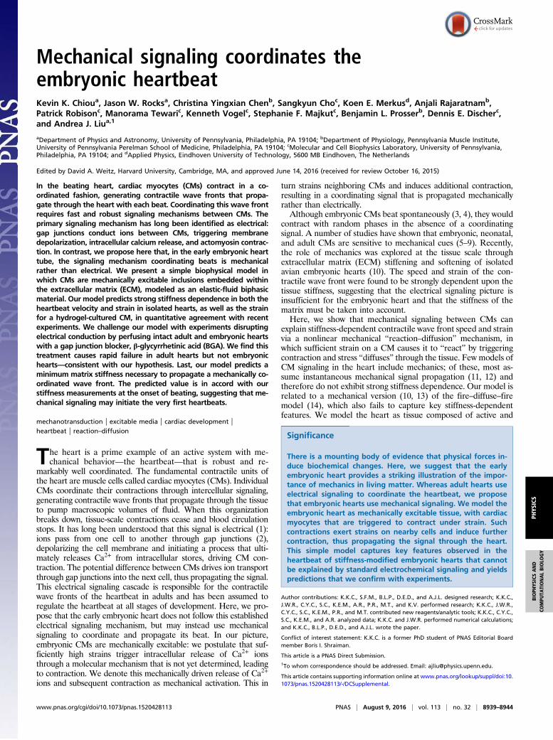
Physical Model of Cardiac Mechanical SignalingThe myocardium of the embryonic heart is composed primarilyof mechanically excitable CMs (that contract when activated)and the surrounding ECM. We treat CMs as elastic inclusionsembedded in the ECM, in accord with recent experiments ofCMs embedded in 3D hydrogels (20). We ignore direct cell–cellmechanical coupling; experimental evidence indicates that stressesare transmitted primarily through cell–matrix adhesions ratherthan cell–cell contacts during development (21), likely due to theprevalence of cell–junction remodeling. In addition, collagenasetreatment indicates that ECM is the primary component of tissuestructural integrity (10). We find that a good approximation(Materials and Methods) is to consider CMs as infinitesimal andarranged in a cubic array, spaced by Δx = 10 μm (Table 1). Weconsider a 3D mechanical version of fire-diffuse-fire signaling.This requires capturing the physics of (i) activated CMs creatingmechanical stress; (ii) stress propagation between CMs; and(iii) activation of quiescent CMs in response to mechanical stressin the ECM.
How Activated CMs Create Stress. We use two models to charac-terize the eigenstrain, i.e., the strain of an active inclusion (CM) inthe absence of external stresses. In the constant eigenstrain (CE)model, we assume that CMs contract with a fixed eigenstrain in-dependent of ECM stiffness. In the saturating eigenstrain (SE)model, the eigenstrain increases linearly with Young’s modulus Eup to a stall stiffness Es, and is independent of E for E>Es (Fig.1A, Inset). This behavior is observed for embryonic and neonatalCMs cultured on hydrogels (5, 7, 8, 22) and has been studiedtheoretically (23).The two models for the strain exerted by a CM when it con-
tracts are as follows:
epij = epf ðE=EsÞQij,fCEðE=EsÞ= 1 ∀E
fSEðE=EsÞ=�E=Es E<Es
1 E≥Es,
[1]
where Qij is the strain tensor representation of a uniaxial con-traction in the x direction (Supporting Information) and ep is themagnitude of the eigenstrain in the CE model or of the eigen-strain for E>Es in the SE model. See Fig. 1A, Inset.The eigenstrain epkl from the activated CM induces a stress in the
matrix. To properly capture the physical effects of differencesin stiffness between CMs and their surrounding ECM, we use
Table 1. Parameter symbols, references, and values
Parameter Symbol Value (fit/ref.)
Mesh/fluid drag Γ 0.4 mPa·s/μm2 (Fit)E4 myocardium modulus Ep 1.6 kPa (10)ECM Poisson ratio ν 0.4 (26)Fluid fraction (average) ϕ 0.8 (50, 51)Fluid viscosity (water, 25 °C) η 0.89 mPa·sCM spacing Δx 10 μm (10)CM modulus Ec 0.75 kPa (fit)CM eigenstrain magnitude ep 0.2 (6)CM strain threshold α 0.11 (fit)CM Poisson ratio νc 0.4 (26)Contraction time (AP duration) τ 250 ms
A
B
Fig. 1. Model for stress propagation in the myocardium. (A, Inset) CE and SEmodels as a function of ECM Young’s modulus, which determines the strengthof contraction (Eq. 1). (Main) The contracting CM (green) acts as a stress sourcefor a quiescent CM (white). An activated cell a contracts with an eigenstraine*cella,ij ðx′, t′Þ, locally inducing a stress σ*a,ijðx′, t′Þ in the ECM that depends on therelative stiffness between the ECM and CMs. We capture these physics via thetensor Tout
ijkl in accordance with the Eshelby theory of elastic inclusions (Sup-porting Information). This stress propagates according to the ECM responsefunction Gijklðx − x′, t − t′Þ (Supporting Information). The matrix stress at ðx, tÞdue to cell a is σa,ijðx, tÞ=
Rd3x′dt′Gijklðx − x′, t − t′Þσ*a,klðx′, t′Þ. This creates
ecella,ij ðx, tÞ, the strain induced in the quiescent CM due to the contraction of a(modified by T in
ijkl). (B) Sketch depicting quiescent (white) and activated (green)CMs in a traveling mechanical wave front at subsequent activation timesseparated by Δt. Arrows represent stresses propagated through the ECM (notall shown) to a quiescent CM, which activates when ecellii ðx, tÞ≥ α.
8940 | www.pnas.org/cgi/doi/10.1073/pnas.1520428113 Chiou et al.
Eshelby’s theory of elastic inclusions (24, 25). We compute thetensor Tout
ijkl ðEÞ, which relates the CM eigenstrain to the stress itinduces in the ECM, shown schematically in Fig. 1A (see Sup-porting Information for detailed calculations). The resulting ECMstress source due to an activated CM takes the following form:
σpa,ijðx, tÞ=Toutijkl e
pklΘðt− taÞΘðτ+ ta − tÞδ3ðx− xaÞ, [2]
where ΘðtÞ is the Heaviside function.
How Stress Propagates Between CMs. At the cellular length scaleand CM contraction velocity scale, the Reynolds number issmall (∼ 10−5). We therefore model the ECM as an overdamped,incompressible biphasic material. See Table 1 for parametervalues. It is composed of a linear elastic mesh [with Young’smodulus E and Poisson ratio ν= 0.4 (10, 26)] and interstitial fluid(of viscosity η similar to water). The fluid and elastic componentsare coupled through incompressibility and a drag term Γ, aneffect of matrix permeability to fluid. Similar approaches wereused to model collagenous tissue (27) and active gels (28). Usingthis model, we calculate the response function Gijklðx, tÞ todescribe propagation of mechanical stress within the ECM(Supporting Information).
How Quiescent CMs Are Activated Mechanically.We assume that, whenthe strain on a quiescent CM exceeds threshold α [ecellkk ðxq, tÞ≥ α],the CM is activated (it contracts). To describe this mathematically,we index noncontracting (quiescent) cells with q and contracting(active) cells with a. Each activated CM contracts for a physiolog-ically relevant amount of time τ before deactivating and becomingrefractory. We assume that the refractory timescale is longer thanmechanical relaxation, allowing us to ignore backpropagation. Letus consider a CM at xa that activated at time ta. Then ta is themoment when this CM’s strain trace first crossed the strain acti-vation threshold ecellkk ðxa, taÞ= α, transforming the originally quies-cent CM into an active one. The active CM contracts, creating aneigenstrain epkl (Eq. 1) for time ta < t< ta + τ, which can be repre-sented as a product of Heaviside functions.To relate the strain on an embedded quiescent CM q at ðxq, tÞ
due to local mechanical stress within the ECM, we compute thetensor T in
ijklðEÞ (also shown schematically in Fig. 1A) using elasticinclusion theory (Supporting Information). The strain contribu-tion on q from an activated CM a is then as follows:
ecella,ij
�xq, t
�=T in
ijkl
ZGklmn
�xq − x′; t− t′
�σpa,mnðx′, t′Þ, [3]
with σpa,mnðx′, t′Þ from Eq. 2. The total strain induced in q is thesum over the contribution from all activated cells ecellij ðxq, tÞ=P
aecella,ij ðxq, tÞ.
ResultsMechanical Signaling Model Yields Contractile Wave Fronts. Fromthe model, we calculate the velocity of the propagating con-tractile wave front as a function of matrix stiffness as follows.When a CM contracts, it creates a stress field σpa,ij in the ECM thatcan induce further contraction by activating quiescent CMs. Ifthe activation process cascades through the tissue, the resultingcontraction wave front can attain a comoving steady stateeijðx, tÞ= eijðx− vtÞ with velocity v. This is unsurprising becausethe model is a mechanical analog of nonlinear reaction-diffusion;such systems are well known to exhibit propagating wave frontsolutions. The activation condition ecellkk ðx, tÞ= α with a comovingsteady state relates the wave front velocity v to the model pa-rameters through an algebraic relation (Supporting Information).Once v is determined, we compute the maximal tissue strain bycoarse-grained solution of the waveform (Supporting Information).
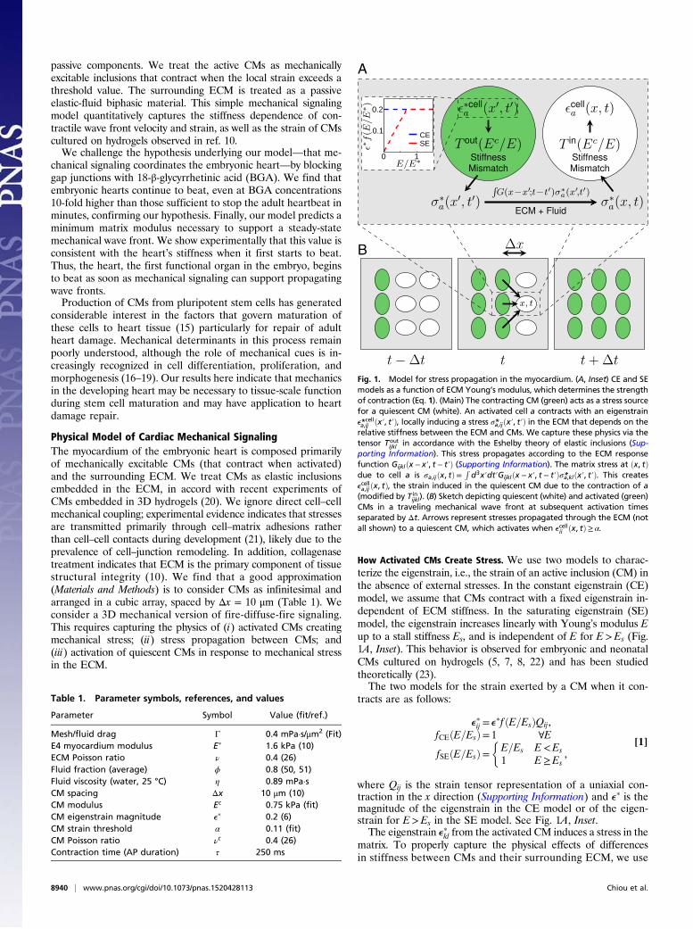
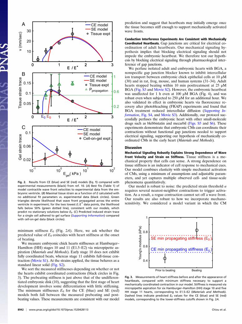
Mechanical Signaling Model Fits Experimental Wave Front Velocitieswith Physiologically Relevant Parameters. We obtain most of thephysiological parameter values from the literature (Table 1). Wetreat CMs as elastic inclusions with Young’s modulus Ec and withthe same Poisson ratio as the surrounding tissue (26) and esti-mate CM eigenstrain magnitude to be ep = 0.2 from intracellularembryonic CM principal strain measurements (6). Stress satu-ration stiffness is estimated to be the cell modulus Es =Ec (5, 6).Three model parameters could not be identified from the liter-ature and are fit via nonlinear regression to wave front velocitydata from ref. 10. These three parameters are the mesh-fluiddrag Γ, the CM activation threshold α, and the effective CMYoung’s modulus Ec. All three fit values (Table 1) fall withinphysiologically sensible ranges. The resulting velocity is plottedagainst ventricle contraction velocity data (black circles, from ref.10) of stiffness-modified embryonic day 4 (E4) hearts in Fig. 2A.No steady-state solution exists below stiffness E0 (which differsbetween CE and SE models). Physically, E0 arises because whenthe tissue is too soft, contracting CMs cannot provide enoughstrain to trigger additional contraction in quiescent CMs. This isconsistent with a significantly reduced likelihood of wave frontpropagation observed in experiment (green triangles in Fig. 2B).Likewise, the wave front velocity vanishes at high tissue stiffness,where the stiffness mismatch between CMs and the surroundingECM prevents contracting CMs from exerting sufficient strain onthe ECM to trigger contraction of quiescent cells.
Calculated Wave Front Strain Agrees with Experimental Observationswith No Additional Fitting Parameters. Using the three parameters(Γ,α,Ec) fit from wave front velocity data, we independently cal-culate the tissue strain of the contractile wave front and comparewith the measured maximal ventricular strain from ref. 10 (Fig.2B). Both the CE (blue) and SE (red) models are in excellentquantitative agreement with the observed behavior (black cir-cles) as a function of tissue stiffness, providing strong evidencein favor of our model. Note that the correct optimum stiffnessnaturally emerges from our model by treating CMs as elasticinclusions (Supporting Information) embedded within a surround-ing matrix of variable stiffness. This quantitative agreement issignificant and nontrivial, as we can observe from the differentvalues of E corresponding to optimum velocity and optimumstrain in experiment and model. Note also that no purely elec-trochemical model can correctly predict strain as a function ofstiffness.
SE Model Is Consistent with Cell-on-Gel Measurements with NoAdditional Fitting Parameters. We further test our model by com-paring to data for beating E4 CMs cultured on polyacrylamide gelwhere gel strain at cell edges was measured for varying gel stiffness(10). We calculate the trace of the 2D projected strain by finite-element simulation (Materials and Methods and Fig. S1) using thefit Ec value and comparing to experiment in Fig. 2C. The failure ofthe CE model on soft gels is expected from cultured CM experi-ments (7, 8, 22). Remarkably, we find agreement between the SEmodel with cell-on-gel measurements, demonstrating that we candeduce this single CM behavior as a function of E quantitativelyfrom collective behavior in tissue.
Mechanical Signaling Model Correctly Predicts Appearance of FirstHeartbeats. The developing heart stiffens with age due to increasedcollagen in the ECM (10). CMs begin periodic contractions at about1.5 d after fertilization (E1.5). At that point, the heart does not beatbut “shivers”; this shivering is similar to behavior observed instrain-activated contractile cell aggregates (29), which lack a sig-naling mechanism to coordinate the phases of the periodicallycontracting cells. The first fully coordinated beats do not occuruntil hours after CMs start contracting. Our model predicts thatcoordinated beats cannot appear until the matrix reaches the
Chiou et al. PNAS | August 9, 2016 | vol. 113 | no. 32 | 8941
PHYS
ICS
BIOPH
YSICSAND
COMPU
TATIONALBIOLO
GY
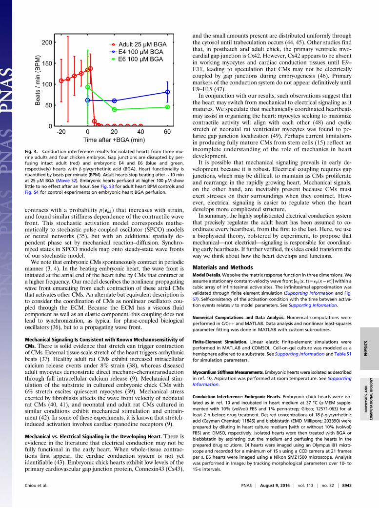
minimum stiffness E0 (Fig. 2A). Here, we ask whether thepredicted value of E0 coincides with heart stiffness at the onsetof beating.We measure embryonic chick hearts stiffnesses at Hamburger–
Hamilton (HH) stages 10 and 11 (E1.5–E2) via micropipette as-piration (Materials and Methods). Early stage 10 does not exhibitfully coordinated beats, whereas stage 11 exhibits full-tissue con-traction (Movie S1). At the strains applied, the tissue behaves as astandard linear solid (Fig. S2).We sort the measured stiffnesses depending on whether or not
the hearts exhibit coordinated contractions (black circles in Fig.3). The prebeating stiffness is just above that of the undifferen-tiated embryonic disk (10), suggesting that the first stage of heartdevelopment involves some differentiation with little stiffening.The minimum stiffnesses E0 for the CE (blue) and SE (red)models both fall between the measured prebeating and post-beating values. These measurements are consistent with our model
prediction and suggest that heartbeats may initially emerge oncethe tissue becomes stiff enough to support mechanically activatedwave fronts.
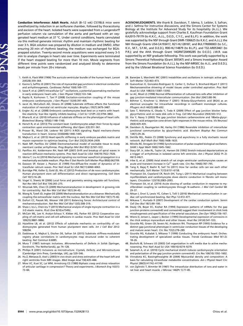
Conduction Interference Experiments Are Consistent with MechanicallyCoordinated Heartbeats. Gap junctions are critical for electrical co-ordination of adult heartbeats. Our mechanical signaling hy-pothesis implies that blocking electrical signaling should notimpede the embryonic heartbeat. We therefore test our hypoth-esis by blocking electrical signaling through pharmacological inter-ference of gap junctions.We perfuse isolated adult and embryonic hearts with BGA, a
nonspecific gap junction blocker known to inhibit intercellularion transport between embryonic chick epithelial cells at 10 μM(30) and in rat, frog, mouse, and human systems (31–34). Adulthearts stopped beating within 10 min posttreatment at 25 μMBGA (Fig. S3 and Movie S2). However, the embryonic heartbeatwas unaffected for 1 h even at 100 μM BGA (Fig. 4), and wasrobust even when subjected to 250 μM for an additional hour. Wealso validated its effect in embryonic hearts via fluorescence re-covery after photobleaching (FRAP) experiments and found thatBGA treatment reduced intercellular diffusion (Supporting In-formation, Fig. S4, and Movie S3). Additionally, our protocol suc-cessfully perfuses the embryonic heart with other small-moleculedrugs such as blebbistatin and mecarbil (Figs. S5 and S6). Theseexperiments demonstrate that embryonic CMs can coordinate theircontractions without functional gap junctions needed to supportelectrical signaling, supporting our hypothesis of mechanically co-ordinated CMs in the early heart (Materials and Methods).
DiscussionMechanical Signaling Robustly Explains Strong Dependence of WaveFront Velocity and Strain on Stiffness. Tissue stiffness is a me-chanical property that cells can sense. A strong dependence ontissue stiffness is an indicator of cell response to mechanical cues.Our model combines elasticity with simple mechanical activationof CMs, using a minimum of assumptions and adjustable param-eters, and yet captures multiple observed cell- and tissue-scalephenomena quantitatively.Our model is robust to noise: the predicted strain threshold α
requires several nearest-neighbor contractions to trigger activa-tion. As a result, a rogue contraction cannot set off a wave front.Our results are also robust to how we incorporate mechano-sensitivity. We considered a model variant in which the CM
Egel( kPa )10
-110
010
110
2
0.02
0.06
0.1CE modelSE modelCell-on-gel expt
10
20
30
0 1 2 3
0.05
0.1
0.15
E / E*
v (m
m/s
ec)
Tiss
ue s
train
trac
eG
el s
train
trac
eA
C
0.2
0.6
1
E0E00 1 2 3E / E*
0 1 2 3E / E*0 1 2 3E / E*
CE modelSE modelTissue expt
CE modelSE modelTissue exptPpropagation
B
Fig. 2. Results from CE (blue) and SE (red) models (Eq. 1) compared withexperimental measurements (black) from ref. 10. (A) Best fits (Table 1) ofmodel contractile wave front velocities to experimental data from the em-bryonic ventricle. (B) Maximal tissue strain as a function of E calculated withno additional fit parameters vs. experimental data (black circles). Greentriangles denote likelihood that wave front propagated across the entireventricle in experiment; for the two lowest E=E* data points, the likelihoodfalls below 50% (green dotted line), consistent with our models, whichpredict no stationary solutions below E0. (C) Predicted induced strain tracefor a single cell adhered to gel surface (Supporting Information) comparedwith cell-on-gel data (black circles).
Prior to beating Beating
Youn
g's
mod
ulus
(kP
a)
0.4
0.6
0.8
1
SE min propagating stiffness (E )0
CE min propagating stiffness (E )0
Fig. 3. Measurements of heart stiffness before and after the appearance ofheartbeats, compared with minimum stiffness necessary to support amechanically coordinated contraction in our model. Stiffness is measured viamicropipette aspiration for six Hamburger–Hamilton (HH) stage 10 and fiveHH stage 11 hearts, corresponding to E1.5–E2 (Materials and Methods).Dashed lines indicate predicted E0 values for the CE (blue) and SE (red)models, corresponding to the lower-stiffness cutoffs shown in Fig. 2A.
8942 | www.pnas.org/cgi/doi/10.1073/pnas.1520428113 Chiou et al.
contracts with a probability pðekkÞ that increases with strain,and found similar stiffness dependence of the contractile wavefront. This stochastic activation model corresponds mathe-matically to stochastic pulse-coupled oscillator (SPCO) modelsof neural networks (35), but with an additional spatially de-pendent phase set by mechanical reaction–diffusion. Synchro-nized states in SPCO models map onto steady-state wave frontsof our stochastic model.We note that embryonic CMs spontaneously contract in periodic
manner (3, 4). In the beating embryonic heart, the wave front isinitiated at the atrial end of the heart tube by CMs that contract ata higher frequency. Our model describes the nonlinear propagatingwave front emanating from each contraction of these atrial CMsthat activates other CMs. An alternate but equivalent description isto consider the coordination of CMs as nonlinear oscillators cou-pled through the ECM. Because the ECM has a viscous fluidcomponent as well as an elastic component, this coupling does notlead to synchronization, as typical for phase-coupled biologicaloscillators (36), but to a propagating wave front.
Mechanical Signaling Is Consistent with Known Mechanosensitivity ofCMs. There is solid evidence that stretch can trigger contractionof CMs. External tissue-scale stretch of the heart triggers arrhythmicbeats (37). Healthy adult rat CMs exhibit increased intracellularcalcium release events under 8% strain (38), whereas diseasedadult myocytes demonstrate direct mechano-chemotransductionthrough full intracellular calcium release (9). Mechanical stim-ulation of the substrate in cultured embryonic chick CMs with6% stretch excites quiescent myocytes (39). Mechanical stressexerted by fibroblasts affects the wave front velocity of neonatalrat CMs (40, 41), and neonatal and adult rat CMs cultured insimilar conditions exhibit mechanical stimulation and entrain-ment (42). In some of these experiments, it is known that stretch-induced activation involves cardiac ryanodine receptors (9).
Mechanical vs. Electrical Signaling in the Developing Heart. There isevidence in the literature that electrical conduction may not befully functional in the early heart. When whole-tissue contrac-tions first appear, the cardiac conduction system is not yetidentifiable (43). Embryonic chick hearts exhibit low levels of theprimary cardiovascular gap junction protein, Connexin43 (Cx43),
and the small amounts present are distributed uniformly throughthe cytosol until trabeculation occurs (44, 45). Other studies findthat, in posthatch and adult chick, the primary ventricle myo-cardial gap junction is Cx42. However, Cx42 appears to be absentin working myocytes and cardiac conduction tissues until E9–E11, leading to speculation that CMs may not be electricallycoupled by gap junctions during embryogenesis (46). Primarymarkers of the conduction system do not appear definitively untilE9–E15 (47).In conjunction with our results, such observations suggest that
the heart may switch from mechanical to electrical signaling as itmatures. We speculate that mechanically coordinated heartbeatsmay assist in organizing the heart: myocytes seeking to maximizecontractile activity will align with each other (48) and cyclicstretch of neonatal rat ventricular myocytes was found to po-larize gap junction localization (49). Perhaps current limitationsin producing fully mature CMs from stem cells (15) reflect anincomplete understanding of the role of mechanics in heartdevelopment.It is possible that mechanical signaling prevails in early de-
velopment because it is robust. Electrical coupling requires gapjunctions, which may be difficult to maintain as CMs proliferateand rearrange in the rapidly growing heart. Mechanical signals,on the other hand, are inevitably present because CMs mustexert stresses on their surroundings when they contract. How-ever, electrical signaling is easier to regulate when the heartdevelops more complicated structure.In summary, the highly sophisticated electrical conduction system
that precisely regulates the adult heart has been assumed to co-ordinate every heartbeat, from the first to the last. Here, we usea biophysical theory, bolstered by experiment, to propose thatmechanical—not electrical—signaling is responsible for coordinat-ing early heartbeats. If further verified, this idea could transform theway we think about how the heart develops and functions.
Materials and MethodsModel Details.We solve thematrix response function in three dimensions. Weassume a stationary constant-velocity wave front [eijðx, tÞ= eijðx − vtÞ] within acubic array of infinitesimal active sites. The infinitesimal approximation wasvalidated through finite element simulation (Supporting Information and Fig.S7). Self-consistency of the activation condition with the time between activa-tion events relates v to model parameters. See Supporting Information.
Numerical Computations and Data Analysis. Numerical computations wereperformed in C/C++ and MATLAB. Data analysis and nonlinear least-squaresparameter fitting was done in MATLAB with custom subroutines.
Finite-Element Simulation. Linear elastic finite-element simulations wereperformed in MATLAB and COMSOL. Cell-on-gel culture was modeled as ahemisphere adhered to a substrate. See Supporting Information and Table S1for simulation parameters.
Myocardium Stiffness Measurements. Embryonic hearts were isolated as describedin ref. 10. Aspiration was performed at room temperature. See SupportingInformation.
Conduction Interference: Embryonic Hearts. Embryonic chick hearts were iso-lated as in ref. 10 and incubated in heart medium at 37 °C (α-MEM supple-mented with 10% (vol/vol) FBS and 1% penn-strep; Gibco; 12571-063) for atleast 2 h before drug treatment. Desired concentrations of 18-β-glycyrrhetinicacid (Cayman Chemical; 11845) and blebbistatin (EMD Millipore; 203390) wereprepared by diluting in heart culture medium [with or without 10% (vol/vol)FBS] and DMSO, respectively. Isolated hearts were then treated with BGA orblebbistatin by aspirating out the medium and perfusing the hearts in theprepared drug solutions. E4 hearts were imaged using an Olympus I81 micro-scope and recorded for a minimum of 15 s using a CCD camera at 21 framesper s. E6 hearts were imaged using a Nikon SMZ1500 microscope. Analysiswas performed in ImageJ by tracking morphological parameters over 10- to15-s intervals.
Time after +BGA (min)
Beat
s / m
in (B
PM)
-20 0 20 40 600
50
100
150
200 Adult 25 μM BGAE4 100 μM BGAE6 100 μM BGA
Fig. 4. Conduction interference results for isolated hearts from three mu-rine adults and four chicken embryos. Gap junctions are disrupted by per-fusing intact adult (red) and embryonic E4 and E6 (blue and green,respectively) hearts with β-glycyrrhetinic acid (BGA). Heart functionality isquantified by beats per minute (BPM). Adult hearts stop beating after ∼10 minat 25 μM BGA (Movie S2). Embryonic hearts perfused at higher 100 μM showlittle to no effect after an hour. See Fig. S3 for adult heart BPM controls andFig. S4 for control experiments on embryonic heart BGA perfusion.
Chiou et al. PNAS | August 9, 2016 | vol. 113 | no. 32 | 8943
PHYS
ICS
BIOPH
YSICSAND
COMPU
TATIONALBIOLO
GY
Conduction Interference: Adult Hearts. Adult (8–12 wk) C57/BL6 mice wereanesthetized by induction in an isoflurane chamber, followed by thoracotomyand excision of the heart. Isolated hearts were suspended from a Langendorffperfusion column via cannulation of the aorta and perfused with an oxy-genated heart medium at 37 °C. Under control conditions, hearts cannulatedvia this method generate intrinsic rhythm and maintain rhythmic beating forover 3 h. BGA solution was prepared by dilution in medium and DMSO. Afterensuring 20 min of rhythmic beating, the medium was exchanged for BGA-prepped solution. Twenty-second movie acquisitions were acquired every 3–5min to analyze changes in heart rate over time. Experiments were terminatedif the heart stopped beating for more than 10 min. Movie segments fromdifferent time points were randomized and analyzed blindly to determinebeats per minute from 20-s intervals.
ACKNOWLEDGMENTS. We thank B. Davidson, T. Idema, S. Leibler, S. Safran,and J. Sethna for instructive discussions, and the Simons Center for SystemsBiology at the Institute for Advanced Study for its hospitality (A.J.L.). Wegratefully acknowledge support from Charles E. Kaufman Foundation GrantKA2015-79179 (to K.K.C., A.J.L., D.E.D., C.Y.C., and B.L.P.). In addition, the workwas supported by the NSF through Grant DMR-1506625 (to K.K.C. and A.J.L.); theNIH through Grants U54-CA193417, R21-HL128187, and 8UL1TR000003 (to S.C.,K.V., M.T., S.F.M., and D.E.D.), R00-HL114879 (to B.L.P.), and T32-AR053461 (toP.R.); and the AHA through Grant 14GRNT20490285 (to D.E.D.). J.W.R. wassupported by an NSF graduate fellowship. This work was partially supported by aSimons Theoretical Fellowship (Grant 305547) and a Simons Investigator Awardfrom the Simons Foundation (to A.J.L.), by the NSF-MRSEC (to A.J.L. and D.E.D.),and by the US/Israel Binational Science Foundation (to D.E.D.).
1. Keith A, Flack MW (1906) The auriculo-ventricular bundle of the human heart. Lancet2(4328):359–364.
2. Kanno S, Saffitz JE (2001) The role of myocardial gap junctions in electrical conductionand arrhythmogenesis. Cardiovasc Pathol 10(4):169–177.
3. Sasse P, et al. (2007) Intracellular Ca2+ oscillations, a potential pacemaking mechanismin early embryonic heart cells. J Gen Physiol 130(2):133–144.
4. Rapila R, Korhonen T, Tavi P (2008) Excitation-contraction coupling of the mouseembryonic cardiomyocyte. J Gen Physiol 132(4):397–405.
5. Jacot JG, McCulloch AD, Omens JH (2008) Substrate stiffness affects the functionalmaturation of neonatal rat ventricular myocytes. Biophys J 95(7):3479–3487.
6. Engler AJ, et al. (2008) Embryonic cardiomyocytes beat best on a matrix with heart-like elasticity: Scar-like rigidity inhibits beating. J Cell Sci 121(Pt 22):3794–3802.
7. Bhana B, et al. (2010) Influence of substrate stiffness on the phenotype of heart cells.Biotechnol Bioeng 105(6):1148–1160.
8. Hersch N, et al. (2013) The constant beat: Cardiomyocytes adapt their forces by equalcontraction upon environmental stiffening. Biol Open 2(3):351–361.
9. Prosser BL, Ward CW, Lederer WJ (2011) X-ROS signaling: Rapid mechano-chemotransduction in heart. Science 333(6048):1440–1445.
10. Majkut S, et al. (2013) Heart-specific stiffening in early embryos parallels matrix andmyosin expression to optimize beating. Curr Biol 23(23):2434–2439.
11. Nash MP, Panfilov AV (2004) Electromechanical model of excitable tissue to studyreentrant cardiac arrhythmias. Prog Biophys Mol Biol 85(2-3):501–522.
12. Panfilov AV, Keldermann RH, Nash MP (2007) Drift and breakup of spiral waves inreaction-diffusion-mechanics systems. Proc Natl Acad Sci USA 104(19):7922–7926.
13. Idema T, Liu AJ (2014) Mechanical signaling via nonlinear wavefront propagation in amechanically excitable medium. Phys Rev E Stat Nonlin Soft Matter Phys 89(6):062709.
14. Dawson SP, Keizer J, Pearson JE (1999) Fire-diffuse-fire model of dynamics of in-tracellular calcium waves. Proc Natl Acad Sci USA 96(11):6060–6063.
15. Burridge PW, Keller G, Gold JD, Wu JC (2012) Production of de novo cardiomyocytes:Human pluripotent stem cell differentiation and direct reprogramming. Cell StemCell 10(1):16–28.
16. Vogel V, Sheetz M (2006) Local force and geometry sensing regulate cell functions.Nat Rev Mol Cell Biol 7(4):265–275.
17. Wozniak MA, Chen CS (2009) Mechanotransduction in development: A growing rolefor contractility. Nat Rev Mol Cell Biol 10(1):34–43.
18. Wang N, Tytell JD, Ingber DE (2009) Mechanotransduction at a distance: Mechanicallycoupling the extracellular matrix with the nucleus. Nat Rev Mol Cell Biol 10(1):75–82.
19. DuFort CC, Paszek MJ, Weaver VM (2011) Balancing forces: Architectural control ofmechanotransduction. Nat Rev Mol Cell Biol 12(5):308–319.
20. Shaw J, Izu L, Chen-Izu Y (2013) Mechanical analysis of single myocyte contraction in a3-D elastic matrix. PLoS One 8(10):e75492.
21. McCain ML, Lee H, Aratyn-Schaus Y, Kléber AG, Parker KK (2012) Cooperative cou-pling of cell-matrix and cell-cell adhesions in cardiac muscle. Proc Natl Acad Sci USA109(25):9881–9886.
22. Hazeltine LB, et al. (2012) Effects of substrate mechanics on contractility of car-diomyocytes generated from human pluripotent stem cells. Int J Cell Biol 2012:508294.
23. Dasbiswas K, Majkut S, Discher DE, Safran SA (2015) Substrate stiffness-modulatedregistry phase correlations in cardiomyocytes map structural order to coherentbeating. Nat Commun 6:6085.
24. Mura T (1987) Isotropic inclusions. Micromechanics of Defects in Solids (Springer,Dordrecht, The Netherlands), pp 74–128.
25. Phillips R (2001) Inclusions as microstructure. Crystals, Defects, and Microstructures(Cambridge Univ Press, Cambridge, UK), pp 520–546.
26. Hu Z, Metaxas D, Axel L (2003) In vivo strain and stress estimation of the heart left andright ventricles from MRI images. Med Image Anal 7(4):435–444.
27. Mow VC, Kuei SC, Lai WM, Armstrong CG (1980) Biphasic creep and stress relaxationof articular cartilage in compression? Theory and experiments. J Biomech Eng 102(1):73–84.
28. Banerjee S, Marchetti MC (2001) Instabilities and oscillations in isotropic active gels.Soft Matter 7(2):463–473.
29. Guevorkian K, Gonzalez-Rodriguez D, Carlier C, Dufour S, Brochard-Wyart F (2011)Mechanosensitive shivering of model tissues under controlled aspiration. Proc NatlAcad Sci USA 108(33):13387–13392.
30. Le AC, Musil LS (1998) Normal differentiation of cultured lens cells after inhibition ofgap junction-mediated intercellular communication. Dev Biol 204(1):80–96.
31. Böhmer C, Kirschner U, Wehner F (2001) 18-beta-Glycyrrhetinic acid (BGA) as anelectrical uncoupler for intracellular recordings in confluent monolayer cultures.Pflugers Arch 442(5):688–692.
32. Sato T, Nishishita K, Okada Y, Toda K (2009) Effect of gap junction blocker beta-glycyrrhetinic acid on taste disk cells in frog. Cell Mol Neurobiol 29(4):503–512.
33. Xia Y, Nawy S (2003) The gap junction blockers carbenoxolone and 18beta-glycyr-rhetinic acid antagonize cone-driven light responses in the mouse retina. Vis Neurosci20(4):429–435.
34. Davidson JS, Baumgarten IM, Harley EH (1986) Reversible inhibition of intercellularjunctional communication by glycyrrhetinic acid. Biochem Biophys Res Commun134(1):29–36.
35. DeVille REL, Peskin CS (2008) Synchrony and asynchrony in a fully stochastic neuralnetwork. Bull Math Biol 70(6):1608–1633.
36. Mirollo RE, Strogatz SH (1990) Synchronization of pulse-coupled biological oscillators.SIAM J Appl Math 50(6):1645–1662.
37. Stacy GP, Jr, Jobe RL, Taylor LK, Hansen DE (1992) Stretch-induced depolarizations asa trigger of arrhythmias in isolated canine left ventricles. Am J Physiol 263(2 Pt 2):H613–H621.
38. Iribe G, et al. (2009) Axial stretch of rat single ventricular cardiomyocytes causes anacute and transient increase in Ca2+ spark rate. Circ Res 104(6):787–795.
39. Tang X, Bajaj P, Bashir R, Saif TA (2011) How far cardiac cells can see each othermechanically. Soft Matter 7(13):6151–6158.
40. Thompson SA, Copeland CR, Reich DH, Tung L (2011) Mechanical coupling betweenmyofibroblasts and cardiomyocytes slows electric conduction in fibrotic cell mono-layers. Circulation 123(19):2083–2093.
41. Thompson SA, et al. (2014) Acute slowing of cardiac conduction in response to my-ofibroblast coupling to cardiomyocytes through N-cadherin. J Mol Cell Cardiol 68:29–37.
42. Nitsan I, Drori S, Lewis YE, Cohen S, Tzlil S (2016) Mechanical communication in car-diac cell synchronized beating. Nat Phys 12:472–477.
43. Mikawa T, Hurtado R (2007) Development of the cardiac conduction system. SeminCell Dev Biol 18(1):90–100.
44. Dealy CN, Beyer EC, Kosher RA (1994) Expression patterns of mRNAs for the gapjunction proteins connexin43 and connexin42 suggest their involvement in chick limbmorphogenesis and specification of the arterial vasculature. Dev Dyn 199(2):156–167.
45. Wiens D, Jensen L, Jasper J, Becker J (1995) Developmental expression of connexins inthe chick embryo myocardium and other tissues. Anat Rec 241(4):541–553.
46. Gourdie RG, Green CR, Severs NJ, Anderson RH, Thompson RP (1993) Evidence for adistinct gap-junctional phenotype in ventricular conduction tissues of the developingand mature avian heart. Circ Res 72(2):278–289.
47. Gourdie RG, Kubalak S, Mikawa T (1999) Conducting the embryonic heart: Orches-trating development of specialized cardiac tissues. Trends Cardiovasc Med 9(1-2):18–26.
48. Bischofs IB, Schwarz US (2003) Cell organization in soft media due to active mecha-nosensing. Proc Natl Acad Sci USA 100(16):9274–9279.
49. Salameh A, et al. (2010) Cyclic mechanical stretch induces cardiomyocyte orientationand polarization of the gap junction protein connexin43. Circ Res 106(10):1592–1602.
50. Vinnakota KC, Bassingthwaighte JB (2004) Myocardial density and composition: Abasis for calculating intracellular metabolite concentrations. Am J Physiol Heart CircPhysiol 286(5):H1742–H1749.
51. von Zglinicki T, Bimmler M (1987) The intracellular distribution of ions and water inrat liver and heart muscle. J Microsc 146(Pt 1):77–85.
8944 | www.pnas.org/cgi/doi/10.1073/pnas.1520428113 Chiou et al.
Supporting InformationChiou et al. 10.1073/pnas.1520428113Linearized Biphasic ModelIn the linearized biphasic model composed of fluid and elasticmesh, we assume that the fluid and elastic components mutuallyexclude each other and that the total volume is fixed. Then for aninfinitesimal volume element at x, if the fluid volume fraction is ϕthe elastic mesh must occupy 1−ϕ. As a result, within the vol-ume element at x neither fluid nor mesh network satisfy theregular incompressible condition, because fluid and mesh mu-tually displace each other. In our model, we take into accountthe fluid dynamic viscosity, mesh elasticity, fluid–mesh drag cou-pling, and the total volume (fluid plus mesh) constraint to linearorder in the dynamic variables:
Γð _ui − viÞ= E2ð1+ νÞ
�∂2ui +
11− 2ν
∂i∂juj�− ð1−ϕÞ∂ip
Γðvi − _uiÞ= η
�∂2vi +
13∂i∂jvj
�−ϕ∂ip
0= ð1−ϕÞ∂i _ui +ϕ∂ivi.
[S1]
Because we are interested primarily in comparing to experimentalwave front speeds and tissue strain trace, for brevity we detail onlythe strain trace and strain rate trace equations here. The fluid andsolid (traces of) strain rates and strain are _ef ,ii = ∂ivi, _es,ii = ∂i _ui, andes,ii = ∂iui. For physiological values of ϕ≈ 0.8 in muscle tissue(Table 1), we solve the equations:
Γ�_es − _ef
�=
Eð1− νÞð1+ νÞð1− 2νÞ∂
2es − ð1−ϕÞ∂2p
Γ�_ef − _es
�=43η∂2 _ef −ϕ∂2p
_ef =− _es1−ϕ
ϕ.
[S2]
Then solving in terms of es, we obtain the equation of motion:"_es −
4ð1−ϕÞ2η3Γ
∂2 _es
#=D∂2es, [S3]
where D=ϕ2Eð1− νÞ=ðΓð1+ νÞð1− 2νÞÞ. We assume that the in-terstitial fluid viscosity is approximately the viscosity of waterat body temperature. Best fits (Results) then indicate that Γ≈45ðη=Δx2Þ, where Δx the spacing between myocytes. As a result,the fluid–mesh drag Γ is dominant over fluid viscosity at physicallength scales of interest, and the second term on the left-handside in Eq. S3 can be dropped. This result is used throughout thetext. For our parameter values, the linearized equations are sta-ble to perturbations at the onset of propagation. In contrast tostandard reaction–diffusion models, but consistent with fire–dif-fuse–fire [14] models, the nonlinear character of transitions be-tween the nonpropagating and propagating phases is a result ofthreshold activation and as a result is not captured by linearstability analysis.
Induced Strain of Eshelby InclusionsHere, we detail the stresses induced by CMs, which are treatedas spherical Eshelby inclusions (elastic inclusions with elasticconstants that may be different from the surrounding matrix).We compute two tensors, Tout
ijkl and T inijkl, which correspond (re-
spectively) to strain induced in the matrix due to an active in-clusion, and the strain felt by a passive inclusion due to strainin the matrix (Fig. 1A). Because we model CM sources as in-finitesimal, incoming stresses are treated as far-field. Note thatwe have checked this approximation using finite-element calcu-lations (SI Materials and Methods, CMs-in-Matrix Finite-ElementMethod Simulations, and Fig. S7), and find that it is quantita-tively quite accurate. We also assume that, near an excited in-clusion, the matrix responds quickly; this allows us to make useof the static Eshelby tensor.Suppose a cell, represented as an ellipsoidal inclusion with
stiffness tensor Cijkl, is embedded in an infinite bulk medium (i.e.,the tissue) with the same stiffness tensor. Although we use thegeneral form, in the isotropic case this tensor is determined byjust two independent parameters, which we call the Young’smodulus E and Poisson ratio ν in the main text. Suppose the cellcontracts uniformly throughout its volume with an eigenstressσpcellij . We equivalently represent this contraction strength as aneigenstrain epcellij , related to the stress by σpcellij =Cijkle
pcellkl . This
eigenstrain can be thought of as how much the cell wouldstrain if the surroundings exerted no stress on the cell. ByEshelby inclusion theory, the observed strain of an embeddedinclusion is related to the eigenstrain by the following:
epij = Sijklepcellkl , [S4]
where Sijkl is the Eshelby tensor [24]. For a spherical inclusion,this tensor is uniform and given by the following:
Sijkl =1+ ν
3ð1− νÞ��v1�ijkl + 2ð4− 5νÞ
15ð1− νÞ��v2�ijkl, [S5]
where jv1iijkl and jv2iijkl are defined for convenience as follows:
��v1�ijkl = 13δijδkl
��v2�ijkl = 12
�δikδjl + δilδjk −
23δijδkl
�.
[S6]
Stress Locally Induced in Matrix by Eshelby Inclusion. Now supposethe cell and tissue have different stiffness tensors Cc
ijkl and Cijkl,respectively. As before, an isotropic medium reduces the in-dependent parameters to E and ν. Assume the cell contracts witha stress σpcellij =Cc
ijklepcellkl . The total stress inside the cell embedded
in the tissue is related to the strain by the following:
σTij =Ccijkl
�epkl − epcellkl
�. [S7]
By superposition, we can map this to a problem to the Eshelbytensor for a homogeneous material with an additional strainsource [24]. This is done by imposing an effective strain sourcein the cell epeffij such that we get identical stresses and strains asthe inhomogeneous problem. The stress–strain relation then be-comes the following:
σTij =Cijkl
�epkl − epeffkl
. [S8]
Note that the effective strain source epeffij does not actually existbut is just a mathematical trick to reduce the inhomogeneous
Chiou et al. www.pnas.org/cgi/content/short/1520428113 1 of 9
inclusion problem to the previously solved homogeneous inclu-sion problem. Eshelby’s solution now tells us that the strainexperienced in the cell embedded in the tissue is as follows:
epij = Sijklepeffkl , [S9]
with the Eshelby tensor calculated using Cijkl. Now we calculatethe effective source epeffij by equating Eqs. S7 and S8, to identifythe (actual) inhomogeneous case with the (constructed) homo-geneous problem:
Ccijkl
�epkl − epcellkl
=Cijkl
�epkl − epeffkl
. [S10]
Substituting in Eq. S9, we get an equation for epeffij ,
h�Ccijkl −Cijkl
Sklmn +Cijmn
�iepoffmn =Cc
ijkl epcellkl . [S11]
Solving for epeffij in terms of epcellij , we find epij (via Eq. S9). We cannow calculate the stress source in the homogeneous case σpij =Cijkle
pkl. We define the tensor Tout
ijkl to relate this stress to our cellstrain source,
σpij =Toutijkl e
pcellkl , [S12]
as in Fig. 1A. For a spherical inclusion and isotropic matrixwith Young’s moduli Ec and E and Poisson ratios νc and ν,respectively,
Toutijkl =
24 E
ð1− 2νÞð1− νÞð1+ νÞ
1+ 2 ð1− 2νcÞð1+ νÞ
EEc
35��v1�ijkl
+
24 15E
4ð1+ νÞð1− νÞð4− 5νÞ
1+ 12ð7− 5νÞð4− 5νÞ
ð1+ νcÞð1+ νÞ
EEc
35��v2�ijkl
=E
3ð1− 2νÞTout1
��v1�ijkl + E2ð1+ νÞT
out2
��v2�ijkl,[S13]
where Tout1 and Tout
2 are dimensionless quantities, with prefactorsof the tissue bulk and shear moduli.
Stress Induced in Eshelby Inclusion by Uniform Stress in Matrix. Asin the previous section, we can likewise compute the strain in acell given some homogeneous stress σij =Cijklekl throughout theneighborhood of tissue surrounding the inclusion. The primarydifference is that this system now exists under a prestress con-dition without a cell source. As before, cell and tissue stiffnesstensors are Cc
ijkl and Cijkl. The total stress inside the cell is givenby the following:
σTij =Ccijkle
cellkl . [S14]
By linearity, the strain in the cell can be broken up into two com-ponents: the homogeneous strain in the surrounding tissue eij, andthe effects of the inclusion’s stiffness mismatch with the tissue eIij,
ecellij = eij + eIij. [S15]
As in the previous section, we map this inhomogeneous problemto a homogeneous one with additional cell strain source epeffij , butthe same total stress and strain:
σTij =Cijkl
�ecellkl − epeffkl
. [S16]
Eshelby’s solution now tells us that the contribution to the strainfrom the inclusion is as follows:
eIij = Sijklepeffkl . [S17]
Equating Eqs. S14 and S16, and using the relation from Eq. S15,
Ccijkl
�ekl + eIkl
�=Cijkl
�ekl + eIkl − epeffkl
. [S18]
Substituting in Eq. S17, we get an equation for epeffij ,h�
Ccijkl −Cijkl
Sklmn +Cijmn
�iepeffmn =
�Cijkl −Cc
ijkl
ekl. [S19]
Solving for epeffij in terms of eij, we find eIij via Eq. S17. This givesthe cell strain ecellij . Using the relation σij =Cijklekl for the homo-geneous case, we express ecellij in terms of σij. We define T in
ijkl torelate this stress to the strain in the cell as follows:
ecellij =T inijklσkl, [S20]
as in Fig. 1A. For a spherical inclusion and isotropic matrix withYoung’s moduli Ec and E and Poisson ratios νc and ν,
T inijkl =
3ð1− 2νÞE
241+
ð1− 2νcÞð1− 2νÞ
EEc − 1
1+ 2 ð1− 2νcÞð1+ νÞ
EEc
35��v1�ijkl
+2ð1+ νÞ
E
241+
ð1+ νcÞð1− νÞ
EEc − 1
1+ 12ð7− 5νÞð4− 5νÞ
ð1+ νcÞð1+ νÞ
EEc
35��v2�ijkl
=3ð1− 2νÞ
ET in1
��v1�ijkl + 2ð1+ νÞE
T in2
��v2�ijkl,
[S21]
where T in1 and T in
2 are dimensionless quantities, with prefactorsof the inverse tissue bulk and shear moduli.
Activation ConditionIn our model, when CMs contract they generate an eigenstresscorresponding to a negative force dipole in the x direction. Thisassumption is based on the observation that E4 CMs exhibit stria-tions, which polarize contraction, and serves to simplify analysis.An activated CM in our model has an eigenstrain epkl and inducesa matrix stress σpij:
epkl = ep
0@−1 0 0
0 νc 00 0 νc
1A, σpij =Tout
ijkl epkl. [S22]
Given a stress source σpa,klðr′, t′Þ indexed by a in the biphasicmedium, the strain trace induced in an included cell at ðr, tÞ isas follows:
ecella,ii ðr, tÞ=1− 2νE
T in1
Zdt′d3r′Giiklðr− r′; t− t′Þσ*a,klðr′, t′Þ, [S23]
for the Green’s function Gijkl. Combining Eqs. S12 and 3,
σpa,ijðr′, t′Þ=Toutijkl e
pa,klðr′, t′Þ
=Toutijkl e
pklΘðt− taÞΘðτ+ ta − tÞδ3ðx− xaÞ.
[S24]
Chiou et al. www.pnas.org/cgi/content/short/1520428113 2 of 9
For a quiescent cell at ðr, tÞ to reach the activation threshold α, weconsider all activated CMs (indexed here by a) as contributing tothe local strain. This requires the following:
ecellii ðr, tÞ=Xa
ecella,ii ðr, tÞ= α. [S25]
Consider a steady-state wave front traveling in the x directionthrough a cubic array of CMs spaced by Δx as in Fig. 1. We labeleach CM with lattice indices ½nx,ny, nz�. Steady state assumesCMs with the same x coordinate activate simultaneously with aconstant (but unknown) time interval Δt between activations atneighboring sites ðx, y, zÞ and ðx+Δx, y, zÞ.We express the activation positions and times as ra = ½nxa, nya,nza�TΔx
and ta = nxaΔt where the index nx is in the propagation directionx. In conjunction with the activation condition (Eq. 3),
EP
aecella,ii
ð1− 2νÞT in1=Xa
Zdt′d3r′Giiklðr− r′; t− t′Þσpa,klðr′, t′Þ
=X
nx , ny , nz
Zdt′Giikl
0BBB@r−
26664nx
ny
nz
37775Δx; t− t′
1CCCA½Θðt′− nxΔtÞ
−Θðt′− nxΔt− τÞ�Toutklmne
pmn,
[S26]
for nx ∈ f1,2, . . . ,∞g and ny, nz ∈ f−∞, . . . ,−1,0,1, . . . ,∞g andthe activation eigenstrain epmn as defined in main-text Eq. 1.Using the Green’s function Gijklðr, tÞ for Eq. S3, we find thatthe response to a Heaviside point force dipole ∼ δ3ðrÞΘðtÞ isas follows:
Hiiklðr, tÞ=Z
dt′Giiklðr, t− t′ÞΘðt′Þ
=1
4πΓDr3
hH1ðr, tÞδkl +H2ðr, tÞ rkrlr2
iΘðtÞ,
H1 = erfc
rffiffiffiffiffiffiffiffi4Dt
p�+
2rffiffiffiffiffiffiffiffiffiffi4πDt
p e−r24Dt
H2 =−3erfc
rffiffiffiffiffiffiffiffi4Dt
p�−
6rffiffiffiffiffiffiffiffiffiffi4πDt
p +4r2
4πDt
�e−
r24Dt,
[S27]
in the limit where Γ � ηq2. Here,D=ϕ2Eð1− νÞ=ðΓð1+ νÞð1− 2νÞÞas before. Combining Eqs. S25–S27, we find a transcendental re-lation for Δt in terms of CM and ECM parameters:
Eα1− 2ν
=T in1
Xa
ðHiiklðr− ra; t− taÞ−Hiiklðr− ra; t− ta − τÞToutklmne
pmn.
[S28]
We truncate the sum when the estimated relative error δ< 10−4,which corresponds to jmj, jnj, joj< 20 for physiological parame-ters. We use Newton–Raphson to solve numerically for Δt toobtain the steady-state wave front velocity, v=Δx=Δt. The cubiclattice approximation was chosen due to its simple analytic so-lution. We found that the steady-state wave front behavior de-pends primarily on average spacing between active sites and notthe chosen lattice.
Tissue Strain CalculationWe calculate the wave front strain trace with a coarse-grained,stationary wave back solution. Consider Eq. S3 in the limitη=ðΓΔx2Þ � 1. We assume a steady-state traveling wave fronteðx, y, z, tÞ= eðx− vt, y, zÞ with a comoving excitation front withvelocity v driven by the activation condition solved above. Be-cause the sites behind the wave front are refractory, there are noother stress sources other than the activation front. The equa-tions of motion then become the following:
−v∂xekkðx− vt, y, zÞ=D∂2ekkðx− vt, y, zÞ
−σpijΓ∂i∂j
Xa
δ3ðx− xa − vt, y− ya, z− zaÞ,
[S29]
where D=ϕ2Eð1− νÞ=ðΓð1+ νÞð1− 2νÞÞ; σpij =Toutijkl e
pkl, the stress
generated in the matrix by each contracting myocyte; and a,the activated sites. Assuming a cubic lattice in the comovingframe, the number of activated sites N in x corresponds to thewidth of the activated front, N = vτ=Δx. So we take the following:
Xa
=X
nx , ny , nz, [S30]
where nx ∈ f1, . . . ,Ng, ny, nz ∈ f−∞, . . . ,∞g. In comoving Four-ier space, we find that the strain trace becomes the following:
~ekk =�−ivkx − k2D
�−1σpijkikjΓ
Xnx , ny , nz
e−iΔxðkxnx+kyny+kznzÞ. [S31]
To compute the tissue-scale limit, we coarse-grain the systemand consider length scales 1=jkj � Δx. We can then express thesum over all activated sites as follows:
Xnx, ny , nz
e−iΔxðkxnx+kyny+kznzÞ = 1
ðΔxÞ3 XN
nx=0
Δxe−inxkxΔx
!
3
X∞ny , nz=−∞
ðΔxÞ2e−iðnyky+nzkzÞΔx!.
[S32]
We then replace the sums with integrals. Let us call x′= nxΔx, y′=nyΔx, z′= nzΔx, Δx the spacing between myocytes. In the contin-uum limit:
1
ðΔxÞ3Z 0
−vτdx′e−ikxx′
Z ∞
−∞dy′dz′e−iðkyy′+kzz′Þ
=ð2πÞ2
ikxðΔxÞ3 1− eikxvτ
�δ�ky�δðkzÞ. [S33]
Substituting this into Eq. S31, we observe that the infinitenumber of excitations in y and z only contribute in the infinitewavelength limit ky, kz → 0 and these components are triviallyintegrated out. The inverse transform in kx is a simple contourintegral with poles no higher than second order. Computing thisgives the tissue scale response in terms of the individual CMexcitation strength:
ekk =σpxx
DΓðΔxÞ3 ×8<:
exp� vDðx− vt+ vτÞ
x< vðt− τÞ
1 vðt− τÞ< x< vt, [S34]
Chiou et al. www.pnas.org/cgi/content/short/1520428113 3 of 9
where σpxx=ðΔxÞ3 is associated with the microscopic stress exertedby each CM. The coefficient in Eq. S34 is the maximal tissuestrain of the contracting wave front. We note that this solutionfails for x> vt because for those positions the tissue is not passive:CMs are primed for activation.
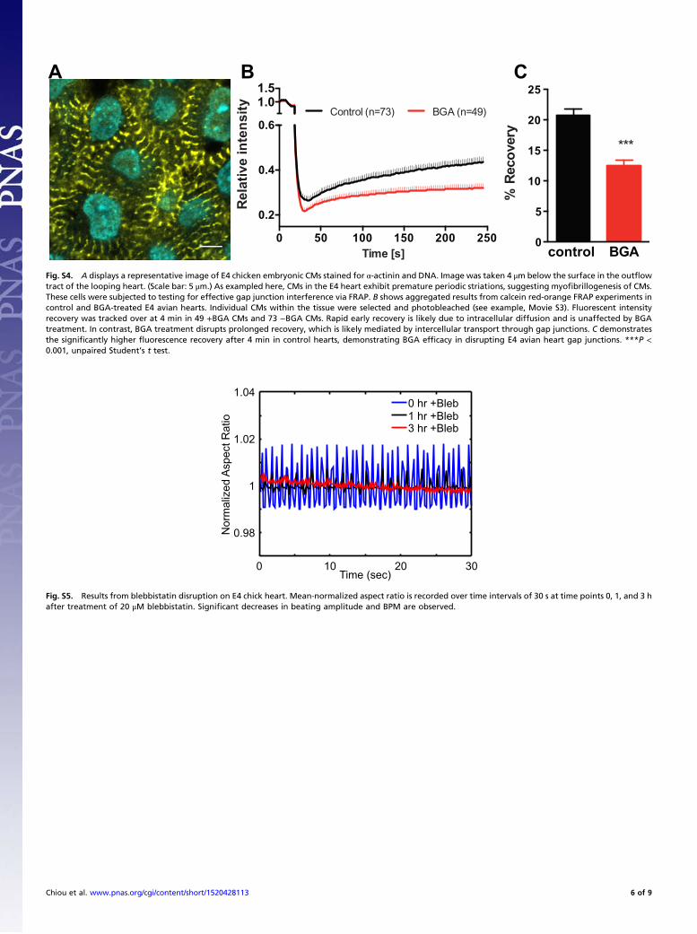
β-Glycyrrhetinic Acid Obstructs Intercellular Transport inEmbryonic Cardiac TissueTo verify that β-glycyrrhetinic acid (BGA) properly interfereswith gap junctions in embryonic CMs, we performed fluores-cence recovery after photobleaching (FRAP) experiments toverify reduced intercellular transport between myocytes. Throughα-actinin staining, we first verified that the cells of interest exhibitedcharacteristic striations found in myocytes and myocyte precursors(Fig. S4A). E4 chick hearts were loaded with calcein red-orangeAM, a cell tracer. This compound is cleaved as it enters cells,rendering it membrane impermeable. Myocytes are selected withinthe tissue and bleached down to 20% of the initial intensity.Fluorescence recovery was recorded and analyzed via (software)and ImageJ (Movie S3). Quantitative FRAP results are shown inFig. S4 B and C. BGA-treated hearts systematically exhibited CMs(n = 49) with significantly reduced fluorescence recovery in rate andintensity compared with control (n = 73), demonstrating that BGAdoes indeed interfere with gap junctions in E4 avian heart tissue.See SI Materials and Methods.
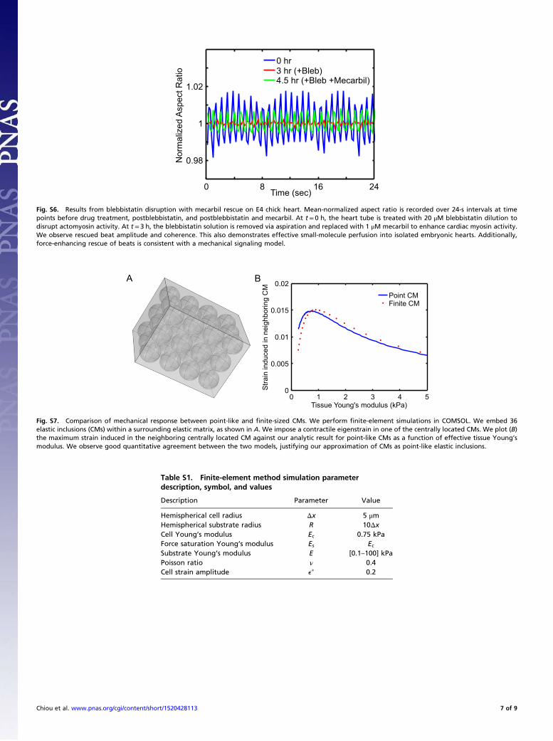
Small-Molecule Drugs Perfuse Embryonic HeartsTreating embryonic hearts with blebbistatin, a drug that blocksmyosin II activity, disrupts the heartbeat (Fig. S5); the heartbeatis then rescued by mecarbil, a cardiac-myosin force-enhancingdrug (Fig. S6). This further indicates that many small-moleculedrugs indeed perfuse the tissue given our experimental protocolsand serves as a control for pharmacologically disrupted (andrescued) beating.
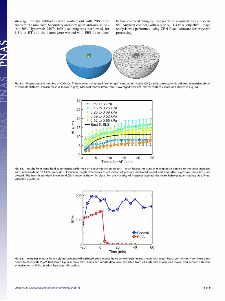
SI Materials and MethodsCell-on-Gel Finite-Element Method Simulations. Finite-element sim-ulations were performed using the Structural Mechanics Modulein COMSOL. An isolated hydrogel-cultured cell was modeled as ahemisphere resting with its flat side fully in contact with a sub-strate. The substrate was also modeled as a hemisphere with thecell placed in the center of its flat surface (Fig. S1). The substrateradius was 10Δx, large enough such that substrate boundary ef-fects are negligible on cell strain. Both components were mod-eled as static linear elastic materials. Under cell contraction, thepartial strain trace (e2d = exx + eyy) in the plane of the substratesurface was calculated and averaged over the cell’s contact sur-face. This procedure was repeated for a range of values forthe substrate Young’s modulus. Both the saturating and con-stant eigenstrain models were used for the contraction strengthof the cell. The “fine” option was used for the element meshsize to ensure convergence. See Table S1 for a list of inputparameters.
CMs-in-Matrix Finite-Element Method Simulations. Finite-elementsimulations were performed using the Structural MechanicsModule in COMSOL. Thirty-six CMs were modeled as linearelastic spherical inclusions arranged in a cubic array and em-bedded within a surrounding elastic matrix of 30 × 30 × 40 μm3
(Fig. S7A), with all model parameters corresponding to Table S1,except that the CM radius is set to 4.6 μm < 5.0 μm to preventissues with meshing in the simulation. We then excited one of thecentrally located CMs by inducing a contractile eigenstrain andcomputed the volume-averaged maximal strain induced in theneighboring central CM as a function of effective tissue Young’smodulus. The effective modulus was determined by simulateduniaxial tensile testing on the composite system. The “fine” elementmesh size was used to ensure convergence. Note the excellent
quantitative agreement between our analytical calculation andthe finite-element calculations for finite-sized inclusions; thisagreement justifies our approximation of CMs as point-like elasticinclusions.
Myocardial Stiffness Measurements. White Leghorn chicken eggs(Charles River Laboratories) were incubated at 37 °C until thedesired developmental stage was reached and isolated as de-scribed in [10]. Embryos were extracted at room temperature(RT) and placed into a Petri dish. The thick albumin was re-moved using blunt forceps and KimWipes. The embryos wereadhered to a Whatman #2 filter paper with an elliptical hole of∼1 × 2 cm cut in it. The embryos were rinsed in PBS and placedventral side up. Prewarmed chick heart media (α-MEM sup-plemented with 10% FBS and 1% penn-strep; Gibco; 12571-063)was added at this point.The heart was extracted with fine forceps severing above the
atrium and below the outflow tract and transferred into a six-wellplate. Micropipettes were pulled from glass capillaries (WorldPrecision Instruments) and broken to final inner diameters of 30–100 μm. Pipettes were filled with PBS and attached to water-filled reservoir. Aspiration was performed at RT in either PBSsupplemented with 3% BSA or heart culture media. Before eachexperiment, pipette tips were placed in the solution for ≤ 20 minto prevent sticking. During aspiration, several pressures wereapplied in the range of 0–0.8 kPa. Imaging was done using aNikon TE300 microscope with a 20× air objective and recordedusing a Cascade Photometric CCD camera. Image analysis wasperformed in ImageJ.
BGA FRAP. Isolated E4 hearts were stained with 0.5 μM CellTraceCalcein Red-Orange, AM (ThermoFisher Scientific; C34851)and Hoechst 33342 in Hepes-buffered α-MEM culture media(Gibco; 12571-063; supplemented with 10% FBS and 1% penn-strep) for 15 min at RT with gentle rocking. Cytochalasin D(Cayman Chemical Company; 11330; 25 μM) was added tominimize contractions and drift during imaging. Calcein andHoechst 33342 were then washed out three times with culturemedia supplemented with cytochalasin D. For BGA treat-ment, embryonic hearts preloaded with calcein were perfusedin 100 μM 18-β-glycyrrhetinic acid (BGA) (Cayman Chemical;11845) for 1 h at RT, with gentle rocking. BGA-treated anduntreated control hearts were placed on 35-mm glass bottomdishes (MatTek; P35G-1.5-10-C) with media filling up thecenter well and capped with a coverslip for FRAP imaging.Confocal time-lapse images were acquired using a Zeiss 880
laser-scanning confocal microscope equipped with a 40× oil, 1.4N.A. objective. Zeiss ZEN Black software was configured toacquire images every 2 s for 6 min and photobleaching startedafter acquiring eight frames and stopped until the intensity droppedto 20% of the original. Average intensity in each bleached regionand a nonbleached region were measured by using ImageJ (https://imagej.nih.gov/ij/download.html) with the time series analyzerplugin (https://imagej.nih.gov/ij/plugins/time-series.html). Intensityof the bleached region was normalized to that of the nonbleachedregion in the same time series to correct overall photobleachingduring imaging. Data were analyzed with a custom C++ code,and graphs were plotted in GraphPad Prism version 6.0d (www.graphpad.com).
Immunostaining of Embryonic Hearts. Isolated E4 hearts were fixedin 4% paraformaldehyde for 15 min at RT on a rocker. Fixed E4hearts were then washed with PBS three times for 5 min each andpermeabilized with 0.1% Triton X in PBS for 30 min. Blockingwas performed in 5% BSA and 0.1% Triton X in PBS for 1.5 h atRT. Hearts were immunostained for α-actinin-2 (Abcam; EA-53mouse monoclonal Ab; 1:200 dilution in blocking solution)and DNA (Hoechst; 33342; 1:1,000) for 3 h at RT with gentle
Chiou et al. www.pnas.org/cgi/content/short/1520428113 4 of 9
shaking. Primary antibodies were washed out with PBS threetimes for 15 min each. Secondary antibody (goat anti-mouse IgGAtto565; Hypermol; 2107; 1:500) staining was performed for1.5 h at RT and the hearts were washed with PBS three times
before confocal imaging. Images were acquired using a Zeiss880 Airyscan confocal with a 40× oil, 1.4 N.A. objective. Imageanalysis was performed using ZEN Black software for Airyscanprocessing.
Fig. S1. Illustration and meshing of COMSOL finite-element–simulated “cell-on-gel” contraction. Active CM (green) contracts while adhered to matrices (blue)of variable stiffness. Chosen mesh is shown in gray. Maximal matrix strain trace is averaged over CM–matrix contact surface and shown in Fig. 2A.
Time after ΔP (sec)
ΔL
(μm
)
0 5 10 15 20 250
5
10
15
20
25
300 to 0.13 kPa0.13 to 0.26 kPa0.26 to 0.39 kPa0.39 to 0.52 kPa0.52 to 0.65 kPaBest fit SLS
Fig. S2. Results from ramp-hold experiments performed on explanted HH stage 10–11 avian hearts. Pressure of micropipette applied to the tissue increaseswith increments of 0.13 kPa every 46 s. Excursion length differences as a function of pressure (indicated colors) and time after a pressure ramp (axis) areplotted. The best-fit standard linear solid (SLS) model is shown in black. For the majority of pressures applied, the heart behaves quantitatively as a linearviscoelastic material.
Time (min)
BPM
-20 0 20 40 600
100
200
ControlBGA
Fig. S3. Beats per minute from isolated Langendorff-perfused adult mouse heart control experiment shown with mean beats per minute from three adulthearts treated with 25 μM BGA (from Fig. 4 in main text). Beats-per-minute data were extracted from 20-s intervals of acquired movie. This demonstrates theeffectiveness of BGA on adult heartbeat disruption.
Chiou et al. www.pnas.org/cgi/content/short/1520428113 5 of 9
0 50 100 150 200 250
0.2
0.4
0.6
1.01.5
Time [s]R
elat
ive
inte
nsity
Control (n=73) BGA (n=49)
control BGA0
5
10
15
20
25
% R
ecov
ery
***
B CA
Fig. S4. A displays a representative image of E4 chicken embryonic CMs stained for α-actinin and DNA. Image was taken 4 μm below the surface in the outflowtract of the looping heart. (Scale bar: 5 μm.) As exampled here, CMs in the E4 heart exhibit premature periodic striations, suggesting myofibrillogenesis of CMs.These cells were subjected to testing for effective gap junction interference via FRAP. B shows aggregated results from calcein red-orange FRAP experiments incontrol and BGA-treated E4 avian hearts. Individual CMs within the tissue were selected and photobleached (see example, Movie S3). Fluorescent intensityrecovery was tracked over at 4 min in 49 +BGA CMs and 73 −BGA CMs. Rapid early recovery is likely due to intracellular diffusion and is unaffected by BGAtreatment. In contrast, BGA treatment disrupts prolonged recovery, which is likely mediated by intercellular transport through gap junctions. C demonstratesthe significantly higher fluorescence recovery after 4 min in control hearts, demonstrating BGA efficacy in disrupting E4 avian heart gap junctions. ***P <0.001, unpaired Student’s t test.
Time (sec)
Nor
mal
ized
Asp
ect R
atio
0 10 20 30
0.98
1
1.02
1.040 hr +Bleb1 hr +Bleb3 hr +Bleb
Fig. S5. Results from blebbistatin disruption on E4 chick heart. Mean-normalized aspect ratio is recorded over time intervals of 30 s at time points 0, 1, and 3 hafter treatment of 20 μM blebbistatin. Significant decreases in beating amplitude and BPM are observed.
Chiou et al. www.pnas.org/cgi/content/short/1520428113 6 of 9
Time (sec)
Nor
mal
ized
Asp
ect R
atio
0 8 16 24
0.98
1
1.02
0 hr3 hr (+Bleb)4.5 hr (+Bleb +Mecarbil)
Fig. S6. Results from blebbistatin disruption with mecarbil rescue on E4 chick heart. Mean-normalized aspect ratio is recorded over 24-s intervals at timepoints before drug treatment, postblebbistatin, and postblebbistatin and mecarbil. At t = 0 h, the heart tube is treated with 20 μM blebbistatin dilution todisrupt actomyosin activity. At t = 3 h, the blebbistatin solution is removed via aspiration and replaced with 1 μM mecarbil to enhance cardiac myosin activity.We observe rescued beat amplitude and coherence. This also demonstrates effective small-molecule perfusion into isolated embryonic hearts. Additionally,force-enhancing rescue of beats is consistent with a mechanical signaling model.
Tissue Young's modulus (kPa)
Stra
in in
duce
d in
nei
ghbo
ring
CM
0 1 2 3 4 50
0.005
0.01
0.015
0.02Point CMFinite CM
B
Fig. S7. Comparison of mechanical response between point-like and finite-sized CMs. We perform finite-element simulations in COMSOL. We embed 36elastic inclusions (CMs) within a surrounding elastic matrix, as shown in A. We impose a contractile eigenstrain in one of the centrally located CMs. We plot (B)the maximum strain induced in the neighboring centrally located CM against our analytic result for point-like CMs as a function of effective tissue Young’smodulus. We observe good quantitative agreement between the two models, justifying our approximation of CMs as point-like elastic inclusions.
Table S1. Finite-element method simulation parameterdescription, symbol, and values
Description Parameter Value
Hemispherical cell radius Δx 5 μmHemispherical substrate radius R 10ΔxCell Young’s modulus Ec 0.75 kPaForce saturation Young’s modulus Es EcSubstrate Young’s modulus E [0.1–100] kPaPoisson ratio ν 0.4Cell strain amplitude ep 0.2
Chiou et al. www.pnas.org/cgi/content/short/1520428113 7 of 9
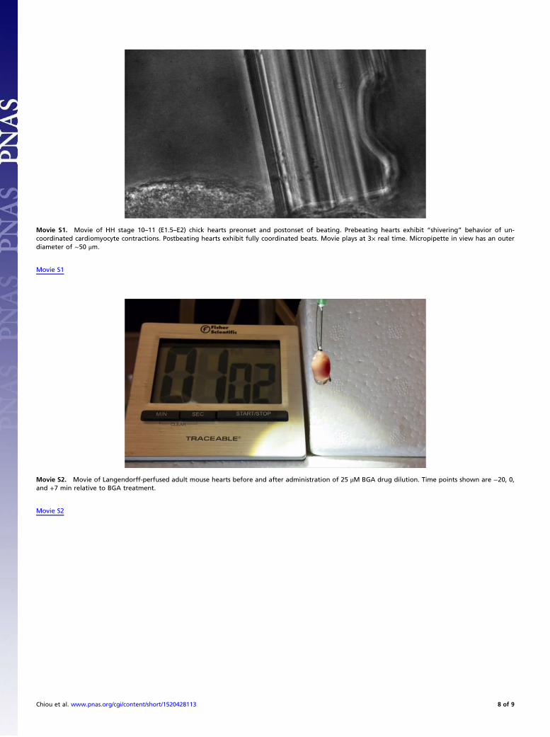
Movie S1. Movie of HH stage 10–11 (E1.5–E2) chick hearts preonset and postonset of beating. Prebeating hearts exhibit “shivering” behavior of un-coordinated cardiomyocyte contractions. Postbeating hearts exhibit fully coordinated beats. Movie plays at 3× real time. Micropipette in view has an outerdiameter of ∼50 μm.
Movie S1
Movie S2. Movie of Langendorff-perfused adult mouse hearts before and after administration of 25 μM BGA drug dilution. Time points shown are −20, 0,and +7 min relative to BGA treatment.
Movie S2
Chiou et al. www.pnas.org/cgi/content/short/1520428113 8 of 9
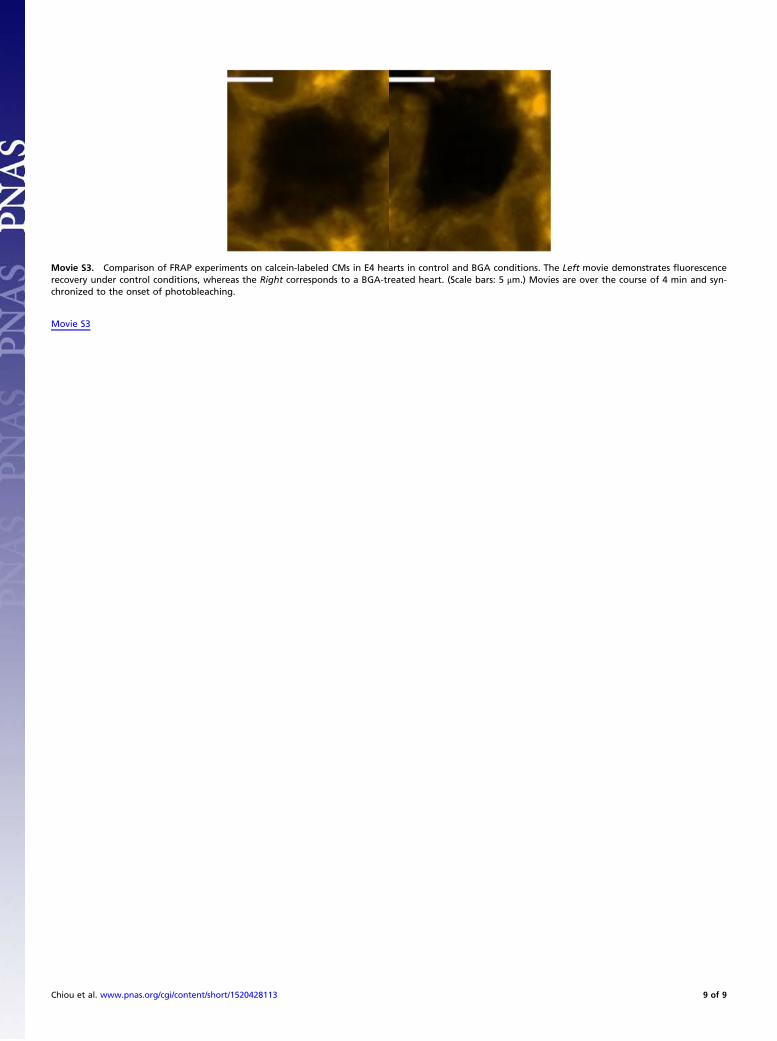
Movie S3. Comparison of FRAP experiments on calcein-labeled CMs in E4 hearts in control and BGA conditions. The Left movie demonstrates fluorescencerecovery under control conditions, whereas the Right corresponds to a BGA-treated heart. (Scale bars: 5 μm.) Movies are over the course of 4 min and syn-chronized to the onset of photobleaching.
Movie S3
Chiou et al. www.pnas.org/cgi/content/short/1520428113 9 of 9