May - Aug 2018 - uv.es · EDITORS: Jesús Salgado Jorge Alegre-Cebollada SBE - Sociedad de...
30
EDITORS: Jesús Salgado Jorge Alegre-Cebollada SBE - Sociedad de Biofísica de España http://biofisica.info/ May - Aug 2018 Xavier Daura Teresa Giráldez <a hreft="http://biofisica.info/"> Courtesy of M. Mivelle Cover image: ISSN 2445-43111 life version at : #11
-
Upload
vuongkhanh -
Category
Documents
-
view
214 -
download
0
Transcript of May - Aug 2018 - uv.es · EDITORS: Jesús Salgado Jorge Alegre-Cebollada SBE - Sociedad de...

EDITORS:Jesús Salgado
Jorge Alegre-Cebollada
SBE - Sociedad de Biofísica de España
http://biofisica.info/
May - Aug 2018
Xavier Daura
Teresa Giráldez
<a hreft="http://biofisica.info/">
Courtesy of M. MivelleCover image:
ISSN 2445-43111
life version at:
#11


EDITORS
Jesús Salgado
Jorge Alegre-Cebollada
Xavier Daura
Teresa Giráldez
ISSN 2445-4311
SPONSORS
CONTACT
SBE - Sociedad de Biofísica de España
Secretaria SBE, IQFR-CSIC,
C/Serrano 119, 28006 Madrid
Email: [email protected]
WEB: http://www.sbe.es
Biofísica M a g a z i n e


Biofísica M a g a z i n e
5
In this issueANALYSISEDITORIAL / BEYOND BIOPHYSICS COOL BIOPHYSICSpage 7 page 11 page 15
The team of Editors Isabel Usón & Claudia MillánThe eyes of chemistry(Dedicated to Geoffrey Roughton)
HIGHLIGHTED PUBLICATIONS
May page 27page 27
page 28page 28June
JulyAugust
A view on peer review Biophysics in microbiology:Lucía García-Ortega
A conversation with Víctor deLorenzo
CONFERENCE REPORT TRIBUTE
On the 6th International Iberian BiophysicsCongress & X Iberoamerican Congressof Biophysics
Tribute to Carlos Gómez-Moreno
( By Vicente M. Aguilella)
( By Milagros Medina & Javier Sancho)
page 23 page 25

EDITORIAL / ANALYSIS
S
Classical peer review is relatively young. Itwas questioned from the beginning and hasnever consolidated as a unique model
A view on peer reviewThe team of Editors.
crutiny of scientific work is a fundamental pillar of Science. Itis not only needed for correctness, in a purely technicalsense, but also takes care of originality, novelty and
significance, ensuring that the scientific work contributes togenerating new knowledge. There are two stages where this task isespecially important: before the start of a scientific investigation(projects) and when the investigation yields results (publications). Atboth stages the dominant (almost exclusive) method for evaluation isreviewing by peers.
A precursor of peer review began in England in the early 19 th
century, with referees commissioned by the Royal Society of Londonto write reports on manuscripts sent for publication in Philosophical Transactions [1]. A standardized referee systemdeveloped from this point among the English scientific societies and slowly spread to independent journals and outsidethe Anglophone world. However, it was in the 1960s when refereeing propagated as the method to objectively judgeScience (although the embracement of this system was delayed by some journals, like Nature, which adopted it in 1973)
[1]. During that period of the 20 th century the scientific activities experienced a huge expansion, especially in the USA,with a massive increase in public funding and a parallel social and political demand for objective methods to judgescientific quality. In fact, the term peer review was first used in evaluation procedures by American funding agencies ofthat time and was later used as a synonym of refereeing for the case of revision of publications. Thus, classical peerreview, either of projects or papers, is a relatively young system.But perhaps most importantly, it has never consolidated as aunique solid model and was, from the beginning, put into question[1]. It is then not surprising that in current times, with technologicalchanges affecting the ways of communicating Science, the future of peer review is subject of a particularly vivid debate.
Overflowed system
Today, the discussion about peer review connects with multiple other problems of Science [ 2] as well as with profoundchanges affecting modern publishing [3, 4]. With the dominance of bibliometrics as the measure to evaluate scientificperformance, publishing has acquired a tremendous importance [5] and peer review, as the method employed to controlwhat is published (and where), has become a crucial variable. Added to this, complex problems have emerged thataffect directly to the quality of Science and thus uncover inefficiencies of the evaluation mechanisms, like poorreproducibility, fabrication of data, plagiarism or even direct corruption of the peer review system [6]. One would expectthat the response of the scientific community to the clear need of stronger and more effective evaluation procedureswould be to strengthen peer review, or even to evolve it into a new system, adapted to the challenges of the present andthe future. However, in the vast majority of cases peer review continues being performed as it was conceived more than50 years ago, despite its well known weaknesses.
Biofísica M a g a z i n e
http://biofisica.info/
7
Biofísica #11, May–Aug 2018

Peer review, as the method to control whatis published (and where), has become acrucial variable
Figure 1. Homage to an anonymous peer reviewer.The monument was placed in May 2017 in thecourtyard of the Institute of Education at Moscow’sHigher School of Economics – HSE [7].
Openness is seen as positive: Fits withtendencies of modern publishing, bringstransparency and provides ways to rewardreviewers
The bibliometric epidemy [5] contributes to inflate the number ofpublications, and with that, the numbers of publishers andjournals have also grown exponentially. This process is fuelled bydigitalization and automation, which makes publishing easier,
cheaper and quicker than ever. But can proper assessment, criticism and discussion of scientific work be similarlyescalated and accelerated? Obviously not. First, this part of the business cannot be easily automatized and needs thecautious time and attention of expert human readers / evaluators. Furthermore, the scientific methodology is quicklychanging and becoming more and more sophisticated. All branches of natural sciences (including, of course, Biophysics)are increasingly becoming multidisciplinary, meaning that diverse and well prepared experts are needed to judge thescientific work. With this requirements it is easy to visualize that the pool of possible (capable) referees must be ofsmaller size than the pool of their peer authors. In practice, the pool of reviewers is further reduced because of the lackof incentives to perform difficult, time constrained and barely recognized reviewing tasks. This all makes findingadequate referees an herculean exercise and accentuates the weakness of the system, in a vicious circle.
Blindness vs transparency?
In the scientific community there is consensus about the importance of a solid peer review system and the need to tailorits traditional scheme for current and future exigencies. However, there is no clear consensus yet about the actions totake –more than paying homage to the stoic anonymous reviewers (Fig. 1) [7].
A major discussion is settled about the convenient level oftransparency [8]. First, we remind that the pioneer refereeingEnglish system, as conceived and exercised originally byWILLIAM WHEWELL [1], started being completely open, withreports published and signed by the referee (in a especialProceedings journal). But it was soon realized that such anoption prevented criticism and discouraged negative reports.Thus, after a couple of years of openness, refereeing becameanonymous and reports were no longer published [1]. Today’sclassical peer review is still mainly single blinded, with theidentity of the authors know by reviewers, who remainunknown to the authors. This system has been attacked in twoopposing directions. On the one hand, it is argued that theuncovered identity of authors facilitates possible discriminationbecause of gender, ethnic background, country of affiliation,personal relations or prestige of previous work. This has led todefend a double-blinded system [9], with unknown identities ofboth authors and referees. Although this option is preferred, according to some studies, it is recognized that is verydifficult in practice to implement, since in many cases the type of work, cited references and other details can betray theidentity of authors [10].
On the other hand, it has been pointed out that a covered identitymakes referees potentially immune from their possible unfair,harsh or unsound criticisms, which is used to defend a completelyopen system. Openness, at different levels, is gaining adepts as itfits well with new tendencies of open publishing and has a
positive connotation, because is seen as a way to bring transparency to the publication process. It also can provideways to reward the reviewers via publication of their reports along with authors’ manuscripts, which helps visualizing theusefulness of the peer reviewing process, even when the identity of the reviewer is kept anonymous. Although still a
8
http://biofisica.info/ A view on peer review | Biofisica #11, May–Aug 2018
Expert scientists are busy people withexhausting responsibilities. Is “sense ofduty” enough to involve them in peerreview?
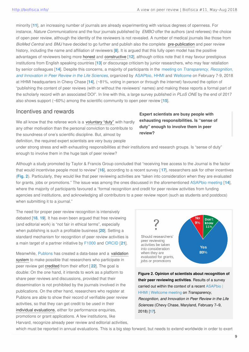
Figure 2. Opinion of scientists about recognition oftheir peer reviewing activities. Results of a surveycarried out within the context of a recent ASAPbio |HHMI | Wellcome meeting on Transparency,Recognition, and Innovation in Peer Review in the LifeSciences (Chevy Chase, Maryland, February 7–9,2018) [17].
minority [11], an increasing number of journals are already experimenting with various degrees of openness. Forinstance, Nature Communications and the four journals published by EMBO offer the authors (and referees) the choiceof open peer review, although the identity of the reviewers is not revealed. A number of medical journals like those fromBioMed Central and BMJ have decided to go further and publish also the complete pre-publication and peer reviewhistory, including the name and affiliation of reviewers [8]. It is argued that this fully open model has the positiveadvantages of reviewers being more honest and constructive [12], although critics note that it may favour prestigiousinstitutions from English speaking countries [13] or discourage criticism by junior researchers, who may fear retaliationby senior colleagues [14]. Despite this concerns, a majority of participants in the meeting on Transparency, Recognition,and Innovation in Peer Review in the Life Sciences, organized by ASAPbio, HHMI and Wellcome on February 7-9, 2018at HHMI headquarters in Chevy Chase [14], (~81%, voting in person or through the internet) favoured the option of“publishing the content of peer reviews (with or without the reviewers’ names) and making these reports a formal part ofthe scholarly record with an associated DOI”. In line with this, a large survey published in PLoS ONE by the end of 2017also shows support (~60%) among the scientific community to open peer review [15].
Incentives and rewards
We all know that the referee work is a voluntary “duty” with hardlyany other motivation than the personal conviction to contribute tothe soundness of one’s scientific discipline. But, almost bydefinition, the required expert scientists are very busy peopleunder strong stress and with exhausting responsibilities at their institutions and research groups. Is “sense of duty”enough to involve them in the huge task of peer review?
Although a study promoted by Taylor & Francis Group concluded that “receiving free access to the Journal is the factorthat would incentivise people most to review” [16], according to a recent survey [ 17], researchers ask for other incentives(Fig. 2). Particularly, they would like that peer reviewing activities are “taken into consideration when they are evaluatedfor grants, jobs or promotions.” The issue was among the ones discussed in the aforementioned ASAPbio meeting [14],where the majority of participants favoured a “formal recognition and credit for peer review activities from fundingagencies and institutions, and acknowledging all contributors to a peer review report (such as students and postdocs)when submitting it to a journal.”
The need for proper peer review recognition is intensivelydebated [18, 19]. It has even been argued that free reviewing(and editorial work) is “not fair in ethical terms”, especiallywhen publishing is such a profitable business [20]. Setting astandard mechanism for recognition of peer review activities isa main target of a partner initiative by F1000 and ORCID [21].
Meanwhile, Publons has created a data-base and a validationsystem to make possible that researchers who participate inpeer review get credited from their effort [ 22]. The goal isdouble: On the one hand, it intends to work as a platform toshare peer reviews and discussions, provided that theirdissemination is not prohibited by the journals involved in thepublications. On the other hand, researchers who register atPublons are able to show their record of verifiable peer reviewactivities, so that they can get credit to be used in theirindividual evaluations, either for performance enquiries,promotions or grant applications. A few institutions, likeHarvard, recognize already peer review and editorial activities,which must be reported in annual evaluations. This is a big step forward, but needs to extend worldwide in order to exert
9
http://biofisica.info/ A view on peer review | Biofisica #11, May–Aug 2018

a significant impact.
In summary, there is little doubt that a deep improvement of the strategies for assessing the quality of Science is a mosturgent need. However, as we just discussed, the general perception is that this crucial and demanding task is notsufficiently rewarded. Very interesting initiatives exist to adapt, at multiple levels, classical peer review. Although thechanges already in place seem mostly experimental, we can foresee that a renewed refereeing system, characterizedby increased openness, rewarding and recognition, will soon crystallize.
References
1. CSISZAR A. “Peer review: Troubled from the start.” Nature, 2016, 532: 306. DOI.
2. BELLUZ J, PLUMER B, RESNICK B. “The 7 biggest problems facing science, according to 270 scientists.” Vox, sep. 2016. URL.
3. ARNS M. “Open access is tiring out peer reviewers.” Nature, 2014, 515: 467. DOI.
4. CURRY S. “Peer review, preprints and the speed of science.” The Guardian, sep. 2015. URL.
5. MACILWAIN C. “Halt the avalanche of performance metrics.” Nature, 2013, 500: 255. DOI.
6. FERGUSON C, MARCUS A, ORANSKY I. “Publishing: The peer-review scam.” Nature, 2014, 515: 480. DOI.
7. SCHIERMEIER Q. “Monument to peer review unveiled in Moscow.” Nature, may 2017. DOI.
8. PULVERER B. “Transparency showcases strength of peer review.” Nature, 2010, 468: 29. DOI.
9. CRESSEY D. “Journals weigh up double-blind peer review.” Nature, jul. 2014. DOI.
10. WARE M. “Peer review: benefits, perceptions and alternatives.” Publishing Research Consortium, 2008, 4: 1.
11. PARKS S, GUNASHEKAR S. “Tracking Global Trends in Open Peer Review.” RAND blog, oct. 2017. URL.
12. DECOURSEY T. “The pros and cons of open peer review.” Nature, 2006. DOI.
13. HOPEWELL S, COLLINS GS, BOUTRON I, YU L-M, COOK J, SHANYINDE M, WHARTON R, SHAMSEER L, ALTMAN DG. “Impact of peer review on
reports of randomised trials published in open peer review journals: retrospective before and after study.” BMJ, 2014, 349: g4145. DOI.
14. BRAINARD J. “Researchers debate whether journals should publish signed peer reviews.” Science, feb. 2018. URL.
15. ROSS-HELLAUER T, DEPPE A, SCHMIDT B. “Survey on open peer review: Attitudes and experience amongst editors, authors and reviewers.”
PLOS ONE, 2017, 12: e0189311. DOI.
16. “Peer review: a global view.” Taylor and Francis, 2015. URL.
17. “Peer-Review Survey.” ASAPbio, 2017. URL.
18. EDITORIAL. “Review rewards.” Nature, 2014, 514: 274. DOI.
19. CURRY S. “Peer review is essential to good science – it’s time to credit expert reviewers.” The Guardian, jun. 2017. URL.
20. DA SILVA JAT, KATAVI&CACUTE; V. “Free editors and peers: squeezing the lemon dry.” Ethics & Bioethics, 2016, 6: 203. DOI.
21. HAAK L. “F1000 and ORCID Partner to Launch Standard for Citing Peer Review Activities.” ORCID blog, may 2015. URL.
22. VAN NOORDEN R. “The scientists who get credit for peer review.” Nature, oct. 2014. DOI.
http://biofisica.info/ A view on peer review | Biofisica #11, May–Aug 2018
10
BEYOND BIOPHYSICS
W
Biology needs to transform from a disciplinebased on phenomena description to onesupported by general rules, standardizedmetrics and therefore able to makepredictions
Biophysics in microbiologyA conversation with Víctor de LorenzoLucía García-Ortega, UCM (Spain).
hen I was proposed to write on this topic it sounded tome like a wonderful idea, since I thought that VÍCTOR, likeme, is a “side effect biophysicist”. In my case, I got into
biophysics as a consequence of my interest in molecular biology,proteins and their interactions with lipids. In VÍCTOR’S case, Iconsidered his quantitative approach to microbiology to be highlybiophysical. However, what a priori I thought it would be the sideeffect of a microbiologist, turned out to be an interesting descriptionof microbiology from biophysical and mathematical points of view.
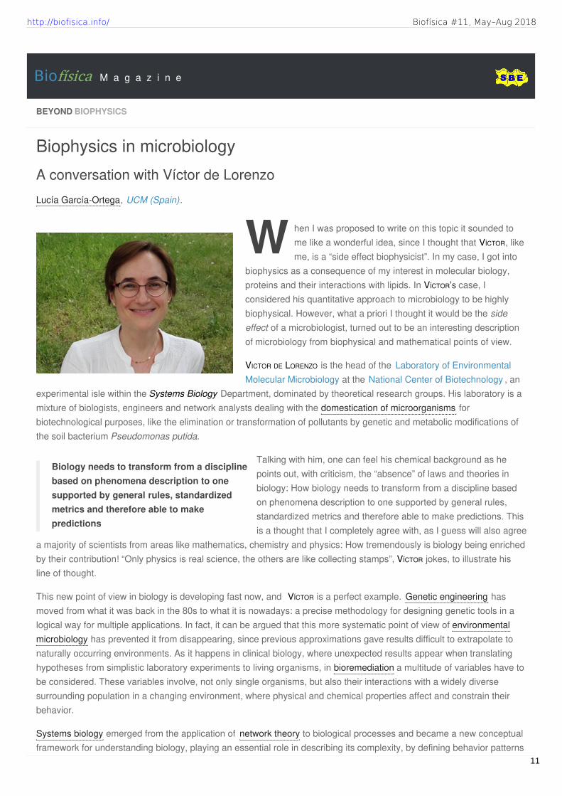
VICTOR DE LORENZO is the head of the Laboratory of EnvironmentalMolecular Microbiology at the National Center of Biotechnology , an
experimental isle within the Systems Biology Department, dominated by theoretical research groups. His laboratory is amixture of biologists, engineers and network analysts dealing with the domestication of microorganisms forbiotechnological purposes, like the elimination or transformation of pollutants by genetic and metabolic modifications ofthe soil bacterium Pseudomonas putida.
Talking with him, one can feel his chemical background as hepoints out, with criticism, the “absence” of laws and theories inbiology: How biology needs to transform from a discipline basedon phenomena description to one supported by general rules,standardized metrics and therefore able to make predictions. Thisis a thought that I completely agree with, as I guess will also agree
a majority of scientists from areas like mathematics, chemistry and physics: How tremendously is biology being enrichedby their contribution! “Only physics is real science, the others are like collecting stamps”, VÍCTOR jokes, to illustrate hisline of thought.
This new point of view in biology is developing fast now, and VÍCTOR is a perfect example. Genetic engineering hasmoved from what it was back in the 80s to what it is nowadays: a precise methodology for designing genetic tools in alogical way for multiple applications. In fact, it can be argued that this more systematic point of view of environmentalmicrobiology has prevented it from disappearing, since previous approximations gave results difficult to extrapolate tonaturally occurring environments. As it happens in clinical biology, where unexpected results appear when translatinghypotheses from simplistic laboratory experiments to living organisms, in bioremediation a multitude of variables have tobe considered. These variables involve, not only single organisms, but also their interactions with a widely diversesurrounding population in a changing environment, where physical and chemical properties affect and constrain theirbehavior.
Systems biology emerged from the application of network theory to biological processes and became a new conceptualframework for understanding biology, playing an essential role in describing its complexity, by defining behavior patterns
Biofísica M a g a z i n e
http://biofisica.info/ Biofísica #11, May–Aug 2018
11
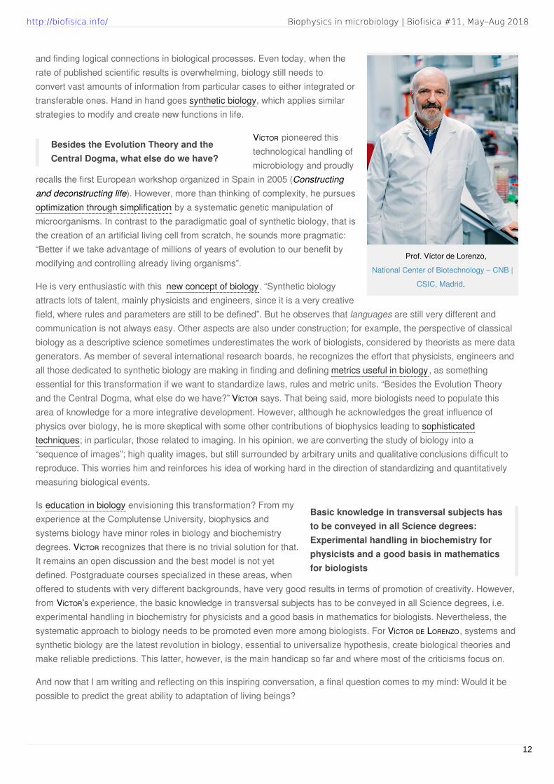
Prof. Víctor de Lorenzo,
National Center of Biotechnology – CNB |
CSIC, Madrid.
Besides the Evolution Theory and theCentral Dogma, what else do we have?
Basic knowledge in transversal subjects hasto be conveyed in all Science degrees:Experimental handling in biochemistry forphysicists and a good basis in mathematicsfor biologists
and finding logical connections in biological processes. Even today, when therate of published scientific results is overwhelming, biology still needs toconvert vast amounts of information from particular cases to either integrated ortransferable ones. Hand in hand goes synthetic biology, which applies similarstrategies to modify and create new functions in life.
VÍCTOR pioneered thistechnological handling ofmicrobiology and proudly
recalls the first European workshop organized in Spain in 2005 (Constructingand deconstructing life). However, more than thinking of complexity, he pursuesoptimization through simplification by a systematic genetic manipulation ofmicroorganisms. In contrast to the paradigmatic goal of synthetic biology, that isthe creation of an artificial living cell from scratch, he sounds more pragmatic:“Better if we take advantage of millions of years of evolution to our benefit bymodifying and controlling already living organisms”.
He is very enthusiastic with this new concept of biology. “Synthetic biologyattracts lots of talent, mainly physicists and engineers, since it is a very creativefield, where rules and parameters are still to be defined”. But he observes that languages are still very different andcommunication is not always easy. Other aspects are also under construction; for example, the perspective of classicalbiology as a descriptive science sometimes underestimates the work of biologists, considered by theorists as mere datagenerators. As member of several international research boards, he recognizes the effort that physicists, engineers andall those dedicated to synthetic biology are making in finding and defining metrics useful in biology, as somethingessential for this transformation if we want to standardize laws, rules and metric units. “Besides the Evolution Theoryand the Central Dogma, what else do we have?” VÍCTOR says. That being said, more biologists need to populate thisarea of knowledge for a more integrative development. However, although he acknowledges the great influence ofphysics over biology, he is more skeptical with some other contributions of biophysics leading to sophisticatedtechniques; in particular, those related to imaging. In his opinion, we are converting the study of biology into a“sequence of images”; high quality images, but still surrounded by arbitrary units and qualitative conclusions difficult toreproduce. This worries him and reinforces his idea of working hard in the direction of standardizing and quantitativelymeasuring biological events.
Is education in biology envisioning this transformation? From myexperience at the Complutense University, biophysics andsystems biology have minor roles in biology and biochemistrydegrees. VÍCTOR recognizes that there is no trivial solution for that.It remains an open discussion and the best model is not yetdefined. Postgraduate courses specialized in these areas, whenoffered to students with very different backgrounds, have very good results in terms of promotion of creativity. However,from VÍCTOR’S experience, the basic knowledge in transversal subjects has to be conveyed in all Science degrees, i.e.experimental handling in biochemistry for physicists and a good basis in mathematics for biologists. Nevertheless, thesystematic approach to biology needs to be promoted even more among biologists. For VÍCTOR DE LORENZO, systems andsynthetic biology are the latest revolution in biology, essential to universalize hypothesis, create biological theories andmake reliable predictions. This latter, however, is the main handicap so far and where most of the criticisms focus on.
And now that I am writing and reflecting on this inspiring conversation, a final question comes to my mind: Would it bepossible to predict the great ability to adaptation of living beings?
Biophysics in microbiology | Biofisica #11, May–Aug 2018http://biofisica.info/
12

LUCÍA GARCÍA-ORTEGA
Departamento de Bioquímica y Biología Molecular,
Universidad Complutense de Madrid – UCM,
Madrid (Spain).
E-mail: [email protected]
VÍCTOR DE LORENZO
Molecular Environmental Microbiology Laboratory,
Systems Biology Programme,
National Center of Biotechnology – CNB | CSIC,
Madrid (Spain).
E-mail: [email protected]
Biophysics in microbiology | Biofisica #11, May–Aug 2018http://biofisica.info/
13

COOL BIOPHYSICS
F
(1)
The eyes of chemistry(Dedicated to Geoffrey Roughton)
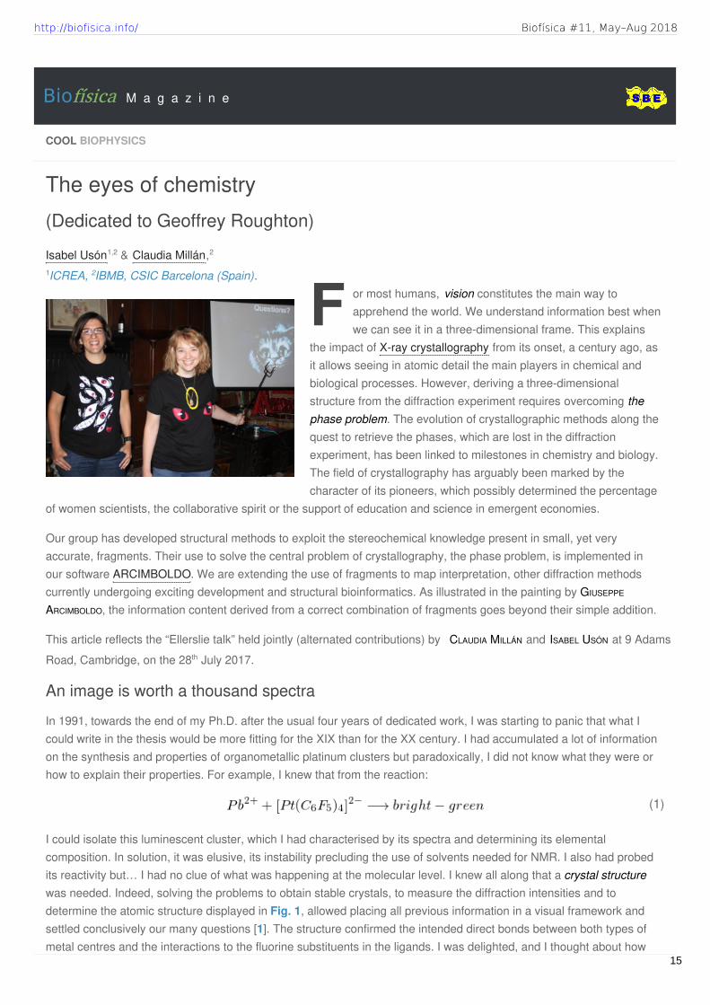
Isabel Usón1,2 & Claudia Millán,2
1ICREA, 2IBMB, CSIC Barcelona (Spain).or most humans, vision constitutes the main way toapprehend the world. We understand information best whenwe can see it in a three-dimensional frame. This explains
the impact of X-ray crystallography from its onset, a century ago, asit allows seeing in atomic detail the main players in chemical andbiological processes. However, deriving a three-dimensionalstructure from the diffraction experiment requires overcoming thephase problem. The evolution of crystallographic methods along thequest to retrieve the phases, which are lost in the diffractionexperiment, has been linked to milestones in chemistry and biology.The field of crystallography has arguably been marked by thecharacter of its pioneers, which possibly determined the percentage
of women scientists, the collaborative spirit or the support of education and science in emergent economies.
Our group has developed structural methods to exploit the stereochemical knowledge present in small, yet veryaccurate, fragments. Their use to solve the central problem of crystallography, the phase problem, is implemented inour software ARCIMBOLDO. We are extending the use of fragments to map interpretation, other diffraction methodscurrently undergoing exciting development and structural bioinformatics. As illustrated in the painting by GIUSEPPE
ARCIMBOLDO, the information content derived from a correct combination of fragments goes beyond their simple addition.
This article reflects the “Ellerslie talk” held jointly (alternated contributions) by CLAUDIA MILLÁN and ISABEL USÓN at 9 Adams
Road, Cambridge, on the 28th July 2017.
An image is worth a thousand spectra
In 1991, towards the end of my Ph.D. after the usual four years of dedicated work, I was starting to panic that what Icould write in the thesis would be more fitting for the XIX than for the XX century. I had accumulated a lot of informationon the synthesis and properties of organometallic platinum clusters but paradoxically, I did not know what they were orhow to explain their properties. For example, I knew that from the reaction:
I could isolate this luminescent cluster, which I had characterised by its spectra and determining its elementalcomposition. In solution, it was elusive, its instability precluding the use of solvents needed for NMR. I also had probedits reactivity but… I had no clue of what was happening at the molecular level. I knew all along that a crystal structurewas needed. Indeed, solving the problems to obtain stable crystals, to measure the diffraction intensities and todetermine the atomic structure displayed in Fig. 1, allowed placing all previous information in a visual framework andsettled conclusively our many questions [1]. The structure confirmed the intended direct bonds between both types ofmetal centres and the interactions to the fluorine substituents in the ligands. I was delighted, and I thought about how
Biofísica M a g a z i n e
http://biofisica.info/ Biofísica #11, May–Aug 2018
15

Figure 1. The three-dimensional view of a moleculein atomic detail. It provides a visual framework to allfunctional knowledge associated, as in the case of the
anion [Pt(C6F5)4]2- [1].
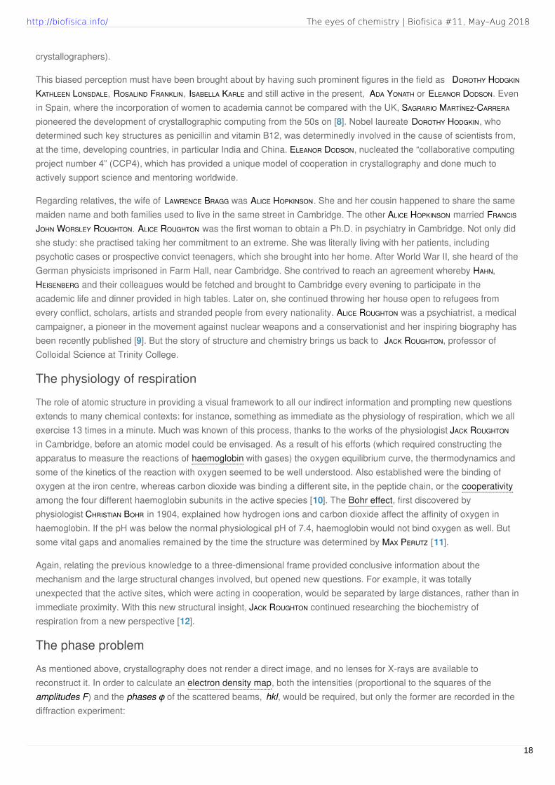
Figure 2. Röntgen’s discovery and geometric illustration of
Bragg’s Law. Left, image recorded by Conrad Röntgen on 22nd
December 1895 showing the hand of his wife, Anna Bertha Ludwig.Right, constructive interference geometry of X-rays incident to a crystalsample.
many others before me must have shared this same joy ofliterally seeing the answers so long pursued. But the structurealso raised new questions: why was the coordination aroundlead, in oxidation state II, linear? Should there not be astereochemically active lone pair of electrons? Why then wasthe structure not bent around the lead centre?
The Braggs shaped the character ofcrystallography
About one hundred years earlier, in 1895, X-rays had beendiscovered by RÖNTGEN [2]. Their ability to penetrate matter andyield image information (Fig. 2, left) had been patent from theonset. Diffraction by crystals, by MAX VON LAUE, WALTHER
FRIEDRICH and PAUL KNIPPING, served to simultaneously establishthe nature of X-rays and crystals [3]. From these findings,WILLIAM BRAGG and his son, sir LAWRENCE BRAGG realized that inthe case of molecules, the natural grating provided by crystalswould be required to amplify the scattering signal, by bringinga large number of equivalent atoms to add their contributions[4]. We cannot see X-rays and if we want to experiment withlight scattering on macroscopic objects and how it reflects periodicity and the underlying blocks making up the periodicobject, we should choose the appropriate wavelength: monochromatic visible light. Among much excellent materialavailable on-line to visualize diffraction on gratings, we would suggest a YouTube video [ 5] (Video 1) showing greenlight scattered on periodic, everyday objects, such as spans of thread, strings of beads, screws, spiral springs mimickingthe double helix structure adopted by DNA or sieves illustrating lattices! As can be appreciated in the video, we are notseeing an image of the illuminated items, but the pattern is obviously related to the periodicity and to the underlyingstructure of the object.
The fundamental law of diffraction is namedBragg’s Law, and establishes the geometry ofthe diffraction pattern (Fig. 2, right). We canthink of diffraction as reflection on sets ofplanes running through the crystal. Only atcertain angles are the waves diffracted fromdifferent planes shifted by a whole number ofwavelengths apart, i.e. in phase. For suchangles, the intensity of the diffracted beamscan be recorded on a detector. At other angles,the waves reflected from different planes areout of phase and cancel out.
LAWRENCE BRAGG, was the first to performstructural analysis by X-ray crystallography,determining the structures of various inorganicsalts, such as NaCl [6]. The BRAGGS analysedthe diffraction pattern and figured out how itrelated to the structure in real space, placingthe atoms that composed structures. Their
work established the grounds for X-ray crystallography, as we know it today and was awarded a Nobel Prize in 1915,
The eyes of chemistry | Biofisica #11, May–Aug 2018http://biofisica.info/
16
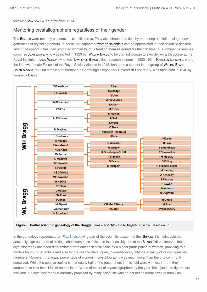
Figure 3. Partial scientific genealogy of the Braggs. Female scientists are highlighted in sepia. Based on [ 7].
following MAX VON LAUE’S prize from 1914.
Mentoring crystallographers regardless of their gender
The BRAGGS were not only pioneers in scientific terms. They also shaped the field by mentoring and influencing a newgeneration of crystallographers. In particular, support of women scientists can be appreciated in their scientific descentand in the opportunities they promoted women to, thus treating them as equals for the first time [7]. Prominent examplescomprise JOAN EVANS, who was invited in 1923 by WILLIAM BRAGG to be the first woman to ever deliver a Discourse at theRoyal Institution; LUCY WILSON, who was LAWRENCE BRAGG’S first research student in 1923-1924; KATHLEEN LONSDALE, one ofthe first two female Fellows of the Royal Society elected in 1945, had been a student in the group of WILLIAM BRAGG;HELEN MEGAW, the first female staff member in Cambridge’s legendary Cavendish Laboratory, was appointed in 1946 byLAWRENCE BRAGG.
In the genealogy reproduced on Fig. 3, displaying part of the scientific descent of the BRAGGS it is noticeable theunusually high numbers of distinguished women scientists. In fact, possibly due to the BRAGGS’ direct intervention,crystallography has been differentiated from other scientific fields by a higher participation of women, providing rolemodels for young scientists and also for the collaborative, open, joy-in-discovery attitude in many of its distinguishedmembers. However, the actual percentage of women in crystallography was much lower than the one commonlyperceived. While the popular feeling is that nearly half of the researchers in this field were women, in truth theyamounted to less than 15% of entries in the World directory of crystallographers by the year 1981 (updated figures areavailable but crystallography is currently practised by many scientists who do not define themselves primarily as
The eyes of chemistry | Biofisica #11, May–Aug 2018http://biofisica.info/
17
crystallographers).
This biased perception must have been brought about by having such prominent figures in the field as DOROTHY HODGKIN
KATHLEEN LONSDALE, ROSALIND FRANKLIN, ISABELLA KARLE and still active in the present, ADA YONATH or ELEANOR DODSON. Evenin Spain, where the incorporation of women to academia cannot be compared with the UK, SAGRARIO MARTÍNEZ-CARRERA
pioneered the development of crystallographic computing from the 50s on [8]. Nobel laureate DOROTHY HODGKIN, whodetermined such key structures as penicillin and vitamin B12, was determinedly involved in the cause of scientists from,at the time, developing countries, in particular India and China. ELEANOR DODSON, nucleated the “collaborative computingproject number 4” (CCP4), which has provided a unique model of cooperation in crystallography and done much toactively support science and mentoring worldwide.
Regarding relatives, the wife of LAWRENCE BRAGG was ALICE HOPKINSON. She and her cousin happened to share the samemaiden name and both families used to live in the same street in Cambridge. The other ALICE HOPKINSON married FRANCIS
JOHN WORSLEY ROUGHTON. ALICE ROUGHTON was the first woman to obtain a Ph.D. in psychiatry in Cambridge. Not only didshe study: she practised taking her commitment to an extreme. She was literally living with her patients, includingpsychotic cases or prospective convict teenagers, which she brought into her home. After World War II, she heard of theGerman physicists imprisoned in Farm Hall, near Cambridge. She contrived to reach an agreement whereby HAHN,HEISENBERG and their colleagues would be fetched and brought to Cambridge every evening to participate in theacademic life and dinner provided in high tables. Later on, she continued throwing her house open to refugees fromevery conflict, scholars, artists and stranded people from every nationality. ALICE ROUGHTON was a psychiatrist, a medicalcampaigner, a pioneer in the movement against nuclear weapons and a conservationist and her inspiring biography hasbeen recently published [9]. But the story of structure and chemistry brings us back to JACK ROUGHTON, professor ofColloidal Science at Trinity College.
The physiology of respiration
The role of atomic structure in providing a visual framework to all our indirect information and prompting new questionsextends to many chemical contexts: for instance, something as immediate as the physiology of respiration, which we allexercise 13 times in a minute. Much was known of this process, thanks to the works of the physiologist JACK ROUGHTON
in Cambridge, before an atomic model could be envisaged. As a result of his efforts (which required constructing theapparatus to measure the reactions of haemoglobin with gases) the oxygen equilibrium curve, the thermodynamics andsome of the kinetics of the reaction with oxygen seemed to be well understood. Also established were the binding ofoxygen at the iron centre, whereas carbon dioxide was binding a different site, in the peptide chain, or the cooperativityamong the four different haemoglobin subunits in the active species [10]. The Bohr effect, first discovered byphysiologist CHRISTIAN BOHR in 1904, explained how hydrogen ions and carbon dioxide affect the affinity of oxygen inhaemoglobin. If the pH was below the normal physiological pH of 7.4, haemoglobin would not bind oxygen as well. Butsome vital gaps and anomalies remained by the time the structure was determined by MAX PERUTZ [11].
Again, relating the previous knowledge to a three-dimensional frame provided conclusive information about themechanism and the large structural changes involved, but opened new questions. For example, it was totallyunexpected that the active sites, which were acting in cooperation, would be separated by large distances, rather than inimmediate proximity. With this new structural insight, JACK ROUGHTON continued researching the biochemistry ofrespiration from a new perspective [12].
The phase problem
As mentioned above, crystallography does not render a direct image, and no lenses for X-rays are available toreconstruct it. In order to calculate an electron density map, both the intensities (proportional to the squares of theamplitudes F) and the phases φ of the scattered beams, hkl, would be required, but only the former are recorded in thediffraction experiment:
The eyes of chemistry | Biofisica #11, May–Aug 2018http://biofisica.info/
18
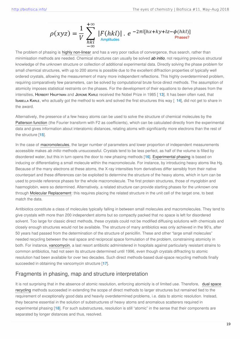
The problem of phasing is highly non-linear and has a very poor radius of convergence, thus search, rather thanminimisation methods are needed. Chemical structures can usually be solved ab initio, not requiring previous structuralknowledge of the unknown structure or collection of additional experimental data. Directly solving the phase problem forsmall chemical structures, with up to 200 atoms is possible due to the excellent diffraction properties of typically wellordered crystals, allowing the measurement of many more independent reflections. This highly overdetermined problem,requiring comparatively few parameters, can be solved by computational brute force direct methods. The assumption ofatomicity imposes statistical restraints on the phases. For the development of their equations to derive phases from theintensities, HERBERT HAUPTMAN and JEROME KARLE received the Nobel Prize in 1985 [ 13]. It has been often rued, thatISABELLA KARLE, who actually got the method to work and solved the first structures this way [ 14], did not get to share inthe award.
Alternatively, the presence of a few heavy atoms can be used to solve the structure of chemical molecules by thePatterson function (the Fourier transform with F2 as coefficients), which can be calculated directly from the experimentaldata and gives information about interatomic distances, relating atoms with significantly more electrons than the rest ofthe structure [15].
In the case of macromolecules, the larger number of parameters and lower proportion of independent measurementsaccessible makes ab initio methods unsuccessful. Crystals tend to be less perfect, as half of the volume is filled bydisordered water, but this in turn opens the door to new phasing methods [16]. Experimental phasing is based oninducing or differentiating a small molecule within the macromolecule. For instance, by introducing heavy atoms like Hg.Because of the many electrons at these atoms, the X-ray intensities from derivatives differ sensibly from their nativecounterpart and these differences can be exploited to determine the structure of the heavy atoms, which in turn can beused to provide reference phases for the whole macromolecule. The first protein structures, those of myoglobin andhaemoglobin, were so determined. Alternatively, a related structure can provide starting phases for the unknown onethrough Molecular Replacement: this requires placing the related structure in the unit cell of the target one, to bestmatch the data.
Antibiotics constitute a class of molecules typically falling in between small molecules and macromolecules. They tend togive crystals with more than 200 independent atoms but so compactly packed that no space is left for disorderedsolvent. Too large for classic direct methods, these crystals could not be modified diffusing solutions with chemicals andclosely enough structures would not be available. The structure of many antibiotics was only achieved in the 90’s, after50 years had passed from the determination of the structure of penicillin. These and other “large small molecules”needed recycling between the real space and reciprocal space formulation of the problem, constraining atomicity inboth. For instance, vancomycin, a last resort antibiotic administered in hospitals against particularly resistant strains tocommon antibiotics, had not seen its structure determined until 1996, even though crystals diffracting to atomicresolution had been available for over two decades. Such direct methods-based dual-space recycling methods finallysucceeded in obtaining the vancomycin structure [17].
Fragments in phasing, map and structure interpretation
It is not surprising that in the absence of atomic resolution, enforcing atomicity is of limited use. Therefore, dual spacerecycling methods succeeded in extending the scope of direct methods to larger structures but remained tied to therequirement of exceptionally good data and heavily overdetermined problems, i.e. data to atomic resolution. Instead,they became essential in the solution of substructures of heavy atoms and anomalous scatterers required inexperimental phasing [18]. For such substructures, resolution is still “atomic” in the sense that their components areseparated by longer distances and thus, resolved.
The eyes of chemistry | Biofisica #11, May–Aug 2018http://biofisica.info/
19

Video 1. Diffraction gratings. Experiments with diffractiongratings made from everyday objects. A telescopemagnifies small bending angles of light so the diffractionpatterns can be easily observed on a screen. StandardYouTube License 2014 [5].
Video 2. 4J5M structure solution usingARCIMBOLDO_SHREDDER spheres. In this video, wedescribe schematically the process of structure solutionwith ARCIMBOLDO_SHREDDER using the x-raydiffraction data from the cargo binding domain fromhuman myosin Vb. Standard YouTube License 2017 [22].
At the typical resolutions reached in macromolecular crystallography, rather than exploiting atomicity as a constraint, itwas necessary to resort to the fact that macromolecular structures contain fragments of known geometry . Therefore, ourgroup developed methods to exploit the stereochemical knowledge present in small, yet very accurate, structural unitssuch as secondary structure fragments and their association into local folds [19]. Their use to solve the phase problem
is implemented in our software ARCIMBOLDO, named in analogy to the portraits this artist painted in the XVI th centuryout of common objects such as fruits and vegetables. We assemble structural hypotheses out of common fragments ofsecondary structure or small local folds, such as alpha helices or small beta sheets. This is achieved with the molecularreplacement methods implemented in PHASER based on Bayesian statistics [20], which provide a sensitive guide todecision making. If one of such substructures, comprising some 6% of the total structure, is correct and accurate to 0.5Årmsd to the true structure, density modification and automatic map interpretation with SHELXE [21] reveals the “portrait”of our protein. As most trials remain a still life, massive, parallel computing is needed in difficult cases. A videoreproducing the process can be seen in our YouTube channel [22] (Video 2). As illustrated in the painting by GIUSEPPE
ARCIMBOLDO, the information content derived from a correct combination of fragments goes beyond their simple addition.This has required developing our own particular toolbox for the very detailed view required in phasing [23], which can beextended to the solution of other problems. In particular, we are also extending this view to map interpretation inautotracing and general structure interpretation. The recent resolution revolution experimented in electron diffractionmethods [24], has brought cryo-electron microscopy into the world of high resolution, and thus quantitative structuredetermination and many computing methods are being developed starting from crystallographic ones.
Acknowledgments
AIRLIE MCCOY is gratefully acknowledged for her lecture on the BRAGG symposium, at the ECM 28 in Warwick, UK. Wethank NICOLAS SOLER for expert feedback.
Movies
20
The eyes of chemistry | Biofisica #11, May–Aug 2018http://biofisica.info/

References
1. USÓN R, FORNIÉS J, FALVELLO LR, USÓN MA, USÓN I. “Synthesis and molecular structure of
bis(tetrabutylammonium)bis[tetrakis(perfluorophenyl)palladium]plumbate(2-), the first lead(II) compound linearly bonded to two metal
atoms.” Inorg Chem, 1992, 31: 3697. DOI.
2. RÖNTGEN WC. “Ueber eine neue Art von Strahlen. Vorläufige Mittheilung”. In: Sonderabbdruck aus den Sitzungsberichten der Würzburger
Physik.-medic. Gesellschaft. Stahel, Würzburg, 1895. URL.
3. FRIEDRICH W, KNIPPING P, VON LAUE M. “Interferenz-Erscheinungen bei Röntgenstrahlen.” Sitz. ber. Bayer. Akad. Wiss., 1912, 1912,14: 303.
URL.
4. BRAGG WH, BRAGG WL. “The Reflection of X-rays by Crystals.” Proc R Soc A, 1913, 88: 428. DOI.
5. “Diffraction gratings.” YouTube (channel: SuperLaser123), 00:03:19 (2014). ID: jzmqeRp_tmk.
6. BRAGG WL. “The structure of some crystals as indicated by their diffraction of X-rays.” Proc R Soc A, 1913, 89: 248. DOI.
7. JULIAN MM. “Women in crystallography”. In: Women of science : righting the record, KASS-SIMON G, FARNES P, NASH D (Ed). Bloomington :
Indiana University Press, 1990, p. 335.
8. MARTÍNEZ-CARRERA S. “Commission on crystallographic apparatus.” Acta Cryst B, 1987, 43: 408. DOI.
9. PUIGGRÒS XM, Alice’s. La biografia de la doctora humanista Alice Roughton en el Cambridge del segle XX . L’art de la memoria, 2018.
10. FERGUSON JKW, ROUGHTON FJW. “The chemical relationships and physiological importance of carbamino compounds of CO2with
haemoglobin.” J Physiol, 1934, 83: 87. DOI.
11. PERUTZ MF, ROSSMANN MG, CULLIS AF, MUIRHEAD H, WILL G, NORTH ACT. “Structure of haemoglobin: a three-dimensional Fourier synthesis
at 5.5-A. resolution, obtained by X-ray analysis.” Nature, 1960, 185: 416. DOI.
12. ROUGHTON FJW. “Some recent work on the interactions of oxygen, carbon dioxide and haemoglobin.” Biochem J, 1970, 117: 801. DOI.
13. KARLE J, HAUPTMAN H. “A theory of phase determination for the four types of non-centrosymmetric space groups 1P222, 2P22, 3P12,
3P22.” Acta Cryst, 1956, 9: 635. DOI.
14. KARLE IL. “Molecular formula, configuration and conformation by X-ray analysis.” Pure Appl Chem, 1977, 49: 1291. DOI.
15. PATTERSON AL. “A Fourier Series Method for the Determination of the Components of Interatomic Distances in Crystals.” Phys Rev, 1934,
46: 372. DOI.
16. HENDRICKSON WA. “Evolution of diffraction methods for solving crystal structures.” Acta Cryst A, 2012, 69: 51. DOI.
17. SHELDRICK GM, HAUPTMAN HA, WEEKS CM, MILLER R, USÓN I. “Ab initio phasing”. In: International Tables for Crystallography, Vol. F:
Crystallography of biological macromolecules, ROSSMANN MG, ARNOLD E (Ed). Springer, Dordrecht, 2006, p. 333. DOI.
18. WEEKS CM, ADAMS PD, BERENDZEN J, BRUNGER AT, DODSON EJ, GROSSE-KUNSTLEVE RW, SCHNEIDER TR, SHELDRICK GM, TERWILLIGER TC,
TURKENBURG MG, USÓN I. “Automatic Solution of Heavy-Atom Substructures”. In: Methods in Enzymology, Vol. 374, CARTER CW, SWEET
RM (Ed). Elsevier, 2003, p. 37. DOI.
19. MILLÁN C, SAMMITO M, USÓN I. “Macromolecularab initiophasing enforcing secondary and tertiary structure.” IUCrJ, 2015, 2: 95. DOI.
20. OEFFNER RD, AFONINE PV, MILLÁN C, SAMMITO M, USÓN I, READ RJ, MCCOY AJ. “On the application of the expected log-likelihood gain to
decision making in molecular replacement.” Acta Cryst D, 2018, 74: 245. DOI.
21. USÓN I, SHELDRICK GM. “An introduction to experimental phasing of macromolecules illustrated by SHELXmathsemicolon new autotracing
features.” Acta Cryst D, 2018, 74: 106. DOI.
22. “4J5M structure solution using ARCIMBOLDO_SHREDDER spheres.” YouTube (channel: ArcimboldoTeam), 00:03:04 (2017). ID:
KdmmujCit3o.
23. SAMMITO M, MILLÁN C, RODRÍGUEZ DD, DE ILARDUYA IM, MEINDL K, MARINO ID, PETRILLO G, BUEY RM, DE PEREDA JM, ZETH K, SHELDRICK GM,
USÓN I. “Exploiting tertiary structure through local folds for crystallographic phasing.” Nat Methods, 2013, 10: 1099. DOI.
24. KUHLBRANDT W. “The Resolution Revolution.” Science, 2014, 343: 1443. DOI.
21
The eyes of chemistry | Biofisica #11, May–Aug 2018http://biofisica.info/

ISABEL USÓN
ICREA Research Professor,
Crystallographic methods group,
Structural Biology Unit – SBU,
Institute of Molecular Biology of Barcelona – IBMB,
Barcelona (Spain).
E-mail: [email protected]
CLAUDIA MILLÁN
Crystallographic methods group,
Structural Biology Unit – SBU,
Institute of Molecular Biology of Barcelona – IBMB,
Barcelona (Spain).
E-mail: [email protected]
The eyes of chemistry | Biofisica #11, May–Aug 2018http://biofisica.info/
22

CONFERENCE REPORT
T
Group picture of 2018 IIBC attendees
On the 6th International Iberian Biophysics Congress & XIberoamerican Congress of BiophysicsVicente M. Aguilella, Chair of the Organizing Committee, UJI, Castellón (Spain) .
he XVII Annual Meeting of the Spanish Biophysical Society (SBE) took place at the Campus of UniversitatJaume I in Castellón, on June 20–22, 2018. This year, the SBE, the Portuguese Biophysical Society ( SPBf) andthe Latin American Federation of Biophysical Societies (LAFeBS) jointly organized this Scientific Conference, so
that it was named as 6th International Iberian Biophysics Congress and X Iberoamerican Congress of Biophysics.
Over 200 scientists, from nearly 20 countries, gathered in Castellón to discuss the state-of-the-art in Biophysics duringthree days. They presented nearly 170 communications, 70 of them in Talks and around a hundred Posters in twosessions. As expected, young participants (PhD students and postdocs) were by far the major part of the audience.Their participation was greatly facilitated by nearly 40 bursaries kindly offered by the SBE, the SPBf, the EuropeanBiophysical Societies Association (EBSA), the International Society of Magnetic Resonance ( ISMAR) and theBiophysical Society International Relations Committee.
Two satellite meetings tookadvantage of the IIBC-2018celebration to attract attendees.On June 19th, Members of theSpanish Ion Channel Initiativehad a scientific session inCastellón with a goodrepresentation of the groupsthat form this Scientific Network.Right after the congress, theSummer School MemBiophysics
2018, was held from 25th to 29th
June in Oporto, Portugal. TheScientific Programme includedsix parallel symposia on thesecond and third day of theConference, covering the main research topics in Molecular Biophysics. Renowned speakers delivered five PlenaryLectures. SERGEY BEZRUKOV, JORDI GARCÍA OJALVO , ROSANGELA ITRI, CELERINO ABAD ZAPATERO and FRED MACKINTOSH stimulatedthe discussion during and after their talks. The first day was devoted to a special session on New and NotableBiophysics —with speakers selected by their recent outstanding contributions— followed by a new stand alonesymposium on Physics in Biology. With this symposium, highly successful looking at the number of abstracts submitted,and with the selection of the Plenary Speakers, we wanted to highlight this year the key role of Physics for quantitativeunderstanding of biological problems, not only focused in the Experimental Methods and Simulation but also, andparticularly, on the theory. As some highly reputed biophysicists wrote,
The connection between biology and physics is a two way street. However, the heavy traffic has gone one way.
Biofísica M a g a z i n e
23
http://biofisica.info/ Biofísica #11, May–Aug 2018
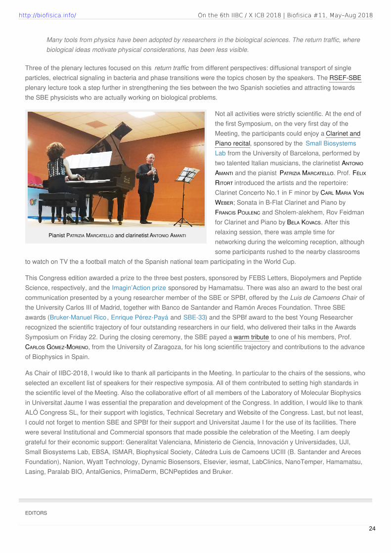
Pianist PATRIZIA MARCATELLO and clarinetist ANTONIO AMANTI
Three of the plenary lectures focused on this return traffic from different perspectives: diffusional transport of singleparticles, electrical signaling in bacteria and phase transitions were the topics chosen by the speakers. The RSEF-SBEplenary lecture took a step further in strengthening the ties between the two Spanish societies and attracting towardsthe SBE physicists who are actually working on biological problems.
Not all activities were strictly scientific. At the end ofthe first Symposium, on the very first day of theMeeting, the participants could enjoy a Clarinet andPiano recital, sponsored by the Small BiosystemsLab from the University of Barcelona, performed bytwo talented Italian musicians, the clarinetist ANTONIO
AMANTI and the pianist PATRIZIA MARCATELLO. Prof. FÉLIX
RITORT introduced the artists and the repertoire:Clarinet Concerto No.1 in F minor by CARL MARIA VON
WEBER; Sonata in B-Flat Clarinet and Piano byFRANCIS POULENC and Sholem-alekhem, Rov Feidmanfor Clarinet and Piano by BELA KOVACS. After thisrelaxing session, there was ample time fornetworking during the welcoming reception, althoughsome participants rushed to the nearby classrooms
to watch on TV the a football match of the Spanish national team participating in the World Cup.
This Congress edition awarded a prize to the three best posters, sponsored by FEBS Letters, Biopolymers and PeptideScience, respectively, and the Imagin’Action prize sponsored by Hamamatsu. There was also an award to the best oralcommunication presented by a young researcher member of the SBE or SPBf, offered by the Luis de Camoens Chair ofthe University Carlos III of Madrid, together with Banco de Santander and Ramón Areces Foundation. Three SBEawards (Bruker-Manuel Rico, Enrique Pérez-Payá and SBE-33) and the SPBf award to the best Young Researcherrecognized the scientific trajectory of four outstanding researchers in our field, who delivered their talks in the AwardsSymposium on Friday 22. During the closing ceremony, the SBE payed a warm tribute to one of his members, Prof.CARLOS GÓMEZ-MORENO, from the University of Zaragoza, for his long scientific trajectory and contributions to the advanceof Biophysics in Spain.
As Chair of IIBC-2018, I would like to thank all participants in the Meeting. In particular to the chairs of the sessions, whoselected an excellent list of speakers for their respective symposia. All of them contributed to setting high standards inthe scientific level of the Meeting. Also the collaborative effort of all members of the Laboratory of Molecular Biophysicsin Universitat Jaume I was essential the preparation and development of the Congress. In addition, I would like to thankALÓ Congress SL, for their support with logistics, Technical Secretary and Website of the Congress. Last, but not least,I could not forget to mention SBE and SPBf for their support and Universitat Jaume I for the use of its facilities. Therewere several Institutional and Commercial sponsors that made possible the celebration of the Meeting. I am deeplygrateful for their economic support: Generalitat Valenciana, Ministerio de Ciencia, Innovación y Universidades, UJI,Small Biosystems Lab, EBSA, ISMAR, Biophysical Society, Cátedra Luis de Camoens UCIII (B. Santander and ArecesFoundation), Nanion, Wyatt Technology, Dynamic Biosensors, Elsevier, iesmat, LabClinics, NanoTemper, Hamamatsu,Lasing, Paralab BIO, AntalGenics, PrimaDerm, BCNPeptides and Bruker.
Many tools from physics have been adopted by researchers in the biological sciences. The return traffic, wherebiological ideas motivate physical considerations, has been less visible.
EDITORS
24
On the 6th IIBC / X ICB 2018 | Biofisica #11, May–Aug 2018http://biofisica.info/

SPECIAL RECOGNITION
Prof. CARLOS GÓMEZ-MORENO
W
Tribute to Carlos Gómez-MorenoMilagros Medina & Javier Sancho , BIFI & UNIZAR, Zaragoza (Spain) .
hen CARLOS GÓMEZ-MORENO assumed his Cátedra (full Professorship) atthe University of Zaragoza (1983) he may not have imagined the futureof the Structural Biology and Biophysics in this city. This makes us look
back to see the very long road that he has travelled and that, fortunately for us, wehave in many occasions intensely walked with him. CARLOS has lived, and keepsliving, by pushing on each of the turns of life to come out in the best possible way;forcing himself to work hard, to make his ideas and actions valuable for all of us.
He did his doctorate in Seville, moving latter to the USA for two postdoctoralperiods, first in Ohio and later in San Francisco. As he likes to say:
In 1983 CARLOS arrived to the recently founded Department of Biochemistry and Molecular and Cell Biology at theUniversity of Zaragoza (DBMCB-UNIZAR), with 35 years, a brand new full Professorship and a test tube containing asample of Anabaena variabilis, the microscopic organism on which he has been working during all this time. When hecame to Zaragoza, he joined a department that was active in two lines of research, metabolism of lipids and cell biology,both of them very different from his own expertise. In this context, he founded his research group with the biologicalmaterial he brought and with many ideas to elucidate the reaction mechanisms of photosynthetic enzymes. This laid thefoundations of Structural Biology and Biophysics at the University of Zaragoza and in the whole region of Aragón, andsome of us had the fortune to be there almost from the very beginning. One of us (JAVIER) joined first, and as CARLOS
recalls, “he was not only a brilliant student, but also strong and able to handle the 600 liters of cultures that we neededto produce proteins, working long days, if you were feeding him well.” He also attracted to this nascent group a newlecturer in Plant Physiology at DBMCB, MARIA LUISA PELEATO, and continued with his “psychological” task to convince therest of us to carry out our PhD under his supervision. In his first 10 years in Zaragoza, MARIA FILLAT, JOSÉ JAVIER PUEYO,MILAGROS MEDINA and TERESA BES, in addition to JAVIER SANCHO, got their PhD in CARLOS’ group, and many more scientistscame later on. These were the fruitful seeds that he planted for Structural Biology and Biophysics in Zaragoza, sincemost of us have made our scientific career and created our own research groups in these areas, in many casesremaining at UNIZAR, thus spreading the field in the form of numerous talented CARLOS’ “grandsons” and“granddaughters” PhDs, doctorated with us.
He can also be very proud of having contributed to the development of two biotechnological companies, based inAragón, that today are selling to hundreds of countries and employing many people trained at UNIZAR. In CARLOS ownwords:
Four years, two postdocs, two sons, many experiences, much of training, anew vision of life and ideas in science to develop onwards, first in a shortperiod in Granada, and latter, for 35 years in Zaragoza, where I developedmost of my scientific life, as well as my family and personal life.
Biofísica M a g a z i n e
25
http://biofisica.info/ Biofísica #11, May–Aug 2018
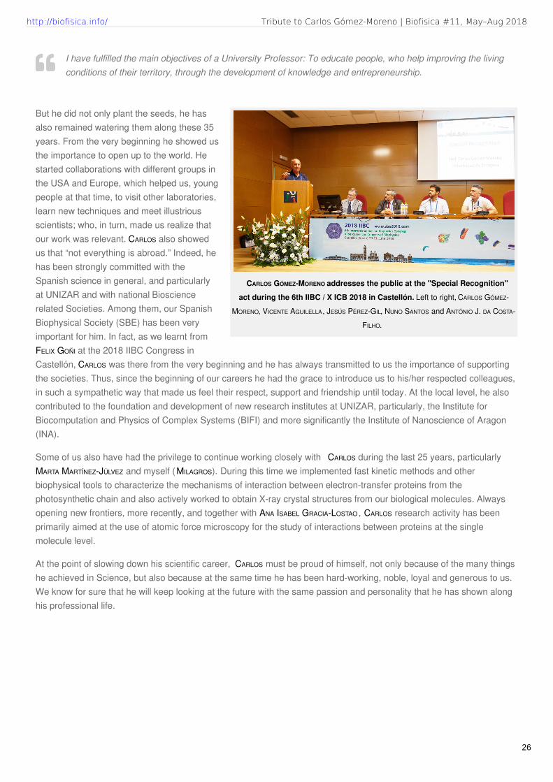
CARLOS GÓMEZ-MORENO addresses the public at the "Special Recognition"
act during the 6th IIBC / X ICB 2018 in Castellón. Left to right, CARLOS GÓMEZ-
MORENO, VICENTE AGUILELLA, JESÚS PÉREZ-GIL, NUNO SANTOS and ANTÓNIO J. DA COSTA-
FILHO.
But he did not only plant the seeds, he hasalso remained watering them along these 35years. From the very beginning he showed usthe importance to open up to the world. Hestarted collaborations with different groups inthe USA and Europe, which helped us, youngpeople at that time, to visit other laboratories,learn new techniques and meet illustriousscientists; who, in turn, made us realize thatour work was relevant. CARLOS also showedus that “not everything is abroad.” Indeed, hehas been strongly committed with theSpanish science in general, and particularlyat UNIZAR and with national Biosciencerelated Societies. Among them, our SpanishBiophysical Society (SBE) has been veryimportant for him. In fact, as we learnt fromFELIX GOÑI at the 2018 IIBC Congress inCastellón, CARLOS was there from the very beginning and he has always transmitted to us the importance of supportingthe societies. Thus, since the beginning of our careers he had the grace to introduce us to his/her respected colleagues,in such a sympathetic way that made us feel their respect, support and friendship until today. At the local level, he alsocontributed to the foundation and development of new research institutes at UNIZAR, particularly, the Institute forBiocomputation and Physics of Complex Systems (BIFI) and more significantly the Institute of Nanoscience of Aragon(INA).
Some of us also have had the privilege to continue working closely with CARLOS during the last 25 years, particularlyMARTA MARTÍNEZ-JÚLVEZ and myself (MILAGROS). During this time we implemented fast kinetic methods and otherbiophysical tools to characterize the mechanisms of interaction between electron-transfer proteins from thephotosynthetic chain and also actively worked to obtain X-ray crystal structures from our biological molecules. Alwaysopening new frontiers, more recently, and together with ANA ISABEL GRACIA-LOSTAO , CARLOS research activity has beenprimarily aimed at the use of atomic force microscopy for the study of interactions between proteins at the singlemolecule level.
At the point of slowing down his scientific career, CARLOS must be proud of himself, not only because of the many thingshe achieved in Science, but also because at the same time he has been hard-working, noble, loyal and generous to us.We know for sure that he will keep looking at the future with the same passion and personality that he has shown alonghis professional life.
I have fulfilled the main objectives of a University Professor: To educate people, who help improving the livingconditions of their territory, through the development of knowledge and entrepreneurship.
Tribute to Carlos Gómez-Moreno | Biofisica #11, May–Aug 2018http://biofisica.info/
26

HIGHLIGHTS 2018 | MAY.
Cryo-EM structure of the adenosineA2A receptor coupled to anengineered heterotrimeric G proteinGarcía-Nafría J, Lee Y, Bai X, Carpenter B, Tate CGeLife 2018 (May), 7:
HIGHLIGHTS 2018 | MAY.
Single-Stranded CondensationStochastically Blocks G-QuadruplexAssembly in Human Telomeric RNAGutiérrez I, Garavís M, de Lorenzo S, Villasante A,González C, Arias-Gonzalez JRJ Phys Chem Lett 2018 (May), 9: 2498
HIGHLIGHTS 2018 | MAY.
Reversible Immobilization ofProteins in Sensors and Solid-StateNanoporesAnanth A, Genua M, Aissaoui N, Díaz L, Eisele NB, Frey S,Dekker C, Richter RP, Görlich DSmall 2018 (May), 14: 1703357
HIGHLIGHTS 2018 | JUN.
Cryo-EM structure of the serotonin5-HT1B receptor coupled toheterotrimeric GoGarcía-Nafría J, Nehmé R, Edwards PC, Tate CGNature 2018 (Jun), 558: 620
Papers of the month by SBE members
HIGHLIGHTED PUBLICATIONS: MAY - SEPTEMBER 2018
Biofísica M a g a z i n e
27
http://biofisica.info/ Biofísica #11, May–Aug 2018

HIGHLIGHTS 2018 | JUN.
Enhancing Magnetic Light Emissionwith All-Dielectric OpticalNanoantennasSanz-Paz M, Ernandes C, Esparza JU, Burr GW, van HulstNF, Maitre A, Aigouy L, Gacoin T, Bonod N, Garcia-ParajoMF, Bidault S, Mivelle MNano Letters 2018 (Jun), 18: 3481
HIGHLIGHTS 2018 | JUN.
Anomalously low dielectric constantof confined waterFumagalli L, Esfandiar A, Fabregas R, Hu S, Ares P,Janardanan A, Yang Q, Radha B, Taniguchi T, Watanabe K,Gomila G, Novoselov KS, Geim AKScience 2018 (Jun), 360: 1339
HIGHLIGHTS 2018 | JUL.
Oxidative stress is tightly regulatedby cytochromecphosphorylation andrespirasome factors in mitochondriaGuerra-Castellano A, Díaz-Quintana A, Pérez-Mejías G,Elena-Real CA, González-Arzola K, García-Mauriño SM, laRosa MAD, Díaz-Moreno IProc Natl Acad Sci USA 2018 (Jul), 115: 7955
HIGHLIGHTS 2018 | AUG.
Photoexcitation of the P4480 StateInduces a Secondary PhotocycleThat Potentially DesensitizesChannelrhodopsin-2Saita M, Pranga-Sellnau F, Resler T, Schlesinger R, HeberleJ, Lorenz-Fonfria VAJ Am Chem Soc 2018 (Aug), 140: 9899
28
http://biofisica.info/ Highlighted Publications | Biofisica #11, May–Aug 2018

HIGHLIGHTS 2018 | AUG.
Water Sculpts the DistinctiveShapes and Dynamics of theTumor-Associated Carbohydrate TnAntigens: Implications for TheirMolecular RecognitionBermejo IA, Usabiaga I, Compañón I, Castro-López J,Insausti A, Fernández JA, Avenoza A, Busto JH, Jiménez-Barbero J, Asensio JL, Peregrina JM, Jiménez-Osés G, et alJ Am Chem Soc 2018 (Aug), 140: 9952
29
Highlighted Publications | Biofisica #11, May–Aug 2018http://biofisica.info/

Biofísica: SBE - Sociedad de Biofísica de España is licensed under a
Creative Commons Attribution 4.0 International License .. Design & technical editing by J. Salgado,
based on a Theme by Alx. Powered by WordPress
Permissions beyond the scope of this license may be available at http://www.sbe.es
Biophysics Magazine by
. Exported to PDF by wkhtmltopdf.