MASTER ASSIGNMENT Treatment outcome in patients with ...
26
MASTER ASSIGNMENT Treatment outcome in patients with anterior crossbites in the student clinic in Tromsø Authors Aleksandra Zakariassen, Marte Notkevich, Ingrid Vevik, Iselin Stallvik Mandal Supervisors Heidi Kerosuo, Professor, IKO Arne Mathisen, Specialist Orthodontist, TkNN UNIVERSITETET I TROMSØ Det helsevitenskapelige fakultet Institutt for klinisk odontologi Juni 2012
Transcript of MASTER ASSIGNMENT Treatment outcome in patients with ...

MASTER ASSIGNMENT
Treatment outcome in patients with anterior crossbites in the
student clinic in Tromsø
Authors
Aleksandra Zakariassen, Marte Notkevich, Ingrid Vevik, Iselin Stallvik Mandal
Supervisors
Heidi Kerosuo, Professor, IKO Arne Mathisen, Specialist Orthodontist, TkNN
UNIVERSITETET I TROMSØ
Det helsevitenskapelige fakultet Institutt for klinisk odontologi
Juni 2012
1
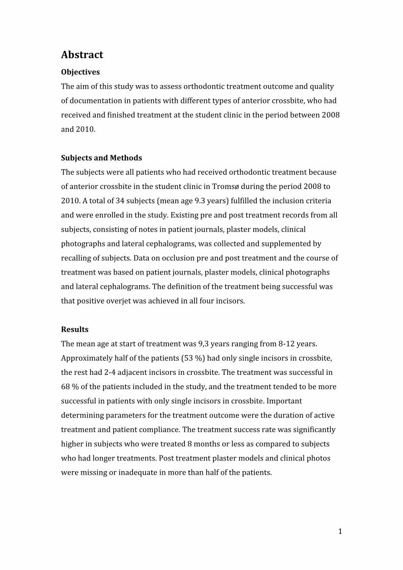
Abstract
Objectives
The aim of this study was to assess orthodontic treatment outcome and quality
of documentation in patients with different types of anterior crossbite, who had
received and finished treatment at the student clinic in the period between 2008
and 2010.
Subjects and Methods
The subjects were all patients who had received orthodontic treatment because
of anterior crossbite in the student clinic in Tromsø during the period 2008 to
2010. A total of 34 subjects (mean age 9.3 years) fulfilled the inclusion criteria
and were enrolled in the study. Existing pre and post treatment records from all
subjects, consisting of notes in patient journals, plaster models, clinical
photographs and lateral cephalograms, was collected and supplemented by
recalling of subjects. Data on occlusion pre and post treatment and the course of
treatment was based on patient journals, plaster models, clinical photographs
and lateral cephalograms. The definition of the treatment being successful was
that positive overjet was achieved in all four incisors.
Results
The mean age at start of treatment was 9,3 years ranging from 8-12 years.
Approximately half of the patients (53 %) had only single incisors in crossbite,
the rest had 2-4 adjacent incisors in crossbite. The treatment was successful in
68 % of the patients included in the study, and the treatment tended to be more
successful in patients with only single incisors in crossbite. Important
determining parameters for the treatment outcome were the duration of active
treatment and patient compliance. The treatment success rate was significantly
higher in subjects who were treated 8 months or less as compared to subjects
who had longer treatments. Post treatment plaster models and clinical photos
were missing or inadequate in more than half of the patients.

2
Conclusions
Our results suggest that 2 out of 3 anterior crossbites were successfully
corrected in the university student clinic in Tromsø, and longer treatment time
seemed to affect the treatment success negatively. The common lack of post-
treatment documentation calls for considerable improvement.

3
Introduction
Anterior crossbite
An occlusal disorder where one or more of the upper incisors are in reverse
overjet relative to the lower arch, which means that the upper incisors are
lingual to the lower incisors, is called anterior crossbite. If all four upper incisors
(or at least both upper central incisors) are lingual to the lower incisors it is
called negative overjet.
Inversion of incisors usually refers to a situation where one to three individual
incisors are in anterior crossbite. The etiology is dentoalveolar, e.g. crowding in
the upper arch or a retained primary incisor, which leads to one or more
palatally displaced incisors from the arch. The prevalence of one or more
inverted teeth was reported to be 11 % in a study on Swedish schoolchildren1.
Anterior crossbite may be due to abnormal inclination of the maxillary and
mandibular incisors, occlusal interferences, or skeletal discrepancies of the
maxilla and/or mandible2. Individuals with Class III malocclusion may have
combinations of skeletal and dento-alveolar components2. In clinical
orthodontics, it is important to make the differential diagnosis between skeletal
and dento-alveolar anterior crossbite due to differences in treatment modalities
in these patients regarding timing, duration and difficulty of treatment, which
affect the success of treatment (Table 1). Sometimes, e.g. in mild Class III cases,
the differential diagnosis is not always clear, because both dento-alveolar and
skeletal Class III characteristics may be present.
Dento-alveolar anterior crossbites
The majority of dento-alveolar anterior crossbites are caused by local
environmental factors. These factors, such as retained primary teeth, odontomas,
trauma, crowding, etc., could change the normal path of eruption allowing the
upper incisors to erupt palatally and/or the lower incisors to erupt labially3. In
children and adolescents in Bogota Colombia the prevalence of Pseudo Class III
was reported 2,1 %4. In a study on Swedish schoolchildren the prevalence of
4
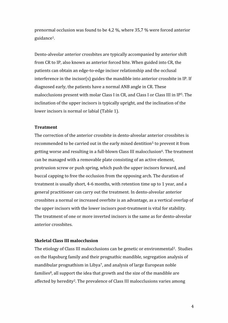
prenormal occlusion was found to be 4,2 %, where 35,7 % were forced anterior
guidance1.
Dento-alveolar anterior crossbites are typically accompanied by anterior shift
from CR to IP, also known as anterior forced bite. When guided into CR, the
patients can obtain an edge-to-edge incisor relationship and the occlusal
interference in the incisor(s) guides the mandible into anterior crossbite in IP. If
diagnosed early, the patients have a normal ANB angle in CR. These
malocclusions present with molar Class I in CR, and Class I or Class III in IP5. The
inclination of the upper incisors is typically upright, and the inclination of the
lower incisors is normal or labial (Table 1).
Treatment
The correction of the anterior crossbite in dento-alveolar anterior crossbites is
recommended to be carried out in the early mixed dentition5 to prevent it from
getting worse and resulting in a full-blown Class III malocclusion6. The treatment
can be managed with a removable plate consisting of an active element,
protrusion screw or push spring, which push the upper incisors forward, and
buccal capping to free the occlusion from the opposing arch. The duration of
treatment is usually short, 4-6 months, with retention time up to 1 year, and a
general practitioner can carry out the treatment. In dento-alveolar anterior
crossbites a normal or increased overbite is an advantage, as a vertical overlap of
the upper incisors with the lower incisors post-treatment is vital for stability.
The treatment of one or more inverted incisors is the same as for dento-alveolar
anterior crossbites.
Skeletal Class III malocclusion
The etiology of Class III malocclusions can be genetic or environmental2. Studies
on the Hapsburg family and their prognathic mandible, segregation analysis of
mandibular prognathism in Libya7, and analysis of large European noble
families8, all support the idea that growth and the size of the mandible are
affected by heredity2. The prevalence of Class III malocclusions varies among

5
different ethnic groups, and the prevalence in the Caucasian population is
approximately 3-5 %2.
Typical occlusal characteristics of skeletal Class III are Angle Class III relation in
molars and canines, and anterior crossbite (Table 1). Dento-alveolar
compensation in the inclination of the incisors can be seen with proclination of
the upper incisors and retroclination of the lower incisors (Table 1). This results
in the incisor relationship being less severe than the underlying skeletal pattern.
Anterior shift from CR to IP, which is commonly found in dento-alveolar anterior
crossbites, can also be present in skeletal Class III malocclusions in the early
mixed dentition. If such a displacement is present, the prognosis for correction of
the anterior crossbite is more favourable9.
The skeletal characteristics of true Class III malocclusions are mandibular
prognathism, maxillary retrognathia, or a combination of both3. The patients
have a skeletal discrepancy, which is seen from their concave profile. In the
cephalometric analysis these patients have either a decreased SNA angle or an
increased SNB angle or both, so that the ANB angle is negative. A posterior
rotation growth pattern of the mandible will camouflage the skeletal Class III
pattern and reduce overbite with growth9. If there is an anterior rotation growth
pattern of the mandible, there will be increased prominence of the chin and
worsening of the skeletal pattern and the negative overjet with growth9.
The skeletal and dental components of Class III malocclusions are present
already in early childhood, and tend to worsen with growth if left untreated, e.g.
increased dento-alveolar compensation by proclination of upper incisors and
retroclination of lower incisors, worsening of mandibular prognathism and
sagittal skeletal discrepancy between the jaws with growth10. In a study among
untreated Class III patients, changes in angle ANB, Wits appraisal and molar
relationship indicated a worsening of the Class III relationship with increasing
skeletal maturity as the mandible outgrew the maxilla10. This, and the fact that
the growth spurt in midfacial length occurs in the prepubertal period10, indicate
early treatment of Class III malocclusions.
6
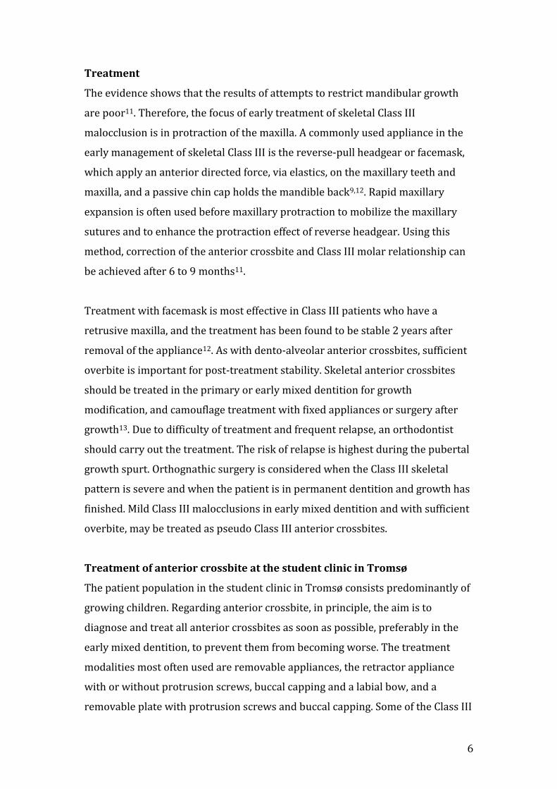
Treatment
The evidence shows that the results of attempts to restrict mandibular growth
are poor11. Therefore, the focus of early treatment of skeletal Class III
malocclusion is in protraction of the maxilla. A commonly used appliance in the
early management of skeletal Class III is the reverse-pull headgear or facemask,
which apply an anterior directed force, via elastics, on the maxillary teeth and
maxilla, and a passive chin cap holds the mandible back9,12. Rapid maxillary
expansion is often used before maxillary protraction to mobilize the maxillary
sutures and to enhance the protraction effect of reverse headgear. Using this
method, correction of the anterior crossbite and Class III molar relationship can
be achieved after 6 to 9 months11.
Treatment with facemask is most effective in Class III patients who have a
retrusive maxilla, and the treatment has been found to be stable 2 years after
removal of the appliance12. As with dento-alveolar anterior crossbites, sufficient
overbite is important for post-treatment stability. Skeletal anterior crossbites
should be treated in the primary or early mixed dentition for growth
modification, and camouflage treatment with fixed appliances or surgery after
growth13. Due to difficulty of treatment and frequent relapse, an orthodontist
should carry out the treatment. The risk of relapse is highest during the pubertal
growth spurt. Orthognathic surgery is considered when the Class III skeletal
pattern is severe and when the patient is in permanent dentition and growth has
finished. Mild Class III malocclusions in early mixed dentition and with sufficient
overbite, may be treated as pseudo Class III anterior crossbites.
Treatment of anterior crossbite at the student clinic in Tromsø
The patient population in the student clinic in Tromsø consists predominantly of
growing children. Regarding anterior crossbite, in principle, the aim is to
diagnose and treat all anterior crossbites as soon as possible, preferably in the
early mixed dentition, to prevent them from becoming worse. The treatment
modalities most often used are removable appliances, the retractor appliance
with or without protrusion screws, buccal capping and a labial bow, and a
removable plate with protrusion screws and buccal capping. Some of the Class III
7
patients who are treated early at the student clinic need specialist treatment
later. Early treatment aims only for anterior crossbite correction, which is
expected to reduce later need and difficulty of treatment14.
The integrated master program in odontology started in Tromsø in 2004. As the
routines have not yet been fully established, some problems related to
organization of treatment at the student clinic have existed. First of all, the
students´ restricted time at the clinic can prolong the duration of treatment for
some patients, especially during the summer where students are not present at
the student clinic. Another problem is that a patient may change operator one or
several times during the treatment, and that different supervisors are involved in
the treatment. The lack of continuity can affect the treatment negatively, thereby
lowering the quality of treatment.
In orthodontics, evaluation of treatment success is usually done on each
individual patient at the end of treatment. However, regarding the quality and
success of treatment outcome, general evaluations of larger patient groups are
needed to maintain quality control in the patient care and to improve the
treatment practices. More comprehensive evaluations are based on patient
documentations and can be carried out retrospectively. So far, evaluations of the
orthodontic treatments carried out in the student clinic, are missing.
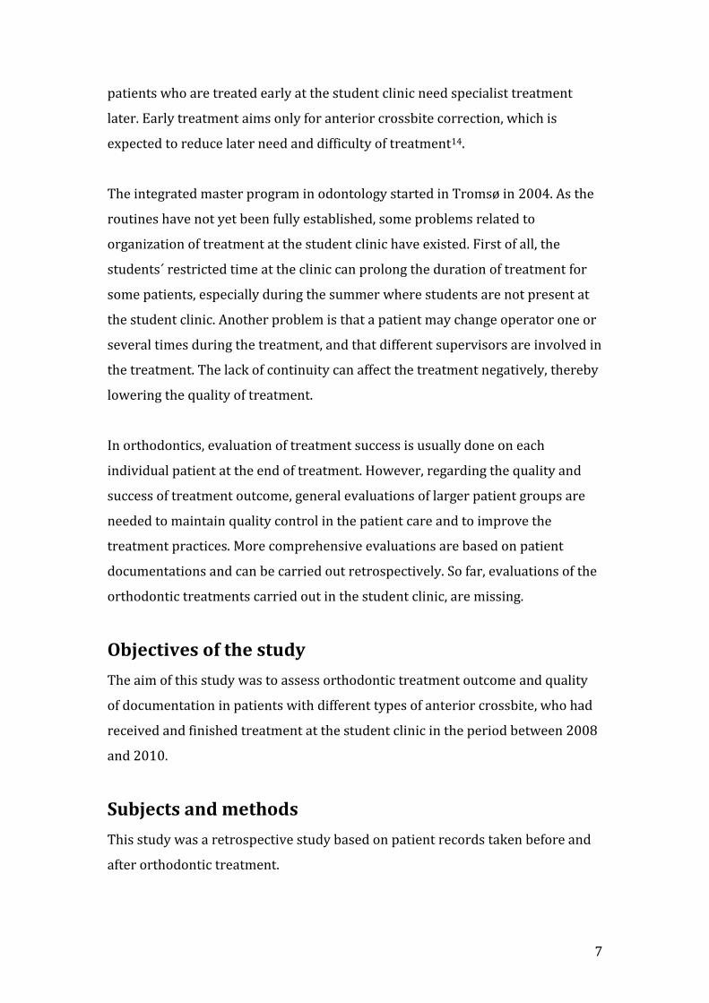
Objectives of the study
The aim of this study was to assess orthodontic treatment outcome and quality
of documentation in patients with different types of anterior crossbite, who had
received and finished treatment at the student clinic in the period between 2008
and 2010.
Subjects and methods
This study was a retrospective study based on patient records taken before and
after orthodontic treatment.
8
Subjects
The subjects were all patients who had received orthodontic treatment because
of anterior crossbite in the student clinic in Tromsø during the period 2008 to
2010. All patient records were manually searched through to find patients with
anterior crossbite diagnoses. Following inclusion criteria were used:
The subject had to have one to four incisors in anterior crossbite, the subject had
received and finished treatment at the student clinic during the period 2008 to
2010 and acceptable pre treatment records encompassing journal notes, plaster
models and/or clinical photographs had to be available.
In case of missing post treatment records the subjects were recalled to the
student clinic for completing the records. The full post treatment records
encompassed necessary notes in the patient journals, plaster models with index,
clinical photographs and lateral cephalogram if pre treatment cephalogram
existed.
The total number of subjects fulfilling with the pre treatment criteria was 34. 16
of them were recalled to the student clinic for completing the post treatment
records. In 1 subject the anterior crossbite was confirmed through clinical
pictures due to missing pre treatment plaster models.
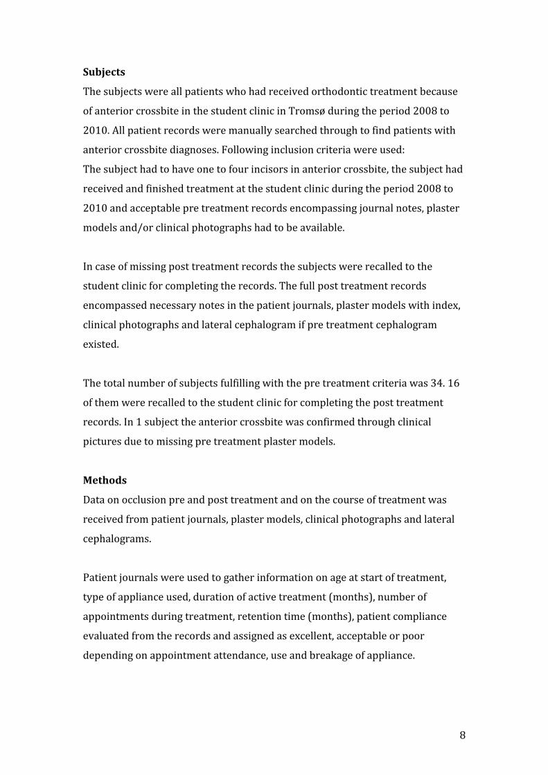
Methods
Data on occlusion pre and post treatment and on the course of treatment was
received from patient journals, plaster models, clinical photographs and lateral
cephalograms.
Patient journals were used to gather information on age at start of treatment,
type of appliance used, duration of active treatment (months), number of
appointments during treatment, retention time (months), patient compliance
evaluated from the records and assigned as excellent, acceptable or poor
depending on appointment attendance, use and breakage of appliance.

9
Plaster models
Data collected from plaster models included the number of centrals and/or
laterals in anterior crossbite, Angle classification in permanent molars, dental
stage assigned as primary, early mixed, late mixed or permanent dentition,
overjet and overbite (mm) on fully erupted teeth, measured on the most
prominent central incisor, and anterior crowding assigned yes or no. Anterior
crossbite was divided into three subgroups based on number of teeth in anterior
crossbite (Figure 1).
1. One central, one lateral incisor or both lateral incisors in anterior
crossbite.
2. One central and one lateral, two central incisors, two central and one
lateral, and one central and two lateral incisors in anterior crossbite.
3. All four incisors in anterior crossbite.
Clinical pictures
Profile pictures were used to evaluate the profile assigned as concave, straight or
convex. Intra oral frontal pictures were used for confirming the diagnosis and
treatment outcome in cases with missing or broken plaster models, which was
the case in 1 subject.
Lateral cephalograms
The parameters that were included and measured on the lateral cephalograms
were the SNA, SNB and ANB angles, inclination upper incisor/NSL, inclination
lower incisor/ML, SN/ML and the inter-incisal angle (Figure 2).
Evaluation of treatment success
The treatment was considered being successful if positive overjet was achieved
in all four incisors. The treatment was considered not successful if one or more
of the following was recorded; the subject did not want to continue the
treatment, positive overjet and overbite was not achieved during treatment, or
the patient was referred to a specialist for further treatment.

10
Analysis of data
The data was recorded and analyzed in SPSS for Windows 19. Means and
frequencies for different variables were calculated. Pearson’s chi square was
used to test the differences between groups. Differences with P-values < 0.05
were considered as statistically significant.
Method error
The examiners were calibrated by an experienced orthodontist to perform the
occlusal measurements. The intra-class correlation coeffisient (ICC) for overjet
and overbite measurements between the two examiner groups ranged between
0.625-0.746 (=moderate to strong agreement) and between the examiner groups
and the calibrating orthodontist 0.744-0.887 (=strong to almost perfect
agreement). For Angle classification (right and left side) there was a 100 %
agreement between the two examiner groups, and between the calibrating
orthodontist and the examiner groups the agreement was 74 %. For evaluating
the intra-examiner reliability, duplicate measurements were carried out on 16
randomly selected cases. The intra-examiner consistency between duplicate
measurements of Angle classification ranged from 94 % to 100 % (κ = 0.763-
1.00), indicating substantial to almost perfect agreement. The ICC for the
examiners duplicate measurements of overjet and overbite ranged between
0.691-0.927.
Cephalometric measurements were done by the authors (MUN, IV, ISM and AZ)
using hand tracing method and by the computerized method by the supervisor
(AM) (Facad version 3.5.0.3.). Because of marked differences between the values
received by the two methods, the values from the Facad analysis method were
used in the analyses.
Results
The mean age of the patients at start of treatment was 9,3 years ranging from 8-
12 years. Seventy-four percent of the subjects were in early mixed dentition, 21
% in late mixed dentition, and 2 subjects (6 %) in permanent dentition at
treatment start (Table 2). Ten subjects (29 %) had a negative overjet pre-

11
treatment. For the rest of the subjects (24) the mean overjet was 1,9 mm (range
0-4 mm). Mean overbite before treatment was 2,1 mm (range 0-4 mm).
Class I molar relationship pre treatment was found in 18 subjects (69 %), and
the molar relationship did not change with treatment. Six subjects had Class II
molar relationship and one of them changed to Class I with treatment. Two
subjects had Class III molar relationship pre treatment, and the molar
relationship changed to Class I in both of them with treatment. Eight subjects
were missing records on angle classification pre and/or post treatment.
The treatment was successful in 68 % of the subjects included in the study. The
treatment success was 57% (12 out of 21) in subjects who had started the
treatment before 10 years of age, and 85 % in the subjects with the treatment
started at 10 years of age or later, although the difference did not reach
statistical significance (Table 3). No difference was seen in treatment success
between different dental stages (Table 2).
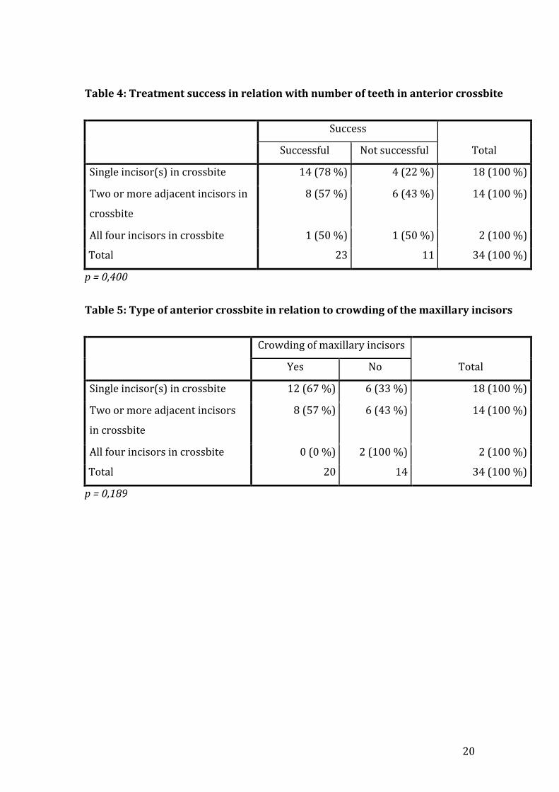
Treatment success
Orthodontic treatment was successful in 78 % of the subjects with only
individual teeth in anterior crossbite as compared to 57 % success rate in
subjects with 2-3 adjacent teeth in crossbite. When all four incisors were in
anterior crossbite 1 out of 2 subjects (50 %) were successful. The differences
were however not statistically significant (Table 4).
Twenty subjects (59 %) had maxillary anterior crowding pre treatment.
Crowding was slightly more common (67 %) in subjects with crossbites of single
teeth than in subjects with several or all anterior teeth in crossbite (50 %) (Table
5).
Caphalometric analysis
The cephalometric results were based on the 9 subjects who had cephalograms
taken before and after treatment (Table 6). Seven of the 9 subjects had several
anterior teeth in crossbite.
Three of the subjects with lateral cephalogram had a prognathic mandible pre
treatment (SNB > 82 degrees) and 4 had a retrognathic maxilla pre treatment
(SNA < 80 degrees).

12
The inclination of the upper incisors/SN increased with treatment in 8 of the 9
subjects with the mean pre-treatment value of 108 degrees and post-treatment
114 degrees. The mean inclination of lower incisors/ML decreased in the same
subjects from 100 to 95 degrees during treatment (Table 6). The mean ANB and
interincisal angles were low both pre and post treatment as compared to the
norm values (Table 6).
Factors related to orthodontic treatment and success rate
The average duration of active treatment was 8,3 months ranging from 2-26
months. The average number of appointments was 6,4 ranging from 2-18. The
treatment success rate was significantly higher (88%) in subjects who were
treated 8 months or less as compared to subjects who had longer treatments
(12%) (Table 7).
Duration of active treatment varied with the type of appliances used. Treatment
time was longest with the retractor appliance, with an average of 9,7 months,
and shortest for the acrylic plate, with an average of 7,1 months. The duration of
active treatment was intermediate for the expansion plate, with an average of 8,4
months.
Subjects with all four incisors or both central incisors in anterior crossbite were
treated with a retractor appliance in 7 out of 8 cases, and with an expansion
plate with protrusion screw(s) in one case. The rest 26 were treated with either
an acrylic plate with protrusion screw(s) or an expansion plate with protrusion
screw(s). The success rate was highest with the acrylic plate with protrusion
screw(s) (88 %) and lowest with the retractor (57%), but the differences were
not statistically significant (Table 8).
Patient compliance was excellent in 42 % of the patients, 52 % showed
acceptable cooperation and 6 % had poor cooperation during the treatment.
There was found no difference in the compliance between the younger and older
patient groups or between the dental stages. Treatment was successful in 87% of
the patients with excellent cooperation as compared to 55 % in patients with
acceptable/poor cooperation (p=0.063).
The patient compliance was almost significantly different between the
appliances used to correct anterior crossbite (p=0.056). Excellent compliance
13
was most often met with the expansion plate with protrusion screw(s) and
acrylic plate with protrusion screw(s), whereas with all subjects using a
retractor the cooperation was somewhat compromised (Table 9).
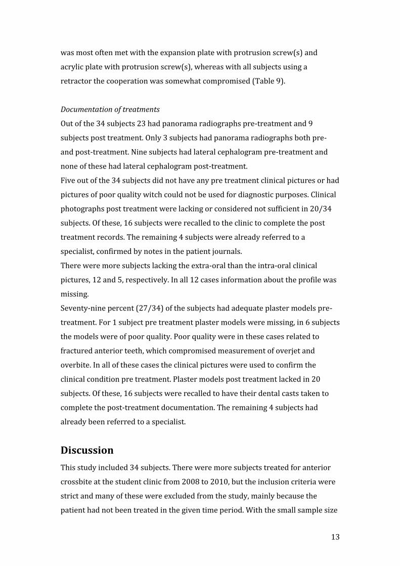
Documentation of treatments
Out of the 34 subjects 23 had panorama radiographs pre-treatment and 9
subjects post treatment. Only 3 subjects had panorama radiographs both pre-
and post-treatment. Nine subjects had lateral cephalogram pre-treatment and
none of these had lateral cephalogram post-treatment.
Five out of the 34 subjects did not have any pre treatment clinical pictures or had
pictures of poor quality witch could not be used for diagnostic purposes. Clinical
photographs post treatment were lacking or considered not sufficient in 20/34
subjects. Of these, 16 subjects were recalled to the clinic to complete the post
treatment records. The remaining 4 subjects were already referred to a
specialist, confirmed by notes in the patient journals.
There were more subjects lacking the extra-oral than the intra-oral clinical
pictures, 12 and 5, respectively. In all 12 cases information about the profile was
missing.
Seventy-nine percent (27/34) of the subjects had adequate plaster models pre-
treatment. For 1 subject pre treatment plaster models were missing, in 6 subjects
the models were of poor quality. Poor quality were in these cases related to
fractured anterior teeth, which compromised measurement of overjet and
overbite. In all of these cases the clinical pictures were used to confirm the
clinical condition pre treatment. Plaster models post treatment lacked in 20
subjects. Of these, 16 subjects were recalled to have their dental casts taken to
complete the post-treatment documentation. The remaining 4 subjects had
already been referred to a specialist.
Discussion
This study included 34 subjects. There were more subjects treated for anterior
crossbite at the student clinic from 2008 to 2010, but the inclusion criteria were
strict and many of these were excluded from the study, mainly because the
patient had not been treated in the given time period. With the small sample size

14
the power of the study was too low to reach statistical significance for the
differences found. Thus, our sample is not representative for generalizing the
results to any other population, but it gives information on local level on the
treatment success at the student clinic in Tromsø.
In orthodontic cephalometrics it is important to keep method error to a
minimum in order to see the valid small changes achieved by treatment15. Hand
tracing is time consuming and errors may arise from acquisition of radiographs,
tracing, landmark identification, and measurements15. In this study it was
decided to use the results of the digital measurements instead of the results of
hand tracings, although Sayinsu et al reported that the validity and
reproducibility of the measurements with the Dolphin Imaging Software and
with the conventional method were highly correlated. This was decided because
of the source of error in finding the landmarks due to poor image quality in the
prints. Also Sayinsu et al concluded that the digitalized method could be
preferred for research purposes. One source of error in this study was also the
inconsistent occlusion position during cephalometric radiographing. Most of the
lateral cephalograms were taken in IP pre- and post treatment.
Occlusal characteristics and treatment outcome
The results showed that the average start of treatment was 9,3 years, and no
correlation was found between age at start of treatment and treatment success.
Treatment of anterior crossbite should be carried out in the early mixed
dentition, particularly if central incisors are involved16. In this study 74 % of the
subjects started treatment in the early mixed dentition with the success rate of
68 %. Somewhat lower success of treatment was found when the subjects
started treatment in the late mixed dentition (57 %). Only 2 subjects started
treatment in the permanent dentition, and their treatment was successful. These
subjects only had one or two laterals in anterior crossbite with no skeletal Class
III involvement, which is expected to be easier to treat than a full anterior
crossbite. The small number of subjects treated in the permanent dentition
makes a poor foundation for comparison, and a less successful result might have
been expected if the sample size was bigger.

15
In our study, the treatment of patients at the student clinic aimed only for
anterior crossbite correction, which is expected to reduce eventual later need
and difficulty of treatment. For the most part, the treatment resulted in anterior
crossbite correction, and all successful cases had a positive overjet in all four
incisors post treatment. The success rate of treatment showed some variation
according to the number of teeth in crossbite with the highest success rate in
subjects with 1 incisor in anterior crossbite. One incisor in anterior crossbite is
generally easier to treat successfully than several incisors in anterior crossbite,
mainly because the probability of skeletal Class III discrepancy increases with
more incisors in crossbite, and therefore correction of skeletal anterior crossbite
with removable appliances had rather poor prognosis in our study. In all
unsuccessful cases, there was a remaining anterior crossbite of 1-2 incisors not
including both central incisors post treatment.
Correction of a Class III molar relationship to a Class I or super Class I molar
relationship was achieved in 2 successful cases, but mainly there were no
changes in molar relationship, indicating that the treatment of anterior crossbite
with removable appliances did not affect the molar relationship.
The results showed that maxillary crowding was somewhat more frequent in
subjects with less than four incisors in anterior crossbite than in subjects with a
full anterior crossbite. This observation may be caused by different etiologies. A
full anterior crossbite is often associated with a skeletal discrepancy13. In cases
with single anterior teeth in crossbite, the crowding itself might be the main
etiological cause, since anterior crowding in the upper jaw often leads to palatal
displacement of individual teeth, which may cause an anterior crossbite13.
Therefore, anterior crossbite may also reflect crowding of teeth, when there are
only one or two incisors involved, not including both central incisors in the
anterior crossbite.
The treatment was unsuccessful in 3 subjects, who showed clear Class III skeletal
characteristics, which complicated the treatment, and was the likely reason for
the unfavorable treatment response. Compliance, insufficient overbite post
treatment, or choice of appliance could also have contributed to the failure. In 4
of the subjects there were an increase in the ANB angle after treatment with a
retractor appliance. This indicates that treatment with a retractor appliance may
16
have some favorable skeletal effects in addition to the dental effects, though
some of the changes can be contributed to the natural growth of the maxilla and
the mandible. The inclination lower incisors/mandibular line decreased in all
subjects after treatment with a retractor appliance, which means that the
treatment retroclined the lower incisors, compensating the skeletal discrepancy.
In cases with an anterior crossbite, an important determinant for stability of the
treatment results is a positive overbite16. If a sufficient overbite is achieved,
retention is theoretically not essential because the occlusion will maintain the
result. In this study, the subjects with an equal or increased overbite post
treatment may have a better prognosis in the long term as compared to subjects
with a reduced overbite.
Treatment practices in relation to treatment outcome
The results showed that there was a significant difference in duration of active
treatment between subjects with a successful and unsuccessful treatment
outcome, 7.4 and 14.4 months, respectively. This shows that the duration of
treatment of unsuccessful subjects was nearly doubled. The number of
appointments did not show difference, as the unsuccessful subjects in average
only had 1.5 more appointments than the successful subjects. This means that
the appointments in the unsuccessful group were distributed over a longer time
span.
There can be different causes for the extended duration of treatment in the
unsuccessful cases, but most likely this is connected to problems with
compliance or difficulty of treatment. Examples of lack of compliance might be
that the subjects did not show up for appointments or that the treatment
continued over the summer. This insufficient follow-up hampers the possibility
for the clinician to control the adjustments of the appliance and subjects’
compliance, both of which affect the treatment outcome. When there was no
success after expected duration of treatment, the treatment was not interrupted
but instead continued a little longer. This can be because the operator expects
the compliance to be the reason, or that the appliance has not been adjusted
correctly, although the underlying reason might also have been the appliance
because of the skeletal components of the anterior crossbite.

17
The removable appliances used at the student clinic to treat the subjects in this
study were a retractor appliance with or without protrusion screw(s), an acrylic
plate with protrusion screw(s), and an expansion plate with protrusion screw(s).
The expansion plate was most often used, as many subjects had an anterior
crossbite in combination with a posterior crossbite. The retractor appliance was
used least often, because it was generally used only in subjects with a full
anterior crossbite or both central incisors in anterior crossbite, who were in the
minority in this study.
The success rates with retractor, acrylic plate and expansion plate were 57 %, 75
%, and 63 %, respectively. The retractor appliance showed the lowest success
rate, while the acrylic plate showed the greatest success rate. Reasons for this
might be that the retractor needs adjustment of the labial bow continuously to be
effective, and because the retractor appliance was most often used in subjects
with all four or both central incisors in anterior crossbite and often with a
skeletal Class III tendency. The acrylic plate may be easier to use, needs little
adjustment to be effective, and it was only used in subjects with 2 incisors in
anterior crossbite not including both central incisors, which generally are less
complicated to treat than a full anterior crossbite.
Compliance was not superior when the treatment started in the early mixed
dentition compared to the late mixed and permanent dentition. The definition of
compliance is relative, since there is no clear measurement for this variable. In
this study, the compliance was divided into three distinct categories: excellent,
acceptable and poor, and the subjects were assigned to the different categories
based on the examiners’ subjective opinions. Subjects in the excellent
cooperation category showed no lack of compliance, while in contrast poor
compliance was associated with broken, lost, or not used appliances, or missed
appointments. Subjects in the acceptable category were considered to be in
between these limits. The results showed that most subjects in the excellent
category had a successful treatment outcome, while roughly only 2 out of 3
subjects ranked in the acceptable category showed treatment success. This is in
accordance with previous reports, which have shown positive correlation
between treatment success and patient compliance17. The category of poor
compliance showed no successful cases. Even though this shows a clear

18
connection between compliance and success rate, there were only two subjects
in the poor compliance category, which makes a weak foundation for
comparison.
Challenges of treating patients at the student clinic in Tromsø
The activities in the student clinic in Tromsø started gradually in 2007 and have
been under development during the last 5 years. Therefore, the routines have
not yet been fully established regarding documentation of patient treatments
and evaluation of the results. An obvious drawback when several students and
supervisors are involved in the same treatment is the big variation seen in the
quality and quantity of pre-treatment and post treatment records. In this study,
the pre-treatment records included more information than the post-treatment
records. The clear shortcoming was the insufficient recording in the patients’
journal in OPUS, both in the pre- and post-records. In general, all pre-treatment
records were present with plaster models, clinical photos and x-rays, but very
often information from these were not described in the records. Information
from patient files, which often were not described, included teeth in anterior
crossbite, family history of malocclusion, evaluation of profile and appliances of
choice. Lateral cephalogram were often missing in pre-treatment records.
Post treatment records were typically insufficient or missing, especially when
the treatment was unsuccessful. This made evaluation of treatment outcome
impossible in many cases. Therefore, nearly half of the subjects had to be invited
to the student clinic only to get their post-treatment records completed to enable
assessment of their treatment result. Inadequate records were probably due to
absence of guidance, which specifies what the records should contain. Fractured
teeth, not fully erupted incisors, or missing plaster models were the most
frequent reasons for why the overjet was not measurable pre-treatment.
Conclusions
Our results suggest that 2 out of 3 anterior crossbites were successfully
corrected in the university student clinic in Tromsø, and longer treatment time
seemed to affect the treatment success negatively. The common lack of post-
treatment documentation calls for considerable improvement.

19
Tables and figures
Table 1: Typical diagnostic signs in differentiating between dento-alveolar and
skeletal Class III
Dento-alveolar anterior crossbite Skeletal anterior crossbite
Family history of Cl III No Yes
Angle Class in CR1 Cl I Cl III
Angle Class in IP2 Cl I or III Cl III
Incisor inclination Upper incisors upright
Lower incisors normal/labial
Upper incisors proclined
Lower incisors retroclined
Anterior shift CR-IP Yes No*
Profile Straight in CR
Can be concave in IP
Concave
*Skeletal Class III can be associated with anterior shift from CR to IP in the early mixed
dentition.
1 CR = centric relation, 2IP = intercuspal position
Table 2: Treatment success in relation to dental stage pre-treatment
Success
Total Successful Not successful
Dental
stage
Early mixed 17 (68 %) 8 (32 %) 25 (100 %)
Late mixed 4 (57 %) 3 (43 %) 7 (100 %)
Permanent dent 2 (100 %) 0 (0,0 %) 2 (100 %)
Total 23 11 34 (100 %)
p = 0,519
Table 3: Treatment success in relation to the age at start of treatment
Success
Total Successful Not successful
Age 8-9 years 12 (57 %) 9 (43 %) 21 (100 %)
Age 10-12 years 11 (85 %) 2 (15 %) 13 (100 %)
Total 23 11 34 (100 %)
p = 0,096
20
Table 4: Treatment success in relation with number of teeth in anterior crossbite
Success
Total Successful Not successful
Single incisor(s) in crossbite 14 (78 %) 4 (22 %) 18 (100 %)
Two or more adjacent incisors in
crossbite
8 (57 %) 6 (43 %) 14 (100 %)
All four incisors in crossbite 1 (50 %) 1 (50 %) 2 (100 %)
Total 23 11 34 (100 %)
p = 0,400
Table 5: Type of anterior crossbite in relation to crowding of the maxillary incisors
Crowding of maxillary incisors
Total Yes No
Single incisor(s) in crossbite 12 (67 %) 6 (33 %) 18 (100 %)
Two or more adjacent incisors
in crossbite
8 (57 %) 6 (43 %) 14 (100 %)
All four incisors in crossbite 0 (0 %) 2 (100 %) 2 (100 %)
Total 20 14 34 (100 %)
p = 0,189

21
Table 6: Mean values with ranges for different cephalometric parameters of the subjects (N=9) according to the cephalometric analysis.
*Normal values according to Facad 3.5.0.3
Table 7: Treatment success in relation to duration of the treatment
Success
Total Successful Not successful
Treatment time ≤8 months 21 (88 %) 3 (12 %) 24 (100 %)
Treatment time ≥9 months 2 (20 %) 8 (80 %) 10 (100 %)
Total 23 11 34 (100 %)
p = 0,000
Table 8: Treatment success in relation to appliance used
Success
Total Successful Not successful
Appliance Retractor 4 (57 %) 3 (43 %) 7 (100 %)
Acrylic plate 7 (88 %) 1 (12 %) 8 (100 %)
Expansion plate with
protrusion
12 (63 %) 7 (37 %) 19 (100 %)
Total 23 11 34 (100 %)
p = 0,374
SNA SNB ANB Incl.up.inc/SN Incl.lo.inc/ML SN/ML Interinc ang
Pre Post Pre Post Pre Post Pre Post Pre Post Pre Post Pre Post
Range 70,
7-
86,
3
74,9-
87,5
72,3-
86,6
73,0-
88,9
-2,4-
3,5
-3,2-
4,1
92,8-
121,3
99,5-
125,9
84,4-
113,4
79,4-
109,6
21,4-
45,1
20,3-
42,8
105-
137,2
107,1-139,2
Mean 78,
9
81,2 78,6 81,7 0,4 -0,5 107,7 113,7 100,4 94,9 32,1 29,6 119,8 121,8
Norm
values*
80-89
75-82 2-4 102 +/- 6 94 +/- 4,5 33 +/- 4 130-150

22
Table 9: Patient compliance in relation to different appliances used
Appliance
Total Retractor Acrylic plate
Expansion +
protrusion plate
Excellent 0 (0 %) 5 (36 %) 9 (64 %) 14 (100 %)
Acceptable 7 (39 %) 3 (17 %) 8 (44 %) 18 (100 %)
Poor 0 (0 %) 0 (0 %) 2 (100 %) 2 (100 %)
Total 7 (21 %) 8 (23 %) 19 (56 %) 34 (100 %)
p = 0,056

23
Figure 1: Anterior crossbite groups
Figure 2: Cephalometric parameters used in the cephalometric analysis (picture adopted from L. Mitchell, 2007)
a) Example group 1 b) Example group 2 c) Example group 3

24
References 1 Birgit Thilander and Nils Myrberg. The prevalence of malocclusion in Swedish schoolchildren. Scand. J. dent. Res. 1973: 81: 12-20. 2 Peter Ngan, DMD, Annie M. Hu, DDS, MS, Henry W. Fields, Jr., DDS, MS, MSD. Treatment of Class III problems begins with differential diagnosis of anterior crossbites. American Academy of Pediatric Dentistry, 19:6, 1997. 3 A.B.M. Rabie, BDS, Cert Ortho, MS, PhDa and Yan Gu, BDSb. Diagnostic criteria for Pseudo Class III malocclusion. American Journal of Orthodontics and Dentofacial Orthopedics, Vol 117, No 1, January 2000. 4 Birgit Thilander1, Lucia Pena2, Clementina Infante2, Sara Stella Parada2 and Clara de Mayorga. Prevalence of malocclusion and orthodontic treatment need in children and adolescents in Bogota, Colombia. An epidemiological study related to different stages of dental development. European Journal of Orthodontics 23 (2001), 153-167. 5 A. Giancotti, A. Maselli, G. Mampieri and E. Spanò. Pseudo Class III malocclusion treatment with Balters` Bionator. Journal of Orthodontics, Vol 30, September 2003, 203-215. 6 A Kapur, HS Chawla, A Utreja, A Goyal. Early Class III occlusal tendency in children and its selective management. Journal of the Indian Society of Pedodontics and Preventive Dentistry, Vol 26, Pg 107-113, 2008. 7 A.A. El-Gheriani B.S. Maher A.S. El-Gheriani, J.J Sciote, F.A. Abu-shahba, R. Al-Azemi and M.L. Marazita. Segregattion Analysis of Manibular Prognathism in Libya. Journal of Dental Research 82(7):523-527, 2003. 8 G Wolf T F Wienker, H Sander. On the genetics of mandibular prognathism: analysis of large European noble families. Journal of Medical Genetics 30(2): 112-116, 1993. 9 Laura Mitchell. An introduction to orthodontics. 3rd ed. 2007, pg 123, 122, 127. 10 Ann E. Zionic Alexander,a James A. McNamara, Jr,b Lorenzo Franchi,c and Tiziano Baccettic. Semilongitudinal cephalometric study of craniofacial growth in untreated Class III malocclusion. American Journal of Orthodontics and Dentofacial Orthopedics, Vol 135, No 6, June 2009. 11 Hideo Mitani D.D.S., M.S., D.D.Sc., Hirofumi Fukazawa D.D.S., D.D.Sc. Effects of chincap force on the timing and amount of mandibular growth associated with anterior reversed occlusion during puberty. American Journal of Orthodontics and Dentofacial Orthopedics Volume 90, Issue 6, December 1986, Pages 454-463. 12 Peter W. Ngan, Urban Hogg, Cynthia Yiu, and Stephen H. Y. Wei. Treatment response and long-term dentofacial adaptations to maxillary expansion and protraction. Seminars in Orthodontics, Vol 3, No 4 (December), 1997: pp 255-264. 13 Sawsan Tabbaa et al. Early diagnosis and treatment of an anterior crossbite, 2012. 14 Laura De Toffola; Chiara Pavonia; Tiziano Baccettib; Lorenzo Franchib; Paola Cozzac. Orthopedic treatment outcomes in Class III malocclusion. A systematic review. Angle Orthodontist, Vol 78, No 3, 2008. 15 Sayinsu et al. European Journal of Orthodontics, 29 (2007), 105-108. 16 Ferro et al. American journal of orthodontics and dentofacial orthopedics, 2003.

25
17 Treatment of simple fixed appliance and reverse headgear in correction of anterior crossbites, Yan Gu BDS, A. Bakr, M. Rabie, BDS, CertOrtho, MS, PhD, and Urban Hägg, DDS, OdontDr. American Journal of Orthodontics and Dentofacial Orthopedics, Vol. 117, No 6 2000