Mannosylated Dextran Derivatives Labeled with fac þ (M...
12
1 Mannosylated Dextran Derivatives Labeled with fac-[M(CO) 3 ] þ 2 (M = 99m Tc, Re) for Specific Targeting of Sentinel Lymph Node 3 Maurício Morais, † Suresh Subramanian, ‡ Usha Pandey, ‡ Grace Samuel, ‡ Meera Venkatesh, ‡ 4 Manuel Martins, § S ergio Pereira, § Jo ~ ao D. G. Correia, † and Isabel Santos* ,† 5 † Unidade de Ci ^ encias Químicas e Radiofarmac^ euticas, ITN, Estrada Nacional 10, 2686-953 Sacav em, Portugal 6 ‡ Radiopharmaceuticals Division, Bhabha Atomic Research Centre, Mumbai, India 400 085 7 § CICECO, Universidade de Aveiro, Portugal 8 b S Supporting Information ’ INTRODUCTION 32 The sentinel lymph node (SLN) is the first lymph node that 33 receives lymphatic drainage from a primary tumor, and the 34 application of this concept to nuclear medicine is well estab- 35 lished. An accurate identification and characterization of SLN is 36 very important as it helps to decide the extension of surgery, the 37 tumor staging, and the establishment of the most adequate 38 therapy. 1-3 39 Lymphoscintigraphy and sentinel node biopsy are the two 40 major methods that widely govern SLN diagnosis. 1,4 Lymphos- 41 cintigraphy is a radionuclide-based technique for imaging re- 42 gional lymph node drainage systems, providing functional and 43 morphological information of the lymphatic network. This 44 technique is typically performed by injecting a radiolabeled 45 colloid and a blue dye. The radiotracer is used to determine 46 the anatomical location of the node with a gamma probe and to 47 guide the dissection. The blue dye is typically injected at the 48 beginning of surgery to facilitate the visualization of the lym- 49 phatic drainage. 3,5 99m Tc-human serum albumin colloids (HSA) 50 and filtered 99m Tc-sulfur colloids (fTcSC) are in clinical use for 51 lymphoscintigraphy, although not universally approved by 52 the regulatory authorities, due to their nonideal properties. 5-7 53 From the clinical point of view, an ideal tracer must combine 54 persistent retention in the SLN, low distal lymph node 55 accumulation, fast clearance rate from the injection site, safe 56 radiation exposure level, and lack of toxicity. The parameters 57 that affect the biological properties of the tracers for SLN 58 detection (SLND) are unclear, but it is considered that nature, 59 size and surface characteristics of the nanoparticles may play a 60 significant role. 5-8 61 Mannose receptors, expressed by lymph node macrophages, 62 have been considered attractive targets to design receptor- 63 specific diagnostic agents for SLND. 9-13 Based on this principle, Received: December 13, 2010 Accepted: February 7, 2011 Revised: January 31, 2011 9 ABSTRACT: Despite being widely used in the clinical setting 10 for sentinel lymph node detection (SLND), 99m Tc-based 11 colloids (e.g., 99m Tc-human serum albumin colloids) present 12 a set of properties that are far from ideal. Aiming to design novel 13 compounds with improved biological properties, we describe 14 herein the first class of fully characterized 99m Tc(CO) 3 -man- 15 nosylated dextran derivatives with adequate features for SLND. 16 Dextran derivatives, containing the same number of pendant 17 mannose units (13) and a variable number (n) of tridentate 18 chelators (9, n = 1; 10, n = 4, 11, n = 12), have been synthesized 19 and fully characterized. Radiolabeled polymers of the type fac-[ 99m Tc(CO) 3 (k 3 -L)] (12,L= 9, 13,L= 10, 14,L= 11) have been 20 obtained quantitatively in high radiochemical purity (g98%) upon reaction of the dextran derivatives with fac-[ 99m Tc(CO) 3 - 21 (H 2 O) 3 ] þ . The highly stable compounds 13 and 14 were identified by comparing their HPLC chromatograms with the ones 22 obtained for the corresponding rhenium surrogates fac-[Re(CO) 3 (k 3 -10)] (13a) and fac-[Re(CO) 3 (k 3 -11)] (14a), which have 23 been characterized both at the chemical (NMR and IR spectroscopy, and HPLC) and physical level (DLS, AFM and LDV). 24 Compounds 13a and 14a present a positive zeta potential (þ 7.1 mV, pH 7.4) and a hydrodynamic diameter in the range 8.4- 25 8.7 nm. Scintigraphic imaging and biodistribution studies in Wistar rats have shown good accumulation in the sentinel node at 60 26 min postinjection (6.71 ( 2.35%, 13; and 7.53 ( 0.69%, 14), with significant retention up to 180 min. A clear delineation of the 27 sentinel lymph node without significant washout to other regions was observed in the scintigraphic images. The popliteal extraction 28 of 94.47 ( 2.45% for 14 at 1 h postinjection, as compared to 61.81 ( 2.4% for 13, indicated that 14 is a very promising compound to 29 be further explored as SLN imaging agent. 30 KEYWORDS: sentinel lymph node imaging, 99m Tc-tricarbonyl, Re-tricarbonyl, dextran, mannose 31 Molecular Pharmaceutics | 3b2 | ver.9 | 16/2/011 | 21:35 | Msc: mp-2010-00425p | TEID: tms00 | BATID: 00000 | Pages: 11.99 ARTICLE pubs.acs.org/molecularpharmaceutics rXXXX American Chemical Society A dx.doi.org/10.1021/mp100425p | Mol. Pharmaceutics XXXX, XXX, 000–000
Transcript of Mannosylated Dextran Derivatives Labeled with fac þ (M...

1 Mannosylated Dextran Derivatives Labeled with fac-[M(CO)3]þ
2 (M = 99mTc, Re) for Specific Targeting of Sentinel Lymph Node3 Maurício Morais,† Suresh Subramanian,‡ Usha Pandey,‡ Grace Samuel,‡ Meera Venkatesh,‡
4 Manuel Martins,§ S�ergio Pereira,§ Jo~ao D. G. Correia,† and Isabel Santos*,†
5†Unidade de Ciencias Químicas e Radiofarmaceuticas, ITN, Estrada Nacional 10, 2686-953 Sacav�em, Portugal
6‡Radiopharmaceuticals Division, Bhabha Atomic Research Centre, Mumbai, India 400 085
7§CICECO, Universidade de Aveiro, Portugal
8 bS Supporting Information
’ INTRODUCTION
32 The sentinel lymph node (SLN) is the first lymph node that33 receives lymphatic drainage from a primary tumor, and the34 application of this concept to nuclear medicine is well estab-35 lished. An accurate identification and characterization of SLN is36 very important as it helps to decide the extension of surgery, the37 tumor staging, and the establishment of the most adequate38 therapy.1-3
39 Lymphoscintigraphy and sentinel node biopsy are the two40 major methods that widely govern SLN diagnosis.1,4 Lymphos-41 cintigraphy is a radionuclide-based technique for imaging re-42 gional lymph node drainage systems, providing functional and43 morphological information of the lymphatic network. This44 technique is typically performed by injecting a radiolabeled45 colloid and a blue dye. The radiotracer is used to determine46 the anatomical location of the node with a gamma probe and to47 guide the dissection. The blue dye is typically injected at the48 beginning of surgery to facilitate the visualization of the lym-49 phatic drainage.3,5 99mTc-human serum albumin colloids (HSA)
50and filtered 99mTc-sulfur colloids (fTcSC) are in clinical use for51lymphoscintigraphy, although not universally approved by52the regulatory authorities, due to their nonideal properties.5-7
53From the clinical point of view, an ideal tracer must combine54persistent retention in the SLN, low distal lymph node55accumulation, fast clearance rate from the injection site, safe56radiation exposure level, and lack of toxicity. The parameters57that affect the biological properties of the tracers for SLN58detection (SLND) are unclear, but it is considered that nature,59size and surface characteristics of the nanoparticles may play a60significant role.5-8
61Mannose receptors, expressed by lymph node macrophages,62have been considered attractive targets to design receptor-63specific diagnostic agents for SLND.9-13 Based on this principle,
Received: December 13, 2010Accepted: February 7, 2011Revised: January 31, 2011
9 ABSTRACT: Despite being widely used in the clinical setting10 for sentinel lymph node detection (SLND), 99mTc-based11 colloids (e.g., 99mTc-human serum albumin colloids) present12 a set of properties that are far from ideal. Aiming to design novel13 compounds with improved biological properties, we describe14 herein the first class of fully characterized 99mTc(CO)3-man-15 nosylated dextran derivatives with adequate features for SLND.16 Dextran derivatives, containing the same number of pendant17 mannose units (13) and a variable number (n) of tridentate18 chelators (9, n = 1; 10, n = 4, 11, n = 12), have been synthesized19 and fully characterized. Radiolabeled polymers of the type fac-[99mTc(CO)3(k
3-L)] (12, L = 9, 13, L = 10, 14, L = 11) have been20 obtained quantitatively in high radiochemical purity (g98%) upon reaction of the dextran derivatives with fac-[99mTc(CO)3-21 (H2O)3]
þ. The highly stable compounds 13 and 14 were identified by comparing their HPLC chromatograms with the ones22 obtained for the corresponding rhenium surrogates fac-[Re(CO)3(k
3-10)] (13a) and fac-[Re(CO)3(k3-11)] (14a), which have
23 been characterized both at the chemical (NMR and IR spectroscopy, and HPLC) and physical level (DLS, AFM and LDV).24 Compounds 13a and 14a present a positive zeta potential (þ 7.1 mV, pH 7.4) and a hydrodynamic diameter in the range 8.4-25 8.7 nm. Scintigraphic imaging and biodistribution studies in Wistar rats have shown good accumulation in the sentinel node at 6026 min postinjection (6.71 ( 2.35%, 13; and 7.53 ( 0.69%, 14), with significant retention up to 180 min. A clear delineation of the27 sentinel lymph node without significant washout to other regions was observed in the scintigraphic images. The popliteal extraction28 of 94.47( 2.45% for 14 at 1 h postinjection, as compared to 61.81( 2.4% for 13, indicated that 14 is a very promising compound to29 be further explored as SLN imaging agent.
30 KEYWORDS: sentinel lymph node imaging, 99mTc-tricarbonyl, Re-tricarbonyl, dextran, mannose
31
Molecular Pharmaceutics | 3b2 | ver.9 | 16/2/011 | 21:35 | Msc: mp-2010-00425p | TEID: tms00 | BATID: 00000 | Pages: 11.99
ARTICLE
pubs.acs.org/molecularpharmaceutics
rXXXX American Chemical Society A dx.doi.org/10.1021/mp100425p |Mol. Pharmaceutics XXXX, XXX, 000–000
64 albumin and dextran functionalized with mannose units were65 synthesized and labeled with 99mTc.14-16 The mannosylated66
99mTc-diethylene triaminepentaacetic acid (DTPA)-labeled dex-67 tran, introduced by Vera et al., has shown the most promising68 In Vivo behavior, being currently in clinical trials.17 However,69 from the chemical and radiochemical point of view, DTPA70 cannot be considered an ideal bifunctional chelator to stabilize71 Tc. Indeed, the chemistry of this metal with DTPA is not72 well-defined, some controversy existing about the nature73 of the complex formed at the non carrier added (nca) level.18
74 Therefore, further improvement is needed to prepare highly75 stable and chemically well-defined target-specific 99mTc com-76 plexes for SLND. Aiming to contribute to this purpose, and77 taking advantage of both the versatile 99mTc-tricarbonyl technol-78 ogy and superior coordination properties of the pyrazolyl-79 diamine (pz) chelator,19-22 we decided to prepare the first80 mannosylated-dextran conjugates labeled with 99mTc-tricarbonyl81 for SLND.82 Herein, we report on the synthesis and characterization of83 novel mannose-dextran conjugates loaded with a different84 number (1, 4, 8 units/mol of dextran) of pyrazolyl-diamine85 chelating units. The labeling of such nanocarriers with
86fac-[M(CO)3]þ (M = 99mTc, Re), their chemical and physical
87characterization, and biological evaluation for SLND will be also88reported.
89’METHODS
90Materials.Dextran (9,500-10,500 g/mol) andmannose were91purchase from Sigma Aldrich. The Boc protected pyrazolyl-92diamine chelator (pzBoc) and cyanomethyl-2,3,4,6-tetra-O-acet-93yl-1-β-mannopyranoside (CNM-thiomannose) were prepared94according to described procedures.22,23 All the other chemicals95not specified were purchased from Aldrich. Na[99mTcO4]96was eluted from a 99Mo/99mTc generator, using 0.9% saline.97The IsoLink kit (Mallinckrodt-Covidien, Petten, Holland)98was used to prepare fac-[99mTc(CO)3(H2O)3]
þ, and fac-[Re-99(CO)3(H2O)3]Br was prepared as described.19,241H and 11C100NMR spectra were recorded at room temperature on a Varian101Unity 300 MHz spectrometer. 1H and 13C chemical shifts102were referenced with the residual solvent resonance relative to103tetramethylsilane. The spectra were assigned based on 2D104experiments (1H-1H correlation spectroscopy, COSY). The105assignment of the 1H and 13C NMR peaks has been made
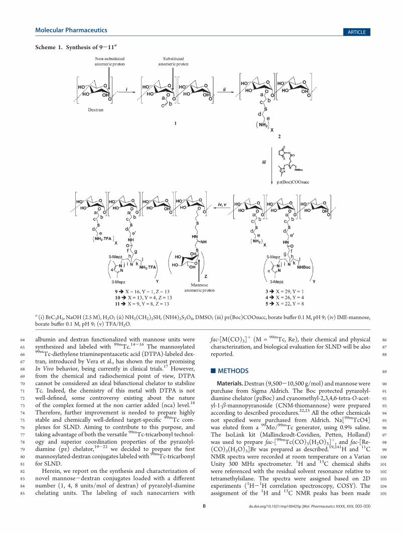
Scheme 1. Synthesis of 9-11a
a (i) BrC3H5, NaOH (2.5 M), H2O; (ii) NH2(CH2)2SH, (NH4)2S2O8, DMSO; (iii) pz(Boc)COOsucc, borate buffer 0.1 M, pH 9; (iv) IME-mannose,borate buffer 0.1 M, pH 9; (v) TFA/H2O.
Molecular Pharmaceutics ARTICLE
B dx.doi.org/10.1021/mp100425p |Mol. Pharmaceutics XXXX, XXX, 000–000

106 as indicated in the reaction scheme, Scheme 1S1 . Infrared107 spectra were recorded as KBr pellets on a Bruker Tensor 27108 spectrometer.109 HPLC Methods. The HPLC analysis was performed on110 Perkin-Elmer equipment coupled to a gamma (Berthold Lb111 509) and to a UV/vis detector (Shimadzu SPD-10 AV or Perkin112 Helmer Lc 290). Analysis was done either by reversed-phase high113 performance liquid chromatography (RP-HPLC) or by size114 exclusion chromatography (SEC-HPLC). RP-HPLC: Supelco115 Discovery Bio Wide Pore C18 25 cm� 4.6 mm, 5 μm analytical116 column; flow, 1 mL/min; eluents, A, TFA 0.1% in H2O; B, TFA117 0.1% in CH3CN. SEC-HPLC: Shodex OHpack SB-803 HQ118 analytical column; flow, 0.5 mL/min; eluent, ammonium acetate119 0.5M. Resolution of the columnwas calculated to be 0.76( 0.29.120 The wavelengths for UV detection were 220 and 254 nm for RP-121 HPLC and SEC-HPLC, respectively.122 Instant Thin Layer Cromatography (ITLC). Analysis was123 performed using PALL Life Sciences (prod. 61886) or Gelman124 Sciences Inc. (prod. 51432) strips and three different eluent125 systems (A, B, C). Radioactivity detection was performed on a126 radio chromatographer (Berthold LB 2723) equipped with 20127 mm diameter NaI(Tl) scintillation crystal.128 System A: Methyl Ethyl Ketone (MEK). [TcO4]
- migrates in129 the front of the solvent (Rf = 1), while [99mTc(CO)3(H2O)3]
þ,130 radioactive nanoconjugates and colloidal species stay at the131 origin (Rf = 0).132 System B: 5% HCl 6 N/MeOH. [99mTc(CO)3(H2O)3]
þ and133 [TcO4]
- migrate in the front of the solvent (Rf = 1), while134 radioactive nanoconjugates and colloidal species stay at the135 origin (Rf = 0).136 System C: C5H5N/AcOH/H2O (3:5:1). Colloidal species stay at137 the origin (Rf = 0). [99mTc(CO)3(H2O)3]
þ, [TcO4]- and
138 radioactive nanoconjugates migrate in the front of the solvent139 (Rf = 1).140 Synthesis of Dextran-allyl (1). Dextran-allyl was prepared141 according to a method described previously with some142 modifications.25 Dextran (1.00 g, 0.10 mmol) in water (7.5 mL)143 and excess of allyl bromide (2.50 mL, 14.5 mmol) reacted for144 6 h. After dialysis against water, the retentate was concentrated145 under reduced pressure and lyophilized, yielding dextran-allyl146 (1) as a white solid (0.98 g, 0.09 mmol, 87%, MWcalculated =147 11,164 g/mol).148
1H NMR (300 MHz, D2O) δH: 5.93 (m, Hb), 5.29 (m, Hc),149 5.02 (0.82 H, s broad, Hsubst.anom.), 4.86 (1 H, s broad, 1H,150 Hanom.), 4.11 (d, H
a), 3.78-3.35 (m, dextran); 13C NMR (75.3151 MHz, D2O) δC: 133.7 (Cb), 118.0 (Cc), 97.5 (Canom.), 95.7152 (Csubst.anom.), 78.2 (C
a), 73.2-65.3 (5C, dextran).153 Synthesis of Dextran-amine (2). A solution of dextran-allyl154 (1) (0.61 g, 0.05 mmol) in dry DMSO (3 mL) reacted with155 aminoethanethiol (0.75 g, 6.60mmol) and ammoniumpersulfate156 (0.1 g, 0.43 mmol) for 3 h at room temperature. The pH of the157 reaction mixture was adjusted to 4 with sodium hydroxide, and158 the volume of the mixture was double with sodium acetate buffer159 0.02 M, pH 4. After dialysis against sodium acetate 0.02 M, pH 4160 and water, the retentate was concentrated and lyophilized,161 yielding dextran-amine (2) as a white solid (0.668 g, 0.05 mmol,162 92%, MWcalculated = 13,320 g/mol).163
1H NMR (75.3 MHz, D2O) δH: 5.05 (s broad, Hsubst.anom.),164 4.87 (s broad, Hanom.), 3.64 (d, H
a), 3.80-3.30 (m), 3.14 (t, Hd),165 2.78 (t, He), 2.60 (s broad, Hc), 1.76 (s, Hb); 13CNMR (75MHz,166 D2O)δC: 99.7 (Canom.), 97.8 (Csubst.anom.), 81.3 (C
a), 75.3-67.4167 (5C, dextran), 40.3 (Ce), 30.7 (Cd), 30.1 (Cc), 29.1 (Cb).
168Quantification of Amine Units on Dextran Backbone. The169number of amine groups per mole of dextran was calculated on170the basis of three determinations, using the following equation:14
amine number=mol of dextran ¼ ½NH2�glucose� �� n ðIÞ
171172where [NH2] = amine concentration determined by the trini-173trobenzene sulfonate assay (TNBS), using cysteamine solutions174as standard;26 [glucose] = glucose concentration determined by175the sulfuric acid-phenol colorimetric assay, using glucose as176standard;27 and n = average number of glucose units/mole of177dextran.178For the trinitrobenzene sulfonic acid (TNBS) assay, cystea-179mine solutions (5-50 μg in 1.5 mL of H2O) were diluted in180borate buffer 0.2M, pH 8 (1.5 mL) and TNBS 5% (0.02 mL) was181added. Themixture was vortexed and, after standing for 15min at182room temperature, absorbance readings were taken at 420 nm.183Based on the calibration curve obtained (y = 0.1296x - 6.9 �18410-2; r2 = 0.9909), the amine concentration of 2was determined185to be 698 ( 0.61 μM, based on three determinations.186For the sulfuric acid-phenol assay, a 5% phenol solution in187H2O (0.5 mL) and concentrated H2SO4 (2.5 mL) were added to188glucose solutions (5-50 μg in 0.5 mL of H2O) and the mixture189was vortexed. After standing for 30 min at room temperature,190absorbance readings were taken at 490 nm. Based on the191calibration curve obtained (y = 0.1236x þ 2.76 � 10-2; r2 =1920.9973), the glucose concentration per mole of dextran was193found to be 1400 ( 5.9 μM, based on three determinations.194Synthesis of the Dextran-amine-[pyrazolyl-diamine-195(Boc)]y (3, y = 1; 4, y = 4; 5, y = 8). General Procedure. The196carboxylic acid of the Boc protected pyrazolyl-diamine chelator197(pz(Boc)) was activated with N,N0-dicyclohexylcarbodiimide198(DCC) and N-hydroxysuccinimide (NHS) in dry CH2Cl2. After199reacting at room temperature for 18 h, the DCC-urea precipi-200tate was removed by filtration and the filtrate was vacuum-dried,201yielding the corresponding activated ester pz(Boc)COOsucc as a202yellow pail oil. This ester was dissolved in CH3CN and added to a203solution of dextran-amine (2) in borate buffer 0.1 M, pH 9, using204different pz(Boc)COOsucc/amine molar ratios (Table 1 T1). After205overnight reaction at room temperature, the mixture was dia-206lyzed, first against borate buffer 0.02 M, pH 9, and finally207against water.2083 (y = 1). pz(Boc) (0.03 g, 0.08mmol) andNHS (0.011 g, 0.09209mmol) were suspended in dry CH2Cl2 (5 mL), and DCC (0.011210g, 0.09 mmol) was added in solid form. After reaction (18 h), the211solvent was evaporated and the resulting activated ester pz-212(Boc)COOsucc was redissolved in CH3CN (1mL) and added to213a solution of 2 (0.174 g, 0.01 mmol) in borate buffer 0.1 M, pH 9214(26 mL). The reaction mixture was dialyzed against borate buffer2150.02 M, pH 9 and water. The retentate was dried under vacuum,216and the solid obtained was washed with chloroform and
Table 1. Molar Ratios Used To Prepare Conjugates 3-5,Experimental Molar Ratios and Reaction Yields
molar ratios pz/NH2
conjugates used obtained reaction yield (%)
3 0.15 0.03( 0.01 96
4 0.25 0.15( 0.01 96
5 0.4 0.3 ( 0.01 94
Molecular Pharmaceutics ARTICLE
C dx.doi.org/10.1021/mp100425p |Mol. Pharmaceutics XXXX, XXX, 000–000

217 methanol, yielding 3 as a white solid (0.17 g, 0.01 mmol, 96%,218 MWcalculated = 13,580 g/mol)219 4 (y = 4). pz(Boc) (0.059 g, 0.161 mmol) and NHS (0.022 g,220 0.193 mmol) were suspended in dry CH2Cl2 (5 mL), and DCC221 (0.022 g, 0.193 mmol) was added in solid form. After reaction222 (18 h), the solvent was evaporated and the resulting activated223 ester pz(Boc)COOsucc was redissolved in 1 mL of CH3CN and224 added to a solution of 2 (0.286 g, 0.022 mmol) in borate buffer225 0.1M, pH 9 (28mL). After workup as indicated for 3, compound226 4 was obtained as a white solid (0.30 g, 0.02 mmol, 96%,227 MWcalculated = 14,630 g/mol).228 5 (y = 8). pz(Boc) (0.103 g, 0.278 mmol) and NHS (0.039 g,229 0.33 mmol) were suspended in CH2Cl2 (5 mL) and DCC (0.134230 g, 0.33 mmol) was added in solid form. After reaction (18 h), the231 solvent was evaporated and the resulting activated ester pz-232 (Boc)COOsucc was redissolved in 1 mL of CH3CN and added233 to a solution 2 (0.300 g, 0.023 mmol) in borate buffer 0.1 M, pH234 9 (30 mL). After workup as indicated for 3, compound 5 was235 obtained as a white solid (0.341 g, 0.021 mmol, 94%, MWcalculated
236 = 16,030 g/mol).237 As an example, the NMR data for the Boc protected com-238 pound 4 is presented 1H NMR (300 MHz, D2O) δH: 5.82 (s,239 H(4)pz), 5.03 (s, broad, Hsubst.anom.), 4.85 (s, broad, Hanom.),240 3.88-3.35 (m, dextran), 3.21 (s, broad), 2.99 (s, broad), 2.81 (s,241 broad), 2.56 (s, broad), 2.08 (s, Mepz), 1.98 (s, Mepz), 1.61 (s,242 broad), 1.31 (s, 9H, CH3).
1HNMR data for the Boc protected 3243 and 5 are given in the Supporting Information.244 Aliquots of 3, 4 and 5 were treated with TFA, to remove the245 Boc protecting group, and the resulting compounds analyzed by246
1H NMR, to determine the number of pyrazolyl chelating units.247 All the spectra gave similar patterns, the main difference being248 related to the intensity ratio of the peaks due to methyl groups of249 the pyrazolyl ring (3,5-Me2pz) and the ones due to the protons250 adjacent to the unsubstituted amines (He).251 As an example, the NMR data obtained for 4, after removing252 the Boc protecting group, is presented. 1H NMR (300 MHz,253 D2O) δH: 5.92 (s, H(4)pz), 4.96 (s, broad, Hsubst.anom.), 4.85 (s,254 broad, Hanom.), 3.88-3.35 (m, dextran), 3.10 (t, Hd), 2.74 (t,255 He), 2.56 (s, broad), 2.27 (t, Hh), 2.21 (s, Mepz), 2.14 (s, Mepz),256 1.85 (s, broad), 1.74 (s, Hb).257 Synthesis of Dextran-amine-[pyrazolyl-diamine(Boc)]y-258 mannose (6, y = 1; 7, y = 4; 8, y = 8). 6: A solution of sodium259 methoxide (10.8 mg, 1 mL,) was added to a dry methanolic260 suspension of CNM-thiomannose (0.7 g in 16 mL of methanol).261 After 20 h at room temperature, the solvent was evaporated,262 affording a golden syrup that reacted with a solution of 3 (0.100263 g, 0.007 mmol) in borate buffer 0.1 M, pH 9.0 (5 mL) for 20 h at264 room temperature. The reaction mixture was concentrated and265 dialyzed first against borate buffer 0.02 M, pH 9.0, and finally266 against water. The retentate was concentrated and lyophilized,267 yielding 6 as a pale yellow solid (0.093 g, 0.006 mmol, 75%, MW268 est. = 16,870 g/mol).269 7: The compound was prepared as above, using conjugate 4270 (0.11 g, 0.01 mmol) to yield 7 as a pale yellow solid (0.096 g,271 0.005 mmol, 74%, MWcalculated = 17,920 g/mol).272 8: The compound was prepared as above, using conjugate 5273 (0.20 g, 0.001 mmol) to yield 8 as a pale yellow solid (0.196 g,274 0.01 mmol, 80%, MWcalculated = 18,850 g/mol).275
1H NMR spectra of 6-8 have similar patterns, the main276 difference being related to the intensity ratio between some277 of the 1H NMR peaks. As an example, the NMR data for 8 is278 presented. 1HNMR (300MHz, D2O) δH: 5.85 (s, H(4)pz), 5.27
279(d, Hanom.mannose), 5.01 (s, broad, Hanom.subst.), 4.82 (s, broad,280Hanom.), 3.99 (s, broad), 3.78-3.35 (m, dextran), 2.99-2.76281(m), 2.59 (s, broad), 2.17 (s, Mepz), 2.07 (s, Mepz), 1.8 (s,282broad), 1.31 (s, 9H, CH3).283Synthesis of Dextran-amine-[pyrazolyl-diamine]y-man-284nose (9, y = 1; 10, y = 4; 11, y = 8). The Boc protecting groups285in 6-8 were removed with TFA/H2O affording the final286conjugates 9-11 in quantitative yield. The compounds were287characterized by SEC-HPLC, NMR and IR spectroscopy.288Based on the intensity ratio between the mannose anomeric289proton (Hanom, δ 5.22), the Me-pz (δ 2.14) and the protons290adjacent to free amine of the dextran backbone (He: δ 2.74), the291number of mannose units per mole of dextran was calculated to292be 13 ( 1.293As an example, the NMR data for 10 is presented. 1H NMR294(300 MHz, D2O) δH: 5.80 (s, H(4)pz), 5.22 (s,broad Hmannose
295anom.), 4.99 (s, broad, Hsubst.anom.), 4.82 (s, broad, Hanom.), 4.39296(t, CH2
j), 3.99 (s, broad), 3.74-3.35 (m, dextran), 3.10 (t, Hd),2972.74 (t, He), 2.52 (s, broad), 2.24 (t, Hh), 2.07 (s, Mepz), 1.98 (s,298Mepz), 1.74 (s, broad, Hb), 1.46 (t, Hg). 13C NMR (75.3 MHz,299D2O) δC: 176.4 (CdO), 165.9 (CdN), 162.8 (q, CF3COO
-),300150.5 (C(3)pz), 143.7 (C(5)pz), 116.5 (q, CF3COO
-), 107.4301(C(4)pz), 99.9 (Canom.), 97.8 (Csubst.anom.), 86.9, 86.7, 82.4, 81.5302(Ca), 75.8-68.9 (m, 11Cdextran/mannose), 68.5, 67.4, 63.1, 62.8303(Ck), 54.5 (Ci), 52.3, 47.9 (Cj), 43.9 (Cl), 40.6 (Ce), 39.3, 35.2,30431.0, 30.8 (Cd), 30.3 (Cc), 29.6, 29.1 (Cb), 24.4 (Cg), 14.3305(Mepz), 12,3 (Mepz). For 1H NMR data of 9 and 11 see the306Supporting Information.307IR (KBr) (ν/cm-1): ν (OH, br) 3418, ν (CdO, s) 1678, ν308(CdO, s, dextran) 1643, 1384, 1261.309SEC-HPLC (λ = 254 nm): 9, tR = 16.8 min; 10, tR = 16.4 min;31011, tR = 15.6 min.311Synthesis of fac-[99mTc(CO)3(L)]
þ (fac-[99mTc(CO)3]-2, L =3122; 12, L = 9; 13, L = 10; 14, L = 11). The precursor fac-[99mTc-313(CO)3(H2O)3]
þ was prepared using the Isolink kit (Covidien)314and its radiochemical purity checked by RP-HPLC.315Compounds fac-[99mTc(CO)3]-2 and 12-14 were obtained by316reacting the dextran derivatives 2 and 9-11with fac-[99mTc(CO)3-317(H2O)3]
þ. Briefly, a solution of fac-[99mTc(CO)3(H2O)3]þ
318(1 mL) was added to a capped vial, previously flushed with N2,319containing 2 (5 � 10-5 M) and 9-11 (2.5 � 10-5 M). The320mixture reacted at 100 �C for 30 min, and the radiochemical purity321of 12-14 was checked by RP-HPLC, SEC-HPLC and ITLC.322Stability Studies in the Presence of Cysteine and Histi-323dine. Aliquots of fac-[99mTc(CO)3]-2 and 12-14 (100 μL, 10-
3245 M) were added to a large molar excess (1:100) of cysteine or
325histidine solutions in PBS (900 μL, 10-3 M, pH 7.4). The326samples were incubated at 37 �C and analyzed by RP-HPLC and327ITLC after 2, 4, and 6 h of incubation. The results are328summarized in Table 2 T2.329Synthesis of fac-[Re(CO)3(k
3-L)]þ (13a, L = 10; 14a, L = 11).330Complexes 13a and 14awere prepared by reacting fac-[Re(CO)3-331(H2O)3]Br
24 with compounds 10 and 11 in H2O, using a rhenium332precursor/pzmolar ratio of 4:1. After stirring at 50 �C for 16 h, the333reaction mixtures were concentrated and dialyzed against water334and the retentate was concentrated and lyophilized. The resulting335pale yellow powder was washed with CH2Cl2 and methanol. The336progress of these reactions was monitored by taking aliquots of the337reaction mixture at several time points. The aliquots were dialyzed338overnight, dried under reduced pressure and washed with chloro-339form and methanol, yielding a yellow solid that was analyzed by340RP-HPLC and 1H NMR spectroscopy (Figure 3). When
Molecular Pharmaceutics ARTICLE
D dx.doi.org/10.1021/mp100425p |Mol. Pharmaceutics XXXX, XXX, 000–000

341 complete, the reaction mixture was treated as referred above and342 the solids obtained were formulated as 13a and 14a, based on RP-343 HPLC, multinuclear NMR and IR spectroscopy.344 The 1H and 13C NMR spectra of 13a and 14a present similar345 patterns, the main differences being the intensity ratio of some346
1H NMR peaks.347 As an example, NMR and IR data for 13a is presented. 1HNMR348 (300MHz, D2O) δH: 6.1 (s, H(4)pz), 5.23 (d, Hmannoseanom.), 4.98349 (s, broad, Hsubst.anom.dextran), 4.80 (s, broad, Hanom.), 4.39 (t, CH2
j),350 3.99 (s, broad), 3.74-3.35 (m, dextran), 3.10 (t, Hd), 2.71 (t, He),351 2.52 (s, broad), 2.21 (s, Mepz), 2.12 (s, Mepz), 1.91 (t, Hg), 1.73352 (s, broad, Hb). 13C NMR (75.3 MHz, D2O) δC: 195.2 (ReCO),353 194.7 (ReCO), 193.1 (ReCO), 176.2 (CdO), 165.4 (CdN),354 168.2 (q, CF3COO-), 153.7 (C(3)pz), 144.3 (C(5)pz), 118.5 (q,355 CF3COO
-), 114.6 (pz), 108.1 (C(4)pz), 97.9 (Canom.), 96.1356 (Csubst.anom.), 85.1, 79.5-65.7 (11C), 60.9, 42.0, 38.7, 33.8, 32.9,357 30.7 (Ch), 30.9, 30.0, 27.3, 20.3, 15.5 (Mepz), 11.1 (Mepz). 1H358 NMR and 13C NMR data for 14a are given in the Supporting359 Information.360 IR (KBr) (ν/cm-1): ν (O-H, br) 3397, ν (C-H, m) 2950, ν361 (CtO, s) 2027, ν (CtO, w) 1999, ν (CtO, s) 1899, ν (CdO, s)362 1678, ν (CdO, s, dextran) 1643, 1556, 1384.363 RP-HPLC: 13a, tR = 12.6 min; 14a, tR = 12.6 min.364 Physical Characterization. The hydrodynamic diameter and365 the zeta potential of dextran (9,500-10,500 g/mol), 2, 10, 11,366 13a, and 14a were determined in phosphate buffer 0.01 M, pH367 7.4, by DLS using a ZetaSizer Nano ZS from Malvern. Particle368 size was measured at 25 �C with a 173� scattering angle. The369 surface charge was determined by electrophoretic mobility using370 laser doppler velocimetry (LDV) and zeta potential cells.371 A Digital Instruments MultiMode scanning probe microscope372 (SPM) with a Nanoscope IIIA controller in tapping mode was373 used for the atomic force microscopy (AFM) measurements.374 Biodistribution studies of fac-[99mTc(CO)3(k
3-L)]þ (13, L =375 10; 14, L = 11). In Vivo evaluation studies of 13 and 14 were376 carried out in a Wistar rat model. All animal experiments377 performed were in accordance with the guidelines of the institu-378 tional animal ethics committee. Female Wistar rats weighing379 200-250 g were used in the experiment. The animals were first380 anesthetized by intraperitoneal injection of a mixture of xylazine381 (70 mg/kg) and ketamine (7 mg/kg). Boosters if subsequently382 required were given using ketamine alone. Then ∼50 μL of the383
99mTc-labeled preparation (∼1.8 MBq containing∼20 μg of the384 ligand) was injected subcutaneously in the footpad region. The385 area of injection was massaged gently with a strip of gauze pad for386 about 2 min to facilitate movement of the radiolabeled prepara-387 tion from the injection site. The rejection criterion followed was388 observation of any bleeding at the site of injection or
389measurement of more than 0.5% of administered dose on the390gauze pad. Postinjection (p.i.), the animals were kept in separate391sets (n = 4 per set) for incubation periods of 15 min, 30 min, 60392min and 180 min under normal conditions, water provided ad393libitum. Ten minutes prior to the end of each incubation period,394the animals under anesthetized condition were given a subcuta-395neous administration of∼50 μL of Patent Blue Dye (1% w/v in396saline) in the same region as the labeled preparation. At the end397of the incubation, the animals were sacrificed and the relevant398organs and tissues, including the popliteal (which serves as the399sentinel lymph node in this protocol) and secondary nodes, were400excised for the determination of In Vivo distribution of 99mTc401activity. Radioactivity measurement was done on Integral Line402flat-bed NaI(Tl) scintillation detector (Harshaw, USA). Activity403retained in each organ/tissue was expressed as a percentage of404the total injected dose (% ID). The results are summarized in405tables 4 T4
406and 5 T5.407Popliteal extraction (PE) was calculated using the following408equation:14
PE ð%Þ ¼ % ID ðpoplitealÞ-% ID ðiliacÞ% ID ðpoplitealÞ � 100 ðIIÞ
Scintigraphic Imaging Studies of the Sentinel LymphNode. Scintigraphic imaging was performed on the Millennium
409MPS medical imaging system (Wipro-GE Healthcare, India).410Scintigraphic imaging studies for 13 and 14 were also performed411in Wistar rat model. ∼37 MBq of the radiolabeled preparation412(in 50 μL volume) was used for each imaging study (n = 3). The413technique employed for anesthesia of animals and administration414of 13 and 14 preparations was the same as for the above-referred415biodistribution studies, excepting that blue dye was not injected416in the imaging protocol. For the acquisition, the animals were417placed with their dorsal side facing the camera. Planar static418images were acquired at 10 min, 30 min, 60 min and 180 min p.i.419using the Genie Acq Image Acquisition software (release 3.0).420Acquisition parameters were as follows: matrix 256� 256, zoom4211.33, 5 min acquisition time. The site of injection was masked422with lead shielding during acquisition. Subsequent image proces-423sing was achieved with the Xeleris image processing software424(version 1.0272). The image results are summarized in Figure 7.
425’RESULTS
426Synthesis and Characterization of Dextran-Mannose-427Pyrazolyl-Diamine Derivatives (9-11). Dextran-mannose428conjugates loaded with one (9), four (10) and eight (11)429pyrazolyl-diamine chelating units per mol of dextran were430synthesized as depicted in Scheme 1.431The first step of the synthetic pathway involved the functio-432nalization of dextran (MW: 9,500-10,500) with allyl groups,433yielding compound 1. Quantitative conversion of the allyl groups434in 1 to amines yielded 2. Reactions of 2 with pz(Boc)COOsucc,435in different molar ratios, yielded the dextran derivatives 3-5436(Table 1).22 A freshly prepared solution of 2-imino-2-methox-437ethyl-1-thio-β-D-mannoside (IME-thiomannose) in borate buf-438fer 0.1 M, pH 9, reacted with 3-5, yielding the final dextran-439mannose-pyrazolyl-diamine conjugates 9-11, after Boc-de-440protection with TFA/H2O.441All dextran derivatives were analyzed by 1H and/or 13C NMR442spectroscopy and, in the case of 2, also by colorimetric assays.
Table 2. In Vitro Stability of 99mTc(CO)3-2 and 12-14 in thePresence of Cysteine and Histidine, at Different Time Points
activity bound to dextran derivatives (%)
cysteine histidine
compds 0 h 2 h 4 h 6 h 2 h 4 h 6 h
99mTc(CO)3-2 78 69 49 nda 61 50 nd
12 98 80 75 nd 89 85 nd
13 98 93 92 90 99 98 97
14 98 94 94 92 99 98 98aNot determined.
Molecular Pharmaceutics ARTICLE
E dx.doi.org/10.1021/mp100425p |Mol. Pharmaceutics XXXX, XXX, 000–000

443 The 1H NMR spectrum of 1 in D2O has shown clearly two444 multiplets (δ 5.93, Hb; δ 5.29 Hc) and one doublet (δ 4.11 Ha)445 due to the protons of the allyl group. We could also clearly446 identify two broad singlets appearing at δ 5.02 and δ 4.86447 assigned to the anomeric protons of the substituted and448 nonsubstituted glucose units, respectively. The intensity ratio449 of these two 1H NMR peaks was used to evaluate the substitu-450 tion degree, which was found to be 50 ( 5%, based on five451 different batches. Reaction of 1with aminoethanethiol has been452 quantitative, as indicated by the absence of the three resonances453 due to the allyl groups in the 1H NMR spectra of 2. The 1H454 NMR peaks assigned to the anomeric protons (substituted and455 unsubstituted glucose units) could be identified in these456 spectra, presenting chemical shifts comparable to those found457 in 1. The amine density per mol of dextran in 2 was also458 estimated using TNBS and sulfuric acid-phenol colorimetric459 assays. The values found were 30( 3 amine groups per mole of460 dextran. As mentioned above, based on the NMR data, 50( 5%461 of all glucose units of the commercial dextran were functiona-462 lized with allyl groups and all these groups were transformed463 into amines. Considering that the dextran used has ca. 60464 glucose units, it was found that ca. 30 of the glucose units have465 been derivatized with amines, a result which completely agrees466 with the colorimetric data. The number of pyrazolyl-diamine467 units in 3-5 was determined by 1H NMR, after Boc deprotec-468 tion of these conjugates. Based on the intensity of the 1H NMR469 peaks attributed to methyl groups of the azolyl ring (δ 2.08, δ
4701.98) and to the protons adjacent to free amine of the dextran471backbone (δ 2.74, He), the number of pyrazolyl chelators in472each compounds was found to be one (3), four (4) and eight473(5) per mole of dextran. Then, the number of free amines was474calculated to be 29 (3), 26 (4) and 22 (5).475The dextran-mannose-pyrazolyl-diamine conjugates 9-47611 (overall yield 63-69%) presented only one peak on the SEC-477HPLC chromatograms, indicating a purity higher than 98%. The478total number of mannose units per mole of dextran was cal-479culated to be 13 ( 1, based on the intensity ratios of the peaks480corresponding to the mannose anomeric proton (δ 5.22), 3,5-481Me2pz (δ 2.08) and protons adjacent to free amines (δ 2.74, He),482easily assigned in the 1HNMR spectra of 9-11. As an illustrative483example, we present in Figure 1 F1the 1H NMR spectrum obtained484for 10, with the assignment of themost relevant peaks. NMRdata485for 3, 5, 9 and 11 are given in the Supporting Information.486Reactions of the Conjugates 9-11 with fac-[M(CO)3-487(H2O)3]
þ (M = 99mTc, Re). The dextran-mannose-pyrazo-488lyl-diamine derivatives 9, 10 and 11 reacted with the precursors489fac-[M(CO)3(H2O)3]
þ (M = 99mTc or Re) leading to the490corresponding metalated derivatives 12-14, 13a and 14a491(Scheme 2 S2).492
99mTc(CO)3-mannosylated dextran derivatives (12-14)493were analyzed by RP-HPLC and ITLC. The RP-HPLC chroma-494tograms of 12 (tR = 12.4 min), 13 (tR = 12.6 min) and 14 (tR =49512.6 min) presented only one species, and no free fac-[99mTc-496(CO)3(H2O)3] (tR = 7.8 min) or any other radiochemical
Table 3. Group Density, Hydrodynamic Diameter (DLS), Zeta Potential and Calculated Molecular Weight of Dextran, 2, 10, 11,13a and 14a
group density (units/mol of dextran)
compd amine pz mannose diama (nm) zeta potentiala (mV) MW calcd (g/mol)
dextran 4.3( 0.4 -9.9( 0.5 10,000
2 30 5.7( 0.5 7.7( 1.3 13,320
10 13 4 13 7.0( 0.3 6.6( 0.3 18,820
11 9 8 13 7.0( 0.7 7.3( 0.6 20,132
13a 13 4 13 8.4( 0.5 7.1( 0.7 19,904
14a 9 8 13 8.7( 0.3 7.1( 0.1 22,300aMean ( SD.
Table 4. Biodistribution Data for 13 in Wistar Rat Model atDifferent Time Points
% ID/organ
organ 15 min 30 min 1 h 3 h
liver 2.57( 1.17 4.06( 0.32 4.80( 1.03 6.49( 0.09
intestine 0.45( 0.10 0.59( 0.04 0.75 ( 0.06 0.95( 0.17
stomach 0.12( 0.03 0.18( 0.01 0.30( 0.05 0.52( 0.17
kidney 0.56( 0.19 0.82( 0.02 1.12( 0.27 1.20 ( 0.01
heart 0.06( 0.02 0.07( 0.00 0.06( 0.01 0.06( 0.02
lungs 0.48( 0.27 0.65( 0.01 0.83 ( 0.16 0.65( 0.04
spleen 0.12( 0.07 0.18( 0.08 0.32( 0.10 0.31( 0.04
blood (whole) 0.97( 0.34 1.47( 0.08 0.91( 0.02 0.96 ( 0.37
1st node 3.96( 0.87 7.43( 1.59 6.71( 2.35 5.98( 1.68
2nd node 0.83( 0.05 3.15( 0.83 2.59 ( 1.06 1.41( 0.50
site of inj 89.06( 0.28 80.93( 2.87 83.85( 1.37 79.50( 5.02
PE (%) 78.60( 3.39 57.85( 2.19 61.81( 2.42 76.65 ( 1.73
Table 5. Biodistribution Data for 14 in Wistar Rat Model atDifferent Time Points
% ID/organ
organ 15 min 30 min 1 h 3 h
liver 2.42( 0.47 3.74( 0.17 5.44( 1.06 6.84( 0.01
intestine 0.55( 0.07 1.21( 0.11 0.61 ( 0.57 1.28( 0.17
stomach 0.15( 0.03 0.27( 0.01 0.42( 0.01 0.74( 0.07
kidney 0.53( 0.02 0.94( 0.01 1.18( 0.20 1.67 ( 0.28
heart 0.04( 0.01 0.08( 0.02 0.09( 0.00 0.09( 0.03
lungs 0.32( 0.01 0.35( 0.05 0.43 ( 0.03 0.60( 0.07
spleen 0.12( 0.03 0.20( 0.10 0.25( 0.11 0.45( 0.04
blood (whole) 2.14( 0.01 2.86( 1.72 1.72( 0.81 1.55 ( 0.65
1st node 4.43( 0.27 4.31( 0.27 7.53( 0.69 5.21( 0.78
2nd node 1.09( 0.40 1.41( 0.28 0.41 ( 0.15 0.59( 0.14
site of inj 89.14( 2.09 87.24( 3.06 81.58( 0.35 81.13( 0.01
PE (%) 68.55( 1.35 76.27( 5.07 94.47( 2.45 87.81 ( 3.75
Molecular Pharmaceutics ARTICLE
F dx.doi.org/10.1021/mp100425p |Mol. Pharmaceutics XXXX, XXX, 000–000

497 impurity could be detected. By ITLC it was also confirmed that498 12-14 are formed with high radiochemical purity (>95%), as no499 [TcO4]
-, fac-[99mTc(CO)3(H2O)3]þ or aggregates were de-
500 tected. As an example, Figure 2F2 shows the RP-HPLC and ITLC501 chromatograms obtained for 13.
502Owing to the existence of donor atoms on 2, with coordination503affinity for fac-[99mTc(CO)3]
þ, we have also labeled directly504this polymeric derivative with fac-[99mTc(CO)3(H2O)3]
þ. The505resulting radioconjugate 99mTc(CO)3-2 was obtained in 78%506yield, due to the presence of some radiochemical impurities,
Scheme 2. Synthesis of fac-[M(CO)3(k3-L)] (M = 99mTc/Re: 12, L = 9; 13/13a, L = 10; 14/14a, L = 11)
Figure 1. 1H NMR spectrum of 10 in D2O.
Figure 2. Radiochromatograms for 13: (I) RP-HPLC chromatogram (tR = 12.6 min); (II) ITLC (Rf = 1) using system C as eluent.
Molecular Pharmaceutics ARTICLE
G dx.doi.org/10.1021/mp100425p |Mol. Pharmaceutics XXXX, XXX, 000–000

507 which were identified by ITLC as aggregates and/or colloidal508 species.509
99mTc(CO)3-2 and 12-14 were incubated with a large excess510 of cysteine and histidine to evaluate their in vitro stability toward511 transchelation. Table 2 summarizes the activity bound to the512 dextran derivatives at different time points.513
99mTc(CO)3-2, where no pyrazolyl-diamine chelating units514 are present, is obtained in relatively low yield (78%) and is not515 stable to transchelation. Compound 12, with only one pyrazolyl-516 diamine chelator per mole of dextran, is obtained in almost517 quantitative yield but presents also a relatively low stability in the518 presence of cysteine and histidine. On the contrary, 13 and 14 are519 highly stable under the tested conditions, presenting in both520 cases high radiochemical purity (g90%), even after long incuba-521 tion times. Based on these results, compounds 13 and 14 were522 selected as the most promising radioactive compounds to pursue523 animal studies. Their characterization has been done by compar-524 ison of their HPLC chromatograms with the ones obtained for525 their rhenium mannosylated-dextran analogues, fac-[Re(CO)3-526 (k3-L)]þ (13a, L = 10; 14a, L = 11), synthesized as depicted in527 Scheme 2. The kinetics of these reactions were relatively slow,528 and their progress was monitored by RP-HPLC and 1H NMR529 spectroscopy at different time points (1 h, 6 h and 16 h). As an530 example, Figure 3F3 shows the RP-HPLC chromatograms and 1H531 NMR spectra for one of these reactions at 1 h and 16 h. From all532 the peaks appearing in the 1H NMR spectra, we have only used533 the ones assigned to the pyrazolyl-diamine chelator, namely, to534 the H(4)pz and the 3,5-Me2pz to evaluate the progress of the535 reaction. After 1 h, the H(4)pz and the 3,5-Me2pz groups appear536 at δ 5.8, δ 2.08 and δ 1.98, respectively. The chemical shifts of537 these NMR peaks correspond to the free conjugate 10, indicating538 that no metalation has taken place. By RP-HPLC it was also539 concluded that the peak at tR = 11.5 min was due to 10. After540 reacting 6 h, the 1H NMR analysis showed that some metalation
541had taken place, but some uncoordinated ligand could still be542detected (results not shown). Only after 16 h the reaction was543complete, as indicated by the three 1H NMR peaks at δ 6.1, δ5442.21 and δ 2.12 due toH(4)pz and 3,5-Me2pz groups of 13a. The545chemical shift of theseNMRpeaks, compared to the values found546in the free conjugate 10, clearly confirmed the coordination of547the metal to the pyrazolyl-diamine chelator. The RP-HPLC548chromatograms also show only one peak for 13a.549In the 13C NMR spectra of 13a and 14a the peaks assigned to550the carbonyl groups of the fac-[Re(CO)3]
þ core (13a, at δ 195.2,551δ 194.7 and δ 193.1; 14a, at δ 195.4, δ 194.9 and δ 193.2) could552also be clearly seen in the expected range. The presence of such a553core was also confirmed by the IR data obtained for 13a and 14a554(ν(CtO) strong bands in the range 2027-1998 cm-1).22
Figure 4. RP-HPLC chromatograms of 13 (γ-detection) and 13a (λ =220 nm).
Figure 3. Progression of the reaction of fac-[Re(CO)3(H2O)3]Br with 10: RP-HPLC (λ = 220 nm) chromatograms and 1H NMR data of the mixtureafter 1 h and 16 h of reagent addition.
Molecular Pharmaceutics ARTICLE
H dx.doi.org/10.1021/mp100425p |Mol. Pharmaceutics XXXX, XXX, 000–000

555 A chromatographic comparative study (RP- and SEC-HPLC)556 of 13/13a and 14/14a allowed the characterization of the557 radioactive nanocompounds. As an example, Figure 4F4 displays558 the RP-HPLC chromatograms obtained for 13 (γ-detection)559 and 13a (UV-detection). Identical results were obtained for560 14/14a.561 Physical Characterization. The hydrodynamic diameter and562 the zeta potential of dextran, 2, 10, 11, 13a and 14a were563 determined in phosphate buffer 0.01 M, pH 7.4, in order to564 mimic the labeling conditions. As an example, Figure 5F5 shows the565 size distribution histograms of 13a and 14a.566 The final composition of the dextran derivatives, their physical567 parameters and the calculated molecular weight for each com-568 pound are summarized in Table 3T3 .569 The hydrodynamic diameter of the particles increases with the570 dextran backbone functionalization. However, the number of571 pyrazolyl-diamine chelating units (4 vs 8) does not affect572 significantly their size, as shown by the results obtained for 10573 and 11. Upon metalation of 10 and 11 a slight increase of the574 particles size was also found (13a, 8.4 ( 0.5 nm; 14a, 8.7 (575 0.3 nm). Analysis of dextran, 13a and 14a by AFM, after576 dispersion on mica substrates, gave molecular diameters of ca.577 4 nm (Figure 6F6 A), 12 nm (Figure 6B), and 8-16 nm (Figure 6C)578 for dextran, 13a and 14a, respectively. AFM and DLS molecular579 measurements compare well for dextran and 13a, the main580 difference being found for 14a. The dispersion found for 14a581 may indicate some aggregation during sample deposition.582 Biodistribution Studies. Tables 4 and 5 show the In Vivo583 distribution results after injection (p.i.) of 13 and 14 in Wistar584 rats, respectively.585 Both radiocompounds show appreciable accumulation in the586 popliteal (sentinel) node. The highest radioactivity uptake in the587 sentinel node occurs between 30 and 60 min p.i., and a major588 portion of this is observed to be retained up to 180 min p.i. The589 popliteal extraction (PE), a parameter that predicts the suitability590 of a preparation as an agent for SLND, was determined according591 to eq II. The values found for 13 and 14, at 1 h p.i, were 61.81(592 2.42% and 94.47 ( 2.45%, respectively. Figure 7F7 displays the593 scintigraphic images of 13 (Figure 7A) and 14 (Figure 7B),594 showing a clear delineation of the sentinel node. In the case of 14595 (Figure 7B), a greater degree of specific retention in the sentinel596 node is observed, with minimal spread to other regions.
597 ’DISCUSSION
59899mTc-labeled mannosylated macromolcules like albumin and
599 dextran of different molecular weights have been explored for600 SLND. For the dextran derivatives it was found that particles with601 size in the range 7-10 nm could be trapped, in a saturable mode,602 by the sentinel node.15,16 Among them, mannosylated dextran603 labeled with Tc, using DTPA as a bifunctional chelating agent604 (Lymphoseek), was the most studied and the most promising for
605In Vivo application. However, the chemical structure of techne-606tium complexes with DTPA is not well-defined, and its chemical607characterization has been the subject of many investigations and608speculation at the macroscopic and nca level in the past few609years.18 Thus, taking into account that the real chemical structure610of 99mTc-DTPA is unknown, there is interest on preparing fully611characterized compounds with high radiochemical purity and612specific activity, and adequate biological properties for SLND.613Previously, we have introduced several pyrazolyl-based bifunc-614tional chelators suitable for the stabilization of the core fac-[M-615(CO)3]
þ (M = 99mTc, Re) and for the quantitative labeling of616several biomolecules.21,22 Profiting from these results, we617decided to expand this technology to the synthesis of the first618dextran-mannose derivatives containing one (9), four (10) and619eight (11) pyrazolyl-diamine chelating units. These nanoconju-620gates, obtained in good overall yields (>70%), were quantitatively621labeled with fac-[99mTc(CO)3]
þ, leading to the nanocomplexes62212-14. The significant difference in labeling yields found for62312-14 (>95%) and 99mTc(CO)3-2 (78%) clearly highlights the624importance of the presence of those chelating units on the dextran625backbone. Moreover, the number of these units seems also to be626crucial for the kinetic inertness of the radioactive nanocompounds,627as shown by the high stability of 13 and 14 compared to 12. Most628probably, when the number of pyrazoly rings is low, as in the case629of 12, the 99mTc(CO)3 binds to dextran through the pyrazolyl630chelator but also through other coordinating groups existing in the631dextran backbone. When this happens the radioactive nanocom-632pound is not stable to transchelation, as shown for the species633
99mTc(CO)3-2, where no chelators are present.634Due to the high labeling yields and stability, compounds 13635and 14 were selected for further biological studies.636Before such studies, 13 and 14 were chemically characterized637at the macroscopic level by comparing their RP-HPLC chroma-638tograms with the ones obtained for the rhenium analogues 13a639and 14a, used as surrogates. These surrogates, formulated as640fac-[Re(CO)3(k
3-L)] (13a, L = 10, 14a, L = 11), based on641multinuclear NMR, HPLC and IR spectroscopy, were synthe-642sized by reacting 10 and 11 with the precursor fac-[Re(CO)3-643(H2O)3]
þ. Additionally, 13a and14a and the respective precursors644(10 and 11) were physically characterized by DLS, AFM and LDV.645The hydrodynamic diameters determined by DLS and AFM are of646the same order of magnitude, and the values found showed an647increase of the particle size due to both dextran functionalization648and metalation. Taking into account the hydrodynamic diameter649found for 10 (7.0 ( 0.3 nm), 11 (7.0 ( 0.7 nm), 13a (8.4 (6500.5 nm) and 14a (8.7 ( 0.3 nm), the radioactive compounds 13651and 14, used for biological studies, certainly have a hydrodyna-652mic diameter in the range 7-9 nm. The hydrodynamic diameters653of 10 and 11 are similar to those found for DTPA-mannosyl-654dextran (7.1 ( 0.9 nm) and MAG3-mannosyl-dextran (5.5 (6552.4 nm), andmuch smaller than the radiopharmaceuticals in clinical
Figure 5. Hydrodynamic size of 13a (8.4 ( 0.5 nm) and 14a (8.7 ( 0.3 nm), determined by dynamic light scattering (detection angle of 173�).
Molecular Pharmaceutics ARTICLE
I dx.doi.org/10.1021/mp100425p |Mol. Pharmaceutics XXXX, XXX, 000–000

Figure 6. Height-scaled atomic force microscopy images and corresponding line profiles of dextran (A); 13a (B); 14a (C).
Figure 7. Scintigraphic images of Wistar rats injected with 13 (A) and 14 (B) at different time points.
Molecular Pharmaceutics ARTICLE
J dx.doi.org/10.1021/mp100425p |Mol. Pharmaceutics XXXX, XXX, 000–000

656 use (80-100 nm).5,14,15 As far as we are aware, no examples of657 mannosylated dextran derivatives have been synthesized and658 characterized at the macroscopic level, 13a and 14a being the first659 examples.660 Zeta potential measurements indicated a negative charge for661 dextran (-9.9 ( 0.5 mV), which changed drastically upon662 functionalization with amines (2, þ7.7 ( 1.3 mV). Our studies663 also indicated that further functionalization of 2 did not affect the664 charge of the nonmetalated (10,þ6.6( 0.3 mV; 11,þ7.3( 0.6665 mV) and metalated final nanocompounds (13a,þ7.1( 0.7 mV;666 14a,þ7.1( 0.1 mV). To the best of our knowledge, from all the667 nanocompounds studied for SLND, only the charge of 99mTc-668 MAG3-mannosyl-dextran was determined by electrophoresis669 (negative overall charge).670 The biodistribution results (Tables 4 and 5) and the scinti-671 graphic images (Figure 7) of 13 and 14 have shown an appreci-672 able accumulation in the popliteal (sentinel) node, the highest673 radioactivity uptake occurring between 30 and 60 min p.i. with a674 major portion of this retained up to 180 min p.i. The scinti-675 graphic images for 13 and 14 clearly agree with the biodistribu-676 tion pattern, as both show a clear delineation of the sentinel node677 and a great specific retention, mainly for 14. For 13 some spread678 to the liver was observed. All biological data taken together679 renders 14 as a very favorable radiotracer for SLN imaging.680 Our biological data cannot be directly compared with other681 nanotracers explored for SLND, namely, filtered 99mTc-sulfur colloid,682
99mTc-DTPA-mannosyl-dextran (Lymphoseek), and 99mTc-HYNIC-683 NMA-tricine2, as these compounds were evaluated in different684 animal models.14,16 However, if we consider the PE parameter,685 14 presents a value (1 h p.i.: 94.47 ( 2.47%) much higher than686 filtered 99mTc-sulfur colloid (78.8 ( 6.5%), and comparable to687 Lymphoseek (90.1 ( 10.7%) and 99mTc-HYNIC-NMA-tricine2688 (92.93( 5.08%).14,16 The clearance from the injection site of 13689 (83.85 ( 1.37%) and 14 (81.58( 0.35%) is comparable to that690 found for 99mTc-sulfur colloid (70.4( 11.0%) and slightly lower691 than for 99mTc-HYNIC-NMA-tricine2 (67.57 ( 8.29%) and692 Lymphoseek (52.6 ( 10.5%), at 1 h p.i.14,16 The charge of 13693 and 14 may explain the moderate clearance from the injection694 site. In fact, 13 and 14 are positively charged, whichmay promote695 electrostactic interactions with the polyanionic glycosaminogly-696 cans or other negatively charged species present in the interstitial697 space.28 However, the high specific activity of the 99mTc-tricar-698 bonyl complexes may also contribute to such behavior. Further699 studies on specific activity/injection site clearance are underway.
700 ’CONCLUSIONS
701 Aiming at the design of innovative radiotracers for SLN702 detection, we have introduced the first class of fully characterized703
99mTc(CO)3-mannosylated dextran derivatives with adequate704 biological features for SLN detection.705 Several dextran derivatives (9-11), containing the same706 number of pendant mannose moieties (13 units) and a variable707 number of tridentate chelators (1 unit, 9; 4 units, 10; 12 units,708 11) have been synthesized and characterized. The radioactive709 nanocompounds of the type fac-[99mTc(CO)3(k
3-L)] (12, L = 9,710 13, L = 10, 14, L = 11) were prepared quantitatively in high711 radiochemical purity (g98%) and specific activity. Unlike 12,712 the nanotracers 13 and 14 displayed very high in vitro stability,713 and have been chosen for biological studies. 13 and 14 were714 identified/characterized by comparing their chromatographic715 behavior with that of the corresponding rhenium surrogates
71613a and 14a, which have been synthesized and characterized at717both the chemical (NMR and IR spectroscopy, and HPLC) and718physical levels (DLS, AFM and LDV). Scintigraphic imaging and719biodistribution studies with 13 and 14 have shown good720accumulation in the sentinel node at 60 min postinjection721(6.71 ( 2.35% and 7.53 ( 0.69%, respectively), with significant722retention up to 180min. A clear delineation of the sentinel lymph723node without significant washout to other regions was observed724in the scintigraphic images. The higher PE of 14 compared to 13725highlights the superior biological properties of 14 to be further726explored as SLN imaging agent.727Despite being evaluated in different animal models, the PE728value found for 14 is higher than that found for fTcSC (78.8 (7296.5%), routinely used for SLND, and comparable to the PE of730Lymphoseek (90.1 ( 10.7%), which emerged as a promising731radiotracer for SLND in recent clinical trials.
732’ASSOCIATED CONTENT
733bS Supporting Information. 1HNMR spectra of 3, 5, 9, and73411, 13C NMR spectra of 14a, and SEC-HPLC chromatograms of73510 and 11. This material is available free of charge via the Internet736at http://pubs.acs.org.
737’AUTHOR INFORMATION
738Corresponding Author739*Instituto Tecnologico e Nuclear, Chemistry, Estrada Nacional,74010, apartado 21, 2686-953 Sacav�em, Portugal. E-mail: [email protected]. Phone: þ351219946201. Fax: 351 21 994 6185.
742’ACKNOWLEDGMENT
743This work is part of the Coordinated Research Project (CRP)744on the “Development of 99mTc radiopharmaceuticals for sentinel745node detection and cancer diagnosis” of the IAEA, which is746acknowledged. The authors also wish to thank John J. Zaknun747and Ambi M. R. Pillai from the NA department of the IAEA,748Vienna, for their active role in this CRP. Roberto Pasqualini is749also acknowledged for fruitful scientific discussions. M.M. ac-750knowledges FCT for a PhD grant (SFRH/BD/48066/2008).
751’REFERENCES
752(1) Jain, R.; Dandekar, P.; Patravale, V. Diagnostic nanocarriers for753sentinel lymph node imaging. J. Controlled Release 2009, 138, 90–102.754(2) Jeong, J. M.; Hong, M. K.; Kim, Y. J.; Lee, J.; Kang, J. H.; Lee,755D. S.; Chung, J.; Lee, M. C. Development of 99mTc-mannosyl human756serum albumin (99mTc-MSA) as a novel receptor binding agent for757sentinel lymph node imaging. Nucl. Med. Commun. 2004, 12,7581211–1217.759(3) Scolyer, R. A.; Murali, R.; Satzger, I.; Thompson, J. F. The760detection and significance of melanoma micrometastases in sentinel761nodes. Surg. Oncol. 2008, 17, 165–174.762(4) Sharma, R.; Wendt, J. A.; Rasmussen, J. C.; Adams, K. E.;763Marshall, M. V.; Muraca, E. M. New horizons for imaging lymphatic764function. Ann. N.Y. Acad. Sci. 2008, 1131, 13–36.765(5) Wilhelm, A. J.; Mijnhout, S.; Franssen, E. J. F. Radiopharmaceu-766ticals in sentinel lymph node detection: an overview. Eur. J. Nucl. Med.7671999, 36–42.768(6) Eshima, D.; Fauconnier, T.; Eshima, L.; Thornback, J. R. Radio-769pharmaceuticals for lymphoscintigraphy: Including dosimetry and radia-770tion considerations. Semin. Nucl. Med. 2000, 1, 25–32.771(7) Hoh, C. K.; Wallace, A. M.; Vera, D. K. Preclinical studies of772
99mTc-DTPA-mannosyl-dextran. Nucl. Med. Biol. 2003, 30, 457–464.
Molecular Pharmaceutics ARTICLE
K dx.doi.org/10.1021/mp100425p |Mol. Pharmaceutics XXXX, XXX, 000–000

773 (8) Jinno, H.; Kubo, A. Sentinel lymph node biopsy in breast cancer774 using technetium-99m tin. Biomed. Pharmacother. 2002, 56, 213–216.775 (9) Liu, Y.; Chirino, A. J.; Misulovin, Z.; Leteux, C.; Nussenzweig,776 M. C.; Bjorkman, P. J. Crystal Structure of the Cysteine-rich Domain of777 Mannose Receptor Complex with sulphated Carbohydrate Ligand.778 J. Exp. Med. 2000, 191, 1105–1115.779 (10) Ponpipom, M. M.; Bugianesi, R. L.; Robbins, J. C.; Doebber,780 T. W.; Shen, T. Y. Cell-specific ligands for selective drug delivery to781 tissue and organs. J. Med. Chem. 1981, 24, 1388–1395.782 (11) Robbins, J. C.; Lam, M. H.; Tripp, C. S.; Bugianesi, R. L.;783 Ponpipom, M. M.; Shen, T. Y. Synthetic glicopeptides substrates for784 receptor-mediated endocytosis by macrophages. Proc. Natl. Acad. Sci. U.785 S.A. 1981, 78, 7294–7298.786 (12) Liu, Y.; Chirino, A. J.; Misulovin, Z.; Leteux, C.; Nussenzweig,787 M. C.; Bjorkman, P. J. Crystal Structure of the Cysteine-rich Domain of788 Mannose Receptor Complex with sulphated Carbohydrate Ligand.789 J. Exp. Med. 2000, 191, 1105–1115.790 (13) Higuchi, Y.; Oka, M.; Kawakami, S.; Hashida, M.Mannosylated791 semiconductor quantum dots for the labeling of macrophages.792 J. Controlled Release 2008, 125, 131–136.793 (14) Vera, D. R.; Wallace, A. M.; Hoh, C. K.; Mattrey, R. F. A794 synthetic macromolcule for sentinel node detection: 99mTc-DTPA-795 Mannosyl-Dextran. J. Nucl. Med. 2001, 42, 951–959.796 (15) Vera, D. R.; Wallace, A. M.; Hoh, C. K. [99mTc]-MAG3-797 Mannosyl-Dextran: a receptor-binding radiopharmaceutical for sentinel798 node detection. J. Nucl. Med. 2001, 28, 493–498.799 (16) Takagi, K.; Uehara, T.; Kaneko, E.; Nakayama,M; Koizumi, M.;800 Endo, K.; Arano, Y. 99mTc-labeled mannosyl-neoglycoalbumin for801 sentinel lymph node identification. Nucl. Med. Biol. 2004, 31, 893–900.802 (17) Wallace, A. M.; Hoh, C. K.; Limmer, K. K.; Darrah, D. D.; Schulties,803 G.; Vera, D. R. Sentinel lymph node accumulation of Lymphoseek and Tc-804 99msulfur colloidusing a “2-day”protocol.Nucl.Med.Biol.2009,36, 687–692.805 (18) Liu, G.; Hnatowich, D. J. Labeling Biomolecules with Radio-806 rhenium - a Review of the Bifunctional Chelators. Anticancer Agents Med807 Chem. 2007, 7, 367–377.808 (19) Alberto, R.; Ortner, K.; Wheatley, N.; Schibli, R.; Schubiger, A.809 Synthesis and properties of boranocarbonate: A convenient in situ CO810 source for aqueous preparation of fac-[99mTc(H2O)3(CO)3]
þ. J. Am.811 Chem. Soc. 2001, 123, 3135–3136.812 (20) Alberto, R. The particular role of radiopharmacy within bioor-813 ganometallic chemistry. J. Organomet. Chem. 2007, 692, 1179–1186.814 (21) Correia, J. D. G.; Paulo, A.; Santos, I. Re and Tc complexes with815 pyrazolyl-containing chelators: from coordination chemistry to target-816 specific delivery of radioactivity. Curr. Radiopharm. 2009, 2, 277–294.817 (22) Alves, S.; Paulo, A.; Correia, J. D. G.; Gano, L.; Smith, C. J.;818 Hoffman, T. J.; Santos, I. Pyrazolyl derivatives as bifunctional chelators819 for labeling tumor-seeking peptides with the fac-[M(CO)3]
þ Moiety820 (M) = 99mTc, Re): synthesis, characterization, and biological behaviour.821 Bioconjugate Chem. 2005, 16, 438–449.822 (23) Lee, Y. C.; Stowell, C. P.; Krantz, M. J. 2-imino-2-methoxy-823 lethyl-1-thioglycosides: new reagents for attaching sugars to proteins.824 Biochemistry 1976, 15, 3956–3963.825 (24) Lazarova, N.; James, S.; Babich, J.; Zubieta, J. A convenient826 synthesis, chemical characterization and reactivity of [Re(CO)3-827 (H2O)3]Br: the crystal and molcular structure of [Re(CO)3-828 (CH3CN)2Br]. Inorg. Chem. Commun. 2004, 7, 1023–1026.829 (25) Holmberg, A.; Meurling, L. Preparation of sulfhydrylborane-830 dextran conjugates for boron neutron capture therapy. Bioconjugate831 Chem. 1993, 4, 570–573.832 (26) Cayot, P.; Tainturier, G. The quantification of protein amino833 groups by the trinitrobenzenesulfonic acid method: a re-examination.834 Anal. Biochem. 1997, 249, 184–200.835 (27) Rao, P.; Pattabiraman, T. N. Reevaluation of the phenol-sulfuric836 acid reaction for the estimation of hexoses and pentoses. Anal. Chem.837 1989, 181, 18–22.838 (28) Wiig, H; Kolmannskog, O.; Tenstad, O.; Bert, J. L. Effect of839 charge on interstitial distribution of albumin in rat dermis in vitro.840 J. Physiol. 2003, 2, 505–514.
Molecular Pharmaceutics ARTICLE
L dx.doi.org/10.1021/mp100425p |Mol. Pharmaceutics XXXX, XXX, 000–000