Low Molecular Weight Dextran Sulfate (ILB®) Administration ...
18
antioxidants Article Low Molecular Weight Dextran Sulfate (ILB ® ) Administration Restores Brain Energy Metabolism Following Severe Traumatic Brain Injury in the Rat Giacomo Lazzarino 1, † , Angela Maria Amorini 2, † , Nicholas M. Barnes 3,4 , Lars Bruce 5 , Alvaro Mordente 6,7 , Giuseppe Lazzarino 2, * , Valentina Di Pietro 3,8, *, Barbara Tavazzi 1,6,7, * , Antonio Belli 3,8 and Ann Logan 9, * 1 UniCamillus-Saint Camillus International University of Health Sciences, Via di Sant’Alessandro 8, 00131 Rome, Italy; [email protected] 2 Department of Biomedical and Biotechnological Sciences, Division of Medical Biochemistry, University of Catania, Via S. Sofia 97, 95123 Catania, Italy; [email protected] 3 National Institute for Health Research Surgical Reconstruction and Microbiology Research Centre, Queen Elizabeth Hospital, Edgbaston, Birmingham B15 2TH, UK; [email protected] (N.M.B.); [email protected] (A.B.) 4 Institute of Clinical Sciences, College of Medical and Dental Sciences, University of Birmingham, Edgbaston, Birmingham B15 2TT, UK 5 Tikomed AB, 26303 Viken, Sweden; [email protected] 6 Department of Basic Biotechnological Sciences, Intensive and Perioperative Clinics, Catholic University of the Sacred Heart of Rome, Largo F. Vito 1, 00168 Rome, Italy; [email protected] 7 Department of Laboratory Sciences and Infectious Disease, Fondazione Policlinico Universitario A. Gemelli IRCCS, Largo A. Gemelli 8, 00168 Rome, Italy 8 Neurotrauma and Ophthalmology Research Group, Institute of Inflammation and Ageing, College of Medical and Dental Sciences, University of Birmingham, Edgbaston, Birmingham B15 2TT, UK 9 Axolotl Consulting Ltd., Droitwich, Worcestershire WR9 0JS, UK * Correspondence: [email protected] (G.L.); [email protected] (V.D.P.); [email protected] (B.T.); [email protected] (A.L.); Tel.: +39-06-3015-5135 (B.T.); Fax: +39-06-3015-4309 (B.T.) † These Authors equally contributed to this work. Received: 28 July 2020; Accepted: 8 September 2020; Published: 10 September 2020 Abstract: Traumatic brain injury (TBI) is the leading cause of death and disability in people less than 40 years of age in Western countries. Currently, there are no satisfying pharmacological treatments for TBI patients. In this study, we subjected rats to severe TBI (sTBI), testing the effects of a single subcutaneous administration, 30 min post-impact, of a new low molecular weight dextran sulfate, named ILB ® , at three different dose levels (1, 5, and 15 mg/kg body weight). A group of control sham-operated animals and one of untreated sTBI rats were used for comparison (each group n = 12). On day 2 or 7 post-sTBI animals were sacrificed and the simultaneous HPLC analysis of energy metabolites, N-acetylaspartate (NAA), oxidized and reduced nicotinic coenzymes, water-soluble antioxidants, and biomarkers of oxidative/nitrosative stress was carried out on deproteinized cerebral homogenates. Compared to untreated sTBI rats, ILB ® improved energy metabolism by increasing ATP, ATP/ adenosine diphosphate ratio (ATP/ADP ratio), and triphosphate nucleosides, dose-dependently increased NAA concentrations, protected nicotinic coenzyme levels and their oxidized over reduced ratios, prevented depletion of ascorbate and reduced glutathione (GSH), and decreased oxidative (malondialdehyde formation) and nitrosative stress (nitrite + nitrate production). Although needing further experiments, these data provide the first evidence that a single post-injury injection of a new low molecular weight dextran sulfate (ILB ® ) has beneficial effects on sTBI metabolic damages. Due to the absence of adverse effects in humans, ILB ® represents a promising therapeutic agent for the treatment of sTBI patients. Antioxidants 2020, 9, 850; doi:10.3390/antiox9090850 www.mdpi.com/journal/antioxidants
Transcript of Low Molecular Weight Dextran Sulfate (ILB®) Administration ...

antioxidants
Article
Low Molecular Weight Dextran Sulfate (ILB®)Administration Restores Brain Energy MetabolismFollowing Severe Traumatic Brain Injury in the Rat
Giacomo Lazzarino 1,†, Angela Maria Amorini 2,† , Nicholas M. Barnes 3,4, Lars Bruce 5,Alvaro Mordente 6,7, Giuseppe Lazzarino 2,* , Valentina Di Pietro 3,8,*, Barbara Tavazzi 1,6,7,* ,Antonio Belli 3,8 and Ann Logan 9,*
1 UniCamillus-Saint Camillus International University of Health Sciences, Via di Sant’Alessandro 8,00131 Rome, Italy; [email protected]
2 Department of Biomedical and Biotechnological Sciences, Division of Medical Biochemistry, University of Catania,Via S. Sofia 97, 95123 Catania, Italy; [email protected]
3 National Institute for Health Research Surgical Reconstruction and Microbiology Research Centre,Queen Elizabeth Hospital, Edgbaston, Birmingham B15 2TH, UK; [email protected] (N.M.B.);[email protected] (A.B.)
4 Institute of Clinical Sciences, College of Medical and Dental Sciences, University of Birmingham, Edgbaston,Birmingham B15 2TT, UK
5 Tikomed AB, 26303 Viken, Sweden; [email protected] Department of Basic Biotechnological Sciences, Intensive and Perioperative Clinics, Catholic University of
the Sacred Heart of Rome, Largo F. Vito 1, 00168 Rome, Italy; [email protected] Department of Laboratory Sciences and Infectious Disease, Fondazione Policlinico Universitario A. Gemelli IRCCS,
Largo A. Gemelli 8, 00168 Rome, Italy8 Neurotrauma and Ophthalmology Research Group, Institute of Inflammation and Ageing, College of
Medical and Dental Sciences, University of Birmingham, Edgbaston, Birmingham B15 2TT, UK9 Axolotl Consulting Ltd., Droitwich, Worcestershire WR9 0JS, UK* Correspondence: [email protected] (G.L.); [email protected] (V.D.P.); [email protected] (B.T.);
[email protected] (A.L.); Tel.: +39-06-3015-5135 (B.T.); Fax: +39-06-3015-4309 (B.T.)† These Authors equally contributed to this work.
Received: 28 July 2020; Accepted: 8 September 2020; Published: 10 September 2020�����������������
Abstract: Traumatic brain injury (TBI) is the leading cause of death and disability in people less than40 years of age in Western countries. Currently, there are no satisfying pharmacological treatmentsfor TBI patients. In this study, we subjected rats to severe TBI (sTBI), testing the effects of a singlesubcutaneous administration, 30 min post-impact, of a new low molecular weight dextran sulfate,named ILB®, at three different dose levels (1, 5, and 15 mg/kg body weight). A group of controlsham-operated animals and one of untreated sTBI rats were used for comparison (each group n = 12).On day 2 or 7 post-sTBI animals were sacrificed and the simultaneous HPLC analysis of energymetabolites, N-acetylaspartate (NAA), oxidized and reduced nicotinic coenzymes, water-solubleantioxidants, and biomarkers of oxidative/nitrosative stress was carried out on deproteinized cerebralhomogenates. Compared to untreated sTBI rats, ILB® improved energy metabolism by increasing ATP,ATP/ adenosine diphosphate ratio (ATP/ADP ratio), and triphosphate nucleosides, dose-dependentlyincreased NAA concentrations, protected nicotinic coenzyme levels and their oxidized over reducedratios, prevented depletion of ascorbate and reduced glutathione (GSH), and decreased oxidative(malondialdehyde formation) and nitrosative stress (nitrite + nitrate production). Although needingfurther experiments, these data provide the first evidence that a single post-injury injection of a newlow molecular weight dextran sulfate (ILB®) has beneficial effects on sTBI metabolic damages. Due tothe absence of adverse effects in humans, ILB® represents a promising therapeutic agent for thetreatment of sTBI patients.
Antioxidants 2020, 9, 850; doi:10.3390/antiox9090850 www.mdpi.com/journal/antioxidants
Antioxidants 2020, 9, 850 2 of 18
Keywords: severe traumatic brain injury; low molecular weight dextran sulfate; mitochondrialdysfunction; energy metabolism; N-acetylaspartate; nicotinic coenzymes; oxidative/nitrosative stress;ascorbate; reduced glutathione (GSH); HPLC
1. Introduction
Traumatic brain injury (TBI) is one of the most common acute neurodegenerative diseases andrepresents the leading cause of death for people less than 45 years of age in Western countries.The incidence of TBI is on the rise, and by 2020 the World Health Organization estimates it will bethe largest cause of disability worldwide [1]. Depending on the severity of the symptoms relatedto TBI (evaluated by the Glasgow Coma Scale), TBI may be classified as mild (mTBI), moderate,or severe (sTBI). Severe TBI is characterized by a high mortality rate and those who survive oftensuffer from profound disabilities with permanent impairment of cognitive, physical, and psychosocialfunctions, associated with a diminished or altered state of consciousness and inability to be independent,work correctly, and maintain social relationships [2].
To date, there are no satisfying pharmacological treatments capable of decreasing mortality/morbidityand improving recovery of sTBI patients [2,3]. Clinical and therapeutic management are complicated by theinhomogeneity of head injured patients, including the diversity of adverse consequences associated withsubdural hematomas, subarachnoid hemorrhages, cerebral hypoxia from respiratory difficulties, and theinvasive neurosurgical interventions used to control cerebral perfusion pressure [4–6]. Damage associatedwith any of the aforementioned complications may augment damage caused by sTBI, concomitantly requiringadditional specific treatments and decreasing the effectiveness of TBI targeted therapies. An additional,but no less important, problem contributing to the lack of effective pharmacological therapies for sTBIpatients is the highly variable time elapsed between TBI occurrence and arrival in Emergency Departmentsfor initiation of patients’ treatment [7].
Even with the absence of the aforementioned variables, TBI is a complicated pathology in whichthe inevitable primary insult (the impact force acting on the brain tissue) is directly responsiblefor the induction of a secondary insult. This is characterized by a rapid cascade of biochemical,metabolic, and molecular changes causing profound modifications of various cerebral cell functions [8,9],often ultimately leading to cell necrosis and apoptosis with significant neuronal loss [10,11]. The severityof this cellular and molecular damage depends on the impact force acting on the cerebral tissue [12].Mitochondrial dysfunction occurs in the post-injured brain [13,14], causing an imbalance of ATPproduction and consumption, with consequent energy crisis [15]. This triggers the intrinsic apoptoticpathway [16], and increasing levels of reactive oxygen species (ROS) and reactive nitrogen species (RNS)with associated decreased levels of cell antioxidants [17] causing an insurgence of oxidative/nitrosativestress [18]. Particularly this last phenomenon is responsible for the activation of microglia leadingto a generalized inflammatory response with the balance shifted to production of proinflammatorycytokines [19–21].
In experimental graded TBI, we previously demonstrated a correlation between the severity ofTBI and the energy deficit associated with an increased rate of anaerobic metabolism [22] andmitochondrial dysfunction [23,24], thereby initiating tissue damage mediated by reactive ROSand RNS [25,26]. Moreover, we identified N-acetylated amino acid N-acetylaspartate (NAA) asa reliable surrogate biomarker to monitor the state of the energetic metabolism in vivo [27,28].Indeed, as mitochondrial NAA biosynthesis has a high indirect energy expenditure, changes in NAAintracerebral concentrations are closely related to changes of parameters related to energy metabolism,such as ATP, guanosine triphosphate (GTP), adenosine diphosphate (ADP), adenosine monophosphate(AMP), Acetyl-CoA, CoA-SH, and oxidized nicotinamide adenine dinucleotide (NAD+), and tomitochondrial phosphorylating capacity (ATP/ADP) [29,30].

Antioxidants 2020, 9, 850 3 of 18
The low molecular weight dextran sulfate (LMW-DS) used in this study (ILB®) is a branchedpolysaccharide with an average molecular weight of 5 kDa and contains molecules spanningapproximately 3–8 kDa, in which the α-d-glucopyranose-2,4-sulfate monomers are linked, in 95%of cases, by α-1,6-glycosidic bonds (the main linear chain) and, in the remaining 5% of cases,by α-1,3-glycosidic bonds (the branches) (see patent publication WO 2016/076780—New dextransulfate). In ILB®, sulfur represents ~17% of its total mass. While high molecular weight dextran sulfatepossesses toxic effects, inducing cancer development and inflammation [31–33], ILB® is a proven safeand well-tolerated LMW-DS. Previous studies showed that LMW-DS dose-dependently limits theinstant blood-mediated inflammatory reaction (IBMIR) [34,35], inhibits E-selectin-mediated adhesion ofneutrophils to endothelial cells [36,37], prevents adherence of peritoneal metastases [38], and improvesthe effectiveness of therapy and reduces mortality in patients with acute cerebral infarction treatedwith the thrombolytic agent urokinase [39]. Of particular mechanistic relevance to TBI, subcutaneousinjection of ILB® induces a rapid release of pharmacologically relevant levels of Hepatocyte GrowthFactor (HGF) into the circulation in laboratory animals and healthy human volunteers, which mayprovide a neurotrophic stimulus to the injured central nervous system (CNS) [35].
In the present study, using the weight drop impact acceleration model of closed-head trauma [40],we assessed the dose-response effects of ILB®, subcutaneously administered 30 min after injury,on mitochondrial functions, energy metabolism, and oxidative/nitrosative stress of the brain tissueof rats experiencing sTBI, by evaluating concentrations of high energy phosphates, oxidized andreduced nicotinic coenzymes, N-acetyl aspartate (NAA), ascorbate, reduced glutathione (GSH),malondialdehyde (MDA), nitrite and nitrate, oxypurines (hypoxanthine, xanthine, and uric acid),and nucleosides (inosine, guanosine, and adenosine).
2. Materials and Methods
2.1. Induction of sTBI, Drug Dosage and Mode of Administration, and Drug Administration Protocol
The experimental study was approved by the Ethical Committee of the Catholic University of theSacred Heart of Rome, Italy (approval 1F295.52, released on 10/20/2017), and by the Ethical Committeeof the Italian Ministry of Health (approval No. 78/2018-PR, released on 02/05/2018).
Male Wistar rats (n = 160) of 300–350 g body weight were used. They were fed a standard laboratorydiet and water ad libitum in a controlled environment. As the accepted anesthetic mixture, animalsreceived 35 mg/kg body weight (b.w.) ketamine and 0.25 mg/kg b.w. midazolam by intramuscularinjection. Diffuse severe TBI (sTBI) was induced according to the “weight drop” impact accelerationmodel of diffuse TBI set up by Marmarou A. et al. [40]. Severe TBI was induced by dropping a 450 gweight from 2 m height onto the rat head protected by a helmet (metal disk previously fixed on theskull using dental cement) in order to uniformly distribute the mechanical force to the brain. Rats wereplaced prone on a bed of specific polyurethane foam inserted in a special container; this foam dissipatesthe major part of the potential energy (deriving from the mechanical forces) and prevents any reboundof the animal after the impact that could produce spinal damages.
Animals suffering from skull fracture, seizures, nasal bleeding, or that did not survive the impact(4/52, with a mortality rate of the 7.7%) were excluded from the study. All animals surviving theimpact survived for the entire observational period, up to 7 days after TBI. After 2 or 7 days fromsTBI induction, rats were anesthetized again and immediately sacrificed. According to our previousstudies [22–30], these time points are coincident with deep biochemical, metabolic, and molecularderangement (2 days) or with a recovery (7 days) of nervous cell functions.
Animals killed at both 2 and 7 days post-injury were divided into four groups, with 12 rats in each:(1) sTBI only; (2) sTBI + 1 mg/kg b.w. ILB®; (3) sTBI + 5 mg/kg b.w. ILB®; (4) sTBI + 15 mg/kg b.w. ILB®.A fifth sham-operated control group also contained 12 rats with brains harvested for analysis at 2 dayspost-procedure, a time point that shows no significant difference in metabolic perturbation from thatmeasured in sham-operated rat brains harvested at 7 days post-procedure (unpublished observations).
Antioxidants 2020, 9, 850 4 of 18
ILB® was provided by Tikomed AB (Tikomed AB, Viken, Sweden) in 10 mL vials containing asolution of 20 mg/mL ILB® (Tikomed AB, Viken, Sweden) in 9 mg/mL NaCl (Carlo Erba ReagentsS.r.l., Cornaredo, Italy). A single batch of drug was used throughout the study (batch 3045586; expirydate 2019-02). ILB® was diluted in sterile 9 mg/mL NaCl and injected subcutaneously (s.c.) in avolume of 0.5 mL in order to obtain a final administration of 1, 5, or 15 mg/kg b.w. of ILB®. The drugwas administered in a single s.c. injection of 0.5 mL at 30 min after sTBI. The control group (n = 12)consisted of sham-operated animals that underwent the same procedure of anesthesia but received nosTBI or any s.c. injection and was sacrificed 2 days after the procedure.
2.2. Cerebral Tissue Processing for Biochemical Analyses
To minimize metabolite loss, an in vivo craniectomy was performed in all animals during terminalanesthesia. The rat skull cap was carefully removed, the brain was exposed, rapidly removed, the cerebralhemispheres dissected and placed on aluminium tongues pre-cooled in liquid nitrogen, freeze-clamped,and then immersed in liquid nitrogen. The freeze-clamping procedure was introduced to acceleratefreezing of the tissue, thus minimizing potential metabolite loss [9,12,15,16]. Tissue homogenization formetabolite analyses was effected as described below. After the wet weight (w.w.) determination, thefrozen hemispheres were placed into an ice-cold, nitrogen-saturated, precipitating solution (1:10 w/v)composed of HPLC-grade CH3CN (Carlo Erba Reagents S.r.l., Cornaredo, Italy)+ 10 mM KH2PO4
(Carlo Erba Reagents S.r.l., Cornaredo, Italy), pH 7.40, (3:1; v:v), and the homogenization was performedusing an Ultra-Turrax homogenizer set at 24,000 rpm/min (Janke & Kunkel, Staufen, Germany).After centrifugation at 20,690× g for 10 min at 4 ◦C, the clear supernatants were saved, pellets weresupplemented with an aliquot of 10 mM KH2PO4 and homogenized again as described above, andsaved overnight at−20 ◦C in order to obtain a complete recovery of aqueous phase from tissue. A secondcentrifugation was performed (20,690× g, for 10 min at 4 ◦C) and the supernatants combined with thosepreviously obtained. Clear protein-free samples were extracted by vigorous agitation with a doublevolume of HPLC-grade CHCl3 (Carlo Erba Reagents S.r.l., Cornaredo, Italy) and centrifuged as above.The upper aqueous phases (containing water-soluble low molecular weight compounds) were collectedand subjected to chloroform washings for two more times (this procedure allowed the removal of allthe organic solvent and of any lipid soluble compound from the buffered tissue extracts), adjusted involumes with 10 mM KH2PO4, pH 7.40, to produce aqueous 10% tissue homogenates that were savedat −80 ◦C until assay.
2.3. HPLC Analysis of Energy Metabolites, Antioxidants, and Oxidative/Nitrosative Stress Biomarkers
Aliquots of each deproteinized aqueous brain sample were filtered through a 0.45 µm HV Milliporefilter and loaded (20 µL) onto a Hypersil C-18, 250 × 4.6 mm, 5 µm particle size column, providedwith its own guard column (ThermoFisher Scientific, Rodano, Milan, Italy) and connected to an HPLCapparatus consisting of a Surveyor System (ThermoFisher Scientific, Rodano, Milan, Italy) with ahighly sensitive diode array detector (equipped with a 5 cm light path flow cell) set-up between 200and 300 nm wavelength. Data acquisition and analysis were automatically performed by a linked PCusing the ChromQuest 5.0® software package provided by the HPLC manufacturer (ThermoFisherScientific, Rodano, Milan, Italy).
The selected metabolites ATP, GTP, uridine triphosphate (UTP), cytidine triphosphate (CTP),ADP, AMP, hypoxanthine, xanthine, uric acid, inosine, guanosine, adenosine, NAA, NAD+, reducednicotinamide adenine dinucleotide (NADH), oxidized nicotinamide adenine dinucleotide phosphate(NADP+), reduced nicotinamide adenine dinucleotide phosphate (NADPH), ascorbic acid, GSH,MDA, nitrite, and nitrate), related to tissue energy state and mitochondrial function, antioxidantdefenses, and oxidative/nitrosative stress, were separated, in a single chromatographic run, accordingto slight modifications of existing ion-pairing HPLC methods formerly set up in our laboratory [20,21].Assignment and calculations of the compounds of interest in chromatographic runs of tissue extractswere carried out at the proper wavelengths (206, 234, and 260 nm) by comparing retention times,
Antioxidants 2020, 9, 850 5 of 18
absorption spectra and areas of peaks with those of peaks of chromatographic runs of freshly preparedultrapure standard mixtures of known concentrations.
2.4. Statistical Analysis
Normal data distribution in each group was determined using the Kolmogorov–Smirnov test.Differences across groups were estimated by the one-way analysis of variance ANOVA. The Tukey’sMultiple Comparison Test was used as the post hoc test. Only two-tailed p-values of less than 0.05were considered statistically significant.
3. Results
3.1. ILB® Restores Cerebral Mitochondrial-Dependent Energy Metabolism and NAA Following sTBI
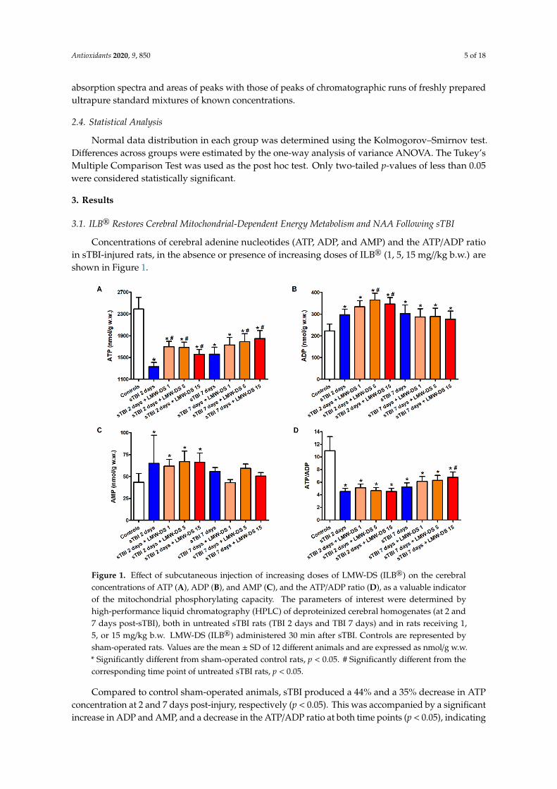
Concentrations of cerebral adenine nucleotides (ATP, ADP, and AMP) and the ATP/ADP ratioin sTBI-injured rats, in the absence or presence of increasing doses of ILB® (1, 5, 15 mg//kg b.w.) areshown in Figure 1.
Antioxidants 2020, 9, x FOR PEER REVIEW 5 of 18
2.4. Statistical Analysis
Normal data distribution in each group was determined using the Kolmogorov–Smirnov test. Differences across groups were estimated by the one-way analysis of variance ANOVA. The Tukey’s Multiple Comparison Test was used as the post hoc test. Only two-tailed p-values of less than 0.05 were considered statistically significant.
3. Results
3.1. ILB® Restores Cerebral Mitochondrial-Dependent Energy Metabolism and NAA Following sTBI
Concentrations of cerebral adenine nucleotides (ATP, ADP, and AMP) and the ATP/ADP ratio in sTBI-injured rats, in the absence or presence of increasing doses of ILB® (1, 5, 15 mg//kg b.w.) are shown in Figure 1.
Figure 1. Effect of subcutaneous injection of increasing doses of LMW-DS (ILB®) on the cerebral concentrations of ATP (A), ADP (B), and AMP (C), and the ATP/ADP ratio (D), as a valuable indicator of the mitochondrial phosphorylating capacity. The parameters of interest were determined by high-performance liquid chromatography (HPLC) of deproteinized cerebral homogenates (at 2 and 7 days post-sTBI), both in untreated sTBI rats (TBI 2 days and TBI 7 days) and in rats receiving 1, 5, or 15 mg/kg b.w. LMW-DS (ILB®) administered 30 min after sTBI. Controls are represented by sham-operated rats. Values are the mean ± SD of 12 different animals and are expressed as nmol/g w.w. * Significantly different from sham-operated control rats, p < 0.05. # Significantly different from the corresponding time point of untreated sTBI rats, p < 0.05.
Compared to control sham-operated animals, sTBI produced a 44% and a 35% decrease in ATP concentration at 2 and 7 days post-injury, respectively (p < 0.05). This was accompanied by a significant increase in ADP and AMP, and a decrease in the ATP/ADP ratio at both time points (p < 0.05), indicating dysfunctional mitochondria as a cause of sustained imbalance of energy metabolism of cerebral cells. A single s.c. injection at 30 min post-injury of each of the three ILB® doses positively affected brain energy metabolism. At 2 and 7 days after impact, cerebral ATP in ILB®-treated animals
Figure 1. Effect of subcutaneous injection of increasing doses of LMW-DS (ILB®) on the cerebralconcentrations of ATP (A), ADP (B), and AMP (C), and the ATP/ADP ratio (D), as a valuable indicatorof the mitochondrial phosphorylating capacity. The parameters of interest were determined byhigh-performance liquid chromatography (HPLC) of deproteinized cerebral homogenates (at 2 and7 days post-sTBI), both in untreated sTBI rats (TBI 2 days and TBI 7 days) and in rats receiving 1,5, or 15 mg/kg b.w. LMW-DS (ILB®) administered 30 min after sTBI. Controls are represented bysham-operated rats. Values are the mean ± SD of 12 different animals and are expressed as nmol/g w.w.* Significantly different from sham-operated control rats, p < 0.05. # Significantly different from thecorresponding time point of untreated sTBI rats, p < 0.05.
Compared to control sham-operated animals, sTBI produced a 44% and a 35% decrease in ATPconcentration at 2 and 7 days post-injury, respectively (p < 0.05). This was accompanied by a significantincrease in ADP and AMP, and a decrease in the ATP/ADP ratio at both time points (p < 0.05), indicating

Antioxidants 2020, 9, 850 6 of 18
dysfunctional mitochondria as a cause of sustained imbalance of energy metabolism of cerebral cells.A single s.c. injection at 30 min post-injury of each of the three ILB® doses positively affected brainenergy metabolism. At 2 and 7 days after impact, cerebral ATP in ILB®-treated animals was significantlyhigher than the values measured in untreated sTBI rats (p < 0.05). Notwithstanding, ATP remained 23%lower than the value recorded in control rats (p < 0.05), even with the highest dose of ILB®. While ADPin ILB®-treated rats was significantly higher than the values of sham-operated controls at both 2 and7 days after injury (p < 0.05), AMP measured at 7 days in ILB®-treated animals was not different fromthe AMP concentrations detected in the sham-operated controls. Consequently, the ATP/ADP ratio inILB®-treated sTBI animals, measuring the mitochondrial phosphorylating capacity [41], was similar tothat of untreated sTBI rats at 2 days after sTBI, but significantly increased at the highest dose tested at7 days after injury (p < 0.05). At this time point, the ATP/ADP ratio was, however, still lower than thevalues measured in the sham-operated control rats (p < 0.05).
The concentration of cerebral NAA in untreated sTBI animals (Figure 2) was decreased dramaticallyat both times post-impact (−39% at 2 days and −48% at 7 days, p < 0.05 compared to the value ofcontrols), further evidencing the steady impairment of mitochondrial functions involving multiplebiochemical processes of brain metabolism. Treatment with ILB® at 30 min post-sTBI positivelyaffected cerebral NAA levels, only when administered at 5 and 15 mg/kg b.w. Both doses attenuatedthe NAA decrease when measured at both 2 and 7 days after injury (p < 0.05 compared to levels atthe corresponding times of untreated sTBI rats), even though these values remained significantlylower than the values measured in sham-operated control animals (p < 0.05). At 7 days post-injury,a dose-dependent effect of ILB® administration was observed.
Antioxidants 2020, 9, x FOR PEER REVIEW 6 of 18
was significantly higher than the values measured in untreated sTBI rats (p < 0.05). Notwithstanding, ATP remained 23% lower than the value recorded in control rats (p < 0.05), even with the highest dose of ILB®. While ADP in ILB®-treated rats was significantly higher than the values of sham-operated controls at both 2 and 7 days after injury (p < 0.05), AMP measured at 7 days in ILB®-treated animals was not different from the AMP concentrations detected in the sham-operated controls. Consequently, the ATP/ADP ratio in ILB®-treated sTBI animals, measuring the mitochondrial phosphorylating capacity [41], was similar to that of untreated sTBI rats at 2 days after sTBI, but significantly increased at the highest dose tested at 7 days after injury (p < 0.05). At this time point, the ATP/ADP ratio was, however, still lower than the values measured in the sham-operated control rats (p < 0.05).
The concentration of cerebral NAA in untreated sTBI animals (Figure 2) was decreased dramatically at both times post-impact (−39% at 2 days and −48% at 7 days, p < 0.05 compared to the value of controls), further evidencing the steady impairment of mitochondrial functions involving multiple biochemical processes of brain metabolism. Treatment with ILB® at 30 min post-sTBI positively affected cerebral NAA levels, only when administered at 5 and 15 mg/kg b.w. Both doses attenuated the NAA decrease when measured at both 2 and 7 days after injury (p < 0.05 compared to levels at the corresponding times of untreated sTBI rats), even though these values remained significantly lower than the values measured in sham-operated control animals (p < 0.05). At 7 days post-injury, a dose-dependent effect of ILB® administration was observed.
Figure 2. Effect of subcutaneous injection of increasing doses of LMW-DS (ILB®) on the cerebral concentrations of NAA, as a valuable indirect indicator of energy wellness and mitochondrial function. NAA was determined by HPLC on deproteinized cerebral homogenates at 2 and 7 days post-impact, both in untreated sTBI rats (TBI 2 days and TBI 7 days) and in rats receiving 1, 5, or 15 mg/kg b.w. LMW-DS (ILB®) administered 30 min after sTBI. Controls are represented by sham-operated rats. Values are the mean ± SD of 12 different animals and are expressed as nmol/g w.w. * Significantly different from sham-operated controls rats, p < 0.05. # Significantly different from the corresponding time point of untreated sTBI rats, p < 0.05. ## Significantly different from LMW-DS 1, p < 0.05. ### Significantly different from LMW-DS 5, p < 0.05.
Figure 2. Effect of subcutaneous injection of increasing doses of LMW-DS (ILB®) on the cerebralconcentrations of NAA, as a valuable indirect indicator of energy wellness and mitochondrial function.NAA was determined by HPLC on deproteinized cerebral homogenates at 2 and 7 days post-impact,both in untreated sTBI rats (TBI 2 days and TBI 7 days) and in rats receiving 1, 5, or 15 mg/kg b.w.LMW-DS (ILB®) administered 30 min after sTBI. Controls are represented by sham-operated rats.Values are the mean ± SD of 12 different animals and are expressed as nmol/g w.w. * Significantlydifferent from sham-operated controls rats, p < 0.05. # Significantly different from the correspondingtime point of untreated sTBI rats, p < 0.05. ## Significantly different from LMW-DS 1, p < 0.05.### Significantly different from LMW-DS 5, p < 0.05.

Antioxidants 2020, 9, 850 7 of 18
3.2. ILB® Improves Cerebral High Energy Phosphate Concentrations and Reduces ATP Catabolism after sTBI
Results of the cerebral triphosphate nucleosides GTP, UTP, and CTP measurements are illustratedin Figure 3.
Antioxidants 2020, 9, x FOR PEER REVIEW 7 of 18
3.2. ILB® Improves Cerebral High Energy Phosphate Concentrations and Reduces ATP Catabolism after sTBI
Results of the cerebral triphosphate nucleosides GTP, UTP, and CTP measurements are illustrated in Figure 3.
Figure 3. Effect of subcutaneous injection of increasing doses of LMW-DS (ILB®) on the cerebral concentrations of GTP (A), UTP (B), and CTP (C), as valuable indicators of the nervous cell energy state. The parameters of interest were determined by HPLC on deproteinized cerebral homogenates at 2 and 7 days post-impact, both in untreated sTBI rats (TBI 2 days and TBI 7 days) and in rats receiving 1, 5, or 15 mg/kg b.w. LMW-DS (ILB®) administered 30 min after sTBI. Controls are represented by sham-operated rats. Values are the mean ± SD of 12 different animals and are expressed as nmol/g w.w. * Significantly different from sham-operated control rats, p < 0.05. # Significantly different from the corresponding time point of untreated sTBI rats, p < 0.05.
At 2 and 7 days post-injury, the brains of untreated sTBI rats showed significant depletions in all of the three high energy phosphates (p < 0.05), with GTP being the compound most evidently affected by the insult (2.8 and 1.5 times lower than the values of controls at 2 and 7 days post-injury, respectively). The administration of all tested ILB® doses produced beneficial effects, particularly evident at 7 days post-sTBI. At this time point, while GTP concentrations were still significantly lower than the values of sham-operated control animals (p < 0.05), levels of cerebral UTP were higher than those of untreated sTBI rats (p < 0.05) and not different from those of sham-operated control rats. Unexpectedly, brain CTP levels in animals receiving ILB® were higher than those measured in both untreated sTBI and sham-operated control rats (p < 0.05).
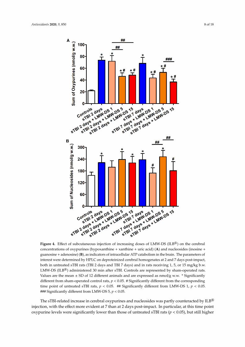
Concentrations of oxypurines (hypoxanthine + xanthine + uric acid) and nucleosides (inosine + guanosine + adenosine), deriving from purine nucleotide dephosphorylation, were significantly higher in the brains of untreated sTBI rats at both times post-injury compared to the values of sham-operated controls (p < 0.05), suggesting sustained imbalance in energy metabolism even at longer times after sTBI (Figure 4).
Figure 3. Effect of subcutaneous injection of increasing doses of LMW-DS (ILB®) on the cerebralconcentrations of GTP (A), UTP (B), and CTP (C), as valuable indicators of the nervous cell energystate. The parameters of interest were determined by HPLC on deproteinized cerebral homogenates at2 and 7 days post-impact, both in untreated sTBI rats (TBI 2 days and TBI 7 days) and in rats receiving1, 5, or 15 mg/kg b.w. LMW-DS (ILB®) administered 30 min after sTBI. Controls are represented bysham-operated rats. Values are the mean ± SD of 12 different animals and are expressed as nmol/g w.w.* Significantly different from sham-operated control rats, p < 0.05. # Significantly different from thecorresponding time point of untreated sTBI rats, p < 0.05.
At 2 and 7 days post-injury, the brains of untreated sTBI rats showed significant depletions in all ofthe three high energy phosphates (p < 0.05), with GTP being the compound most evidently affected bythe insult (2.8 and 1.5 times lower than the values of controls at 2 and 7 days post-injury, respectively).The administration of all tested ILB® doses produced beneficial effects, particularly evident at 7 dayspost-sTBI. At this time point, while GTP concentrations were still significantly lower than the values ofsham-operated control animals (p < 0.05), levels of cerebral UTP were higher than those of untreatedsTBI rats (p < 0.05) and not different from those of sham-operated control rats. Unexpectedly, brainCTP levels in animals receiving ILB® were higher than those measured in both untreated sTBI andsham-operated control rats (p < 0.05).
Concentrations of oxypurines (hypoxanthine + xanthine + uric acid) and nucleosides (inosine +
guanosine + adenosine), deriving from purine nucleotide dephosphorylation, were significantly higherin the brains of untreated sTBI rats at both times post-injury compared to the values of sham-operatedcontrols (p < 0.05), suggesting sustained imbalance in energy metabolism even at longer times aftersTBI (Figure 4).
Antioxidants 2020, 9, 850 8 of 18Antioxidants 2020, 9, x FOR PEER REVIEW 8 of 18
Figure 4. Effect of subcutaneous injection of increasing doses of LMW-DS (ILB®) on the cerebral concentrations of oxypurines (hypoxanthine + xanthine + uric acid) (A) and nucleosides (inosine + guanosine + adenosine) (B), as indicators of intracellular ATP catabolism in the brain. The parameters of interest were determined by HPLC on deproteinized cerebral homogenates at 2 and 7 days post-impact, both in untreated sTBI rats (TBI 2 days and TBI 7 days) and in rats receiving 1, 5, or 15 mg/kg b.w. LMW-DS (ILB®) administered 30 min after sTBI. Controls are represented by sham-operated rats. Values are the mean ± SD of 12 different animals and are expressed as nmol/g w.w. * Significantly different from sham-operated control rats, p < 0.05. # Significantly different from the corresponding time point of untreated sTBI rats, p < 0.05. ## Significantly different from LMW-DS 1, p < 0.05. ### Significantly different from LMW-DS 5, p < 0.05.
The sTBI-related increase in cerebral oxypurines and nucleosides was partly counteracted by ILB® injection, with the effect more evident at 7 than at 2 days post-impact. In particular, at this time point oxypurine levels were significantly lower than those of untreated sTBI rats (p < 0.05), but still
Figure 4. Effect of subcutaneous injection of increasing doses of LMW-DS (ILB®) on the cerebralconcentrations of oxypurines (hypoxanthine + xanthine + uric acid) (A) and nucleosides (inosine +
guanosine + adenosine) (B), as indicators of intracellular ATP catabolism in the brain. The parameters ofinterest were determined by HPLC on deproteinized cerebral homogenates at 2 and 7 days post-impact,both in untreated sTBI rats (TBI 2 days and TBI 7 days) and in rats receiving 1, 5, or 15 mg/kg b.w.LMW-DS (ILB®) administered 30 min after sTBI. Controls are represented by sham-operated rats.Values are the mean ± SD of 12 different animals and are expressed as nmol/g w.w. * Significantlydifferent from sham-operated control rats, p < 0.05. # Significantly different from the correspondingtime point of untreated sTBI rats, p < 0.05. ## Significantly different from LMW-DS 1, p < 0.05.### Significantly different from LMW-DS 5, p < 0.05.
The sTBI-related increase in cerebral oxypurines and nucleosides was partly counteracted by ILB®
injection, with the effect more evident at 7 than at 2 days post-impact. In particular, at this time pointoxypurine levels were significantly lower than those of untreated sTBI rats (p < 0.05), but still higher

Antioxidants 2020, 9, 850 9 of 18
than the values of sham-operated controls (p < 0.05), while cerebral nucleoside concentrations werelower than those of untreated sTBI rats (p < 0.05) and not significantly different from the values ofsham-operated animals.
3.3. Redox Metabolism of Cerebral Nicotinic Coenzymes after sTBI Is Improved by ILB®
As already observed in previous studies (12), sTBI caused a progressive time-dependent depletionof oxidized (NAD+ and NADP+) and reduced (NADH and NADPH) nicotinic coenzymes, which isparticularly evident at 7 days post-injury (Figure 5).
Antioxidants 2020, 9, x FOR PEER REVIEW 9 of 18
higher than the values of sham-operated controls (p < 0.05), while cerebral nucleoside concentrations were lower than those of untreated sTBI rats (p < 0.05) and not significantly different from the values of sham-operated animals.
3.3. Redox Metabolism of Cerebral Nicotinic Coenzymes after sTBI is Improved by ILB®
As already observed in previous studies (12), sTBI caused a progressive time-dependent depletion of oxidized (NAD+ and NADP+) and reduced (NADH and NADPH) nicotinic coenzymes, which is particularly evident at 7 days post-injury (Figure 5).
Figure 5. Effect of subcutaneous injection of increasing doses of LMW-DS (ILB®) on the cerebral concentrations of NAD+ (A), NADH (B), NADP+ (C), and NADPH (D), and of the NAD+/NADH (E) and NADP+/NADPH (F) ratios as valuable indicators of oxido-reductive metabolism of the nervous cell energy state. The parameters of interest were determined by HPLC on deproteinized cerebral homogenates at 2 and 7 days post-impact, both in untreated sTBI rats (TBI 2 days and TBI 7 days) and in rats receiving 1, 5, or 15 mg/kg b.w. LMW-DS (ILB®) administered 30 min after sTBI. Controls are represented by sham-operated rats. Values are the mean ± SD of 12 different animals and are expressed as nmol/g w.w. * Significantly different from sham-operated control rats, p < 0.05. # Significantly different from the corresponding time point of untreated sTBI rats, p < 0.05. ## Significantly different from LMW-DS 1, p < 0.05. ### Significantly different from LMW-DS 5, p < 0.05.
At this latter time point, the total pool of cerebral nicotinic coenzymes (NAD+ + NADH + NADP+ + NADPH) was 1.89 times lower (280.96 ± 36.75 nmol/g w.w.) than the value calculated in sham-operated controls (530.99 ± 39.52 nmol/g w.w., p < 0.05). Of relevance, the NAD+/NADH ratio was significantly lower at both time points after sTBI (−15% and −25%, respectively, at 2 and 7 days post-injury, p < 0.05 compared to sham-operated controls) suggesting an increased glycolytic rate during mitochondrial dysfunction. The beneficial effect of ILB® administration was particularly evident, both at 2 and 7 days post-injury, with the highest dose tested (15 mg/kg b.w.) and involved each of the four nicotinic coenzymes (Figure 4). At this dosage, the total pool of cerebral nicotinic coenzymes (NAD+ + NADH + NADP+ + NADPH) was 512.83 ± 84.30 and 540.24 ± 73.70 nmol/g w.w. at 2 and 7 days after sTBI, respectively, i.e., equal to the value in the sham-operated control group and significantly higher than that in untreated sTBI rats (p < 0.05). It is worth underlining that the
Figure 5. Effect of subcutaneous injection of increasing doses of LMW-DS (ILB®) on the cerebralconcentrations of NAD+ (A), NADH (B), NADP+ (C), and NADPH (D), and of the NAD+/NADH (E)and NADP+/NADPH (F) ratios as valuable indicators of oxido-reductive metabolism of the nervouscell energy state. The parameters of interest were determined by HPLC on deproteinized cerebralhomogenates at 2 and 7 days post-impact, both in untreated sTBI rats (TBI 2 days and TBI 7 days) andin rats receiving 1, 5, or 15 mg/kg b.w. LMW-DS (ILB®) administered 30 min after sTBI. Controls arerepresented by sham-operated rats. Values are the mean ± SD of 12 different animals and are expressedas nmol/g w.w. * Significantly different from sham-operated control rats, p < 0.05. # Significantlydifferent from the corresponding time point of untreated sTBI rats, p < 0.05. ## Significantly differentfrom LMW-DS 1, p < 0.05. ### Significantly different from LMW-DS 5, p < 0.05.
At this latter time point, the total pool of cerebral nicotinic coenzymes (NAD+ + NADH +
NADP+ + NADPH) was 1.89 times lower (280.96 ± 36.75 nmol/g w.w.) than the value calculatedin sham-operated controls (530.99 ± 39.52 nmol/g w.w., p < 0.05). Of relevance, the NAD+/NADHratio was significantly lower at both time points after sTBI (−15% and −25%, respectively, at 2 and7 days post-injury, p < 0.05 compared to sham-operated controls) suggesting an increased glycolyticrate during mitochondrial dysfunction. The beneficial effect of ILB® administration was particularlyevident, both at 2 and 7 days post-injury, with the highest dose tested (15 mg/kg b.w.) and involvedeach of the four nicotinic coenzymes (Figure 4). At this dosage, the total pool of cerebral nicotiniccoenzymes (NAD+ + NADH + NADP+ + NADPH) was 512.83 ± 84.30 and 540.24 ± 73.70 nmol/g w.w.at 2 and 7 days after sTBI, respectively, i.e., equal to the value in the sham-operated control group

Antioxidants 2020, 9, 850 10 of 18
and significantly higher than that in untreated sTBI rats (p < 0.05). It is worth underlining that theNAD+/NADH ratio increased in the brains of all three groups of ILB®-treated animals when measuredat both time points following sTBI, in comparison with the values measured at the correspondingtimes in traumatized untreated rats (p < 0.05). At 7 days, the highest dose of ILB® had significantlyhigher values than those calculated in rats receiving 1 or 5 mg/kg b.w. ILB® (p < 0.05). The effectof on the NAD+/NADH ratio suggests a remarkable drug-induced tendency to decrease glycolysisand to recover mitochondrial metabolic oxidative processes finalized to energy production, namely,through the tricarboxylic acid cycle (TCA cycle), the electron transfer chain (ETC), and oxidativephosphorylation (OXPHOS).
3.4. ILB® Ameliorates Changes in Cerebral Water-Soluble Antioxidants and Decreases Oxidative/NitrosativeStress after sTBI
Animals receiving sTBI displayed a drastic depletion of the main water-soluble brain antioxidants,ascorbate, and GSH (Figure 6).
Antioxidants 2020, 9, x FOR PEER REVIEW 10 of 18
NAD+/NADH ratio increased in the brains of all three groups of ILB®-treated animals when measured at both time points following sTBI, in comparison with the values measured at the corresponding times in traumatized untreated rats (p < 0.05). At 7 days, the highest dose of ILB® had significantly higher values than those calculated in rats receiving 1 or 5 mg/kg b.w. ILB® (p < 0.05). The effect of on the NAD+/NADH ratio suggests a remarkable drug-induced tendency to decrease glycolysis and to recover mitochondrial metabolic oxidative processes finalized to energy production, namely, through the tricarboxylic acid cycle (TCA cycle), the electron transfer chain (ETC), and oxidative phosphorylation (OXPHOS).
3.4. ILB® Ameliorates Changes in Cerebral Water-Soluble Antioxidants and Decreases Oxidative/Nitrosative Stress after sTBI
Animals receiving sTBI displayed a drastic depletion of the main water-soluble brain antioxidants, ascorbate, and GSH (Figure 6).
Figure 6. Effect of subcutaneous injection of increasing doses of LMW-DS (ILB®) on the cerebral concentration of the water-soluble low molecular weight antioxidants ascorbate (A) and GSH (B) and also of the biomarkers of oxidative/nitrosative stress, MDA (C) and nitrite + nitrate (D), which are valuable indicators of the nervous cell antioxidant defenses and ROS- and RNS-mediated damage. The parameters of interest were determined by HPLC on deproteinized cerebral homogenates at 2 and 7 days post-impact, both in untreated sTBI rats (TBI 2 days and TBI 7 days) and in rats receiving 1, 5, or 15 mg/kg b.w. LMW-DS (ILB®) administered 30 min after sTBI. Controls are represented by sham-operated rats. Values are the mean ± SD of 12 different animals and are expressed as nmol/g w.w. * Significantly different from sham-operated control rats, p < 0.05. # Significantly different from the corresponding time point of untreated sTBI rats, p < 0.05. ## Significantly different from LMW-DS 1, p < 0.05. ### Significantly different from LMW-DS 5, p < 0.05.
At 2 and 7 days post-injury, levels of cerebral ascorbate in untreated sTBI rats decreased by 22% and 32%, respectively, compared to the values measured in the brains of sham-operated controls. Similarly, the concentration of GSH, the main cellular reducing compound of the free -SH groups in proteins, underwent a 44% and 50% decrease at 2 and 7 days post-impact, respectively (p < 0.05
Figure 6. Effect of subcutaneous injection of increasing doses of LMW-DS (ILB®) on the cerebralconcentration of the water-soluble low molecular weight antioxidants ascorbate (A) and GSH (B) andalso of the biomarkers of oxidative/nitrosative stress, MDA (C) and nitrite + nitrate (D), which arevaluable indicators of the nervous cell antioxidant defenses and ROS- and RNS-mediated damage.The parameters of interest were determined by HPLC on deproteinized cerebral homogenates at 2and 7 days post-impact, both in untreated sTBI rats (TBI 2 days and TBI 7 days) and in rats receiving1, 5, or 15 mg/kg b.w. LMW-DS (ILB®) administered 30 min after sTBI. Controls are represented bysham-operated rats. Values are the mean ± SD of 12 different animals and are expressed as nmol/g w.w.* Significantly different from sham-operated control rats, p < 0.05. # Significantly different from thecorresponding time point of untreated sTBI rats, p < 0.05. ## Significantly different from LMW-DS 1,p < 0.05. ### Significantly different from LMW-DS 5, p < 0.05.
At 2 and 7 days post-injury, levels of cerebral ascorbate in untreated sTBI rats decreased by 22% and32%, respectively, compared to the values measured in the brains of sham-operated controls. Similarly,the concentration of GSH, the main cellular reducing compound of the free -SH groups in proteins,

Antioxidants 2020, 9, 850 11 of 18
underwent a 44% and 50% decrease at 2 and 7 days post-impact, respectively (p < 0.05 compared tothe values in the sham-operated controls). The decrease in antioxidant defenses was accompaniedby an increase in the biomarkers representative of oxidative/nitrosative stress, with MDA increasing12- and 8-fold and the sum of nitrite + nitrate increasing 1.6- and 1.7-fold, at 2 and 7 days post-injury,respectively (p < 0.05 compared to the corresponding values detected in sham-operated controls).The treatment of sTBI rats with ILB® was effective to protect cerebral levels of both of the water-solublebrain antioxidants, but only when administered at the highest dose tested (15 mg/kg b.w.). At thisdosage, ILB®-treated rats had higher values of ascorbate and GSH concentrations than those measuredin untreated sTBI animals, at both time points post-impact (p < 0.05). However, cerebral concentrationsof both ascorbate and GSH remained significantly lower than the values in sham-operated animals(p < 0.05). ILB® treatment decreased the extent of oxidative/nitrosative stress in the injured brain,particularly at 7 days after trauma, when cerebral levels of MDA and the sum of nitrite + nitrate werestill higher than the corresponding values seen in the brains of sham-operated control rats (p < 0.05),but were significantly lower than concentrations measured in equivalent untreated sTBI rats (p < 0.05).
4. Discussion
The complex pathobiological mechanisms, the individual variability, the myriad of different causesof head injuries, and the consequent multitude of variables in the forces acting on the brain tissue at thetime of impact render TBI as a difficult cerebral pathology to treat, with effective pharmacotherapiesessentially absent [42,43]. For these reasons, the results from preclinical studies exploring efficacy andmechanisms of action of new potential treatments for TBI are of great interest.
The weight-drop closed-head impact acceleration model, set up by Marmarou et al. in 1994 [40],was used to induce reproducible brain damage characterized, at the biochemical, metabolic, molecular,and gene levels, in previous studies from our laboratories [12,22–26,29,30]. In the present study,we showed that a single post-injury administration of ILB®, a novel LMW-DS (5–8 kDa), decreased thedamaging metabolic and oxidative/nitrosative stress induced in the rat brain by experimental sTBI,with a tendency to manifest dose-dependent effects on various metabolic parameters.
In the current experiments, we confirmed previous observations that cerebral metabolism inuntreated sTBI rats underwent profound changes, particularly involving the capacity of an adequatesupply of high-energy phosphates for nervous cell energy requirements. Compared to sham-operatedcontrols, a decrease in cerebral levels of ATP, GTP, UTP, and CTP, and, most importantly, in the ATP/ADPratio, were evident at both observational post-injury time points (2 and 7 days). The metabolic index(ATP/ADP ratio) is particularly relevant to metabolic function since it represents a valid index toevaluate the correct functioning of mitochondria through assessment of their main biochemical role,i.e., the coupling of the ETC to OXPHOS that ensures adequate ATP production for cell energydemand [41,44,45]. The imbalance between production and consumption of ATP leads to a remarkableincrease in the level of its dephosphorylated products (oxypurines and nucleosides) measurable at both2 and 7 days post-sTBI. The general metabolic derangement in brain tissue of untreated sTBI rats wasalso evidenced by the decreased concentrations of nicotinic coenzymes (compromising the efficiency ofoxido-reductive reactions) and by the changes in their oxidized/reduced ratios. In particular, the decreasein the NAD+/NADH ratio corroborates the evidence of altered mitochondrial oxidative metabolismand indicates a switch to glycolytic-based metabolism for ATP supply in the post-injured brain.Dysfunctional mitochondria leading to energy failure are certainly related to the progressive decreasein NAA concentrations observed at 2 and 7 days post-sTBI, thus confirming previous observationsdemonstrating a strict connection between NAA homeostasis and mitochondrial function [29,30,44,45].The altered ability to manage the tetravalent reduction of molecular oxygen to H2O in the ETC ofthe sTBI brain is probably one of the major determinants of sustained oxidative/nitrosative stress,generating the striking increase in cerebral levels of MDA and nitrite + nitrate, with the concomitantdecrease in levels of the main water-soluble low molecular weight antioxidants (ascorbate and GSH).

Antioxidants 2020, 9, 850 12 of 18
In an attempt to counteract the aforementioned damaging effects of dysregulated brainmetabolism in this study, we administered one single s.c. injection of increasing doses of ILB®
(1, 5, and 15 mg/kg b.w.) at 30 min after the induction of sTBI. The beneficial effects of ILB® onmitochondrial-related energy metabolism in the post-injured brain were evident due to higher cerebralconcentrations, not only of ATP and of the ATP/ADP ratio, but also of the triphosphate nucleosides(GTP, UTP, and CTP). This suggests that ILB® administration improved mitochondrial function aftersTBI, compared to that within the brains of untreated sTBI rats, permitting a general restoration ofnecessary brain energy metabolites required for a plethora of repair/biosynthetic cell processes [46,47].The raised NAA concentrations found in the post-injured brain of ILB®-treated rats may have beendue to acetyl-CoA, generated by the enhanced activity of the pyruvate dehydrogenase (PDH) complex,thereby regenerating NAA via the NAT8L-dependent biosynthetic reaction [48,49]. Recently, we foundthat, following sTBI, the genes controlling the sophisticated enzymatic mechanism regulating the PDHcomplex activity are expressed in an inhibitory mode causing an overall inhibition of the PDH complexand compromising restoration of energy metabolism of the post-injured brain [50]. Therefore, ILB®
treatment may disinhibit the PDH complex and potentiate acetyl-CoA production at an adequate rate tosupply the TCA cycle, as well as to allow NAT8L-mediated condensation of acetyl-CoA with aspartateto form NAA. However, it should also be considered that higher values of NAA in ILB®-treatedanimals might have been due also to higher neuron integrity compared to that of untreated sTBI rats.
The improved mitochondrial-dependent cerebral metabolism of sTBI rats receiving ILB® at 30 minpost-impact supports the maintenance of higher levels of nicotinic coenzymes in treated brains comparedto levels measured in untreated sTBI animals. The ILB®-induced increase in availability of NAD+ andNADH ensures glycolytic flux (via the NAD+-dependent glyceraldehyde-3-phosphate dehydrogenasereaction), PDH complex activity (via the NAD+-dependent acetyl-CoA formation), TCA cyclefunctioning (via the NAD+-dependent isocitrate dehydrogenase, α-ketoglutarate dehydrogenase,and malate dehydrogenase reactions), and enhanced electron flow through the ETC [51,52]. In addition,the increased availability of NADP+ and NADPH allows the tissue to efficiently perform pentosephosphate pathway and biosynthetic reactions (fatty acid biosynthesis for myelin regeneration), and alsoto catalyze important reductive reactions involved in cell mechanisms of defense (NADPH-dependentGSH-reductase reaction) [53,54]. This last important link was evidenced by the higher values ofcerebral GSH measured in ILB®-treated animals, compared to those found in untreated sTBI rats.Higher cerebral GSH levels coupled to an increase in ascorbate, allowed animals treated with ILB® toundergo lower oxidative/nitrosative stress-mediated tissue damage induced by sTBI, as indicated bythe lower concentrations of MDA and nitrite + nitrate.
Dysfunctional mitochondria are considered as the main intracellular source of ROS [55,56].However, additional contributors to oxidative stress, such as NADPH oxidase [57], activated microgliaand macrophages during neuroinflammation [58], and an imbalance in iron metabolism [59], have allbeen shown to participate in ROS overproduction following TBI. RNS formation depends both onNO formation by the three isoforms of nitric oxide synthases (endothelial, eNOS; neuronal, nNOS;and inducible, iNOS) and by the concomitant overproduction of ROS [60]. It has also been shownthat microglia, activated as part of the neuroinflammatory response to ROS formation [58], are largelyresponsible for most of the RNS production in the post-traumatized brain [61]. As the brain tissueof our untreated sTBI rats clearly showed biochemical signatures of mitochondrial dysfunction(decreased ATP/ADP ratio) and oxidative nitrosative stress (increased MDA and nitrite + nitrate),all the aforementioned sources of ROS and RNS production were likely contributing to the metabolicdysregulation during the post-injury period.
Post-sTBI administration of the sulfated polysaccharide ILB® interfered with multipletargets, causing a significant decrease of mitochondrial dysfunction, energy derangement,and oxidative/nitrosative stress. Attenuation of oxidative/nitrosative stress may be the basis ofthe overall positive effect of ILB® on brain metabolism. In fact, studies using various experimentalmodels of cell toxicity have shown that different sulfated polysaccharides (SP) are able to attenuate

Antioxidants 2020, 9, 850 13 of 18
oxidative stress by acting as efficient antioxidants [62], with scavenging properties directed towardssuperoxide anions and hydroxyl radicals [63] and some chelating capacity shown towards iron and,more evident, copper [64], as well as decreasing ROS formation thanks to their marked reducingpotential [65]. Our results with ILB® suggest that the concomitant decrease in lipid peroxidation(evidenced by the decrease in cerebral MDA) and NO production (decrease in nitrite + nitrate), and theincrease in cell antioxidant defenses (increase in ascorbate and GSH) are mediated, at least in part,by the ability of ILB® to act as an efficient ROS scavenger. Therefore, amelioration of mitochondrialactivities (particularly TCA cycle, ETC, and OXPHOS) might be the result of the lower degree ofoxidative/nitrosative stress. However, it should be underlined that previous studies performed withneuroblastoma cells in vitro and in the rodent brain after sTBI demonstrated that ILB® dose-dependentlymodulates the gene expression and activation of multiple cytokines (e.g., IL1-β and TGF-β family)and growth factors (e.g., HGF, BDNF, and TNF), consistent with an anti-inflammatory phenotype(unpublished data). It is worth mentioning that Patel et al. [66] demonstrated the involvement ofincreasing levels of TGF-β1 in the stimulation of neuroinflammation, oxidative stress, and apoptosis ina model of rodent TBI, thus indicating that modulating the neuroinflammatory response following TBIhas beneficial effects on multiple biochemical processes of the post-injured brain. It is also possible that,like some other dextran sulfates, ILB® acts as a heparin mimetic, releasing and activating numerousheparin-binding growth factors sequestered in the extracellular matrix and prolonging their half-life insoluble form [67]. The release and activation of multiple growth factors from peripheral and centraltissues would initiate a program of diverse downstream cellular responses relating to metabolism,tissue repair, and regeneration. For example, ILB® induces release of Hepatocyte Growth Factor (HGF)from sequestered stores [35,68], a growth factor known to be neuroprotective and to stimulate glucosetransport, metabolism and oxidative stress [69,70].
Previous studies, carried out in experimental models characterized by mitochondrial dysfunction,have also demonstrated that SP extracted from different biological sources had positive effects onmitochondrial membrane potential and apoptosis [71], as well as on the activity of the TCA cycle andROS formation [72]. Interestingly, a recent study showed that the intraperitoneal administration of1, 10, or 50 mg/kg b.w. of low molecular weight fucoidan (a sulfated polysaccharide extracted frombrown algae) to rats before or after TBI induced by controlled cortical impact significantly attenuatesmitochondrial dysfunction and brain oxidative stress [73]. This study also provided information onthe therapeutic window for the SP administration, showing that the maximal effects of the drug wereobserved when the i.p. SP injection occurred between 0 and 2 h from injury [73]. Our results confirmedthe correctness of the time of s.c. ILB® administration (30 min after sTBI) and demonstrated thatsignificant amelioration of the consequent mitochondrial dysfunction, deranged energy metabolism,and oxidative/nitrosative stress were obtained using lower doses of the drug compared to thoseadministered in the study of Wang et al. [73]. Although direct indications of BBB penetration byILB® is currently difficult to be demonstrated as there are no valid tests to assay this compound intissue samples (unless using radiolabeled administration of the drug), it is also possible to speculate,from the coincident results of this and previous study [73], that an aliquot of administered ILB®
and SP penetrated the blood–brain barrier (BBB), as BBB integrity is known to be disrupted afterTBI, allowing BBB penetration by compounds like ILB® characterized by low molecular mass [74].Notwithstanding, Barzò et al. found restoration of BBB functions after 30 min from sTBI [75]; it isworth underlining that studies using the same [76] or similar closed impact models of sTBI [77,78],and evaluating BBB dysfunction for longer times post-impact, showed altered BBB permeabilityfor up to 48 h post-injury. Furthermore, evidence suggests that molecules up to 70 kDa cross anopened blood–brain barrier, with smaller molecular weight compounds passing more freely [79].Therefore, it seems quite reasonable to assume that dysfunctional BBB occurred even in our sTBI ratsallowing a facilitating access of aliquots of the drug to the brain tissue in those animals receiving ILB®
administration 30 min after injury.

Antioxidants 2020, 9, 850 14 of 18
5. Conclusions
In conclusion, the present study provides evidence that the therapeutic administration of ILB® aftersTBI dose-dependently improves brain metabolism, inhibits oxidative/nitrosative stress, and avoidsthe depletion of low molecular weight antioxidants. Particularly relevant is the restoration ofmitochondrial function, allowing resolution of the sTBI-induced energy crisis with rescuing of theATP/ADP ratio and triphosphate nucleotide concentrations. Although these results do not establishwhether ILB® effects are due to a direct influence on mitochondrial activity (secondarily, the improvedmitochondrial functions led to a decrease of ROS and RNS production, oxidative/nitrosative stress,and neuroinflammation), to a direct powerful antioxidant capacity (secondarily, the decrease ofROS and RNS production led to improved mitochondrial functions, oxidative/nitrosative stressinhibition, and reduction of neuroinflammation), or to a neuroimmunomodulator activity (secondarily,the decrease of neuroinflammation led to improved mitochondrial functions, decrease of ROS and RNSproduction, and oxidative/nitrosative stress inhibition), it is possible to affirm that ILB® represents apromising therapeutic agent to decrease the tissue damage associated with sTBI. Further studies tobetter characterize ILB® effects and mechanisms of action are in progress.
Author Contributions: Conceptualization, A.L., G.L. (Giuseppe Lazzarino), V.D.P., and B.T.; methodology,G.L. (Giacomo Lazzarino), A.M.A., and A.M.; software, A.B.; validation, A.L., N.M.B., and A.B.; formalanalysis, G.L. (Giacomo Lazzarino), A.M.A., and B.T.; data curation, G.L. (Giacomo Lazzarino), A.M.A.,G.L. (Giuseppe Lazzarino), and B.T.; writing—original draft preparation, G.L. (Giuseppe Lazzarino) and B.T.;writing—review and editing, A.L., N.M.B., A.B., and V.D.P.; supervision, A.L., G.L. (Giuseppe Lazzarino), V.D.P.,and B.T.; funding acquisition, L.B., A.L., and B.T. All authors have read and agreed to the published version ofthe manuscript.
Funding: This research received no external funding besides the funds received by Tikomed AB, Viken, Sweden.
Acknowledgments: The authors would like to thank Zsuzsanna Nagy for invaluable help with data interpretation.
Conflicts of Interest: Patents pertaining to this work have been filed by Tikomed AB. Lars Bruce is co-inventorof ILB® and an employee of Tikomed AB. N.M.B. and A.B. declare consultancy payments from Tikomed ABfor services outside the submitted work. A.L. declares consultancy payments from Tikomed AB and AxolotlConsulting Ltd. The other authors declare that they have no competing interests. The funders had no role in thedesign of the study; in the collection, analyses, or interpretation of data; in the writing of the manuscript; or in thedecision to publish the results.
References
1. Iaccarino, C.; Carretta, A.; Nicolosi, F.; Morselli, C. Epidemiology of severe traumatic brain injury.J. Neurosurg. Sci. 2018, 62, 535–541. [PubMed]
2. Abdelmalik, P.A.; Draghic, N.; Ling, G.S.F. Management of moderate and severe traumatic brain injury.Transfusion 2019, 59, 1529–1538. [CrossRef] [PubMed]
3. Scerrati, A.; de Rosa, S.; Mongardi, L.; Cavallo, M.A.; Trapella, G.; de Bonis, P. Standard of care, controversies,and innovations in the medical treatment of severe traumatic brain injury. J. Neurosurg. Sci. 2018, 62,574–583. [PubMed]
4. McDonald, S.J.; Sun, M.; Agoston, D.V.; Shultz, S.R. The effect of concomitant peripheral injury on traumaticbrain injury pathobiology and outcome. J. Neuroinflamm. 2016, 13, 90. [CrossRef] [PubMed]
5. Stocchetti, N.; Picetti, E.; Berardino, M.; Buki, A.; Chesnut, R.M.; Fountas, K.N.; Horn, P.; Hutchinson, P.J.;Iaccarino, C.; Kolias, A.G.; et al. Clinical applications of intracranial pressure monitoring in traumatic braininjury: Report of the Milan consensus conference. Acta Neurochir. 2014, 156, 1615–1622. [CrossRef] [PubMed]
6. Hutchinson, P.J.; Kolias, A.G.; Timofeev, I.S.; Corteen, E.A.; Czosnyka, M.; Timothy, J.; Anderson, I.;Bulters, D.O.; Belli, A.; Eynon, C.A.; et al. Trial of decompressive craniectomy for traumatic intracranialhypertension. N. Engl. J. Med. 2016, 375, 1119–1130. [CrossRef]
7. Cristofori, L.; Tavazzi, B.; Gambin, R.; Vagnozzi, R.; Vivenza, C.; Amorini, A.M.; Di Pierro, D.; Fazzina, G.;Lazzarino, G. Early onset of lipid peroxidation after human traumatic brain injury: A fatal limitation for thefree radical scavenger pharmacological therapy? J. Investig. Med. 2001, 49, 450–458. [CrossRef]
8. Ladak, A.A.; Enam, S.A.; Ibrahim, M.T. A review of the molecular mechanisms of traumatic brain injury.World Neurosurg. 2019, 131, 126–132. [CrossRef]

Antioxidants 2020, 9, 850 15 of 18
9. Burda, J.E.; Bernstein, A.M.; Sofroniew, M.V. Astrocyte roles in traumatic brain injury. Exp. Neurol. 2016, 275,305–315. [CrossRef]
10. Bao, Z.; Fan, L.; Zhao, L.; Xu, X.; Liu, Y.; Chao, H.; Liu, N.; You, Y.; Liu, Y.; Wang, X.; et al. Silencing of A20aggravates neuronal death and inflammation after traumatic brain injury: A potential trigger of necroptosis.Front. Mol. Neurosci. 2019, 12, 222. [CrossRef]
11. Lorente, L.; Martín, M.M.; Pérez-Cejas, A.; González-Rivero, A.F.; Argueso, M.; Ramos, L.; Solé-Violán, J.;Cáceres, J.J.; Jiménez, A.; García-Marín, V. Serum caspase-3 levels during the first week of traumatic braininjury. Med. Intensiva 2019. [CrossRef] [PubMed]
12. Tavazzi, B.; Signoretti, S.; Lazzarino, G.; Amorini, A.M.; Delfini, R.; Cimatti, M.; Marmarou, A.; Vagnozzi, R.Cerebral oxidative stress and depression of energy metabolism correlate with severity of diffuse brain injuryin rats. Neurosurgery 2005, 56, 582–589. [CrossRef] [PubMed]
13. Kumar Sahel, D.; Kaira, M.; Raj, K.; Sharma, S.; Singh, S. Mitochondrial dysfunctioning andneuroinflammation: Recent highlights on the possible mechanisms involved in traumatic brain injury.Neurosci. Lett. 2019, 710. [CrossRef]
14. Czigler, A.; Toth, L.; Szarka, N.; Berta, G.; Amrein, K.; Czeiter, E.; Lendvai-Emmert, D.; Bodo, K.; Tarantini, S.;Koller, A.; et al. Hypertension exacerbates cerebrovascular oxidative stress induced by mild traumatic braininjury: Protective effects of the mitochondria-targeted antioxidative peptide ss-31. J. Neurotrauma 2019, 36,3309–3315. [CrossRef]
15. Carteri, R.B.; Kopczynski, A.; Rodolphi, M.S.; Strogulski, N.R.; Sartor, M.; Feldmann, M.; de Bastiani, M.A.;Duval Wannmacher, C.M.; de Franceschi, I.D.; Hansel, G.; et al. Testosterone administration aftertraumatic brain injury reduces mitochondrial dysfunction and neurodegeneration. J. Neurotrauma 2019, 36,2246–2259. [CrossRef]
16. Darwish, R.S.; Amiridze, N.S. Detectable levels of cytochrome C and activated caspase-9 in cerebrospinalfluid after human traumatic brain injury. Neurocrit. Care 2010, 12, 337–341. [CrossRef] [PubMed]
17. Tan, D.; Yu, X.; Chen, M.; Chen, J.; Xu, J. Lutein protects against severe traumatic brain injury throughanti-inflammation and antioxidative effects via ICAM-1/Nrf-2. Mol. Med. Rep. 2017, 16, 4235–4240. [CrossRef]
18. Ma, M.W.; Wang, J.; Dhandapani, K.M.; Brann, D.W. Deletion of NADPH oxidase 4 reduces severity oftraumatic brain injury. Free Radic. Biol. Med. 2018, 117, 66–75. [CrossRef] [PubMed]
19. Chen, Y.; Meng, J.; Bi, F.; Li, H.; Chang, C.; Ji, C.; Liu, W. EK7 regulates NLRP3 inflammasome activation andneuroinflammation post-traumatic brain injury. Front. Mol. Neurosci. 2019, 12, 202. [CrossRef]
20. Kerr, N.; Lee, S.W.; Perez-Barcena, J.; Crespi, C.; Ibañez, J.; Bullock, M.R.; Dietrich, W.D.; Keane, R.W.; deRivero Vaccari, J.P. Inflammasome proteins as biomarkers of traumatic brain injury. PLoS ONE 2018, 13,e0210128. [CrossRef]
21. Morganti-Kossmann, M.C.; Semple, B.D.; Hellewell, S.C.; Bye, N.; Ziebell, J.M. The complexity ofneuroinflammation consequent to traumatic brain injury: From research evidence to potential treatments.Acta Neuropathol. 2019, 137, 731–755. [CrossRef] [PubMed]
22. Amorini, A.M.; Lazzarino, G.; di Pietro, V.; Signoretti, S.; Lazzarino, G.; Belli, A.; Tavazzi, B. Metabolic,enzymatic and gene involvement in cerebral glucose dysmetabolism after traumatic brain injury.Biochim. Biophys. Acta 2016, 1862, 679–687. [CrossRef]
23. Vagnozzi, R.; Tavazzi, B.; Signoretti, S.; Amorini, A.M.; Belli, A.; Cimatti, M.; Delfini, R.; di Pietro, V.;Finocchiaro, A.; Lazzarino, G. Temporal window of metabolic brain vulnerability to concussions:Mitochondrial-related impairment—Part I. Neurosurgery 2007, 61, 379–388. [CrossRef] [PubMed]
24. Di Pietro, V.; Lazzarino, G.; Amorini, A.M.; Signoretti, S.; Hill, L.J.; Porto, E.; Tavazzi, B.; Lazzarino, G.;Belli, A. Fusion or fission: The destiny of mitochondria in traumatic brain injury of different severities.Sci. Rep. 2017, 7, 9189. [CrossRef] [PubMed]
25. Di Pietro, V.; Lazzarino, G.; Amorini, A.M.; Tavazzi, B.; D’Urso, S.; Longo, S.; Vagnozzi, R.; Signoretti, S.;Clementi, E.; Giardina, B.; et al. Neuroglobin expression and oxidant/antioxidant balance after gradedtraumatic brain injury in the rat. Free Radic. Biol. Med. 2014, 69, 258–264. [CrossRef] [PubMed]
26. Tavazzi, B.; Vagnozzi, R.; Signoretti, S.; Amorini, A.M.; Belli, A.; Cimatti, M.; Delfini, R.; di Pietro, V.;Finocchiaro, A.; Lazzarino, G. Temporal window of metabolic brain vulnerability to concussions: Oxidativeand nitrosative stresses—Part II. Neurosurgery 2007, 61, 390–395. [CrossRef]

Antioxidants 2020, 9, 850 16 of 18
27. Vagnozzi, R.; Signoretti, S.; Tavazzi, B.; Floris, R.; Ludovici, A.; Marziali, S.; Tarascio, G.; Amorini, A.M.;di Pietro, V.; Delfini, R.; et al. Temporal window of metabolic brain vulnerability to concussion: Apilot 1H-magnetic resonance spectroscopic study in concussed athletes—part III. Neurosurgery 2008, 62,1286–1295. [CrossRef]
28. Vagnozzi, R.; Signoretti, S.; Cristofori, L.; Alessandrini, F.; Floris, R.; Isgrò, E.; Ria, A.; Marziale, S.;Zoccatelli, G.; Tavazzi, B.; et al. Assessment of metabolic brain damage and recovery following mildtraumatic brain injury: A multicentre, proton magnetic resonance spectroscopic study in concussed patients.Brain 2010, 133, 3232–3242. [CrossRef]
29. Vagnozzi, R.; Marmarou, A.; Tavazzi, B.; Signoretti, S.; di Pierro, D.; del Bolgia, F.; Amorini, A.M.; Fazzina, G.;Sherkat, S.; Lazzarino, G. Changes of cerebral energy metabolism and lipid peroxidation in rats leading tomitochondrial dysfunction after diffuse brain injury. J. Neurotrauma 1999, 16, 903–913. [CrossRef]
30. Di Pietro, V.; Amorini, A.M.; Tavazzi, B.; Vagnozzi, R.; Logan, A.; Lazzarino, G.; Signoretti, S.; Lazzarino, G.;Belli, A. The molecular mechanisms affecting N-acetylaspartate homeostasis following experimental gradedtraumatic brain injury. Mol. Med. 2014, 20, 147–157. [CrossRef]
31. Tanaka, T. Development of an inflammation-associated colorectal cancer model and its application forresearch on carcinogenesis and chemoprevention. Int. J. Inflamm. 2012, 2012, 658786. [CrossRef] [PubMed]
32. Shi, Y.J.; Gong, H.F.; Zhao, Q.Q.; Liu, X.S.; Liu, C.; Wang, H. Critical role of toll-like receptor 4 (TLR4)in dextran sulfate sodium (DSS)-Induced intestinal injury and repair. Toxicol. Lett. 2019, 315, 23–30.[CrossRef] [PubMed]
33. Yassin, M.; Sadowska, Z.; Djurhuus, D.; Nielsen, B.; Tougaard, P.; Olsen, J.; Pedersen, A.E. Upregulation ofPD-1 follows tumour development in the AOM/DSS model of inflammation-induced colorectal cancer inmice. Immunology 2019, 158, 35–46. [CrossRef] [PubMed]
34. Johansson, H.; Goto, M.; Dufrane, D.; Siegbahn, A.; Elgue, G.; Gianello, P.; Korsgren, O.; Nilsson, B. Lowmolecular weight dextran sulfate: A strong candidate drug to block IBMIR in clinical islet transplantation.Am. J. Transplant. 2006, 6, 305–312. [CrossRef] [PubMed]
35. Von Zur-Mühlen, B.; Lundgren, T.; Bayman, L.; Berne, C.; Bridges, N.; Eggerman, T.; Foss, A.; Goldstein, J.;Jenssen, T.; Jorns, C.; et al. Open randomized multicenter study to evaluate safety and efficacy of lowmolecular weight sulfated dextran in islet transplantation. Transplantation 2019, 103, 630–637. [CrossRef]
36. Ley, K.; Allietta, M.; Bullard, D.C.; Morgan, S. Importance of E-selectin for firm leukocyte adhesion in vivo.Circ. Res. 1998, 83, 287–294. [CrossRef]
37. Matsumiya, A.; Yamaguchi, M.; Nakano, H.; Takeda, M.; Kumada, K. Dextran sulfate inhibitsE-selectin-mediated neutrophil adhesion to endotoxin-activated vascular endothelial cells. Life Sci. 1999, 64,9–17. [CrossRef]
38. Hagiwara, A.; Sawai, K.; Sakakura, C.; Shirasu, M.; Ohgaki, M.; Imanishi, T.; Yamasaki, J.; Togawa, T.;Takahashi, T. Prevention of peritoneal metastasis of cancer with dextran sulfate—An experimental study inmice. Anticancer Drugs 1997, 8, 894–897. [CrossRef]
39. Fujishima, M.; Omae, T.; Tanaka, K.; Iino, K.; Matsuo, O.; Mihara, H. Controlled trial of combinedurokinase and dextran sulfate therapy in patients with acute cerebral infarction. Angiology 1986, 37, 487–498.[CrossRef] [PubMed]
40. Marmarou, A.; Foda, M.A.; van den Brink, W.; Campbell, J.; Kita, H.; Demetriadou, K. A new model ofdiffuse brain injury in rats. Part I: Pathophysiology and biomechanics. J. Neurosurg. 1994, 80, 291–300.[CrossRef] [PubMed]
41. Maldonado, E.N.; Lemasters, J.J. ATP/ADP ratio, the missed connection between mitochondria and theWarburg effect. Mitochondrion 2014, 19, 78–84. [CrossRef] [PubMed]
42. Froese, L.; Batson, C.; Gomez, A.; Dian, J.; Zeiler, F.A. the limited impact of current therapeutic interventionson cerebrovascular reactivity in traumatic brain injury: A narrative overview. Neurocrit. Care 2020. [CrossRef]
43. Ng, S.Y.; Lee, A.Y.W. Traumatic brain injuries: Pathophysiology and potential therapeutic targets.Front. Cell Neurosci. 2019, 13, 528. [CrossRef] [PubMed]
44. Casey, P.A.; McKenna, M.C.; Fiskum, G.; Saraswati, M.; Robertson, C.L. Early and sustained alterationsin cerebral metabolism after traumatic brain injury in immature rats. J. Neurotrauma 2008, 25, 603–614.[CrossRef] [PubMed]

Antioxidants 2020, 9, 850 17 of 18
45. Signoretti, S.; Marmarou, A.; Aygok, G.A.; Fatouros, P.P.; Portella, G.; Bullock, R.M. Assessment ofmitochondrial impairment in traumatic brain injury using high-resolution proton magnetic resonancespectroscopy. J. Neurosurg. 2008, 108, 42–52. [CrossRef] [PubMed]
46. Sacoman, J.L.; Dagda, R.Y.; Burnham-Marusich, A.R.; Dagda, R.K.; Berninsone, P.M. Mitochondrial O-GlcNActransferase (mOGT) regulates mitochondrial structure, function, and survival in HeLa cells. J. Biol. Chem.2017, 292, 4499–4518. [CrossRef] [PubMed]
47. Akella, N.M.; Ciraku, L.; Reginato, M.J. Fueling the fire: Emerging role of the hexosamine biosyntheticpathway in cancer. BMC Biol. 2019, 17, 52. [CrossRef]
48. Wang, Q.; Zhao, M.; Parungao, G.G.; Viola, R.E. Purification and characterization of aspartateN-acetyltransferase: A critical enzyme in brain metabolism. Protein Expr. Purif. 2016, 119, 11–18. [CrossRef]
49. Tahay, G.; Wiame, E.; Tyteca, D.; Courtoy, P.J.; van Schaftingen, E. Determinants of the enzymatic activityand the subcellular localization of aspartate N-acetyltransferase. Biochem. J. 2012, 441, 105–112. [CrossRef]
50. Lazzarino, G.; Amorini, A.M.; Signoretti, S.; Musumeci, G.; Lazzarino, G.; Caruso, G.; Pastore, F.S.; diPietro, V.; Tavazzi, B.; Belli, A. Pyruvate dehydrogenase and tricarboxylic acid cycle enzymes are sensitivetargets of traumatic brain injury induced metabolic derangement. Int. J. Mol. Sci. 2019, 20, 5774. [CrossRef]
51. Kulikova, V.A.; Gromyko, D.V.; Nikiforov, A.A. The regulatory role of NAD in human and animal cells.Biochem. Mosc. 2018, 83, 800–812. [CrossRef] [PubMed]
52. Yang, Y.; Sauve, A.A. NAD+ metabolism: Bioenergetics, signaling and manipulation for therapy. Biochim.Biophys. Acta 2016, 1864, 1787–1800. [CrossRef]
53. Xiao, W.; Loscalzo, J. Metabolic responses to reductive stress. Antioxid. Redox Signal 2020, 32,1330–1347. [CrossRef]
54. Mejía, S.Á.; Gutman, L.A.B.; Camarillo, C.O.; Navarro, R.M.; Becerra, M.C.S.; Santana, L.D.; Cruz, M.;Pérez, E.H.; Flores, M.D. Nicotinamide prevents sweet beverage-induced hepatic steatosis in rats byregulating the G6PD, NADPH/NADP+ and GSH/GSSG ratios and reducing oxidative and inflammatorystress. Eur. J. Pharmacol. 2018, 818, 499–507. [CrossRef]
55. Bakthavachalam, P.; Shanmugam, P.S.T. Mitochondrial dysfunction–Silent killer in cerebral ischemia.J. Neurol. Sci. 2017, 375, 417–423. [CrossRef] [PubMed]
56. Yonutas, H.M.; Vekaria, H.J.; Sullivan, P.G. Mitochondrial specific therapeutic targets following brain injury.Brain Res. 2016, 1640, 77–93. [CrossRef] [PubMed]
57. Ma, M.W.; Wang, J.; Dhandapani, K.M.; Wang, R.; Brann, D.W. NADPH oxidases in traumatic braininjury-Promising therapeutic targets? Redox Biol. 2018, 16, 285–293. [CrossRef] [PubMed]
58. Devanney, N.A.; Stewart, A.N.; Gensel, J.C. Microglia and macrophage metabolism in CNS injury anddisease: The role of immunometabolism in neurodegeneration and neurotrauma. Exp. Neurol. 2020, 329,113310. [CrossRef]
59. Nisenbaum, E.J.; Novikov, D.S.; Lui, Y.W. The presence and role of iron in mild traumatic brain injury:An imaging perspective. J. Neurotrauma 2014, 31, 301–307. [CrossRef]
60. Di Pietro, V.; Yakoub, K.M.; Caruso, G.; Lazzarino, G.; Signoretti, S.; Barbey, A.K.; Tavazzi, B.; Lazzarino, G.;Belli, A.; Amorini, A.M. Antioxidant therapies in traumatic brain injury. Antioxidants 2020, 9, 260. [CrossRef]
61. Robinson, S.; Berglass, J.B.; Denson, J.L.; Berkner, J.; Anstine, C.V.; Winer, J.L.; Maxwell, J.R.; Qiu, J.; Yang, Y.;Sillerud, L.O.; et al. Microstructural and microglial changes after repetitive mild traumatic brain injury inmice. J. Neurosci. Res. 2017, 95, 1025–1035. [CrossRef] [PubMed]
62. Josephine, A.; Veena, C.K.; Amudha, G.; Preetha, S.P.; Varalakshmi, P. Evaluating the effect of sulphatedpolysaccharides on cyclosporine a induced oxidative renal injury. Mol. Cell Biochem. 2006, 287,101–108. [CrossRef]
63. Chen, L.; Huang, G. Antioxidant activities of sulfated pumpkin polysaccharides. Int. J. Biol. Macromol. 2019,126, 743–746. [CrossRef] [PubMed]
64. Presa, F.B.; Marques, M.L.M.; Viana, R.L.S.; Nobre, L.T.D.B.; Costa, L.S.; Rocha, H.A.O. The protective roleof sulfated polysaccharides from green seaweed udotea flabellum in cells exposed to oxidative damage.Mar. Drugs 2018, 16, 135. [CrossRef] [PubMed]
65. Ma, X.T.; Sun, X.Y.; Yu, K.; Gui, B.S.; Gui, Q.; Ouyang, J.M. Effect of content of sulfate groups in seaweedpolysaccharides on antioxidant activity and repair effect of subcellular organelles in injured HK-2 cells.Oxid. Med. Cell Longev. 2017, 2017, 2542950. [CrossRef]

Antioxidants 2020, 9, 850 18 of 18
66. Patel, R.K.; Prasad, N.; Kuwar, R.; Haldar, D.; Abdul-Muneer, P.M. Transforming growth factor-beta 1signaling regulates neuroinflammation and apoptosis in mild traumatic brain injury. Brain Behav. Immun.2017, 64, 244–258. [CrossRef]
67. Barritault, D.; Gilbert-Sirieix, M.; Rice, K.L.; Siñeriz, F.; Papy-Garcia, D.; Baudouin, C.; Desgranges, P.;Zakine, G.; Saffar, J.L.; van Neck, J. RGTA® or ReGeneraTing Agents mimic heparan sulfate in regenerativemedicine: From concept to curing patients. Glycoconj. J. 2017, 34, 325–338. [CrossRef]
68. Schmidt, P.; Magnusson, C.; Lundgren, T.; Korsgren, O.; Nilsson, B. Low molecular weight dextran sulfate iswell tolerated in humans and increases endogenous expression of islet protective hepatocyte growth factor.Transplantation 2008, 86, 1523–1530. [CrossRef]
69. Sun, W.; Funakoshi, H.; Nakamura, T. Overexpression of HGF retards disease progression and prolongs lifespan in a transgenic mouse model of ALS. J. Neurosci. 2002, 22, 6537–6548. [CrossRef]
70. Perdomo, G.; Martinez-Brocca, M.A.; Bhatt, B.A.; Brown, N.F.; O’Doherty, R.M.; Garcia-Ocaña, A. Hepatocytegrowth factor is a novel stimulator of glucose uptake and metabolism in skeletal muscle cells. J. Biol. Chem.2008, 283, 13700–13706. [CrossRef]
71. Vasantharaja, R.; Stanley Abraham, L.; Gopinath, V.; Hariharan, D.; Smita, K.M. Attenuation of oxidativestress induced mitochondrial dysfunction and cytotoxicity in fibroblast cells by sulfated polysaccharide fromPadina gymnospora. Int. J. Biol. Macromol. 2019, 124, 50–59. [CrossRef] [PubMed]
72. Josephine, A.; Amudha, G.; Veena, C.K.; Preetha, S.P.; Rajeswari, A.; Varalakshmi, P. Beneficial effects ofsulfated polysaccharides from Sargassum wightii against mitochondrial alterations induced by CyclosporineA in rat kidney. Mol. Nutr. Food Res. 2007, 51, 1413–1422. [CrossRef] [PubMed]
73. Wang, T.; Zhu, M.; He, Z.Z. Low-molecular-weight fucoidan attenuates mitochondrial dysfunctionand improves neurological outcome after traumatic brain injury in aged mice: Involvement of Sirt3.Cell Mol. Neurobiol. 2016, 36, 1257–1268. [CrossRef]
74. Cunningham, T.L.; Cartagena, C.M.; Lu, X.C.; Konopko, M.; Dave, J.R.; Tortella, F.C.; Shear, D.A. Correlationsbetween blood-brain barrier disruption and neuroinflammation in an experimental model of penetratingballistic-like brain injury. J. Neurotrauma 2014, 31, 505–514. [CrossRef] [PubMed]
75. Barzó, P.; Marmarou, A.; Fatouros, P.; Corwin, F.; Dunbar, J. Magnetic resonance imaging-monitoredacute blood-brain barrier changes in experimental traumatic brain injury. J. Neurosurg. 1996, 85,1113–1121. [CrossRef]
76. Nimmo, A.J.; Cernak, I.; Heath, D.L.; Hu, X.; Bennett, C.J.; Vink, R. Neurogenic inflammation is associatedwith development of edema and functional deficits following traumatic brain injury in rats. Neuropeptides2004, 38, 40–47. [CrossRef]
77. Farbood, Y.; Sarkaki, A.; Dianat, M.; Khodadadi, A.; Haddad, M.K.; Mashhadizadeh, S. Ellagic acid preventscognitive and hippocampal long-term potentiation deficits and brain inflammation in rat with traumaticbrain injury. Life Sci. 2015, 124, 120–127. [CrossRef]
78. Si, D.; Li, J.; Liu, J.; Wang, X.; Wei, Z.; Tian, Q.; Wang, H.; Liu, G. Progesterone protects blood-brain barrierfunction and improves neurological outcome following traumatic brain injury in rats. Exp. Ther. Med. 2014,8, 1010–1014. [CrossRef]
79. Choi, J.J.; Wang, S.; Tung, Y.S.; Konofagou, E.E. Molecules of various pharmacologically-relevant sizescan cross the ultrasound-induced blood-brain barrier opening in vivo. Ultrasound Med. Biol. 2010, 36,58–67. [CrossRef]
© 2020 by the authors. Licensee MDPI, Basel, Switzerland. This article is an open accessarticle distributed under the terms and conditions of the Creative Commons Attribution(CC BY) license (http://creativecommons.org/licenses/by/4.0/).