Maize Endochitinase Expression in Response to Fall ...
18
Page 1/18 Maize Endochitinase Expression in Response to Fall Armyworm Herbivory Yang Han ( [email protected] ) Penn State: The Pennsylvania State University https://orcid.org/0000-0002-2261-2943 ERIN B TAYLOR The University of Mississippi Medical Center DAWN LUTHE Penn State: The Pennsylvania State University Research Article Keywords: Maize, Chitinase, Herbivore, Plant resistance, Peritrophic matrix, Trichome Posted Date: February 17th, 2021 DOI: https://doi.org/10.21203/rs.3.rs-204531/v1 License: This work is licensed under a Creative Commons Attribution 4.0 International License. Read Full License
Transcript of Maize Endochitinase Expression in Response to Fall ...
Page 1/18
Maize Endochitinase Expression in Response to Fall Armyworm HerbivoryYang Han ( [email protected] )
Penn State: The Pennsylvania State University https://orcid.org/0000-0002-2261-2943ERIN B TAYLOR
The University of Mississippi Medical CenterDAWN LUTHE
Penn State: The Pennsylvania State University
Research Article
Keywords: Maize, Chitinase, Herbivore, Plant resistance, Peritrophic matrix, Trichome
Posted Date: February 17th, 2021
DOI: https://doi.org/10.21203/rs.3.rs-204531/v1
License: This work is licensed under a Creative Commons Attribution 4.0 International License. Read Full License
Page 2/18
AbstractA large percentage of crop loss is due to insect damage yearly, especially caterpillar damage. Plant chitinases are considered excellent candidates to combatthese insects since they can catalyze chitin degradation in peritrophic matrix (PM), an important protective structure in caterpillar midgut. Compared tochemical insecticides, chitinases could improve host plant resistance and be both economically and environmentally advantageous. The focus of thisresearch was to �nd chitinase candidates that could improve plant resistance by effectively limiting caterpillar damage. Five classes of endochitinase (I-V)genes were characterized in the maize genome, and we further isolated and cloned four chitinase genes (chitinase A, chitinase B, chitinase I, and PRm3)present in two maize (Zea mays L.) inbred lines Mp708 and Tx601, with different levels of resistance to caterpillar pests. Further, we investigated the role ofthese maize chitinases in response to fall armyworm (Spodoptera frugiperda, FAW) attacks. Results from gene expression and enzyme assays from maizeleaves indicated that both chitinase transcript abundance and enzymatic activity increased in response to FAW feeding and mechanicalwounding. Furthermore, chitinase retained activity inside the caterpillar’s midgut since speci�c activity was detected in both the food bolus and frass. Whenexamined under scanning electron microscopy, PMs from Tx601-fed caterpillars showed structural damage when compared to diet controls. Analysis ofchitinase transcript abundance after caterpillar feeding and proteomic analysis of maize leaf trichomes in the two inbreds suggested that the chitinasePRm3 in Tx601 has potential insecticidal properties.
IntroductionHerbivorous insects are one major cause of crop losses worldwide. Maize (Zea mays L.), as one of the most important crops, suffers great damage from awide range of insect pests especially in tropical regions (McMullen et al. 2009), and fall armyworm (Spodoptera frugiperda, FAW) is one of them. To controlthis devastating pest, current pest control methods largely rely on chemical insecticides or genetically modi�ed crops (GMOs) (Aranda et al. 1996; Bohorovaet al. 2001; Bokonon-Ganta et al. 2003; Huang 2020). However, the major problem associated with the above methods, in addition to environmental healthsafety risks and public acceptance of GMOs, is that FAW has quickly developed resistance over the years (Bernardi et al. 2015; Flagel et al. 2018; Gutierrez-Moreno et al. 2019; Horikoshi et al. 2016; Storer et al. 2010; Storer et al. 2012; Virla et al. 2008; Zhu et al. 2015). Sustainable strategies for insect control areurgently needed since they are target-speci�c and environmentally friendly. One approach is enhancing resistance by developing more pest-resistant plants(Corrado et al. 2008). For this purpose, more attention should be given to the identi�cation and characterization of endogenous genes that can increasemaize resistance against herbivore pests.
Chitinases are enzymes that catalyze the degradation of chitin. Chitin, a linear polymer of β-1,4-N-acetyl-D-glucosamine (GlcNAc) residues, is one of the mostabundant polysaccharides in nature and a structural component of many organisms. Because chitin is often localized in speci�c parts of organisms such asfungal cell walls and the exoskeleton and gut linings of insects, it is an excellent target for pest control management (Kramer and Muthukrishnan 1997).Chitinases are produced by a large number of organisms regardless if they contain chitin or not, including bacteria, fungi, plants, invertebrates, andvertebrates (Tronsmo and Harman 1993). Chitinases from different organisms have different biological functions (Table S1). In bacteria, chitinases providenutrients by degrading chitin; in fungi, chitinases are used for cell growth and division; in insects, chitinases are used when they molt their exoskeleton andwhen they form the peritrophic matrix (PM). Unlike others, there is no natural substrate for chitinase in plants, however, plant chitinolytic enzymes play animportant role in defense against pathogens and herbivorous pests (Flach et al. 1992; Gooday 1999; Hamid et al. 2013; Kramer and Muthukrishnan 1997;Lawrence and Novak 2006; Sharma et al. 2011). Reports of plants using chitinases to �ght against fungal pathogens are well documented, some have directantifungal activity because fungal chitin is a key structural component for their hyphal walls (Graham and Sticklen 1994; Peethambaran et al. 2010; vanLoon et al. 2006), and some indirectly affect defenses by releasing oligomers from fungal chitin that act as elicitors for pathogen-responsive defensepathway (Grover 2012). In short, chitinases are involved in many plant growth and development processes and are induced under various biotic/abioticstresses (Grover 2012).
Classi�cations of chitinases are based on protein sequences, domain compositions, and catalytic properties. Glycoside hydrolases (GH) families 18 and 19are regarded as endochitinases (EC 3.2.1.14) which cleave the chitin chain randomly (Henrissat 1999; Kesari et al. 2015; Shoresh and Harman 2008). WhileGH family 20, which cleaves GlcNAc residues from the non-reducing end of the chitin molecule, are considered as exochitinases (β-hexosaminidase, EC3.2.1.52) (Shoresh and Harman 2008). GH19 chitinases, with an α-helix rich lysozyme-like catalytic domain (Oyeleye and Normi 2018), carry out thehydrolysis of the β-1,4-glycosidic linkage utilizing an inverting mechanism; while GH18 chitinases, which contain triosephosphate isomerase (TIM) barrel (β/α)8 domains in their catalytic regions(Hamid et al. 2013; Oyeleye and Normi 2018), cut the chitin polymer through a retaining mechanism (Iseli et al. 1996).Chitinases can be further subdivided into �ve major classes (class I, II, III, IV, and V) taking into account their structural and functional characteristics(Beintema 1994; Hamel et al. 1997). Members from class I, II, and IV contain globular domains (Hamid et al. 2013), which are of plant origin and belong toGH19 family. Whereas, members from class III and V belong to GH18 family. Class III chitinases are typical of plant and fungal origin, while class Vchitinases are typical of bacterial origin. It is worth noticing that GH18 and 19 chitinases share no sequence similarity and differ in catalytic mechanisms,thus resulting in different activity and substrate speci�cities. GH19 chitinases have been well documented to have more antifungal properties than GH18chitinases (Oyeleye and Normi 2018). There is limited information regarding the role of plant chitinases in response to insect pests. In one case, transgenictomato expressing poplar chitinase WIN6, a class 19 chitinase, retarded the development of the Colorado potato beetle (Lawrence and Novak 2006).However, several studies have shown that chitinases from GH18 family can enhance the plant resistance against herbivory pests by targeting thedegradation of insect peritrophic matrix (PM) (Corrado et al. 2008; Kramer and Muthukrishnan 1997). The PM is a semipermeable lining in the intestine ofmany invertebrates that is primarily composed of chitin and glycoproteins (Hegedus et al. 2009). PMs are normally produced during feeding and physicallyprotect the midgut epithelium against food abrasion, digestive enzymes, toxins, and microorganisms (Hegedus et al. 2009; Lehane 1997; Mason et al. 2019).Since the insect PM is continuously exposed to ingested plant materials, compounds having the potential to damage PM would impair normal digestiveprocesses and biological functions (Hegedus et al. 2009; Zhu-Salzman et al. 2008).

Page 3/18
In maize, four plant endochitinase genes have been found previously: chitinase A (Chaudet et al. 2014; Huynh et al. 1992; Peethambaran et al. 2010; Ray etal. 2016), chitinase B (Huynh et al. 1992), chitinase I (Hawkins et al. 2015; Peethambaran et al. 2010; Wu et al. 1994), and chitinase PRm3 (Didierjean et al.1996; Peethambaran et al. 2010). Chitinase A and chitinase B are from class IV chitinases, and chitinase I is an acidic member of the class I chitinase, whichall belong to GH19. However, PRm3 is a member of class III chitinases and belongs to GH18. Previous reports showed that expression of ChiA, ChiI, andPRm3 was induced in response to abiotic or biotic stresses (Didierjean et al. 1996; Pechanova et al. 2011; Peethambaran et al. 2010; Ti�n 2004). Proteomicanalysis of frass collected from FAW larvae that fed on different maize inbreds (Mp708, Tx601, and B73) indicated that maize chitinase peptides werepresent in the frass (Chuang et al. 2014a; Ray et al. 2016). This is noteworthy because it has been shown that proteins involved in plant defense againstherbivores are not completely degraded in the insect gut and can be detected in the frass whereas non-defensive proteins such as Rubisco are completelydigested (Chen et al. 2007; Chen et al. 2005). Plants that accumulate high levels of chitinases in response to insect feeding could interrupt nutrient uptake inthe insect by disrupting the PM and thereby enhance plant resistance to the herbivore. However, there is little research de�nitively documenting that plantchitinases have a detrimental effect on lepidopteran larvae or their PMs.
The overall objective of our study was to determine if maize chitinases play a role against caterpillar herbivores in plant defense. This study focused on theendochitinases that cleave polymers internally and hence may be instrumental in damaging the insect PM. Four maize endochitinases detected from aprevious proteomic study (Chuang et al. 2014a; Ray et al. 2016) were cloned and sequenced from two maize inbred lines-Mp708 and Tx601 that differ innative host plant resistance to FAW. These included chitinase A (ChiA), chitinase B (ChiB), chitinase I (ChiI), and PRm3. The �rst goal was to determine ifchitinase expression and activity were induced in maize leaves in response to herbivore feeding and mechanical wounding and if maize chitinase couldsurvive digestion and retain activity inside the FAW midguts. The second goal was to determine if the PM of plant-fed FAW showed signs of damage duringthis process. While both maize inbreds tested in this study mount inducible defenses against FAW feeding, such as rapid accumulation of transcripts fordefensive proteins (Shivaji et al. 2010), Mp708 mounts a more rapid and robust response than Tx601. So the third goal was to determine if chitinaseexpression/activity pro�le was differently regulated in the two inbreds responding to herbivore feeding or mechanical wounding.
Materials And MethodsPlant Cultivation and Sample Collection. Seeds for maize inbreds, Mp708 and Tx601, and the hybrid Mp704 x Mp708 were obtained from W. P. Williams(USDA-ARS Corn Host Plant Resistance Research Unit) at Mississippi State University (Mississippi State, MS). These inbred lines show differential resistanceto FAW feeding. Resistant inbred Mp708 was developed for resistance to fall armyworm and other leaf-feeding caterpillars using traditional breedingstrategies (Williams et al. 1990). Tx601, which was used as one of the breeding parents for developing Mp708, is relatively susceptible to FAW feeding whencompared to Mp708. Mp708 was selected from a cross between Tx601 and the other resistant inbred Mp704 (Williams et al. 1990). The Mp704 x Mp708hybrid also is resistant. Seeds were sown in 18 L pots �lled with topsoil (Hagerstown Loam). Plants were grown in the Plant Science greenhouse at ThePennsylvania State University (University Park, PA), the average temperature was maintained at 28°C under a 16 hr photoperiod. Plants were harvestedaround the V8 to V9 (Ritchie et al. 1984) leaf stage. Following FAW infestations (see below) tissue adjacent to the feeding sites was collected, immediatelyfrozen in liquid nitrogen, and stored at -80°C. Samples from undamaged control plants were collected at the same time. For wounding experiments, plantswere wounded with a specially designed wounding tool (Bosak 2011).
Insect and Sample Collection. FAW eggs were also obtained from USDA-ARS Corn Host Plant Resistance Research Laboratory at Mississippi State University.After they hatched, larvae were reared on an arti�cial diet (Peiffer and Felton 2005) at 27°C with a 16 hr photoperiod. Larvae used in the experimentsmeasuring chitinase expression and activity were reared until the �fth-instar, then three to �ve larvae were starved for 1 hr and carefully placed in the maizewhorls. After 24 hr of infestation, larvae were retrieved, immobilized on ice, and dissected. The food bolus and PM in the midguts were removed, frozen inliquid nitrogen, and stored at -80°C. Frass collection was performed as described by Chuang et al (2014a). Larvae used for scanning electron microscopy(SEM) experiments were reared on the arti�cial diet until the early second-instar, then starved for 1 hr and three larvae per plant were transferred to the whorlregion of Tx601. Three days after transfer to plants, larvae were collected, weighed, immobilized on ice, dissected and processed for the microscopicprocedures. Relative growth rate (RGR) was calculated using the formula RGR=(W2-W1)/((W2 + W1)/2xdays) g g-1 d-1 (Hoffmann and Poorter 2002; Mohanet al. 2008).
cDNA Cloning and Sequencing. Four chitinase genes (chitinase A, chitinase B, chitinase I, and PRm3) were cloned from cDNA of two maize inbreds Mp708and Tx601. Genes of interest were ampli�ed with Taq polymerase (New England Biolabs, Beverly, MA) using gene-speci�c primers. Products from PCRampli�cation, approximately 1 kb in size, were gel-puri�ed and ligated into the pGEM®-T Vectors (cat. No. A1360, Promega). After transformation intocompetent Escherichia Coli TOP10 cells (Invitrogen), white colonies were picked, and followed by sequencing using vector-speci�c primers T7(GTAATACGACTCACTATAGGG) and SP6 (GCTATTTAGGTGACACTATAG) on an Applied Biosystems 3100 DNA sequencer. The DNA sequences of thechitinase genes were then analyzed using the SeqMan from DNASTAR. All primers used in this study are listed in Table S6.
Chitinase Sequences from Maize Genome Database. To identify candidate chitinase (EC 3.2.1.14) genes in maize (B73), maizeGDB (Schaeffer et al. 2011),maize Gramene database (Monaco et al. 2014), Pfam (Finn et al. 2014), and Cazy (Lombard et al. 2014) databases were used to search homologoussequences containing conserved catalytic domains (GH18 and GH19). After manually eliminating the redundant sequences, the retrieved sequences werecon�rmed by InterPro (Hunter et al. 2012). A literature search was also performed in Pubmed. Nucleotide sequences of maize chitinase genes were obtainedfrom the maizeGDB (Table S2), other chitinase genes were obtained from GenBank. Tandem duplication of chitinase genes was de�ned as adjacenthomologous genes on the same chromosome, but with one or no intervening gene in maize chromosomes (Zhang et al. 2012).
Classi�cation of Chitinases. Deduced amino acid sequences were used for the classi�cation of maize chitinases based on their structural features (Hamel etal. 1997). As described in Hamel et al (1997), Class I chitinases encoded the signal peptide (SP), chitin-binding domain (CBD), a catalytic domain, and acarboxy-terminal extension (CTE). Some class I chitinases lacked the CTE. Class II chitinases lacked the coding sequences for both CBD and CTE. Class IV
Page 4/18
chitinases contained the CBD but CTE was absent, they also shared several deletions in the coding sequences of catalytic domains comparing with class Icatalytic domains. Class III and V chitinases encoded an SP and a catalytic domain, lacking the CBD, and shared no similarity with other chitinase classes.
Phylogenetic Tree. Phylogenetic analysis was conducted using MEGA version 6 (Hall 2013; Tamura et al. 2013). The multiple maize chitinase amino acidsequences were aligned by MUSCLE. The resulting alignment was used for phylogenetic analysis. The best substitution model (WAG + G) was selected forMaximum Likelihood (ML) inference. ML method was then used for phylogenetic tree construction, with 1000 bootstrap replicates.
RNA Extraction and cDNA Synthesis. Total RNA from leaf samples was extracted using TRIzol Reagent (Invitrogen) and re-suspended in DEPC(diethylpyrocarbonate) water. The RNA was then treated with DNase (Progema Corp., Madison, WI) following the standard protocol. Total RNA wasquanti�ed by absorbance at 260nm, and an aliquot of 1 µg of total RNA was reverse transcribed with ABI high capacity cDNA reverse transcription kit (FosterCity, CA), 2.5 µM oligo-dT20 was used in the standard reaction.
Quantitative RT-PCR (qRT-PCR). Maize chitinase gene expression was measured using quantitative RT-PCR (qRT-PCR). Primer Express software (version 3,ABI) was used to design primers for qRT-PCR, using SYBR as a detection dye. All primers were tested in absolute quanti�cation and con�rmed that slopeswere approximately − 3.3 and R2 values were close to 1. qRT-PCR reactions, containing 10 µl of the reaction mix, were performed in an Applied Biosystems(ABI) 7500 Fast Real-Time PCR machine. All reactions were run in triplicates under the default conditions: 50°C for 2 min, 95°C for 10 min; 95°C for 15 s, and40 cycles of 60°C for 1 min; 72°C for 10 min. At the end of each run, a dissociation curve was conducted using the manufacturer’s default setting. Relativegene expression levels were analyzed by ABI 7500 Fast SDS Software (version 1.4), normalized using actin as the reference gene, and the fold-changesrelative to the control sample were determined. All primers used in this study are listed in Table S7.
Protein Extraction and Chitinase assay. Mp708 and Tx601 plants were either mechanically wounded or infested with �fth-instar FAW larvae for 24 hr. FAWlarvae that fed on maize leaves were collected and the food bolus was dissected from the midguts. Frass from FAW larvae was also collected. The sampleswere frozen in liquid nitrogen and ground to �ne powder using Geno/Grinder 2000 (SPEX Certi-Prep, Metuchen, NJ). Then they were homogenized inextraction buffer (Bekesiova et al. 2008) and centrifuged at 13,000 rpm at 4°C for 20 min. The supernatants were transferred to 1.5 ml microcentrifuge tubesand the protein concentrations were measured using the modi�ed Bradford method (Bradford 1976).
Chitinase activity from leaves, food bolus, and frass of insects was measured using chitin-azure (cat. No. C3020, Sigma) as a substrate. The reaction wascarried out in 2 ml microcentrifuge tubes containing 1 mg of chitin-azure, 950 µl of 0.2 M sodium phosphate buffer (pH 7.2), and 50 µl of the enzymeextracts. The mixture was incubated at 37°C for 24 hr, and the samples were centrifuged for 10 min at 10,000 rpm. The absorbance of the supernatants wasmeasured at 570 nm using a sample processed identically without enzyme as blank. One unit of chitinase activity was de�ned as the amount of enzymethat results in an increase of 0.01 in absorbance. Chitinase speci�c activity was calculated as units per milligram of total protein (U/mg).
Scanning Electron Microscopy (SEM). Damage to PMs from FAW larvae fed on Tx601 plants was examined by scanning electron microscopy (SEM). Themidguts were dissected from cold anesthetized larvae. The caterpillar was pinned, cut longitudinally from the dorsal side; the midgut was gently teased openwith �ne forceps to expose the PM and food bolus, and the PMs containing food bolus were isolated. The samples were �xed in primary �xative (1.5 %formaldehyde and 2.5 % glutaraldehyde in 0.1 M phosphate buffer, pH 7.4) overnight at 4°C, then washed in the same buffer and post�xed in 1 % osmiumtetroxide for 1 hr at room temperature. Following serial steps of ethanol dehydration, samples were critical point dried in a Baltec CPD-030 Critical Point Dryer(Techno Trade, Manchester, NH), mounted on aluminum stubs with double-sided carbon tape, and sputter-coated with gold-palladium in a Baltec SCD-050Sputter Coater (Techno Trade, Manchester, NH). Detailed experimental steps for SEM were described in Plymale et al. (2008). The samples were thenexamined with JSM 5400 (JEOL, Peabody, MA) at an accelerating voltage of 20kV, and digital images were recorded using image archiving software (IMIX-PC v.10, Princeton, NJ). For SEM, four to six PMs per treatment were thoroughly examined at several magni�cations. The same method for SEM was usedfor trichome number counting and length measurements (see below).
Trichome Number Counting and Length Measurement. Three to four V8-V9 stage Mp708 and Tx601 plant leaves were used for trichome measurements. Apaper punch was used to cut two leaf disks the yellow-green portion of each leaf, a total of six to eight leaf disks from each inbred were collected, treated,and observed under SEM. Each leaf disk was measured twice at two different places. Then trichome density was calculated as the total number of trichomesper mm2. Trichome length was measured using Sigma Scan Pro 5.0 software (Systat Software, Inc, San Jose, CA).
Trichome Isolation. Mp704 x Mp708 and Tx601 plants were grown in Baccto potting soil (Michigan Peat Company) at Mississippi State University. Plants atthe V8 stage were used for trichome isolation. To remove the trichomes, all leaves that contained trichomes were �rst removed from the plant and rinsed withdistilled water to minimize contamination in the samples. Then leaves were cut into small pieces and dipped into liquid nitrogen. Using a small paintbrushdipped in phosphate buffered saline (pH 7.4) containing 0.005% Triton X-100, 0.1% protease inhibitor cocktail (Sigma), and 0.5% polyvinylpyrrolidone, thetrichomes were gently removed onto a Petri dish. The trichomes were collected in the dish and transferred into a microcentrifuge tube. The samples wereweighed and stored at -80°C for further analysis.
Proteomic Analysis of Trichomes. The trichomes were �rst ground into a �ne powder in liquid nitrogen using a mortar and pestle and the proteins wereextracted using the phenol extraction method (Hurkman and Tanaka 1986; Pechanova et al. 2008). Proteins were quanti�ed using the RC DC Protein Assay(BioRad) with bovine serum albumin as a standard. The resulting proteins were digested with sequencing-grade modi�ed trypsin (Promega) (Donaldson etal. 2009).
Three technical replicates of each genotype were analyzed by (1-D) LC/MS/MS using the ProteomeX Workstation (ThermoFinnigan) as described inPechanova et al. (2010). The raw mass spectrometry data were subjected to database searching with SEQUEST (Bioworks 3.1, ThermoElectron) to identifythe proteins based on the tryptically digested peptides in the sample. The ProtIDer maize database was used (AgBase; http://agbase.msstate.edu). The

Page 5/18
ProtIDer maize database was created by the translation of assembled (TCs) and singleton ESTs from the TIGR maize gene index. For each sequence, theprotein in the UniProt Knowledge Base that gave the highest match using blastx (e-value of less than e-10) was used as the template for translation. Theresulting database was supplemented with the non-redundant rice protein dataset from NCBI and also with mitochondrial proteins from Arabidopsis andchloroplast proteins from rice and maize. The SEQUEST report was then subjected to �ltering using the PepSort program. Only proteins with tryptic peptideswith a delta correlation (Cn) of greater than 0.08 and a correlation (Xcorr) greater than 1.9, 2.2, and 3.75 for charged states of + 1, +2, and + 3, respectivelywere kept. After using the PepSort program, the list of proteins was manually inspected, and only those proteins with at least one unique peptide were kept.
Statistical Analysis. For qRT-PCR data, values for relative expression were were Box-Cox transformed (LaLonde 2005; Osborne 2010) to increasehomogeneity. A general linear model (PROC GLM) was used to assess the chitinase gene expression changes. Least signi�cant difference (LSD) test wasused to determine signi�cant differences (α = 0.05) among treatments. The same method was used for chitinase activity data analysis, FAW growth analysis,and trichome analysis. All analyses were performed using SAS software version 9.3 (SAS Institute, Cary, NC).
ResultsCloning and Classi�cation of Chitinases Genes from Maize Inbreds Mp708 and Tx601. To investigate the aforementioned maize chitinases from the inbredlines used in our lab, four cDNAs encoding maize chitinases A (GRMZM2G051943), chitinase B (GRMZM2G005633), chitinase I (GRMZM2G14561), andPRm3 (GRMZM2G453805) were cloned and sequenced from Mp708 and Tx601. Classi�cations and properties of these chitinase genes are shown inTable 1, such as chromosome location and amino acid numbers. The deduced amino acid sequences of cloned chitinases were then aligned with thecorresponding sequences from the maize inbred B73 (Schaeffer et al. 2011; Schnable et al. 2009) and the conserved domains were pointed out in thealignments (Supplemental Fig. 1), such as signal peptide (SP), chitin-binding domain (CBD), and the catalytic domain. Although the maize inbreds tested inthis study had different levels of resistance, chitinase sequences were almost identical: they shared an apparent 98%-100% sequence identity, at bothnucleotide and amino acid levels (Supplemental Fig. 1). When comparing all cloned maize sequences, chitinases from the same family had a highersequence identity. Class I chitinase (ChiI, GH19) shared > 58% nucleotide sequence identity and > 38% peptide sequence identity with class IV chitinases(ChiA and ChiB, GH19), however, class III chitinase (PRm3, GH18) only had > 40% and > 11% nucleotide and peptide sequence identity with class IVchitinases, respectively.
Table 1Classi�cation of endochitinases in maize
Namea Family Class BinNo.b
Protein (aa) Exon Intron SPc CBDd CATALYTICe Organf Stageg QTLsh
Mp708 Tx601 B73
ChitinaseI
19 I 6.01 321 321 261 1 0 y cd06921 cd00325 l, h S, R
ChitinaseA
19 IV 2.04 277 277 280 2 1 y cd00035 cd00325 s S
ChitinaseB
19 IV 10.04 282 282 281 2 1 y cd00035 cd00325 s S FAW/SWCB
PRm3 18 III 3.08 294 295 295 1 0 y n cd02877 l S SWCB
a Nomenclature of chitinase genes from reference literature inPubMed
b Chromosome number and bin location from maizeGDB
c Signal peptide sequences predicted from SignalP: y, yes
d Conserved chitin binding domain sequences predicted from GenBank: n,none
e Conserved catalytic domain sequences predicted fromGenBank
f Organs with highest expression from maizeGBD: h, husk; l, leaf;s, seed
g Developmental stage with highest expression from maizeGDB: S, seedling; R,reproductive
h Possible insect resistance traits from two QTLs mapping studies (Brooks et al.,2007)
The lengths of the predicted mature proteins from the cloned maize chitinases were 277 amino acids corresponding to a molecular mass of approximately29 kDa for ChiA, 282 amino acids corresponding to a mass of 29 kDa for ChiB, 321 amino acids with a molecular mass of 33 kDa for ChiI, and 294/295(Mp708/Tx601) amino acid with a molecular mass of 30 kDa for PRm3. ChiA and ChiB appeared to be the product of a recent duplication event (Ti�n2004), and they shared > 92% nucleotide sequence identity and > 85% peptide sequence identity. Based on the predicted chromosomal bin number (Schaefferet al. 2011) for each cloned gene, it was possible to determine if the chitinase gene mapped in regions associated with known QTL for resistance to FAW and
Page 6/18
southwestern corn borer (SWCB) (Brooks et al. 2007; Brooks et al. 2005). For example, chitinase B was located in the SWCB and FAW QTL on chromosome10, bin 0.04, PRm3 was in the SWCB QTL on chromosome 3, bin 0.08, and chitinase I was near two QTL (SWCB and FAW QTL) on chromosome 6, bin 0.01.
Genomewide Identi�cation and Classi�cation of Putative Maize Chitinase Genes. In addition to the chitinases listed above, we were also interested inidentifying other potential chitinase genes in maize in silico. Homologous chitinase sequences were found in the maize genome through the extendedsearches described in Materials and Methods. After manual elimination of redundant and unspeci�c sequences, thirty-three sequences (Table S2) wereidenti�ed as possible candidate endochitinase genes: eighteen from GH18 and �fteen from GH19 chitinase family. The candidate genes went through aliterature search, and the descriptions used in this study followed those used in previous papers. Many annotated sequences from search results shared highpercentages of homology in the catalytic region, however, they belonged to other families of chitinolytic enzymes (i.e. lysozyme, EC 3.2.1.17; endo-β-N-acetylglucosaminidase, EC 3.2.1.96) rather than endochitinases (EC 3.2.1.14) speci�cally, thus these sequences were excluded from our study. For thisreason, although our result was in agreement with the previous study (Hawkins et al. 2015; Shoresh and Harman 2008), GRMZM2G312226 (chitinase 8-like),GRMZM2G412577 (chitinase 10-like), and GRMZM2G400497 (chitinase 10-like) were excluded here. We found several chitinase genes tandemly arranged oron the opposite strand of DNA in a nearly identical position, and they shared a high percentage of sequence similarity. Five such gene clusters were found inthe maize genome: chitinase I cluster GRMZM2G145461 and GRMZM2G145518; endochitinase A cluster GRMZM2G051943, GRMZM2G051921,GRMZM2G052175 and GRMZM2G122708; Hevamine-A cluster GRMZM2G837822, GRMZM2G430936, GRMZM2G430942, and GRMZM2G023650;Xylanase inhibitor protein cluster GRMZM2G328171 and GRMZM2G162359; and chitinase 2 cluster GRMZM2G162505 and GRMZM2G057093. It wassuggested this could be due to recent duplication events in maize genomes (Hawkins et al. 2015). Transposable elements (TE) were also identi�ed to bewidely present within or �anking these similar sequences when blasted against the Maize Transposable Elements Database (Wessler et al. 2009). Accordingto the maize transcriptional analysis conducted by Sekhon et al. (2011), the highest chitinase expression levels were found in seeds, silks, or leaves. Therewas no clear correlation between sequence similarity and gene expression pattern. Likewise, seven loci were found in regions of FAW QTL and nine loci werefound in regions of SWCB QTL (Table S2).
Phylogenetic analysis using deduced protein sequences of all putative chitinases from B73 and cloned sequences from Mp708 and Tx601 are presented inFig. 1. All �ve classes of endochitinases were present in maize genome based on domain features, and each GH family comprised evolutionary-conservedindividual classes as predicted previously, since chitinases from the same classes tended to cluster together and had similar splicing patterns. For instance,although there were a few exceptions, normally class I and III had one to two exons, class IV and class V had two to three exons, and class II typically hadthree exons, as shown in Table S2. Our results also showed a large evolutionary distance between GH18 and GH19 families, which suggested a subsequentdiversi�cation from an ancient origin. Besides, the distribution of chitinase classes was different for these two: GH18 family consisted mostly of class IIIchitinases, only two (GRMZM2G141456 and GRMZM2G090441) belonged to class V; while GH19 family had a relatively equal gene number from class I, II,and IV.
Chitinase Gene Expression and Enzymatic Activity were Induced by Mechanical Wounding and Insect Herbivory. Maize chitinase genes were reported to beexpressed in many organs such as leaves, husks, roots, seeds, silks and tassels, and during different development stages including seedling, vegetative andreproductive stages (Table S2). However, far less is known about their expression in response to herbivory (Ray et al. 2016; White et al. 1997). In this study,we examined the expression of chitinase genes (ChiA, ChiB, ChiI, and PRm3) in Mp708 and Tx601 in response to mechanical wounding and FAW feeding. Ingeneral, qRT-PCR results from Fig. 2 showed that transcript abundance for all four chitinase genes increased dramatically in response to wounding andfeeding in both maize inbreds, compared to undamaged controls. FAW infestation resulted in a slightly greater transcript accumulation than mechanicalwounding, although the differences were not statistically signi�cant, except for ChiI. Since FAW feeding process combines wounding with saliva deposition(Chuang et al. 2014a), our data suggested that saliva deposited was needed for maximum induction of ChiI gene expression (Fig. 2c).
When the gene expression level was compared between two inbreds, differences were observed even in controls. It appeared that Mp708 and Tx601 haddifferent chitinase expression pro�les. In Mp708, ChiA and ChiI had signi�cantly higher basal expression levels than Tx601 (Fig. 2a and 2c). Plus, there wasmore ChiB gene expression in Mp708 than in Tx601(Fig. 2b) under both treatments, whereas PRm3 transcripts accumulated to a signi�cantly higherabundance in Tx601 (Fig. 2d). Interestingly, it was noted that the fold-change of ChiB and ChiI transcript induction were generally much greater (about 10-fold) than those of ChiA and PRm3 (see y-axis scales in Fig. 2).
Because chitinase genes were differentially expressed in response to FAW infestation and wounding, we also measured chitinase activity in maize leavesunder the same experimental conditions (Table S3). The data were reported as total chitinase speci�c activity because the enzymatic activity assay usedhere cannot discriminate among different types of chitinases. Unexpectedly, our results showed that activity signi�cantly increased only in Tx601 in responseto FAW feeding when comparing with undamaged control. Unlike the relatively large increase in chitinase transcript abundance in Mp708, it appears thatthere was only a marginal increase in total chitinase activity in FAW-fed Mp708 leaves, and it was not statistically greater than the control.
Chitinase Activity was Detected in Plant-fed FAW Midguts and Frass. Although we found both chitinase gene expression and activity were up-regulated inmaize leaves upon insect damage, it remains debatable if these enzymes would retain activity inside the caterpillar since pH is typically alkaline in themidgut and plant chitinases have been shown to have little or no activity at high pH (Dow 1992; Pegg and Young 1982; Zhe-fu et al. 1992). Previously, Chenet al (2007; 2005) reported that several plant defense enzymes could retain enzymatic activity in the midgut. Another indirect clue supporting maizechitinases might maintain enzyme activity was the presence of chitinase peptides in FAW frass (Chuang et al. 2014a; Ray et al. 2016), this �nding suggestedchitinases could withstand insect’s digestive process. To test this hypothesis, contents from food bolus and frass were collected from FAW larvae reared onMp708, Tx601, or arti�cial diet, and chitinase speci�c activity was tested using chitin-azure method. Results in Fig. 3 clearly showed chitinase activity waspresent in food bolus and frass from plant-fed FAW larvae, and its value was dramatically higher compared to the enzymatic activity in both FAW-fed and un-fed maize leaves. This is probably due to the degradation of plant proteins as leaf materials move through FAW digestive system and thus resulting in higherspeci�c activity. As expected, plant-fed larvae had signi�cantly higher chitinase activity both in food bolus and frass compared to diet-fed caterpillars. The

Page 7/18
basal activity detected in diet samples was possibly due to endochitinase in wheat germ that was used to make insect diet (Molano et al. 1979). Whencomparing between two genotypes, Tx601-fed FAW larvae again had higher chitinase speci�c activity in the food bolus than Mp708-fed ones, however, thevalue was not signi�cantly different. However, speci�c activity in frass from Tx601-fed larvae was signi�cantly higher than Mp708-fed larvae even thoughTx601 is less resistant than Mp708. It is unlikely the chitinase activity in the food bolus was insect-derived because no activity was detected in the PM afterremoving all the plant debris (data not shown).
Tx601-fed FAW Larvae Shown Growth Retardation and PM Damage under SEM. In response to herbivore feeding, defense enzymes that degrade PM proteinsand/or disrupt the chitin network could cause direct deleterious effects on the structural integrity of the PM (Corrado et al. 2008; Gongora et al. 2001;Gopalakrishnan et al. 1995; Huber et al. 1991; Martinez et al. 2012; Pechan et al. 2002). Since chitinase activity was detected in the FAW midgut, we furtherexplored the effect of maize chitinases on the PM by examining PM structure using scanning electron microscopy (SEM) after FAW larvae fed on Tx601plants or arti�cial diet for three days (Fig. 4). Tx601 was selected because it had higher chitinase activity in frass and food bolus than Mp708 and does notexpress the protease, Mir1-CP (Pechan et al. 2000), whose proteolytic activity could mask the effects of chitinases. Since past studies have shown thatMp708-fed FAW larvae had damaged PMs that were attributed to the accumulation of Mir1-CP, a cysteine protease that attacks the PM scaffold proteininsect intestinal mucin (IIM) (Fescemyer et al. 2013; Mohan et al. 2006; Pechan et al. 2002). In this study, the ectoperitrophic layer (closest to the midgutmicrovilli) of the PMs was �rst examined at lower magni�cations to visualize their typical morphology, then they were examined at higher magni�cations toobserve damaged areas. All PMs showed signs of mechanical cracks or tears that occurred during the preparation procedures (data not shown), which couldbe easily distinguished since the edges of the damaged areas were clean and showed no signs of degradation (Pechan et al. 2002). PMs from diet-fed larvaewere used as control.
At lower magni�cation, SEMs of control PMs showed a wrinkled, unbroken surface, without any apparent disruptions (Fig. 4a). Even at higher magni�cation,PM surfaces looked continuous with no signs of damage (Fig. 4b). On the contrary, PMs dissected from Tx601-fed caterpillars showed more severe damagethan control. They had noticeable roughened surfaces, as well as perforations and abrasions on the epithelial side (Fig. 4c). Areas of discontinuity were alsofound scattered along the examined surface. Damage varied from mild to severe depending on the regions of the PM examined. The scattered �akes of PM,one example of severe damage, were further examined at higher magni�cation and additional damage was visualized including holes, cracks, structuraldiscontinuities, and abrasions (Fig. 4d). The discontinuous areas exhibited multiple layers of membranes with loosely aggregated spherical particles, shownin Fig. 4e and 4f, which could be disintegrated protein or lipid components of the PM formed during the degeneration process (Adang and Spence 1981).
Both diet- and Tx601-fed FAW larvae had fully �lled PMs, therefore gut contents were readily observed in the food bolus. Plant materials from Tx601-fed PMswere of various sizes and shapes and most of the leaves were cut into roughly rectangular or square shapes approximately 200 x 500 µm in size (data notshown). Certain leaf structures, such as trichomes were still visible on the leaf fragments. In many cases, the prickle hair class of trichomes (Freeling andLane 1994) were observed penetrating PMs (Fig. 4g and 4h), which had an average length of approximately 44 µm. The perforation could have occurredbecause PM was weakened by the induced plant chitinases, thus allowing the trichomes to pierce the PM more easily. Larvae weights were also measuredbefore and after the feeding and relative growth rates were calculated for each treatment. Results from Table 2 showed that Tx601-fed larvae weight 67%less compared to diet-fed ones, and had a signi�cantly slower growth rate as well. Although the mortality rate of FAW larva feeding on Tx601 plants was notas high as Mp708 (Chang et al. 2000), it did signi�cantly delay the caterpillar growth when compared to diet-fed ones (Chuang et al. 2014b). Altogether ourresults showed that the induction of chitinases by insect feeding could contribute to the plant’s resistance to herbivory since chitinases from maize leavesremained active in the insect midgut, which allowed them to continuously degrade chitin in the PM layers.
Table 2FAW weight and RGRa after feeding on dietb or Tx601c plants for three days
FAW
Initial/g Final/g RGRc
Dieta 0.0165 ± 0.0045 0.0855 ± 0.0229* 0.45*
Tx601b 0.0138 ± 0.0027 0.0276 ± 0.0064 0.22
a RGR=(W2-W1)/((W2 + W1)/2xdays) g g-1 d-1
b twenty 2nd -3rd instar FAW caterpillar to start with, 17–20 caterpillars retrieved later, most of them were around 4th -5th instar
c twenty 2nd -3rd instar FAW caterpillars to start with, 10–15 caterpillars retrieved later, most of them were around 3rd -4th instar
* The data represented are mean values with error bars (+ SE, n = 20). Asterisks indicate signi�cant differences (P < 0.05)
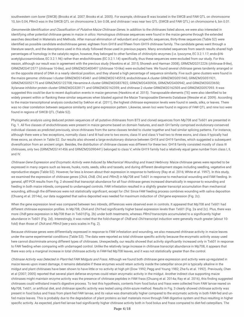
Since trichomes were easily visible on leaf fragments in the food bolus, we also examined the structure and density of trichomes on maize leaves from theyellow-green portion of the whorl, which was the primary FAW feeding site (Pechan et al. 2000). Three types of trichomes, macrohair, prickle hair, andbicellular microhair, were present on maize leaf (Freeling and Lane 1994; Poethig 1990). SEM analysis of trichomes from Mp708 and Tx601 was shown inFig. 5 and representatives of each trichome class were labeled. The appearance of trichomes differed between the two inbreds. Macrohairs on Tx601 leaveswere almost twice as long as those on Mp708 (Table 3). Likewise, the Tx601 prickle hairs and bicellular mircohairs also were signi�cantly longer than thosein Mp708. Measurements of trichome density indicated that the macrohairs were dramatically denser on Tx601 while the bicellular microhairs were denser inMp708.
Page 8/18
Table 3Maize leaf trichomea length and density for Mp708 and Tx601
Length (micron) Density (No./mm2)
mh ph bm mh ph bm
Mp708 455.83 ± 14.35 32.06 ± 0.49 38.3 ± 0.3 3.37 ± 0.2 59.04 ± 3.32 35.67 ± 1.58 *
Tx601 823.5 ± 29.17 * 44.06 ± 0.76 * 48.24 ± 0.44 * 4.56 ± 0.16 * 56.49 ± 3.54 30.02 ± 1.5
a Abbreviation: mh, macrohair; ph, prickle hair; bm, bicelluar microhair (Freeling and Lane 1994)
* The data represented are mean values with error bars (+ SE, n = 6 or 8). Letters indicate signi�cant differences by least signi�cant difference test (P < 0.05).
Supplementary Materials
Supplemental Table 1 List of Endochitinases
Supplemental Table 2 List of maize endochitinase genes
Supplemental Table 3 Speci�c activity of maize endochitinase from various tissues measured using chitin-azurea
Supplemental Table 4 Proteins Identi�ed in Mp704 x Mp708 Trichomes
Supplemental Table 5 Parameters of proteins identi�ed in Tx601 trichomes
Supplemental Table 6 Primers used in chitinase gene cloning
Supplemental Table 7 Primers used in qRT-PCR
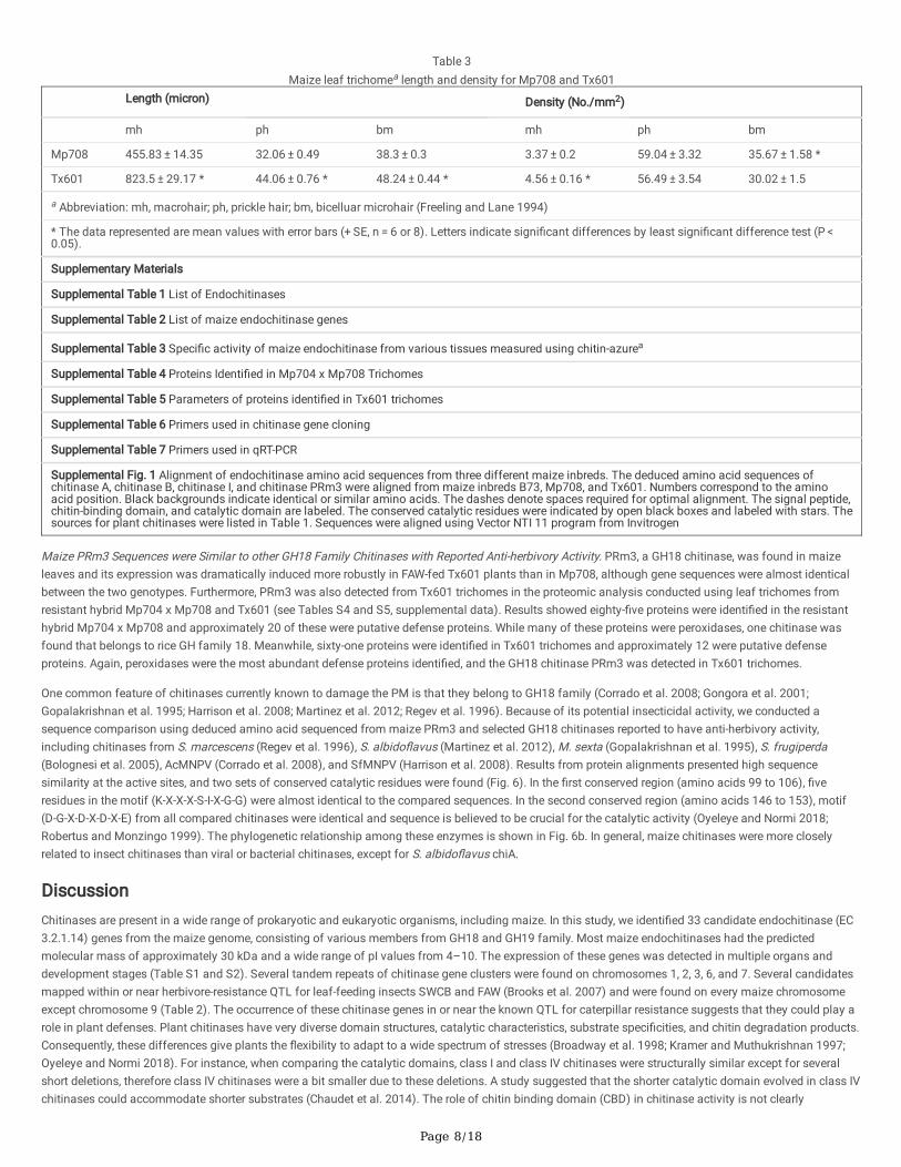
Supplemental Fig. 1 Alignment of endochitinase amino acid sequences from three different maize inbreds. The deduced amino acid sequences ofchitinase A, chitinase B, chitinase I, and chitinase PRm3 were aligned from maize inbreds B73, Mp708, and Tx601. Numbers correspond to the aminoacid position. Black backgrounds indicate identical or similar amino acids. The dashes denote spaces required for optimal alignment. The signal peptide,chitin-binding domain, and catalytic domain are labeled. The conserved catalytic residues were indicated by open black boxes and labeled with stars. Thesources for plant chitinases were listed in Table 1. Sequences were aligned using Vector NTI 11 program from Invitrogen
Maize PRm3 Sequences were Similar to other GH18 Family Chitinases with Reported Anti-herbivory Activity. PRm3, a GH18 chitinase, was found in maizeleaves and its expression was dramatically induced more robustly in FAW-fed Tx601 plants than in Mp708, although gene sequences were almost identicalbetween the two genotypes. Furthermore, PRm3 was also detected from Tx601 trichomes in the proteomic analysis conducted using leaf trichomes fromresistant hybrid Mp704 x Mp708 and Tx601 (see Tables S4 and S5, supplemental data). Results showed eighty-�ve proteins were identi�ed in the resistanthybrid Mp704 x Mp708 and approximately 20 of these were putative defense proteins. While many of these proteins were peroxidases, one chitinase wasfound that belongs to rice GH family 18. Meanwhile, sixty-one proteins were identi�ed in Tx601 trichomes and approximately 12 were putative defenseproteins. Again, peroxidases were the most abundant defense proteins identi�ed, and the GH18 chitinase PRm3 was detected in Tx601 trichomes.
One common feature of chitinases currently known to damage the PM is that they belong to GH18 family (Corrado et al. 2008; Gongora et al. 2001;Gopalakrishnan et al. 1995; Harrison et al. 2008; Martinez et al. 2012; Regev et al. 1996). Because of its potential insecticidal activity, we conducted asequence comparison using deduced amino acid sequenced from maize PRm3 and selected GH18 chitinases reported to have anti-herbivory activity,including chitinases from S. marcescens (Regev et al. 1996), S. albido�avus (Martinez et al. 2012), M. sexta (Gopalakrishnan et al. 1995), S. frugiperda(Bolognesi et al. 2005), AcMNPV (Corrado et al. 2008), and SfMNPV (Harrison et al. 2008). Results from protein alignments presented high sequencesimilarity at the active sites, and two sets of conserved catalytic residues were found (Fig. 6). In the �rst conserved region (amino acids 99 to 106), �veresidues in the motif (K-X-X-X-S-I-X-G-G) were almost identical to the compared sequences. In the second conserved region (amino acids 146 to 153), motif(D-G-X-D-X-D-X-E) from all compared chitinases were identical and sequence is believed to be crucial for the catalytic activity (Oyeleye and Normi 2018;Robertus and Monzingo 1999). The phylogenetic relationship among these enzymes is shown in Fig. 6b. In general, maize chitinases were more closelyrelated to insect chitinases than viral or bacterial chitinases, except for S. albido�avus chiA.
DiscussionChitinases are present in a wide range of prokaryotic and eukaryotic organisms, including maize. In this study, we identi�ed 33 candidate endochitinase (EC3.2.1.14) genes from the maize genome, consisting of various members from GH18 and GH19 family. Most maize endochitinases had the predictedmolecular mass of approximately 30 kDa and a wide range of pI values from 4–10. The expression of these genes was detected in multiple organs anddevelopment stages (Table S1 and S2). Several tandem repeats of chitinase gene clusters were found on chromosomes 1, 2, 3, 6, and 7. Several candidatesmapped within or near herbivore-resistance QTL for leaf-feeding insects SWCB and FAW (Brooks et al. 2007) and were found on every maize chromosomeexcept chromosome 9 (Table 2). The occurrence of these chitinase genes in or near the known QTL for caterpillar resistance suggests that they could play arole in plant defenses. Plant chitinases have very diverse domain structures, catalytic characteristics, substrate speci�cities, and chitin degradation products.Consequently, these differences give plants the �exibility to adapt to a wide spectrum of stresses (Broadway et al. 1998; Kramer and Muthukrishnan 1997;Oyeleye and Normi 2018). For instance, when comparing the catalytic domains, class I and class IV chitinases were structurally similar except for severalshort deletions, therefore class IV chitinases were a bit smaller due to these deletions. A study suggested that the shorter catalytic domain evolved in class IVchitinases could accommodate shorter substrates (Chaudet et al. 2014). The role of chitin binding domain (CBD) in chitinase activity is not clearly

Page 9/18
understood, however, this domain might contribute to substrates binding at active sites and hydrolysis (Li et al. 2005). Furthermore, chitinases from two GHfamilies (18 and 19) shared little to no sequence similarity to each other, and are evolutionary divergent as supported by phylogenetic analysis (Fig. 1).Variations in the topology of active sites for chitinases from a particular GH family (Oyeleye and Normi 2018) suggests that different chitinases may havespeci�c functions in maize defense responses.
Since peptides from chitinase A, B, I, and PRm3 were identi�ed from maize-fed FAW frass (Chuang et al. 2014a; Ray et al. 2016), we further cloned andcompared the coding sequences of these genes in maize inbreds Mp708 and Tx601 (Table 1). Results from sequence comparisons showed that nucleotidesequences from the two inbreds were almost identical except for a few polymorphisms. ChiA, ChiB, and ChiI were more closely related to each other thanPRm3 (Supplemental Fig. 1). Interestingly, when compared to the maize ancestor teosinte (Zea mays ssp parviglumis), both nucleotide and amino acidsequences of GH19 chitinases ChiA, ChiB, and ChiI were 97%-100% identical to the corresponding sequences from teosinte, which indicates they were quiteconserved during the domestication of maize. On the other hand, none of the teosinte subspecies exhibited sequence identities to the GH18 chitinase PRm3.Sequence similarities shared with PRm3 were rather seen from other monocot crops such as sorghum (~ 90%) and rice (~ 80%).
Despite the extensive reviews of plant chitinases and their antifungal activity, few have been studied for the role in insect resistance (Lawrence and Novak2006; Ray et al. 2016). Chitinases have been reported to disrupt the PM structure and consequently may be one of the defense protein plants use to increasetheir insect resistance (Herrera-Estrella and Chet 1999; Tellam et al. 1999; White et al. 1997). Since maize ChiA, ChiB, ChiI, and PRm3 were highly expressed inleaf, silk, and husk tissues, they may serve as a defense for the developing ear against FAW and fungal pathogens (Table 1). In our study, we investigated thegene expressions of the aforementioned chitinases in two maize inbreds in response to herbivore feeding and mechanical wounding. Surprisingly, chitinasegene expression was differently regulated in Mp708 and Tx601 despite the sequence identity between the two. Although transcript levels all signi�cantlyincreased in maize leaves when challenged by FAW infestation, ChiA, ChiB, and ChiI (all GH19, known for antifungal activity) were more abundant in Mp708-fed leaves, while PRm3 (GH18) was dramatically higher in Tx601 (Fig. 2). Similarly, under wounding treatments, ChiB expression was signi�cantly greater inMp708, and PRm3 was constantly higher in Tx601. It was not surprising to �nd that wounding alone could signi�cantly induce the chitinase expression, as aprevious study (Ray et al. 2016) showed that wounding in maize leaves suppressed the growth of fungal pathogen Cochliobolus heterostrophus. Oneexplanation was maize plants might prime themselves to inhibit opportunistic fungal pathogen since wounding caused by herbivore feeding could providean opening for them (Clarke et al. 1998). In addition, chitin oligomers from the degraded PM could also serve as an elicitor to activate the pathogen defensepathway and further prevent diseases from entering the wounded area (Cohen-Kupiec and Chet 1998; Eckardt 2008; Ray et al. 2016). It is unlikely that thedifferences in gene expression between Mp708 and Tx601 were due to polymorphisms in coding sequences of the respective genes, but could be due tovariations in the promoter regions or signals that interacting with them. For instance, Mp708 plants have higher constitutive JA levels than Tx601 and thusare primed and ready for insect attack (Luthe et al. 2011; Shivaji et al. 2010). This priming could explain why Mp708 has higher basal chitinase expressionfor ChiA and ChiI than Tx601, except PRm3. In a recent study, exogenously applied ChiA reduced the growth of Tx601-fed FAW larvae by 80% (Mason et al.2019). In another study, Pr4 and ChiA found in B73-fed FAW frass suppressed pathogen growth by inducing pathogen defense pathways, however, they alsoattenuated insect-induced responses in maize, and exogenously applying ChiA had no impact on caterpillar growth (Ray et al. 2016). Taken together, itseems that chitinase gene regulation most likely depends on the genetic background of the maize plant.
In addition to gene expression, we also found chitinase speci�c activity was induced in maize by both wounding and feeding treatments (Table S3). Next, wedetermined if chitinases were active in the insect midgut and if the midgut lining showed signs of damage after ingesting maize leaves. Results from activityanalysis showed that speci�c activity from both the FAW food bolus and frass increased signi�cantly after 24 hr of feeding in both inbred lines whencompared to diet-fed control (Fig. 3). This large increase in speci�c activity inside FAW midgut suggested that maize chitinases were stable and active wheningested and processed in the midgut. This �nding is signi�cant, since it has always been questioned if plant chitinases retain activity in the alkalinelepidopteran midgut (Broadway et al. 1998). One possible explanation for the retention of chitinase activity is the micro-environment created inside themidgut that may provide suitable conditions for enzyme activity. In our study, SEM images (Fig. 4) clearly shown that partially digested leaves were insidethe midgut and hence, they could sustain a favorable pH range for the chitinases. However, which chitinase or combinations of chitinases contributed to theactivity could not be determined due to the limitation of the enzymatic assay method used. Since both chitinase transcript levels and enzyme activities wereinduced after feeding, it suggests that a combination of chitinases could play a role in maize defense against herbivory.
Chitinase activity was also differently regulated in each maize genotype, in addition to gene expression. Unlike Mp708, signi�cantly greater enzyme activitywas detected in Tx601 compared to control in response to feeding. Also, samples collected from the food bolus and frass of Tx601-fed caterpillars hadhigher chitinase speci�c activity than Mp708-fed ones (Fig. 3). There are several possible causes for this increased speci�c activity in Tx601 despite thegreater transcripts increase in Mp708. Mp708 expresses a potent defense protein, Mir1-CP (Pechan et al. 2002), which can dramatically damage the PM bydegrading the integral PM protein, insect intestinal mucin (IIM) (Fescemyer et al. 2013). Enhancin, a baculovirus metalloprotease, has been also shown toattack this important PM structural protein (Wang and Granados 1997). As a result, high level of Mir1-CP presenting in the food bolus of Mp708-fed larvaemay have inactivated or degraded the chitinases induced by FAW infestation. A previous study also shows that the digestion process is impaired in Mp708-fed FAW larvae (Chang et al. 2000), so caterpillars fed on Tx601 could have digested more protein in the food bolus resulting in lower overall protein contentand higher speci�c activity. Last but not least, it is possible PRm3, which was more abundant in Tx601, is either has more potent enzymatic activity or ismore resistant to degradation in the midgut than the other chitinases induced during infestation (Chen et al. 2007; Koga et al. 1999).
Previous literature shows certain GH18 chitinases that are widely distributed in both prokaryotes and eukaryotes have antiherbivore activity. For example, achitinase from Serratia marcescens was shown to perforate Spodoptera larval midgut PM (Regev et al. 1996). Streptomyces albido�avus chitinases werefound to enhance resistance both to cabbage looper (Gongora et al. 2001) and coffee berry borer (Martinez et al. 2012). Manduca sexta chitinase had beenfound to increase the mortality of tobacco budworm when expressed in a recombinant baculovirus (Gopalakrishnan et al. 1995). Moreover, chitinases fromthe virus, Autographa californica nuclear polyhedrosis virus (AcMNPV), and Spodoptera frugiperda nucleopolyhedrovirus (SfMNPV) were found to enhance

Page 10/18
mortality of Lepidoptera larvae (Corrado et al. 2008; Harrison et al. 2008). As a member of class III chitinase, maize PRm3 had ~ 80% sequence identity at theconserved catalytic domains (K-X-X-X-S-I-X-G-G motif and catalytic motif DXDXE, Fig. 6) to other GH18 chitinases with anti-herbivory activity (Corrado et al.2008; Gongora et al. 2001; Gopalakrishnan et al. 1995; Harrison et al. 2008; Martinez et al. 2012; Regev et al. 1996). This conservation of sequence amongchitinases having insecticidal activity and PRm3 suggests that it also may be involved herbivore resistance. It will be interesting to perform additionalexperiments with puri�ed PRm3 chitinase to see if it has direct deleterious effects on caterpillar growth and PMs.
Morphological observations also suggest that the PRm3 enzyme is a good candidate for insect defense in Tx601. When the damaged PM dissected fromlarvae reared on Tx601 were examined, it appeared that the hydrolytic enzymes from the ingested plant material had weakened the PMs since these PMswere not only more fragile than the diet-fed ones during the sample processing, but showed obvious signs of physical damage compared to those reared onarti�cial diets. Detailed structural abnormalities were observed including membrane �akes, multiple holes, spheroid-shaped particles embedded in membranelayers, and trichome piercing (Fig. 3). The spheroid-shaped particles on the PM surface have been previously seen, but without known function (Adang andSpence 1981; Brandt et al. 1978; Pechan et al. 2002). Trichome structure was clearly visible under SEM, and we also observed the phenomenon of trichomesperforating the PM. This was typically observed near the areas where degenerated PM patches were seen (Fig. 4). This could be explained by the increasedPRm3 chitinase levels present in Tx601 leaves causing PM degradation. It was also shown that Tx601 trichomes contained PRm3 that could directly deliverthis enzyme to the midgut after leaves were consumed.
Trichomes are hair-like structures developed from the aerial epidermis on the leaves of many plant species. They can be glandular or non-glandular,unicellular, or multicellular and vary in size, number, shape, and chemical composition (Johnson 1975; Serna and Martin 2006; Tian et al. 2012; Werker 2000;Yoshida et al. 2009). Previous studies show that trichomes not only act as a physical barrier that interferes with insect herbivore movement and feeding, theyalso poison, trap or repel insect herbivores by releasing toxic chemicals (Elle and Hare 2000; Holeski et al. 2010; Levin 1973; Peiffer et al. 2009; Tian et al.2012; Wagner 1991). In this study, our data also suggested that trichomes may be related to insect defense by facilitating PM penetration and resulting ingrowth delay of FAW. For example, growth retardation was seen in FAW reared directly on Tx601 leaves (this study) or those exogenously treated with ChiA orbacteria on Tx601 leaves (Mason et al. 2019). However, in another inbred line B73, directly applying ChiA did not slow the caterpillar growth (Ray et al. 2016).One reason for the differences could be due to the maize trichome structure and composition. Since Tx601 has signi�cantly longer and denser trichomesthan Mp708 and B73, it can damage PM by physical penetration and further altering the caterpillar gut microbiome, which in turn reduces larvaeperformance (Mason et al. 2019; Williams et al. 2000). Overall, our results strongly indicated that the maize chitinases could disrupt the PM and have thepotential to signi�cantly disrupt insect digestion or nutrient intake, thus increase the susceptibility to abrasion or pathogens (Aranda et al. 1996; Gongora etal. 2001; Hegedus et al. 2009).
ConclusionsChitinases are reported to adversely affect insect pests, together with other natural products like toxins, lectins, proteases, and α-amylase inhibitors(Broadway et al. 1998; Kramer and Muthukrishnan 1997; Oyeleye and Normi 2018). In this study, we found distinctive groups of chitinases in maize withdifferent molecular structures and catalytic mechanisms in support of previous studies, and here we focused on exploring the role of endochitinases inmaize insect defense. Variations were observed in chitinases gene and activity regulation in response to insect feeding and mechanical wounding fromdifferent GH families, and some chitinases may be more effective in defense than others. When comparing maize inbreds with different levels of herbivoreresistance, we found those chitinase sequence identities were extremely high. Nevertheless, each inbred line had a speci�c induction pro�le for chitinasessuggesting that different strategies may be deployed for herbivore defense. The physical/physiological effects of plant chitinases allow us to evaluate itsimpact as a target-speci�c natural resistance factor, and among all four chitinases tested, PRm3 could be a better candidate for a potential pest controlagent.
Declarations:
Declarations
Competing interestsThe authors have no con�icts of interest to declare that are relevant to the content of this article
Ethics approvalNot applicable
Consent for publicationNot applicable
FundingThis study was funded by the Pennsylvania State University.

Page 11/18
Authors' contributionsYang Han and Dawn Luthe contributed to the study conception and design. Material preparation, data collection and analysis were performed by Yang Han.Proteomic analysis of trichomes was carried out by Erin Taylor. The �rst draft of the manuscript was written by Yang Han and Dawn Luthe commented onprevious versions of the manuscript. All authors read and approved the �nal manuscript.
AcknowledgmentsSpecial thanks to M. Peiffer and Penn State Microscopy facility (University Park, PA, U.S.A.) for their technical support.
References1. Adang MJ, Spence KD (1981) Surface morphology of peritrophic membrane formation in the cabbage looper. Trichoplusia ni Cell Tissue Res 218:141–
147. doi:10.1007/bf00210100
2. Aranda E, Sanchez J, Peferoen M, Guereca L, Bravo A (1996) Interactions of Bacillus thuringiensis crystal proteins with the midgut epithelial cells ofSpodoptera frugiperda (Lepidoptera: Noctuidae. J Invertebr Pathol 68:203–212. doi:10.1006/jipa.1996.0087
3. Beintema JJ (1994) Structural features of plant chitinases and chitin-binding proteins. FEBS Lett 350:159–163
4. Bekesiova B, Hraska S, Libantova J, Moravcikova J, Matusikova I (2008) Heavy-metal stress induced accumulation of chitinase isoforms in plants. MolBiol Rep 35:579–588. doi:10.1007/s11033-007-9127-x
5. Bernardi D et al (2015) Cross-Resistance between Cry1 Proteins in Fall Armyworm (Spodoptera frugiperda) May Affect the Durability of CurrentPyramided Bt Maize Hybrids in Brazil. PLoS One 10:e0140130. doi:10.1371/journal.pone.0140130
�. Bohorova N et al (2001) Novel synthetic Bacillus thuringiensiscry1B gene and the cry1B-cry1Ab translational fusion confer resistance to southwesterncorn borer, sugarcane borer and fall armyworm in transgenic tropical maize. Theoret Appl Genetics 103:817–826. doi:10.1007/s001220100686
7. Bokonon-Ganta A, Bernal JS, Pietrantonio PV, SÉTamou M (2003) Survivorship and development of fall armyworm, Spodoptera frugiperda (J. E. Smith)(Lepidoptera: Noctuidae), on conventional and transgenic maize cultivars expressing Bacillus thuringiensis Cry9C and Cry1A(b). endotoxinsInternational Journal of Pest Management 49:169
�. Bolognesi R, Arakane Y, Muthukrishnan S, Kramer KJ, Terra WR, Ferreira C (2005) Sequences of cDNAs and expression of genes encoding chitinsynthase and chitinase in the midgut of Spodoptera frugiperda Insect. Biochem Mol Biol 35:1249–1259.doi:http://dx.doi.org/10.1016/j.ibmb.2005.06.006
9. Bosak EJ (2011) Using a developmental comparison to decipher priming of induced defenses in maize and its effects on a generalist herbivore. ThePennsylvania State University
10. Bradford MM (1976) A rapid and sensitive method for the quantitation of microgram quantities of protein utilizing the principle of protein-dye binding.Anal Biochem 72:248–254. doi:http://dx.doi.org/10.1016/0003-2697(76)90527-3
11. Brandt CR, Adang MJ, Spence KD (1978) The peritrophic membrane: Ultrastructural analysis and function as a mechanical barrier to microbial infectionin Orgyia pseudotsugata. J Invertebr Pathol 32:12–24
12. Broadway R et al (1998) Novel Chitinolytic Enzymes with Biological Activity Against Herbivorous Insects. J Chem Ecol 24:985–998.doi:10.1023/a:1022346301626
13. Brooks TD, Bushman BS, Williams WP, McMullen MD, Buckley PM (2007) Genetic basis of resistance to fall armyworm (Lepidoptera: Noctuidae) andsouthwestern corn borer (Lepidoptera: Crambidae) leaf-feeding damage in maize. J Econ Entomol 100:1470–1475
14. Brooks TD, Willcox MC, Williams WP, Buckley PM (2005) Quantitative Trait Loci Conferring Resistance to Fall Armyworm and Southwestern Corn BorerLeaf Feeding Damage This paper is a joint contribution of USDA-ARS and the Mississippi Agricultural and Forestry Experiment Station and is publishedas journal no. J10582 of the Miss. Agric. and Forestry Exp. Stn Crop Sci 45:2430–2434. doi:10.2135/cropsci2004.0656
15. Chang YM, Luthe DS, Davis FM, Williams WP (2000) In�uence of whorl region from resistant and susceptible corn genotypes on fall armyworm(Lepidoptera: Noctuidae) growth and development. J Econ Entomol 93:477–483
1�. Chaudet MM, Naumann TA, Price NPJ, Rose DR (2014) Crystallographic structure of ChitA, a glycoside hydrolase family 19, plant class IV chitinase fromZea mays Protein Science: A Publication of. the Protein Society 23:586–593. doi:10.1002/pro.2437
17. Chen H, Gonzales-Vigil E, Wilkerson CG, Howe GA (2007) Stability of Plant Defense Proteins in the Gut of Insect. Herbivores Plant Physiol 143:1954–1967. doi:10.2307/40065405
1�. Chen H, Wilkerson CG, Kuchar JA, Phinney BS, Howe GA (2005) Jasmonate-inducible plant enzymes degrade essential amino acids in the herbivoremidgut. Proc Natl Acad Sci U S A 102:19237–19242. doi:10.1073/pnas.0509026102
19. Chuang WP, Herde M, Ray S, Castano-Duque L, Howe GA, Luthe DS (2014a) Caterpillar attack triggers accumulation of the toxic maize protein RIP2. NewPhytol 201:928–939. doi:10.1111/nph.12581
20. Chuang WP, Ray S, Acevedo FE, Peiffer M, Felton GW, Luthe DS (2014b) Herbivore Cues from the Fall Armyworm (Spodoptera frugiperda) Larvae TriggerDirect Defenses in Maize Mol. Plant Microbe Interact 27:461–470. doi:10.1094/MPMI-07-13-0193-R
21. Clarke HRG, Lawrence SD, Flaskerud J, Korhnak TE, Gordon MP, Davis JM (1998) Chitinase accumulates systemically in wounded poplar trees. PhysiolPlant 103:154–161. doi:10.1034/j.1399-3054.1998.1030202.x

Page 12/18
22. Cohen-Kupiec R, Chet I (1998) The molecular biology of chitin digestion. Curr Opin Biotechnol 9:270–277
23. Corrado G et al (2008) The Chitinase A from the baculovirus AcMNPV enhances resistance to both fungi and herbivorous pests in tobacco. TransgenicRes 17:557–571. doi:10.1007/s11248-007-9129-4
24. Didierjean L, Frendo P, Nasser W, Genot G, Marivet J, Burkard G (1996) Heavy-metal-responsive genes in maize: identi�cation and comparison of theirexpression upon various forms of. abiotic stress Planta 199:1–8
25. Donaldson JR, Nanduri B, Burgess SC, Lawrence ML (2009) Comparative Proteomic Analysis of Listeria monocytogenes Strains F2365 and EGDApplied. and environmental microbiology 75:366–373. doi:10.1128/aem.01847-08
2�. Dow J (1992) pH gradients in Lepidopteran midgut. J Exp Biol 172:355–375
27. Eckardt NA (2008) Chitin signaling in plants: insights into the perception of fungal pathogens and rhizobacterial symbionts. Plant Cell 20:241–243.doi:10.1105/tpc.108.058784
2�. Elle E, Hare JD (2000) No Bene�t of Glandular Trichome Production in Natural Populations of. Datura wrightii? Oecologia 123:57–65.doi:10.2307/4222591
29. Fescemyer HW, Sandoya GV, Gill TA, Ozkan S, Marden JH, Luthe DS (2013) Maize toxin degrades peritrophic matrix proteins and stimulatescompensatory transcriptome responses in fall armyworm midgut. Insect Biochem Mol Biol 43:280–291. doi:10.1016/j.ibmb.2012.12.008
30. Finn RD et al (2014) Pfam: the protein families database. Nucleic Acids Res 42:D222–D230. doi:10.1093/nar/gkt1223
31. Flach J, Pilet PE, Jolles P (1992) What's new in chitinase research? Experientia 48:701–716
32. Flagel L et al (2018) Mutational disruption of the ABCC2 gene in fall armyworm, Spodoptera frugiperda, confers resistance to the Cry1Fa and Cry1A.105insecticidal. proteins Scienti�c Reports 8:7255. doi:10.1038/s41598-018-25491-9
33. Freeling M, Lane B (1994) The Maize Leaf. In: Freeling M, Walbot V (eds) The Maize Handbook. Springer Lab Manuals. Springer New York, pp 17–28.doi:10.1007/978-1-4612-2694-9_3
34. Gongora CE, Wang S, Barbehenn RV, Broadway RM (2001) Chitinolytic enzymes from Streptomyces albido�avus expressed in tomato plants: effects onTrichoplusia ni Entomologia experimentalis et applicata 99:193–204 doi:10.1046/j.1570-7458.2001.00817.x
35. Gooday GW (1999) Aggressive and defensive roles for chitinases Exs 87:157–169
3�. Gopalakrishnan B, Muthukrishnan S, Kramer KJ (1995) Baculovirus-mediated expression of a Manduca sexta chitinase gene: Properties of therecombinant protein Insect. Biochem Mol Biol 25:255–265. doi:http://dx.doi.org/10.1016/0965-1748(94)00070-X
37. Graham LS, Sticklen MB (1994) Plant chitinases Canadian. Journal of Botany 72:1057–1083. doi:10.1139/b94-132
3�. Grover A (2012) Plant Chitinases: Genetic Diversity and Physiological Roles Critical Reviews. in Plant Sciences 31:57–73.doi:10.1080/07352689.2011.616043
39. Gutierrez-Moreno R, Mota-Sanchez D, Blanco CA, Whalon ME, Teran-Santo�mio H, Rodriguez-Maciel JC, DiFonzo C (2019) Field-Evolved Resistance ofthe Fall Armyworm (Lepidoptera: Noctuidae) to Synthetic Insecticides in Puerto Rico and Mexico. J Econ Entomol 112:792–802. doi:10.1093/jee/toy372
40. Hall BG (2013) Building Phylogenetic Trees from Molecular Data with MEGA Mol Biol Evol doi:10.1093/molbev/mst012
41. Hamel F, Boivin R, Tremblay C, Bellemare G (1997) Structural and Evolutionary Relationships Among Chitinases of Flowering Plants. J Mol Evol 44:614–624. doi:10.1007/pl00006184
42. Hamid R, Khan MA, Ahmad M, Ahmad MM, Abdin MZ, Musarrat J, Javed S (2013) Chitinases: An update. J Pharm Bioallied Sci 5:21–29.doi:10.4103/0975-7406.106559
43. Harrison RL, Puttler B, Popham HJR (2008) Genomic sequence analysis of a fast-killing isolate of Spodoptera frugiperda multiple nucleopolyhedrovirus.J Gen Virol 89:775–790. doi:10.1099/vir.0.83566-0
44. Hawkins LK et al (2015) Characterization of the Maize Chitinase Genes and Their Effect on Aspergillus �avus and A�atoxin Accumulation. ResistancePLoS One 10:e0126185. doi:10.1371/journal.pone.0126185
45. Hegedus D, Erlandson M, Gillott C, Toprak U (2009) New insights into peritrophic matrix synthesis, architecture, and function. Annu Rev Entomol 54:285–302. doi:10.1146/annurev.ento.54.110807.090559
4�. Henrissat B (1999) Classi�cation of chitinases modules. In: Chitin and Chitinases. Springer, pp 137–156
47. Herrera-Estrella A, Chet I (1999) Chitinases in biological control. In: Chitin and Chitinases. Springer, pp 171–184
4�. Hoffmann WA, Poorter H (2002) Avoiding bias in calculations of relative growth rate. Ann Bot 90:37–42. doi:10.1093/aob/mcf140
49. Holeski LM, Chase-Alone R, Kelly JK (2010) The genetics of phenotypic plasticity in plant defense: trichome production in Mimulus. guttatus Am Nat175:391–400. doi:10.1086/651300
50. Horikoshi RJ et al (2016) Effective dominance of resistance of Spodoptera frugiperda to Bt maize and cotton varieties: implications for resistancemanagement. Sci Rep 6:34864. doi:10.1038/srep34864
51. http://www.nature.com/articles/srep34864#supplementary-information
52. Huang F (2020) Resistance of the fall armyworm, Spodoptera frugiperda, to transgenic Bacillus thuringiensis Cry1F corn in the America: lessons andimplications for Bt corn IRM in China Insect Science doi:10.1111/1744-7917.12826
53. Huber M, Cabib E, Miller LH (1991) Malaria parasite chitinase and penetration of the mosquito peritrophic membrane Proceedings of the NationalAcademy of Sciences 88:2807–2810 doi:10.1073/pnas.88.7.2807

Page 13/18
54. Hunter S et al (2012) InterPro in 2011: new developments in the family and domain prediction database. Nucleic Acids Res 40:D306–D312.doi:10.1093/nar/gkr948
55. Hurkman WJ, Tanaka CK (1986) Solubilization of plant membrane proteins for analysis by two-dimensional gel electrophoresis. Plant Physiol 81:802–806
5�. Huynh QK, Hironaka CM, Levine EB, Smith CE, Borgmeyer JR, Shah DM (1992) Antifungal proteins from plants. Puri�cation, molecular cloning, andantifungal properties of chitinases from maize seed. J Biol Chem 267:6635–6640
57. Iseli B, Armand S, Boller T, Neuhaus JM, Henrissat B (1996) Plant chitinases use two different hydrolytic mechanisms. FEBS Lett 382:186–188
5�. Johnson HB (1975) Plant Pubescence: An Ecological. Perspective Botanical Review 41:233–258. doi:10.2307/4353882
59. Kesari P, Patil DN, Kumar P, Tomar S, Sharma AK (2015) Structural and functional evolution of chitinase-like proteins. from plants Proteomics 15:1693–1705. doi:10.1002/pmic.201400421
�0. Koga D, Mitsutomi M, Fau - Kono M, Kono M, Fau - Matsumiya M, Matsumiya M (1999) Biochemistry of chitinases. In: Chitin and Chitinases. vol 1023-294X (Print). Springer, pp 111–123
�1. Kramer KJ, Muthukrishnan S (1997) Insect chitinases: molecular biology and potential use as biopesticides. Insect Biochem Mol Biol 27:887–900
�2. LaLonde SM Transforming variables for normality and linearity—when, how, why and why not’s. In: SAS Conference Proceedings NESUG (2005) pp 11–14
�3. Lawrence S, Novak N (2006) Expression of Poplar Chitinase in Tomato Leads to Inhibition of Development in Colorado Potato. Beetle Biotechnol Lett28:593–599. doi:10.1007/s10529-006-0022-7
�4. Lehane MJ (1997) Peritrophic matrix structure and function. Annu Rev Entomol 42:525–550. doi:10.1146/annurev.ento.42.1.525
�5. Levin DA (1973) The Role of Trichomes in Plant Defense. Q Rev Biol 48:3–15. doi:10.2307/2822621
��. Li Q, Wang F, Zhou Y, Xiao X (2005) Putative exposed aromatic and hydroxyl residues on the surface of the N-terminal domains of Chi1 from Aeromonascaviae CB101 are essential for chitin binding and hydrolysis. Appl Environ Microbiol 71:7559–7561. doi:10.1128/AEM.71.11.7559-7561.2005
�7. Lombard V, Golaconda Ramulu H, Drula E, Coutinho PM, Henrissat B (2014) The carbohydrate-active enzymes database (CAZy) in 2013. Nucleic AcidsRes 42:D490–D495. doi:10.1093/nar/gkt1178
��. Luthe DS et al (2011) Aboveground to belowground herbivore defense signaling in maize: a two-way street? Plant Signal Behav 6:126–129
�9. Martinez C, Echeverri C, Florez J, Gaitan A, Gongora C (2012) In vitro production of two chitinolytic proteins with an inhibiting effect on the insect coffeeberry borer, Hypothenemus hampei (Ferrari) (Coleoptera: Curculionidae) and the fungus Hemileia vastatrix the most limiting pests of coffee crops. AMBExpress 2:22
70. Mason CJ et al (2019) Plant defenses interact with insect enteric bacteria by initiating a leaky gut syndrome. Proc Natl Acad Sci U S A 116:15991–15996. doi:10.1073/pnas.1908748116
71. McMullen M, Frey M, Degenhardt J (2009) Genetics and Biochemistry of Insect Resistance in Maize. In: Bennetzen J, Hake S (eds) Handbook of Maize:Its Biology. Springer, New York, pp 271–289. doi:10.1007/978-0-387-79418-1_14
72. Mohan S, Ma PW, Pechan T, Bassford ER, Williams WP, Luthe DS (2006) Degradation of the S. frugiperda peritrophic matrix by an inducible maizecysteine protease. J Insect Physiol 52:21–28. doi:10.1016/j.jinsphys.2005.08.011
73. Mohan S, Ma PW, Williams WP, Luthe DS (2008) A naturally occurring plant cysteine protease possesses remarkable toxicity against insect pests andsynergizes Bacillus thuringiensis toxin. PLoS One 3:e1786. doi:10.1371/journal.pone.0001786
74. Molano J, Polacheck I, Duran A, Cabib E (1979) An endochitinase from wheat germ. Activity on nascent preformed chitin J Biol Chem 254:4901–4907
75. Monaco MK et al (2014) Gramene 2013: comparative plant genomics resources. Nucleic Acids Res 42:D1193–D1199. doi:10.1093/nar/gkt1110
7�. Osborne JW (2010) Improving your data transformations: Applying the Box-Cox transformation Practical Assessment. Research Evaluation 15:1–9
77. Oyeleye A, Normi YM (2018) Chitinase: diversity, limitations, and trends in engineering for suitable applications Biosci Rep 38doi:10.1042/BSR20180323
7�. Pechan T, Cohen A, Williams WP, Luthe DS (2002) Insect feeding mobilizes a unique plant defense protease that disrupts the peritrophic matrix ofcaterpillars Proceedings of the National Academy of Sciences 99:13319–13323 doi:10.1073/pnas.202224899
79. Pechan T et al (2000) A unique 33-kD cysteine proteinase accumulates in response to larval feeding in maize genotypes resistant to fall armyworm andother. Lepidoptera Plant Cell 12:1031–1040
�0. Pechanova O, Pechan T, Ozkan S, McCarthy FM, Williams WP, Luthe DS (2010) Proteome pro�le of the developing maize (Zea mays L.). rachisProteomics 10:3051–3055. doi:10.1002/pmic.200900833
�1. Pechanova O, Pechan T, Williams WP, Luthe DS (2011) Proteomic analysis of the maize rachis: potential roles of constitutive and induced proteins inresistance to Aspergillus �avus infection and a�atoxin accumulation. Proteomics 11:114–127. doi:10.1002/pmic.201000368
�2. Pechanova O, Stone WD, Monroe W, Nebeker TE, Klepzig KD, Yuceer C (2008) Global and comparative protein pro�les of the pronotum of the southernpine beetle. Dendroctonus frontalis Insect Mol Biol 17:261–277. doi:10.1111/j.1365-2583.2008.00801.x
�3. Peethambaran B, Hawkins L, Windham GL, Williams WP, Luthe DS (2010) Anti-fungal activity of maize silk proteins and role of chitinases in Aspergillus�avus resistance. Toxin Reviews 29:27–39 doi. doi:10.3109/15569540903402874
�4. Pegg GF, Young DH (1982) Puri�cation and characterization of chitinase enzymes from healthy and Verticillium albo-atrum-infected tomato plants, andfrom V. albo-atrum Physiological Plant Pathology 21:389–409. doi:http://dx.doi.org/10.1016/0048-4059(82)90074-1

Page 14/18
�5. Peiffer M, Felton GW (2005) The host plant as a factor in the synthesis and secretion of salivary glucose oxidase in larval Helicoverpa zea. Arch InsectBiochem Physiol 58:106–113. doi:10.1002/arch.20034
��. Peiffer M, Tooker JF, Luthe DS, Felton GW (2009) Plants on Early Alert: Glandular Trichomes as Sensors for. Insect Herbivores New Phytologist 184:644–656. doi:10.2307/27735815
�7. Plymale R, Grove MJ, Cox-Foster D, Ostiguy N, Hoover K (2008) Plant-mediated alteration of the peritrophic matrix and baculovirus infection inlepidopteran larvae. J Insect Physiol 54:737–749. doi:http://dx.doi.org/10.1016/j.jinsphys.2008.02.005
��. Poethig RS (1990) Phase change and the regulation of shoot morphogenesis. in plants Science 250:923–930. doi:10.1126/science.250.4983.923
�9. Ray S et al (2016) Turnabout Is Fair Play: Herbivory-Induced Plant Chitinases Excreted in Fall Armyworm Frass Suppress Herbivore Defenses in Maize.Plant Physiol 171:694–706. doi:10.1104/pp.15.01854
90. Regev A et al (1996) Synergistic activity of a Bacillus thuringiensis delta-endotoxin and a bacterial endochitinase against Spodoptera littoralis larvae.Appl Environ Microbiol 62:3581–3586
91. Ritchie SW, Hanway JJ, Service ISUCE (1984) How a Corn Plant Develops. Iowa State University of Science and Technology, Cooperative ExtensionService
92. Robertus JD, Monzingo AF (1999) The structure and action of chitinases. In: Chitin and Chitinases. vol 1023-294X (Print). Springer, pp 125–135
93. Schaeffer ML et al (2011) MaizeGDB: curation and outreach go hand-in-hand Database 2011 doi:10.1093/database/bar022
94. Schnable PS et al (2009) The B73 maize genome: complexity, diversity. and dynamics Science 326:1112–1115. doi:10.1126/science.1178534
95. Sekhon RS, Lin H, Childs KL, Hansey CN, Buell CR, de Leon N, Kaeppler SM (2011) Genome-wide atlas of transcription during maize development. PlantJ 66:553–563. doi:10.1111/j.1365-313X.2011.04527.x
9�. Serna L, Martin C (2006) Trichomes: different regulatory networks lead to convergent structures. Trends Plant Sci 11:274–280.doi:10.1016/j.tplants.2006.04.008
97. Sharma N, Sharma K, Gaur R, Gupta V (2011) Role of chitinase in plant defense. Asian J Biochem 6:29–37
9�. Shivaji R et al (2010) Plants on constant alert: elevated levels of jasmonic acid and jasmonate-induced transcripts in caterpillar-resistant maize. J ChemEcol 36:179–191. doi:10.1007/s10886-010-9752-z
99. Shoresh M, Harman GE (2008) Genome-wide identi�cation, expression and chromosomal location of the genes encoding chitinolytic enzymes in Zeamays. Mol Genet Genomics 280:173–185. doi:10.1007/s00438-008-0354-1
100. Storer NP, Babcock JM, Schlenz M, Meade T, Thompson GD, Bing JW, Huckaba RM (2010) Discovery and characterization of �eld resistance to Bt maize:Spodoptera frugiperda (Lepidoptera: Noctuidae) in Puerto Rico. J Econ Entomol 103:1031–1038
101. Storer NP, Kubiszak ME, Ed King J, Thompson GD, Santos AC (2012) Status of resistance to Bt maize in Spodoptera frugiperda: lessons from PuertoRico. J Invertebr Pathol 110:294–300. doi:10.1016/j.jip.2012.04.007
102. Tamura K, Stecher G, Peterson D, Filipski A, Kumar S (2013) MEGA6: Molecular Evolutionary Genetics Analysis Version 6.0. Mol Biol Evol 30:2725–2729.doi:10.1093/molbev/mst197
103. Tellam RL, Wijffels G, Willadsen P (1999) Peritrophic matrix proteins Insect. Biochem Mol Biol 29:87–101
104. Tian D, Tooker J, Peiffer M, Chung SH, Felton GW (2012) Role of trichomes in defense against herbivores: comparison of herbivore response to woollyand hairless trichome mutants in tomato (Solanum lycopersicum). Planta 236:1053–1066. doi:10.1007/s00425-012-1651-9
105. Ti�n P (2004) Comparative evolutionary histories of chitinase genes in the Genus zea and Family poaceae. Genetics 167:1331–1340.doi:10.1534/genetics.104.026856
10�. Tronsmo A, Harman GE (1993) Detection and quanti�cation of N-acetyl-beta-D-glucosaminidase, chitobiosidase, and endochitinase in solutions and ongels. Anal Biochem 208:74–79. doi:10.1006/abio.1993.1010
107. van Loon LC, Rep M, Pieterse CM (2006) Signi�cance of inducible defense-related proteins in infected plants. Annu Rev Phytopathol 44:135–162.doi:10.1146/annurev.phyto.44.070505.143425
10�. Virla EG, Álvarez A, Loto F, Pera LM, Baigorí M (2008) Fall Armyworm Strains (Lepidoptera: Noctuidae) in Argentina, Their Associate Host Plants andResponse to Different Mortality. Factors in Laboratory 91:63–69. doi:10.1653/0015-4040(2008)091[0063:faslni]2.0.co;2
109. Wagner GJ (1991) Secreting Glandular Trichomes: More than Just. Hairs Plant Physiol 96:675–679. doi:10.1104/pp.96.3.675
110. Wang P, Granados RR (1997) An intestinal mucin is the target substrate for a baculovirus enhancin. Proc Natl Acad Sci U S A 94:6977–6982
111. Werker E (2000) Trichome diversity and development. In: Advances in Botanical Research, vol Volume 31. Academic Press, pp 1–35.doi:http://dx.doi.org/10.1016/S0065-2296(00)31005-9
112. Wessler S, Bennetzen J, Dawe R, Jiang N, SanMiguel P, Freeman B (2009) Maize Transposable Element Database. http://maizetedb.org/~maize/.http://maizetedb.org/~maize/
113. White F, Kramer K, Johnson L, Muthukrishnan S (1997) Chitinases for insect control Advances in insect control, the role of transgenic plants CRC Press,Boca Raton ISBN:978-970
114. Williams W, Buckley P, Davis F (2000) Vegetative phase change in maize and its association with resistance to fall armyworm. Maydica 45:215–219
115. Williams WP, Davis FM, Windham GL (1990) Registration of Mp708 Germplasm Line of. Maize Crop Sci 30:757–757.doi:10.2135/cropsci1990.0011183X003000030082x
11�. Wu S, Kriz AL, Widholm JM (1994) Molecular analysis of two cDNA clones encoding acidic class I chitinase in maize. Plant Physiol 105:1097–1105

Page 15/18
117. Yoshida Y, Sano R, Wada T, Takabayashi J, Okada K (2009) Jasmonic acid control of GLABRA3 links inducible defense and trichome patterning in.Arabidopsis Development 136:1039–1048. doi:10.1242/dev.030585
11�. Zhang Y, Gao M, Singer SD, Fei Z, Wang H, Wang X (2012) Genome-wide identi�cation and analysis of the TIFY gene family in grape. PLoS One7:e44465. doi:10.1371/journal.pone.0044465
119. Zhe-fu L, Danqi W, Annuo L, Weiqin Z (1992) Chitinases from Seeds of Zea mays and Coix lachryma-jobi L. Puri�cation Some Properties ProcessBiochemistry 27:83–88. doi:http://dx.doi.org/10.1016/0032-9592(92)80014-T
120. Zhu-Salzman K, Luthe DS, Felton GW (2008) Arthropod-inducible proteins: broad spectrum defenses against multiple herbivores. Plant Physiol 146:852–858. doi:10.1104/pp.107.112177
121. Zhu YC, Blanco CA, Portilla M, Adamczyk J, Luttrell R, Huang F (2015) Evidence of multiple/cross resistance to Bt and organophosphate insecticides inPuerto Rico population of the fall armyworm. Spodoptera frugiperda Pesticide Biochemistry Physiology 122:15–21.doi:http://dx.doi.org/10.1016/j.pestbp.2015.01.007
Figures
Figure 1
Phylogenetic tree of maize chitinases. The tree was constructed using the amino acid sequences by Maximum Likelihood methods with MEGA version 6, thenumbers on the branch indicate bootstrap values from 1000 replicates. Filled circles indicate the sequence partition between family 19 and family 18chitinases. Members belonging to the same class were within each bracket. Sources of the amino acid sequences are listed in Table S2

Page 16/18
Figure 2
Relative expression of chitinase transcripts in maize leaves in response to various treatments. Quantitative RT-PCR was performed for (a) chitinase A, (b)chitinase B, (c) chitinase I, and (d) chitinase PRm3. Whorls from V8 stage Mp708 and Tx601 were mechanically wounded (W), or fed (F) by fall armywormlarvae for 24 hr. Undamaged plants were used as control (C). Total RNA was isolated from control whorls and the damaged areas on the leaves. Relativetranscript abundance was calculated using actin as the reference gene. The data represented were mean values with error bars (+SE, n=9). Letters indicatesigni�cant differences by the least signi�cant difference test (P<0.05)
Figure 3

Page 17/18
Chitinase speci�c activity in maize leaves and fall armyworm food bolus and frass after feeding for 24 hr on Mp708 or Tx601. V8 maize whorls from Mp708and Tx601 were infested with FAW larvae for 24 hr (L). Undamaged plants were used as control (C). FAW larvae that fed on these plants were collected andfood bolus (FB) was dissected from the midguts. Frass (F) from FAW larvae was also collected. Chitinase activity was measured in the collected materialsusing chitin-azure as a substrate as described in Materials and Methods. Food bolus and frass from larvae fed on arti�cial diet were used as a referencecontrol. The data represented are mean values with error bars (+SE, n=3 or 6). Letters indicated signi�cant differences by the least signi�cant difference test(P<0.05)
Figure 4
Scanning electron micrographs showing the peritrophic matrix (PM) of fall armyworm caterpillars. (a) and (b) PM from FAW larvae reared on arti�cial diet(magni�cation X100, and X500 respectively). (c) and (d) PM from FAW larvae reared on Tx601 plant showing the damaged area (magni�cation X100, andX500 respectively). (e) and (f) Enlargement of PM �akes from FAW larvae reared on Tx601 showing perforations and spherical particles (magni�cationX5000). (g) and (h) showing trichomes penetrating the peritrophic matrix (PM) of fall armyworm larvae reared on Tx601 (magni�cation X100). The scalewas indicated on each micrograph
Figure 5
Scanning electron micrographs of trichomes from maize inbreds Mp708 and Tx601. Three types of maize trichomes were observed: macrohair (circled),prickle hair (black arrows), and bicellular microhair (white arrows). Trichome morphology on the leaf surface of Mp708 (a) and Tx601 (b) is shown at 75Xmagni�cation. Enlargement of trichome macrohair (circled) from Mp708 (c) and Tx601 (d) is shown at 150X and100X magni�cation, respectively.Enlargement of trichome prickle hair (black arrows) and bicellular microhair (white arrows) from Mp708 (e, g) and Tx601 (f, h), are shown at 2000Xmagni�cation respectively. The scale was indicated on each micrograph

Page 18/18
Figure 6
Phylogenetic analysis of selected family 18 endochitinases. (a) Comparison of the conserved region of the amino acid sequences. Numbers correspond tothe amino acid position. Gray backgrounds indicate identical or similar amino acids. (b) Phylogenetic tree of chitinases. The tree was constructed using theamino acid sequences by Maximum Likelihood methods with MEGA 6, the numbers on the branch indicated bootstrap values from 1000 replicates.Sequences listed here were deduced sequence of chitinase from maize inbred B73, Mp708, and Tx601 (this work), Serratia marcescens (Regev et al., 1996),Streptomyces albido�avus (Martinez et al., 2012), Autographa californica nuclear polyhedrosis virus (AcMNPV) (Corrado et al., 2008), Spodoptera frugiperdanucleopolyhedrovirus (SfMNPV) (Harrison et al., 2008), Manduca sexta (Gopalakrishnan et al., 1995), and Spodoptera frugiperda (Bolognesi et al., 2005).Sequences were analyzed using MEGA 6
Supplementary Files
This is a list of supplementary �les associated with this preprint. Click to download.
SupplementalFig1.pdf
SupplementalTable17.xlsx