Luke J. Rogers*, Andrew John Lake, Katherine White ... · A saline-filled balloon was placed in the...
8
Bioscience Horizons Volume 7 2014 10.1093/biohorizons/hzt012 © The Author 2014. Published by Oxford University Press. This is an Open Access article distributed under the terms of the Creative Commons Attribution Non-Commercial License (http://creativecommons.org/licenses/by-nc/2.5), which permits unrestricted non-commercial use, distribution, and reproduction in any medium, provided the original work is properly cited. 1 Effects of superoxide donor menadione in adult Rat myocardium are associated with increased diastolic intracellular calcium Luke J. Rogers*, Andrew John Lake, Katherine White, Matthew Hardy and Ed White School of Biomedical Sciences, University of Leeds, Leeds LS2 9JT, UK *Corresponding author: Luke J. Rogers, School of Biomedical Sciences, University of Leeds, Leeds LS2 9JT, UK. Email: [email protected] Supervisor: Ed White, School of Biomedical Sciences, University of Leeds, Leeds LS2 9JT, UK. Superoxide anions have been associated with many aspects of cardiovascular disease. Menadione is a superoxide anion donor that alters the heart’s electrical and mechanical functions. The aim of this study was to demonstrate simultaneous changes in intracellular Ca 2+ ([Ca 2+ ]i) and mechanical activity in intact adult cardiac myocytes, and mechanical activity and electrical activ- ity in isolated whole hearts in order to provide greater insight into the mechanisms associated with the detrimental effects of menadione on the myocardium. Isolated hearts from adult male Wistar rats (n = 11, 200–250 g) were Langendorff perfused at 38°C with a Krebs–Henseleit solution. A saline-filled balloon was placed in the left ventricle (LV) in order to measure diastolic and developed pressure. Monophasic action potentials were simultaneously recorded from the epicardial surface. External stimulation at 5 Hz and intrinsic pacing were used throughout a 10 min control period and 30 min exposure to 50 µM mena- dione. Single LV myocytes (n = 7 from n = 4 animals) were loaded with the Ca 2+ -indicator Fura4-AM, stimulated at 1 Hz and exposed to 50 µM menadione. Myocyte length was simultaneously measured with [Ca 2+ ]i using a video edge detection sys- tem. In isolated hearts, exposure to menadione significantly decreased contractility and action potential duration (with a simi- lar time course); intrinsic heart rate and rhythmicity. Diastolic pressure was significantly increased. In single adult myocytes, menadione caused a significant increase in diastolic [Ca 2+ ]i and a decrease in resting cell length and led to spontaneous release of [Ca 2+ ]i. We conclude that the effects of menadione upon electrical and mechanical activity of the heart are at least in part a consequence of dysregulation of [Ca 2+ ]i handling and the subsequent increase in diastolic [Ca 2+ ] alterations in [Ca 2+ ]i are consistent with the generation of delayed after depolarization arrhythmias. Key words: menadione, calcium, rat, superoxide, contractility, electrophysiology Received 22 July 2013; revised 3 December 2013; accepted 16 December 2013 Introduction Cardiovascular disease is a major contributor to mortality and morbidity throughout the world (Roger et al., 2012). In the case of acute ischaemic events current treatment guidelines prioritize the rapid restoration of circulatory function to reverse cardiomyocyte injury and limit cell death, thus signifi- cantly reducing cardiac mortality and morbidity (Pollack, Antman, and Hollander , 2008). However, the advent of cardiac thrombolysis through the use of agents such as streptokinase and percutaneous coronary interventions has led to the characterization of a paradoxical phenomenon known as ‘reperfusion injury’. This is believed to occur as a result of the rapid production of reactive oxygen species (ROS), which overwhelm the endogenous antioxidant sys- tems leading to cell death via necrosis and apoptotic path- ways. The superoxide anion in particular is believed to have a critical role as a mediator of post-ischaemic contractile dys- function, dysrhythmias and chronic cardiovascular disease (Kevin, Novalija and Stowe, 2005). Research article at University of Leeds on May 24, 2016 http://biohorizons.oxfordjournals.org/ Downloaded from
Transcript of Luke J. Rogers*, Andrew John Lake, Katherine White ... · A saline-filled balloon was placed in the...
BioscienceHorizons Volume 7 2014 10.1093/biohorizons/hzt012
© The Author 2014. Published by Oxford University Press. This is an Open Access article distributed under the terms of the Creative CommonsAttribution Non-Commercial License (http://creativecommons.org/licenses/by-nc/2.5), which permits unrestricted non-commercial use, distribution,and reproduction in any medium, provided the original work is properly cited.
1
Effects of superoxide donor menadione in adult Rat myocardium are associated with increased diastolic intracellular calciumLuke J. Rogers*, Andrew John Lake, Katherine White, Matthew Hardy and Ed White
School of Biomedical Sciences, University of Leeds, Leeds LS2 9JT, UK*Corresponding author: Luke J. Rogers, School of Biomedical Sciences, University of Leeds, Leeds LS2 9JT, UK. Email: [email protected]
Supervisor: Ed White, School of Biomedical Sciences, University of Leeds, Leeds LS2 9JT, UK.
Superoxide anions have been associated with many aspects of cardiovascular disease. Menadione is a superoxide anion donor that alters the heart’s electrical and mechanical functions. The aim of this study was to demonstrate simultaneous changes in intracellular Ca2+ ([Ca2+]i) and mechanical activity in intact adult cardiac myocytes, and mechanical activity and electrical activ-ity in isolated whole hearts in order to provide greater insight into the mechanisms associated with the detrimental effects of menadione on the myocardium. Isolated hearts from adult male Wistar rats (n = 11, 200–250 g) were Langendorff perfused at 38°C with a Krebs–Henseleit solution. A saline-filled balloon was placed in the left ventricle (LV) in order to measure diastolic and developed pressure. Monophasic action potentials were simultaneously recorded from the epicardial surface. External stimulation at 5 Hz and intrinsic pacing were used throughout a 10 min control period and 30 min exposure to 50 µM mena-dione. Single LV myocytes (n = 7 from n = 4 animals) were loaded with the Ca2+-indicator Fura4-AM, stimulated at 1 Hz and exposed to 50 µM menadione. Myocyte length was simultaneously measured with [Ca2+]i using a video edge detection sys-tem. In isolated hearts, exposure to menadione significantly decreased contractility and action potential duration (with a simi-lar time course); intrinsic heart rate and rhythmicity. Diastolic pressure was significantly increased. In single adult myocytes, menadione caused a significant increase in diastolic [Ca2+]i and a decrease in resting cell length and led to spontaneous release of [Ca2+]i. We conclude that the effects of menadione upon electrical and mechanical activity of the heart are at least in part a consequence of dysregulation of [Ca2+]i handling and the subsequent increase in diastolic [Ca2+] alterations in [Ca2+]i are consistent with the generation of delayed after depolarization arrhythmias.
Key words: menadione, calcium, rat, superoxide, contractility, electrophysiology
Received 22 July 2013; revised 3 December 2013; accepted 16 December 2013
Introduction
Cardiovascular disease is a major contributor to mortality and morbidity throughout the world (Roger et al., 2012). In the case of acute ischaemic events current treatment guidelines prioritize the rapid restoration of circulatory function to reverse cardiomyocyte injury and limit cell death, thus signifi-cantly reducing cardiac mortality and morbidity (Pollack, Antman, and Hollander, 2008). However, the advent of cardiac thrombolysis through the use of agents such as
streptokinase and percutaneous coronary interventions has led to the characterization of a paradoxical phenomenon known as ‘reperfusion injury’. This is believed to occur as a result of the rapid production of reactive oxygen species (ROS), which overwhelm the endogenous antioxidant sys-tems leading to cell death via necrosis and apoptotic path-ways. The superoxide anion in particular is believed to have a critical role as a mediator of post-ischaemic contractile dys-function, dysrhythmias and chronic cardiovascular disease (Kevin, Novalija and Stowe, 2005).
Research article
at University of L
eeds on May 24, 2016
http://biohorizons.oxfordjournals.org/D
ownloaded from
Research article Bioscience Horizons • Volume 7 2014
Menadione is a potent superoxide donor (Choi et al., 2005) that has a negative inotropic effect on isolated hearts, which, it has been suggested, is linked to intracellular Ca2+ ([Ca2+]i) regulation (Anderson and Dutta, 1991). In addi-tion, it has been demonstrated that menadione alters the electrical response of cardiac tissue preparations (Choi et al., 2005; Ha et al., 2007). However, the simultaneous measure-ment of the mechanical and electrical effects of menadione has not previously been reported, nor has its effects on [Ca2+]i transients in intact adult myocytes been measured. The objective was to test the hypothesis that the mechanical and electrical effects of the superoxide anion donor menadione can be explained by the presence of dysfunctional [Ca2+]i regulation.
Materials and Methods
Isolated whole heartsAdult male Wistar rats (n = 13, 200–250 g) were killed by stunning and cervical dislocation in accordance with the UK Home Office regulations (Animal [Scientific Procedures] Act 1986). Hearts were isolated, weighed and Langendorff per-fused (Stones et al., 2009) with a Krebs–Henseleit (K–H) solution at 38°C at a constant flow rate of 7 ml min−1 g−1 heart weight. A saline-filled balloon, connected to a pressure transducer, was placed in the left ventricle (LV) via the dis-sected left atrium to measure diastolic and developed pres-sure (DP). The balloon was inflated (typically to a volume of 0.1 ml) until diastolic pressure began to register and transient systolic pressures were visible. Monophasic action potentials (MAPs) were simultaneously recorded from the epicardial surface of the LV (Benoist et al., 2011). Alternating, 5 min periods of external stimulation, delivered via platinum con-tact electrodes at a frequency of 5 Hz and intrinsic pacing (no external stimulation), were used throughout a 10 min control and 30 min exposure to 50-µM menadione (Sigma, Aldrich) followed by a return to menadione-free solution. Alternatively, after 10 min control solution, hearts (n = 2) were exposed to glibenclamide (a blocker of ATP-modulated potassium cur-rent, IKATP) for 10 min followed by glibenclamide plus mena-dione for 30 min. Pressure values and rates of pressure development and MAP durations were measured with Lab Chart 7 software (ADInstruments, Australia).
Single LV myocytesLV myocytes were isolated from n = 4 adult Wistar rat hearts as described previously by McCrossan, Billeter and White (2004). Myocytes selected for study were quiescent when not stimulated and had clear and regular striations. Myocytes were loaded with the Ca2+-indicator Fura4-AM (2 µM for 20 min) stimulated at 1 Hz and exposed to 50 µM menadi-one (n = 7). Cells were alternately excited by light at 340 and 380 nm (optoscan monochromator, Cairn Research, UK) and the ratio of emitted light at 510 nm was our index of [Ca2+]i. Myocyte length was simultaneously measured using a video
edge detection system (Crescent Electronics, Sandy, UT, USA). [Ca2+]i transients and cell shortening were analysed with pClamp 9 (Axon Instruments).
SolutionsThe K–H solution contained (in mM) NaCl 118.5; NaHCO3 25.0; KCl 4.2; KH2PO4 1.2 mM; MgSO47H2O 1.2; glucose 11.1 and CaCl2 2.0. Menadione was dissolved in methanol to make a 50 mM stock solution which was added to K–H solu-tion to give a final concentration of 50 µM menadione and 0.1% methanol. Glibenclamide, at a final concentration of 50 µM, was dissolved in 0.1% methanol. Exposure to 0.1% methanol in K–H solution had no statistically significant effects on isolated whole hearts or single myocytes. The effects of menadione were not reversible on removal of the agent.
Statistical analysisStatistical significance was tested using one-way repeated measures ANOVA (RMANOVA) unless stated otherwise, P values < 0.05 were regarded as significant. Data are expressed as mean ± SEM.
Results
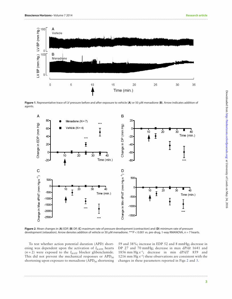
Isolated whole heartsMenadione caused a significant increase in diastolic pressure approximately 10 min after exposure compared to vehicle alone (Fig. 1). The end-diastolic pressure (EDP) rose from 19.7 ± 12.84 mmHg, prior to exposure, to 50.7 ± 9.9 mmHg, (P < 0.001) after 25 min. As a consequence of this, DP fell from 76.5 ± 15.72 to 17.5 ± 3.0 mmHg over the same time period (P < 0.001). The mean changes for EDP and DP are shown in Fig. 2A and B, respectively.
Exposure to menadione for 25 min also significantly reduced the rate of peak pressure development (max dP/dT) (from 4037.0 ± 446.2 to 2254.2 ± 98.0 mmHg s−1, P < 0.001) and the peak rate of relaxation (min dP/dT) (from −3126.6 ± 259.0 to −2240.0 ± 36.5 mmHg s−1, P < 0.001). The mean changes of max and min dP/dT are shown in Fig. 2C and D, respectively.
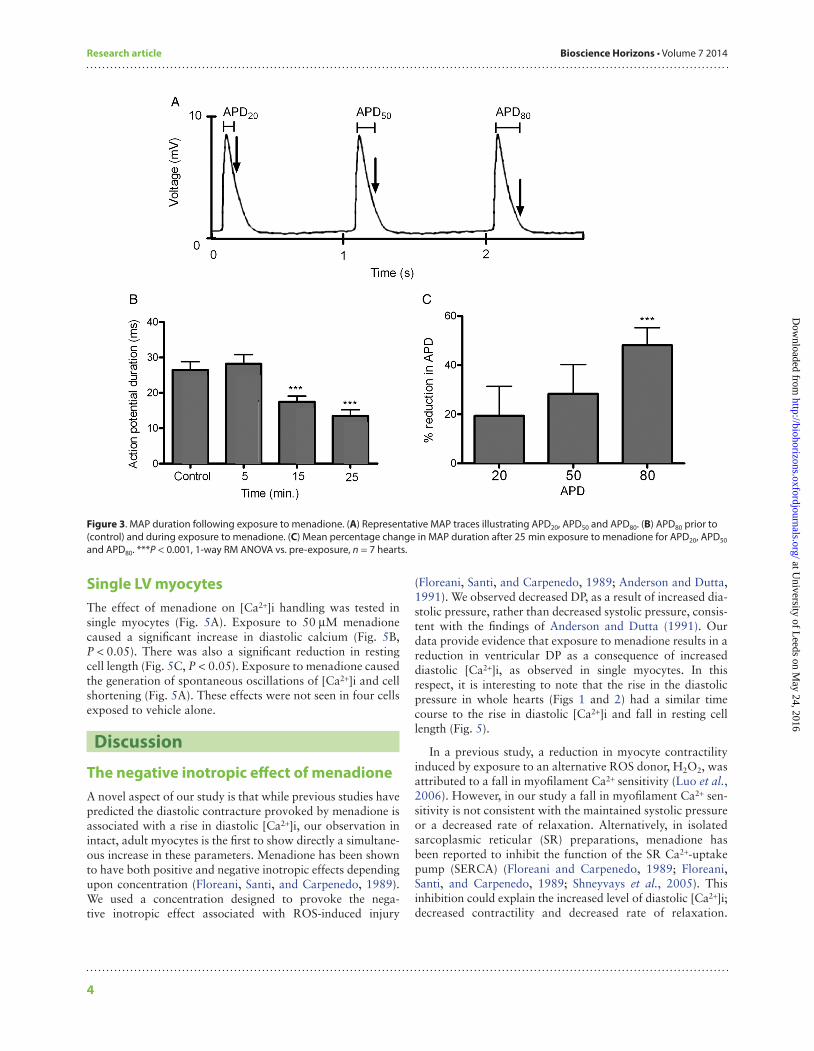
MAP durations at 20, 50 and 80% repolarization (APD20, APD50, APD80, respectively) were simultaneously recorded with LV pressure (Fig. 3A). There was a significant reduction in APD80 after 25 min exposure to menadione (Fig. 3B and C).
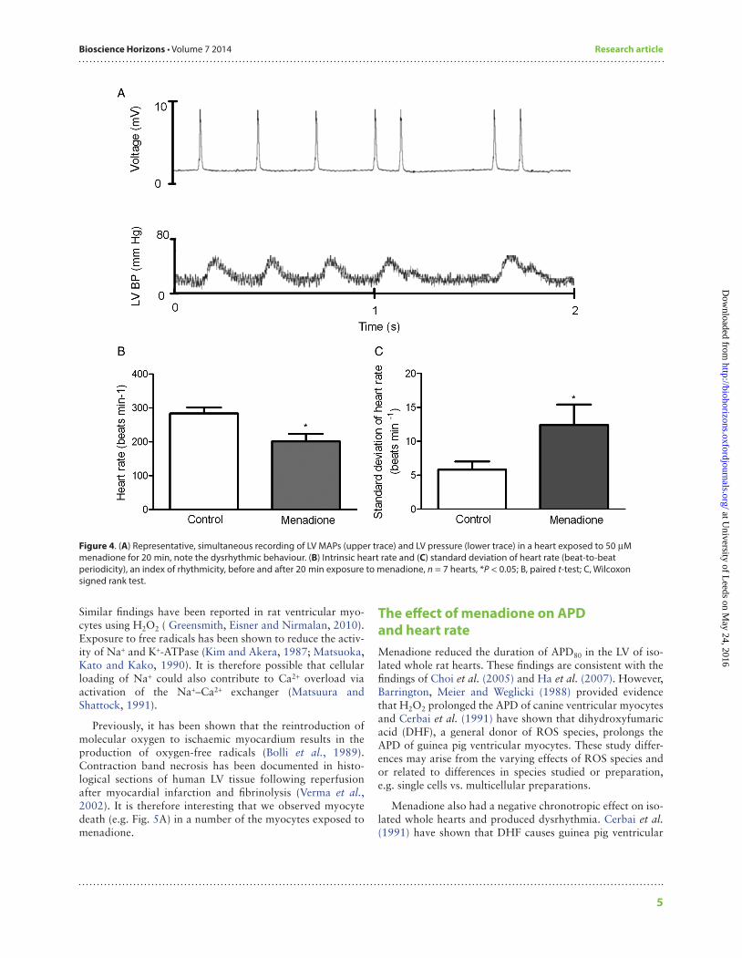
In the absence of external stimulation, menadione affected the rate and rhythmicity of intrinsic pacing (Fig. 4A). Heart rate was significantly reduced after 20 min of exposure (Fig. 4B, P < 0.05, paired t-test). There was a significant decrease in rhythmicity, indicated by an increase in the standard devia-tion of beat-to-beat periodicity (an established index of rhythmicity, Fig. 4C, P < 0.05, Wilcoxon signed rank test).
2
at University of L
eeds on May 24, 2016
http://biohorizons.oxfordjournals.org/D
ownloaded from
Bioscience Horizons • Volume 7 2014 Research article
To test whether action potential duration (APD) short-ening was dependent upon the activation of IKATP, hearts (n = 2) were exposed to the IKATP blocker glibenclamide. This did not prevent the mechanical responses or APD80 shortening upon exposure to menadione (APD80 shortening
59 and 38%; increase in EDP 52 and 8 mmHg; decrease in DP 27 and 70 mmHg; decrease in max dP/dt 1641 and 1856 mm Hg s−1; decrease in min dP/dT 859 and 1216 mm Hg s−1) these observations are consistent with the changes in these parameters reported in Figs 2 and 3.
3
Figure 1. Representative trace of LV pressure before and after exposure to vehicle (A) or 50 µM menadione (B). Arrow indicates addition of agents.
Figure 2. Mean changes in (A) EDP, (B) DP, (C) maximum rate of pressure development (contraction) and (D) minimum rate of pressure development (relaxation). Arrow denotes addition of vehicle or 50 µM menadione. ***P < 0.001 vs. pre-drug, 1-way RMANOVA, n = 7 hearts.
at University of L
eeds on May 24, 2016
http://biohorizons.oxfordjournals.org/D
ownloaded from
Research article Bioscience Horizons • Volume 7 2014
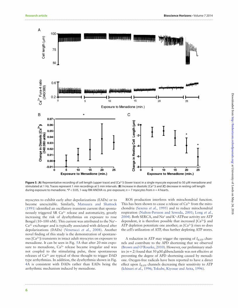
Single LV myocytesThe effect of menadione on [Ca2+]i handling was tested in single myocytes (Fig. 5A). Exposure to 50 µM menadione caused a significant increase in diastolic calcium (Fig. 5B, P < 0.05). There was also a significant reduction in resting cell length (Fig. 5C, P < 0.05). Exposure to menadione caused the generation of spontaneous oscillations of [Ca2+]i and cell shortening (Fig. 5A). These effects were not seen in four cells exposed to vehicle alone.
Discussion
The negative inotropic effect of menadioneA novel aspect of our study is that while previous studies have predicted the diastolic contracture provoked by menadione is associated with a rise in diastolic [Ca2+]i, our observation in intact, adult myocytes is the first to show directly a simultane-ous increase in these parameters. Menadione has been shown to have both positive and negative inotropic effects depending upon concentration (Floreani, Santi, and Carpenedo, 1989). We used a concentration designed to provoke the nega-tive inotropic effect associated with ROS-induced injury
(Floreani, Santi, and Carpenedo, 1989; Anderson and Dutta, 1991). We observed decreased DP, as a result of increased dia-stolic pressure, rather than decreased systolic pressure, consis-tent with the findings of Anderson and Dutta (1991). Our data provide evidence that exposure to menadione results in a reduction in ventricular DP as a consequence of increased diastolic [Ca2+]i, as observed in single myocytes. In this respect, it is interesting to note that the rise in the diastolic pressure in whole hearts (Figs 1 and 2) had a similar time course to the rise in diastolic [Ca2+]i and fall in resting cell length (Fig. 5).
In a previous study, a reduction in myocyte contractility induced by exposure to an alternative ROS donor, H2O2, was attributed to a fall in myofilament Ca2+ sensitivity (Luo et al., 2006). However, in our study a fall in myofilament Ca2+ sen-sitivity is not consistent with the maintained systolic pressure or a decreased rate of relaxation. Alternatively, in isolated sarcoplasmic reticular (SR) preparations, menadione has been reported to inhibit the function of the SR Ca2+-uptake pump (SERCA) (Floreani and Carpenedo, 1989; Floreani, Santi, and Carpenedo, 1989; Shneyvays et al., 2005). This inhibition could explain the increased level of diastolic [Ca2+]i; decreased contractility and decreased rate of relaxation.
4
Figure 3. MAP duration following exposure to menadione. (A) Representative MAP traces illustrating APD20, APD50 and APD80. (B) APD80 prior to (control) and during exposure to menadione. (C) Mean percentage change in MAP duration after 25 min exposure to menadione for APD20, APD50 and APD80. ***P < 0.001, 1-way RM ANOVA vs. pre-exposure, n = 7 hearts. at U
niversity of Leeds on M
ay 24, 2016http://biohorizons.oxfordjournals.org/
Dow
nloaded from
Bioscience Horizons • Volume 7 2014 Research article
Similar findings have been reported in rat ventricular myo-cytes using H2O2 ( Greensmith, Eisner and Nirmalan, 2010). Exposure to free radicals has been shown to reduce the activ-ity of Na+ and K+-ATPase (Kim and Akera, 1987; Matsuoka, Kato and Kako, 1990). It is therefore possible that cellular loading of Na+ could also contribute to Ca2+ overload via activation of the Na+–Ca2+ exchanger (Matsuura and Shattock, 1991).
Previously, it has been shown that the reintroduction of molecular oxygen to ischaemic myocardium results in the production of oxygen-free radicals (Bolli et al., 1989). Contraction band necrosis has been documented in histo-logical sections of human LV tissue following reperfusion after myocardial infarction and fibrinolysis (Verma et al., 2002). It is therefore interesting that we observed myocyte death (e.g. Fig. 5A) in a number of the myocytes exposed to menadione.
The effect of menadione on APD and heart rateMenadione reduced the duration of APD80 in the LV of iso-lated whole rat hearts. These findings are consistent with the findings of Choi et al. (2005) and Ha et al. (2007). However, Barrington, Meier and Weglicki (1988) provided evidence that H2O2 prolonged the APD of canine ventricular myocytes and Cerbai et al. (1991) have shown that dihydroxyfumaric acid (DHF), a general donor of ROS species, prolongs the APD of guinea pig ventricular myocytes. These study differ-ences may arise from the varying effects of ROS species and or related to differences in species studied or preparation, e.g. single cells vs. multicellular preparations.
Menadione also had a negative chronotropic effect on iso-lated whole hearts and produced dysrhythmia. Cerbai et al. (1991) have shown that DHF causes guinea pig ventricular
5
Figure 4. (A) Representative, simultaneous recording of LV MAPs (upper trace) and LV pressure (lower trace) in a heart exposed to 50 µM menadione for 20 min, note the dysrhythmic behaviour. (B) Intrinsic heart rate and (C) standard deviation of heart rate (beat-to-beat periodicity), an index of rhythmicity, before and after 20 min exposure to menadione, n = 7 hearts, *P < 0.05; B, paired t-test; C, Wilcoxon signed rank test.
at University of L
eeds on May 24, 2016
http://biohorizons.oxfordjournals.org/D
ownloaded from
Research article Bioscience Horizons • Volume 7 2014
myocytes to exhibit early after depolarizations (EADs) or to become unexcitable. Similarly, Matsuura and Shattock (1991) identified an oscillatory transient current that sponta-neously triggered SR Ca2+ release and automaticity, greatly increasing the risk of dysrhythmias on exposure to rose Bengal (10–100 nM). This current was attributed to the Na+–Ca2+ exchanger and is typically associated with delayed after depolarizations (DADs) (Venetucci et al., 2008). Another novel finding of this study is the demonstration of spontane-ous [Ca2+]i transients in intact adult myocytes on exposure to menadione. It can be seen in Fig. 5A that after 20 min expo-sure to menadione, Ca2+ release became irregular and was not coupled to the stimulating pulse, these spontaneous releases of Ca2+ are typical of those thought to trigger DAD type arrhythmias. In addition, the dysrhythmia shown in Fig. 4A is consistent with DADs rather than EADs being the arrhythmic mechanism induced by menadione.
ROS production interferes with mitochondrial function. This has been shown to cause a release of Ca2+ from the mito-chondria (Saxena et al., 1995) and to reduce mitochondrial respiration (Nulton-Persson and Szweda, 2001; Long et al., 2004). Both SERCA, and Na+ and K+-ATPase activity are ATP dependent, it is therefore possible that increased [Ca2+]i and ATP depletion potentiate one another; as [Ca2+]i rises so does the cell’s utilization of ATP, thus further depleting ATP stores.
A reduction in ATP may trigger the opening of IKATP chan-nels and contribute to the APD shortening that we observed (Brown and O’Rourke, 2010). However, our preliminary stud-ies (n = 2) found that 50 µM glibenclamide was not effective at preventing the degree of APD shortening caused by menadi-one. Oxygen-free radicals have been reported to have a direct effect upon IKATP channels decreasing their sensitivity to ATP (Ichinari et al., 1996; Tokube, Kiyosue and Arita, 1996).
6
Figure 5. (A) Representative recording of cell length (upper trace) and [Ca2+]i (lower trace) in a single myocyte exposed to 50 µM menadione and stimulated at 1 Hz. Traces represent 1 min recordings at 5 min intervals. (B) Increase in diastolic [Ca2+]i and (C) decrease in resting cell length during exposure to menadione. *P < 0.05, 1-way RM ANOVA vs. pre-exposure; n = 7 myocytes from n = 4 hearts.
at University of L
eeds on May 24, 2016
http://biohorizons.oxfordjournals.org/D
ownloaded from
Bioscience Horizons • Volume 7 2014 Research article
As modification of contractility and electrical activity are interrelated it is useful to measure these parameters simulta-neously. A further novel finding is that APD shortening occurs alongside the fall in DP and is likely to contribute to this effect because a shorter APD will lead to less Ca2+ influx through L-type Ca2+ channels, a smaller SR Ca2+ load and thus a smaller SR Ca2+ release, in turn smaller SR Ca2+ release will generate less inward Na+–Ca2+ exchange current, short-ening the APD (Bouchard, Clark and Giles, 1995).
Study limitationsDue to the instability of physiological buffer solutions, there is currently no method of exposing cardiac tissue to a known concentration of ROS. Consequently, the concentration of menadione administered was a proxy for the concentration of superoxide anions. Furthermore, although menadione is the most specific superoxide anion donor (Choi et al., 2005), potential effects relating to H2O2 and OH− cannot be excluded.
ConclusionThe data presented provide evidence that exposure to mena-dione, at 50 µM, exerts a negative inotropic effect as a conse-quence of an increase in diastolic pressure. Moreover, we have shown that in single LV myocytes this decrease in rest-ing cell length occurs in parallel with an increase in [Ca2+]i, which we suggest is a result of the actions of superoxide on Na+–Ca2+ exchange and SERCA function.
FundingSupported by the University of Leeds.
Author biographyL.J.R. studied pharmacology in relation to medicine as part of an intercalated degree; he subsequently completed his MBChB at the University of Leeds, passing with honours. He began work as a Foundation Year 1 doctor in August 2013 and is currently considering an academic and research career in Cardiology or Cardiac Surgery. He contributed equally to the conduct of experiments and analysis of data, alongside A.L. and K.W. He also wrote the paper and had primary responsibility for its final content. Aside from the conduct of experiments and analysis of data A.L. and K.W. also edited the paper before final submis-sion. A.L. provided the illustrations. Professor Ed White designed this project as a dissertation for L.J.R., A.L. and K.W.
ReferencesAnderson, G. F. and Dutta, S. (1991) Electromechanical effects of mena-
dione on isolated rat heart in relation to oxidative stress, Free Radical Biology & Medicine, 11, 169–177.
Barrington, P. L., Meier, C. F., Jr., and Weglicki, W. B. (1988) Abnormal elec-trical activity induced by free radical generating systems in isolated
cardiocytes, Journal of Molecular and Cellular Cardiology, 20, 1163–1178.
Benoist, D., Stones, R., Drinkhill, M. et al. (2011) Arrhythmogenic sub-strate in hearts of rats with monocrotaline-induced pulmonary hypertension and right ventricular hypertrophy, American Journal of Physiology Heart and Circulatory Physiology, 300, H2230–H2237.
Bolli, R., Jeroudi, M. O., Patel, B. S. et al. (1989) Direct evidence that oxy-gen-derived free radicals contribute to postischemic myocardial dysfunction in the intact dog, Proceedings of the National Academy of Sciences of the United States of America, 86, 4695–4699.
Bouchard, R. A., Clark, R. B., and Giles, W. R. (1995) Effects of action potential duration on excitation-contraction coupling in rat ven-tricular myocytes. Action potential voltage-clamp measurements, Circulation Research, 76, 790–801.
Brown, D. A. and O’Rourke, B. (2010) Cardiac mitochondria and arrhyth-mias, Cardiovascular Research, 88, 241–249.
Cerbai, E., Ambrosio, G., Porciatti, F. et al. (1991) Cellular electrophysiolog-ical basis for oxygen radical-induced arrhythmias. A patch-clamp study in guinea pig ventricular myocytes, Circulation, 84, 1773–1782.
Choi, B. H., Ha, K. C., Park, J. A. et al. (2005) Regional differences of super-oxide dismutase activity enhance the superoxide-induced electrical heterogeneity in rabbit hearts, Basic Research in Cardiology, 100, 355–364.
Floreani, M. and Carpenedo, F. (1989) Inhibition of cardiac sarcoplasmic reticulum Ca2 + -ATPase activity by menadione, Archives of Biochemistry and Biophysics, 270, 33–41.
Floreani, M., Santi, S. E., and Carpenedo, F. (1989) Effects of 2-methyl-1,4-naphthoquinone (menadione) on myocardial contractility and cardiac sarcoplasmic reticulum Ca-ATPase, Naunyn-Schmiedeberg’s Archives of Pharmacology, 339, 448–455.
Greensmith, D. J., Eisner, D. A., and Nirmalan, M. (2010) The effects of hydrogen peroxide on intracellular calcium handling and contractil-ity in the rat ventricular myocyte, Cell Calcium, 48, 341–351.
Ha, K. C., Kwak, Y. G., Piao, C. S. et al. (2007) Differential effects of super-oxide radical on the action potentials in ventricular muscles, Purkinje fibers and atrial muscles in the heart of different aged rats, Archives of Pharmacal Research, 30, 1088–1095.
Ichinari, K., Kakei, M., Matsuoka, T. et al. (1996) Direct activation of the ATP-sensitive potassium channel by oxygen free radicals in guinea-pig ventricular cells: its potentiation by MgADP, Journal of Molecular and Cellular Cardiology, 28, 1867–1877.
Kevin, L. G., Novalija, E. and Stowe, D. F. (2005) Reactive oxygen species as mediators of cardiac injury and protection: the relevance to anes-thesia practise, Anesthesia & Analgesia, 101, 1275–1287.
Kim, M. S. and Akera, T. (1987) O2 free radicals: cause of ischemia- reperfusion injury to cardiac Na + -K + -ATPase, The American Journal of Physiology, 252, H252–H257.
Long, X., Goldenthal, M. J., Wu, G. M. et al. (2004) Mitochondrial Ca2+ flux and respiratory enzyme activity decline are early events in cardio-
7
at University of L
eeds on May 24, 2016
http://biohorizons.oxfordjournals.org/D
ownloaded from
Research article Bioscience Horizons • Volume 7 2014
myocyte response to H2O2, Journal of Molecular and Cellular Cardiology, 37, 63–70.
Luo, J., Xuan, Y. T., Gu, Y. et al. (2006) Prolonged oxidative stress inverts the cardiac force-frequency relation: role of altered calcium han-dling and myofilament calcium responsiveness, Journal of Molecular and Cellular Cardiology, 40, 64–75.
Matsuoka, T., Kato, M., and Kako, K. J. (1990) Effect of oxidants on Na,K,ATPase and its reversal, Basic Research in Cardiology, 85, 330–341.
Matsuura, H. and Shattock, M. J. (1991) Membrane potential fluctua-tions and transient inward currents induced by reactive oxygen intermediates in isolated rabbit ventricular cells, Circulation Research, 68, 319–329.
McCrossan, Z. A., Billeter, R., and White, E. (2004) Transmural changes in size, contractile and electrical properties of SHR left ventricular myo-cytes during compensated hypertrophy, Cardiovascular Research, 63, 283–292.
Nulton-Persson, A. C. and Szweda, L. I. (2001) Modulation of mitochon-drial function by hydrogen peroxide, The Journal of Biological Chemistry, 276, 23357–23361.
Pallandi, R. T., Perry, M. A., and Campbell, T. J. (1987) Proarrhythmic effects of an oxygen-derived free radical generating system on action potentials recorded from guinea pig ventricular myocardium: a possible cause of reperfusion-induced arrhythmias, Circulation Research, 61, 50–54.
Pollack, C. V., Jr., Antman, E. M., and Hollander, J. E. (2008) 2007 focused update to the ACC/AHA guidelines for the management of patients
with ST-segment elevation myocardial infarction: implications for emergency department practice, Annals of Emergency Medicine, 52, 344–355.
Roger, V. L., Go, A. S., Lloyd-Jones, D. M. et al. (2012) Heart disease and stroke statistics–2012 update: a report from the American Heart Association, Circulation, 125, e2–e220.
Saxena, K., Henry, T. R., Solem, L. E. et al. (1995) Enhanced induction of the mitochondrial permeability transition following acute menadi-one administration, Archives of Biochemistry and Biophysics, 317, 79–84.
Shneyvays, V., Lesham, D., Shmist, Y. et al. (2005) Effects of menadione and its derivative on cultured cardiomyocyes with mitochondrial disorders, Journal of Molecular and Cellular Cardiology, 39, 149–158.
Stones, R., Billeter, R., Zhang, H. et al. (2009) The role of transient out-ward K+ current in electrical remodelling induced by voluntary exercise in female rat hearts, Basic Research in Cardiology, 104, 643–652.
Tokube, K., Kiyosue, T., and Arita, M. (1996) Openings of cardiac KATP channel by oxygen free radicals produced by xanthine oxidase reac-tion, The American Journal of Physiology, 271, H478–H489.
Venetucci, L. A., Trafford, A. W., O’Neill, S. C. et al. (2008) The sarcoplasmic reticulum and arrhythmogenic calcium release, Cardiovascular Research, 77, 285–292.
Verma, S., Fedak, P. W., Weisel, R. D. et al. (2002) Fundamentals of reperfusion injury for the clinical cardiologist, Circulation, 105, 2332–2336.
8
at University of L
eeds on May 24, 2016
http://biohorizons.oxfordjournals.org/D
ownloaded from