Look at My Chest!
23
LOOK AT MY CHEST!
-
Upload
hid-dymple-ahmad -
Category
Documents
-
view
218 -
download
0
Transcript of Look at My Chest!

8/8/2019 Look at My Chest!
http://slidepdf.com/reader/full/look-at-my-chest 1/23
LOOK AT MY CHEST!
8/8/2019 Look at My Chest!
http://slidepdf.com/reader/full/look-at-my-chest 2/23
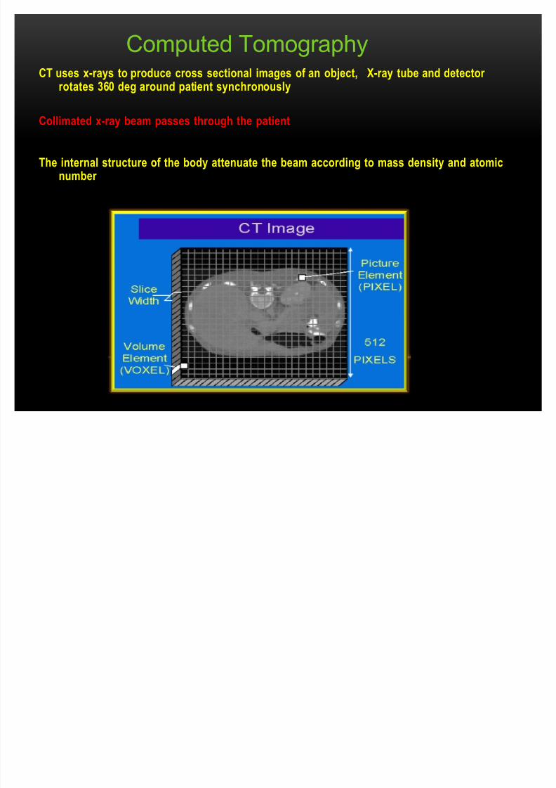
CT uses x-rays to produce cross sectional images of an object, X-ray tube and detector rotates 360 deg around patient synchronously
Collimated x-ray beam passes through the patient
The internal structure of the body attenuate the beam according to mass density and atomicnumber
Computed Tomography

8/8/2019 Look at My Chest!
http://slidepdf.com/reader/full/look-at-my-chest 3/23
X
Y
Z
ISOCENTER

8/8/2019 Look at My Chest!
http://slidepdf.com/reader/full/look-at-my-chest 4/23
HOW CT WORK
With CT scanning, numerous x-ray beams
and a set of electronic x-ray detectors
rotate around you, measuring the amount
of radiation being absorbed throughout
your body.At the same time, the examination table is
moving through the scanner, so that the x-
ray beam follows a spiral path.
A special computer program processes this
large volume of data to create two-
dimensional cross-sectional images of yourbody, which are then displayed on a
monitor.
This technique is called helical or spiral CT.

8/8/2019 Look at My Chest!
http://slidepdf.com/reader/full/look-at-my-chest 5/23
COMPONENTS
8/8/2019 Look at My Chest!
http://slidepdf.com/reader/full/look-at-my-chest 6/23
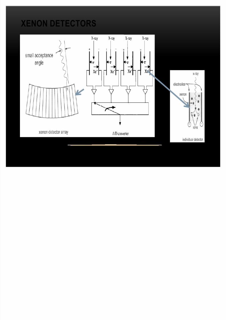
XENON DETECTORS

8/8/2019 Look at My Chest!
http://slidepdf.com/reader/full/look-at-my-chest 7/23
Resulting array is aimage of bodyattenuation
FBP or other reconstruction methods -
Attenuation measurements are used to
produce cross sectional array of tissue
co-efficients
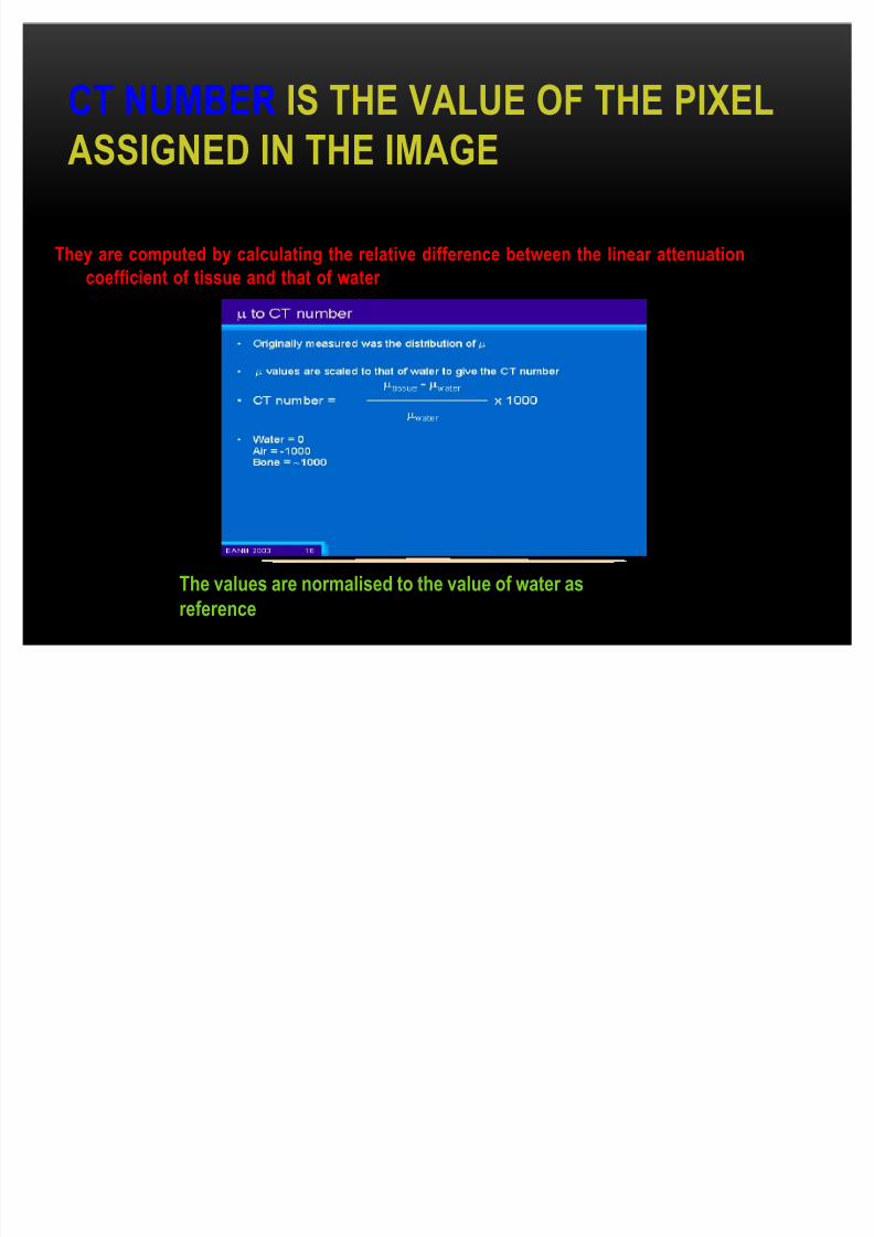
Data are converted to CT number or Hounsfield Unit for display
8/8/2019 Look at My Chest!
http://slidepdf.com/reader/full/look-at-my-chest 8/23
CT NUMBER IS THE VALUE OF THE PIXEL
ASSIGNED IN THE IMAGE
They are computed by calculating the relative difference between the linear attenuation
coefficient of tissue and that of water
The values are normalised to the value of water as
reference
8/8/2019 Look at My Chest!
http://slidepdf.com/reader/full/look-at-my-chest 9/23
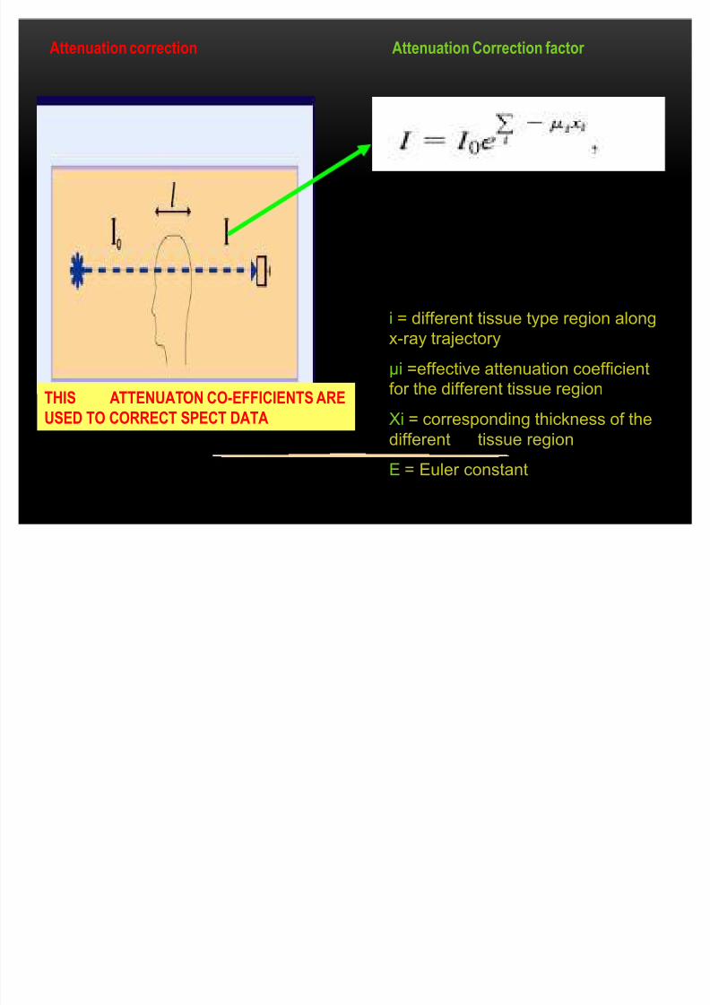
i = different tissue type region along
x-ray trajectory
µi =effective attenuation coefficientfor the different tissue region
Xi = corresponding thickness of the
different tissue region
E = Euler constant
Attenuation Correction factor
´I0 ´ is initial ray &
´Iµ is the measured ray
corrected by attenuationfactor
Attenuation correction
I
THIS ATTENUATON CO-EFFICIENTS ARE
USED TO CORRECT SPECT DATA

8/8/2019 Look at My Chest!
http://slidepdf.com/reader/full/look-at-my-chest 10/23
The CT number for water
has a value of 0
because:( tissue - water )/ water =
0
For tissue and that of
water the equation is:
CTTissue
= (tissue
-
water )/ water x 1000
Based on CT numbers images are displayed in a 512*512 matrix
HOUNSFIELD SCALE

8/8/2019 Look at My Chest!
http://slidepdf.com/reader/full/look-at-my-chest 11/23
WINDOW
The process of changing CT image gray scale is known as windowing
Bone window long
gray scale, low
contrast
Soft tissue window
short gray scale,
high contrast.

8/8/2019 Look at My Chest!
http://slidepdf.com/reader/full/look-at-my-chest 12/23
PIXEL VS VOXEL
PIXEL
VOXEL
PIXEL Based onMATRIX SIZE
FOV
VOXEL based onFOV
MATRIX SIZE
SLICE THICKNESS

8/8/2019 Look at My Chest!
http://slidepdf.com/reader/full/look-at-my-chest 13/23
1st Generation
Pencil beam Rotate Translate
Single detector
2ND GenerationFan beam
Rotate Translate Detector
Array
8/8/2019 Look at My Chest!
http://slidepdf.com/reader/full/look-at-my-chest 14/23
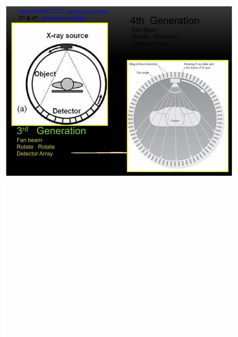
3rd GenerationFan beam
Rotate Rotate
Detector Array
4th GenerationFan Beam
Rotate Stationary
Detector Array
Hybrid SPECT/CT camera uses his
3rd & 4th generation design
8/8/2019 Look at My Chest!
http://slidepdf.com/reader/full/look-at-my-chest 15/23
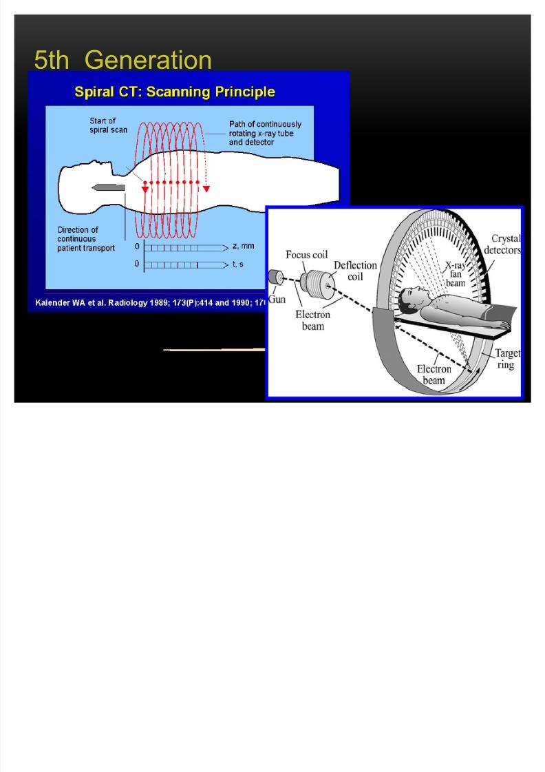
5th Generation
8/8/2019 Look at My Chest!
http://slidepdf.com/reader/full/look-at-my-chest 16/23
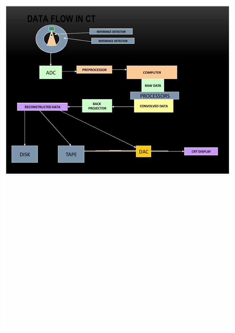
DATA FLOW IN CTREFERENCE DETECTOR
REFERENCE DETECTOR
ADCPREPROCESSOR
COMPUTER
RAW DATA
CONVOLVED DATABACK
PROJECTORRECONSTRUCTED DATA
PROCESSORS
DISK TAPEDAC CRT DISPLAY
8/8/2019 Look at My Chest!
http://slidepdf.com/reader/full/look-at-my-chest 17/23
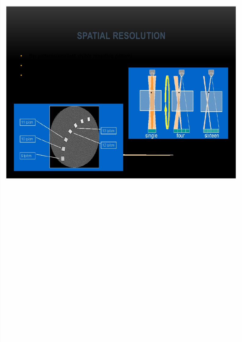
SPATIAL RESOLUTION
Bar patterns(smallest visible repeating pattern)
Edge, wire or bead
Phantom alignment
8/8/2019 Look at My Chest!
http://slidepdf.com/reader/full/look-at-my-chest 18/23
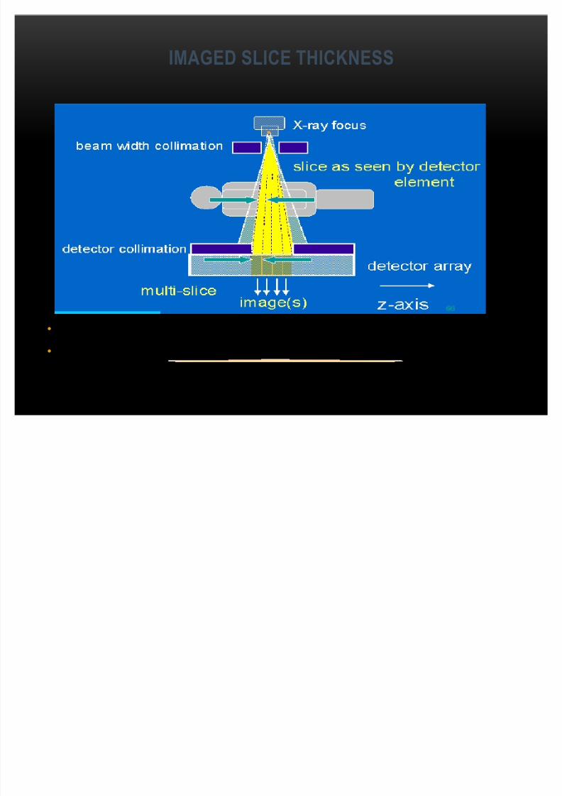
IMAGED SLICE THICKNESS
Multi-slice look at different detector combinations
Important imaging parameter
8/8/2019 Look at My Chest!
http://slidepdf.com/reader/full/look-at-my-chest 19/23
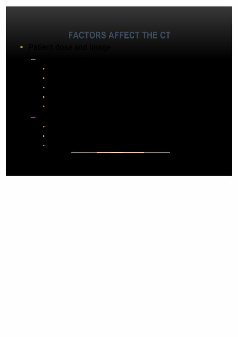
FACTORS AFFECT THE CT
Patient dose and image
± X-ray beam parameters
KVp
mA
Filtration
Collimation
Detection efficiency
± Image parameters
Noise
Spatial resolution
Slice thickness

8/8/2019 Look at My Chest!
http://slidepdf.com/reader/full/look-at-my-chest 20/23

8/8/2019 Look at My Chest!
http://slidepdf.com/reader/full/look-at-my-chest 21/23
BENEFIT AND RISKBenefits:
-CT scanning is painless, non-invasive and accurate-ability to image bone, soft tissue and blood vessels all at the same time.
-provides very detailed images of many types of tissue
-CT is less sensitive to patient movement than MRI
-No radiation remains in a patient's body after a CT examination
Risks:
-There is always a slight chance of cancer from excessive exposure toradiation
-The effective radiation dose from this procedure ranges from approximatelytwo to 10 mSv, which is about the same as the average person receives frombackground radiation in to 8 months to three years.
-Women should always inform their physician and x-ray or CT technologist if there is any possibility that they are pregnant
-CT scanning is, in general, not recommended for pregnant women unlessmedically necessary because of potential risk to the baby.
8/8/2019 Look at My Chest!
http://slidepdf.com/reader/full/look-at-my-chest 22/23
HOW WILL THE TUBE CURRENT OF THE
SOURCE IN CT SCANNER AFFECTS THE IMAGE?
Minimal tube current required for good image quality with the least radiation dose
8/8/2019 Look at My Chest!
http://slidepdf.com/reader/full/look-at-my-chest 23/23
CONCLUSION
CT can visualize small density differences, e.g. grey matter, white matter, and CSF.CT can detect and diagnose disease that cannot be seen with X-ray.
More expensive than X-ray, lower resolution.
Ionizing radiation.
HOW WILL THE TUBE CURRENT OF THE
SOURCE IN CT SCANNER AFFECTS THE IMAGE? Minimal tube current required for good image quality with the least radiation dose