LNCS 7510 - Simulation of Pneumoperitoneum for ... · Simulation of Pneumoperitoneum for...
8
Simulation of Pneumoperitoneum for Laparoscopic Surgery Planning J. Bano 1,2 , A. Hostettler 1 , S.A. Nicolau 1 , S. Cotin 3 , C. Doignon 2 , H.S. Wu 4 , M.H. Huang 4 , L. Soler 1 , and J. Marescaux 1 1 IRCAD, Virtual-Surg, Place de l’Hopital 1, 67091 Strasbourg Cedex, France 2 LSIIT (UMR 7005 CNRS), University of Strasbourg, Parc d’Innovation, Boulevard S. Brant, BP 10412 67412 Illkirch Cedex, France 3 SHACRA Group, Inria, France 4 IRCAD Taiwan, Medical Imaging Team, 1-6 Lugong Road, Lukang 505, Taiwan [email protected] Abstract. Laparoscopic surgery planning is usually realized on a preop- erative image that does not correspond to the operating room conditions. Indeed, the patient undergoes gas insufflation (pneumoperitoneum) to al- low instrument manipulation inside the abdomen. This insufflation moves the skin and the viscera so that their positions do no longer correspond to the preoperative image, reducing the benefit of surgical planning, more particularly for the trocar positioning step. A simulation of the pneu- moperitoneum influence would thus improve the realism and the quality of the surgical planning. We present in this paper a method to simu- late the movement of skin and viscera due to the pneumoperitoneum. Our method requires a segmented preoperative 3D medical image as- sociated to realistic biomechanical parameters only. The simulation is performed using the SOFA simulation engine. The results were evalu- ated using computed tomography [CT] images of two pigs, before and after pneumoperitoneum. Results show that our method provides a very realistic estimation of skin, viscera and artery positions with an average error within 1 cm. Keywords: simulation, surgical planning, pneumoperitoneum. 1 Introduction 1.1 Clinical Context Laparoscopic surgery has become a common procedure in the sugical commu- nity. It involves inserting an endoscopic camera and several instruments through small incisions made on the abdomen. In order to create a working space, gas is injected into abdominal cavity (pneumoperitoneum). Surgical planning is usually performed on a preoperative image, or in the best case, using a three-dimensional model. Part of the planning requires determining the trocar positioning so that the surgeon has a good triangulation between his instruments as well as good camera viewpoint. However, the preoperative model does not take deformation N. Ayache et al. (Eds.): MICCAI 2012, Part I, LNCS 7510, pp. 91–98, 2012. c Springer-Verlag Berlin Heidelberg 2012
Transcript of LNCS 7510 - Simulation of Pneumoperitoneum for ... · Simulation of Pneumoperitoneum for...
Simulation of Pneumoperitoneum
for Laparoscopic Surgery Planning
J. Bano1,2, A. Hostettler1, S.A. Nicolau1, S. Cotin3, C. Doignon2, H.S. Wu4,M.H. Huang4, L. Soler1, and J. Marescaux1
1 IRCAD, Virtual-Surg, Place de l’Hopital 1, 67091 Strasbourg Cedex, France2 LSIIT (UMR 7005 CNRS), University of Strasbourg, Parc d’Innovation,
Boulevard S. Brant, BP 10412 67412 Illkirch Cedex, France3 SHACRA Group, Inria, France
4 IRCAD Taiwan, Medical Imaging Team, 1-6 Lugong Road, Lukang 505, [email protected]
Abstract. Laparoscopic surgery planning is usually realized on a preop-erative image that does not correspond to the operating room conditions.Indeed, the patient undergoes gas insufflation (pneumoperitoneum) to al-low instrument manipulation inside the abdomen. This insufflation movesthe skin and the viscera so that their positions do no longer correspond tothe preoperative image, reducing the benefit of surgical planning, moreparticularly for the trocar positioning step. A simulation of the pneu-moperitoneum influence would thus improve the realism and the qualityof the surgical planning. We present in this paper a method to simu-late the movement of skin and viscera due to the pneumoperitoneum.Our method requires a segmented preoperative 3D medical image as-sociated to realistic biomechanical parameters only. The simulation isperformed using the SOFA simulation engine. The results were evalu-ated using computed tomography [CT] images of two pigs, before andafter pneumoperitoneum. Results show that our method provides a veryrealistic estimation of skin, viscera and artery positions with an averageerror within 1 cm.
Keywords: simulation, surgical planning, pneumoperitoneum.
1 Introduction
1.1 Clinical Context
Laparoscopic surgery has become a common procedure in the sugical commu-nity. It involves inserting an endoscopic camera and several instruments throughsmall incisions made on the abdomen. In order to create a working space, gas isinjected into abdominal cavity (pneumoperitoneum). Surgical planning is usuallyperformed on a preoperative image, or in the best case, using a three-dimensionalmodel. Part of the planning requires determining the trocar positioning so thatthe surgeon has a good triangulation between his instruments as well as goodcamera viewpoint. However, the preoperative model does not take deformation
N. Ayache et al. (Eds.): MICCAI 2012, Part I, LNCS 7510, pp. 91–98, 2012.c© Springer-Verlag Berlin Heidelberg 2012

92 J. Bano et al.
due to gas injection into consideration. Since the pneumoperitoneum shifts theskin up and moves the abdominal viscera by several centimeters [9], the plannedtrocar positioning becomes inconsistent with the true position in the operatingroom, which drastically reduces the usefulness of the preoperative planning.
In this paper, we propose a individualized simulation of pneumoperitoneumand viscera motion using a preoperative medical image (CT) that can be usedfor laparoscopic surgery planning. The realism of the simulation method is eval-uated with ground truth data and we show our method can even predict realpneumoperitoneum with an accuracy close to 0.5 cm which is accurate enoughfor trocar positioning planning. We highlight that in case the CT is injectedwith contrast medium, the abdominal wall arteries (cf. Fig. 1 for abdominalwall artery definition) are visible in the CT image and their position after pneu-moperitoneum can also be simulated. This information can be crucial to avoidinjury of abdominal wall arteries during trocar insertion, which is a commoncomplication in laparoscopic surgery [2,10,7,4].
1.2 Previous Work
To our knowledge, only Kitasaka et al. [3,5] have explored pneumoperitoneumsimulation for surgical planning. They simulated the deformation due to gasinjection by applying forces that seem to be antero-posterior, on the inner surfaceof a portion of the abdominal wall. Recently, they evaluated the accuracy of theirresults on eight patients by comparing thirteen skin landmark positions afterpneumoperitoneum and with simulation results [6]. Their simulation methoduses two parameters which are empirically chosen to fit the in vivo data. Withan optimally chosen set of parameters, the minimal mean error obtained was13.8mm. Although their simulation provides interesting results, its usefulness isstill limited since they do not take the viscera motion into account. Moreoverthe parameters of their model have no biomechanical meaning and cannot beadapted for a particular patient shape.
Soler et al. [11] have presented a patient-specific simulator for laparoscopicsurgery. However in this paper, skin and viscera motions due to gas injection arealso not taken into account. Viscera shape corresponds to viscera segmentationin the preoperative image (without pneumoperitoneum), and trocar positions arechosen without being related to skin position after pneumoperitoneum. There-fore, integrating realistic skin simulation after pneumoperitoneum would improvethe realism of such simulator.
This paper proposes a method to simulate a pneumoperitoneum from a preop-erative image, using realistic biomechanical parameters. In Section 2, we presentthe anatomical structures that we need to model from the preoperative imageand how we choose the biomechanical parameters. In Section 3, we explain howthe influence of gas pressure on the abdominal cavity is modeled and how we ac-count for multiple contacts between moving structures in the abdomen. Finally,we evaluate in Section 4 our simulation realism using porcine data and we showour simulation accuracy is within 4mm on average for the abdominal wall andviscera and 5mm for abdominal wall arteries.

Simulation of Pneumoperitoneum for Laparoscopic Surgery Planning 93
2 Model Generation
The input model of our simulation is created from an abdo-thoracic CT in threesteps: segmentation, mesh generation and mechanical parameterization.
Segmentation Step. The preoperative image is segmented by dividing it intothree regions: the abdo-thoracic wall, the abdominal viscera and the thoracicviscera (Fig. 1). In case they are visible, the arteries in the abdominal wallare also segmented. Note that they are not mandatory for the model. Thesesegmentations are done semi-manually using custom-developed software and takeone hour on average. Improvements are planned in future works to reduce therequired time.
(a) Axial slice (b) Sagittal slice
Fig. 1. On these two slices extracted from a preoperative pig CT acquisition, one cansee the anatomical structures of our simulation model: the abdo-thoracic wall (blue),its internal surface (green line), the abdominal viscera (red), the thoracic viscera (cyan)and the diaphragm (black line). On the left figure, the abdominal wall arteries, locatedin the abdo-thoracic wall, are colored in yellow.
Mesh Generation. Volume and surface meshes are generated using the CGALlibrary1 for the abdo-thoracic wall, the abdominal viscera and the thoracic vis-cera. Volumetric meshes are required for the finite element approach used inthe computation of soft tissue deformation, while surface meshes are used forcollision detection. The mesh generation needs a few seconds only from the seg-mented images.
Mechanical Parameters. Our simulation is based on a finite-element ap-proach using a geometrically non-linear elastic law and a co-rotational formula-tion. The simulation is parametrized by associating Young’s modulus [YM] andPoisson ratio to each volume mesh: the abdominal wall volume mesh is associ-ated with a YM equal to 24 kPa (cf. Song et al. paper [12]) and a Poisson ratioof 0.49 (we assume that the abdominal wall is almost incompressible). Althoughabdominal viscera contain various organs with different stiffnesses, we chose, forsimplicity reasons, to consider it as a homogeneous set associated to standard1 Computational Geometry Algorithms Library, http://www.cgal.org
94 J. Bano et al.
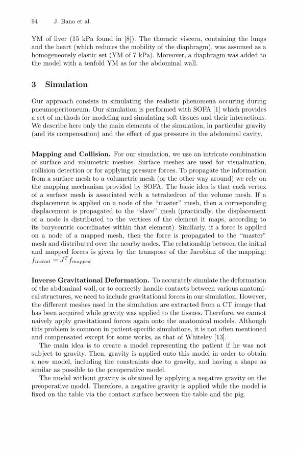
YM of liver (15 kPa found in [8]). The thoracic viscera, containing the lungsand the heart (which reduces the mobility of the diaphragm), was assumed as ahomogeneously elastic set (YM of 7 kPa). Moreover, a diaphragm was added tothe model with a tenfold YM as for the abdominal wall.
3 Simulation
Our approach consists in simulating the realistic phenomena occuring duringpneumoperitoneum. Our simulation is performed with SOFA [1] which providesa set of methods for modeling and simulating soft tissues and their interactions.We describe here only the main elements of the simulation, in particular gravity(and its compensation) and the effect of gas pressure in the abdominal cavity.
Mapping and Collision. For our simulation, we use an intricate combinationof surface and volumetric meshes. Surface meshes are used for visualization,collision detection or for applying pressure forces. To propagate the informationfrom a surface mesh to a volumetric mesh (or the other way around) we rely onthe mapping mechanism provided by SOFA. The basic idea is that each vertexof a surface mesh is associated with a tetrahedron of the volume mesh. If adisplacement is applied on a node of the “master” mesh, then a correspondingdisplacement is propagated to the “slave” mesh (practically, the displacementof a node is distributed to the vertices of the element it maps, according toits barycentric coordinates within that element). Similarly, if a force is appliedon a node of a mapped mesh, then the force is propagated to the “master”mesh and distributed over the nearby nodes. The relationship between the initialand mapped forces is given by the transpose of the Jacobian of the mapping:finitial = JT fmapped
Inverse Gravitational Deformation. To accurately simulate the deformationof the abdominal wall, or to correctly handle contacts between various anatomi-cal structures, we need to include gravitational forces in our simulation. However,the different meshes used in the simulation are extracted from a CT image thathas been acquired while gravity was applied to the tissues. Therefore, we cannotnaively apply gravitational forces again onto the anatomical models. Althoughthis problem is common in patient-specific simulations, it is not often mentionedand compensated except for some works, as that of Whiteley [13].
The main idea is to create a model representing the patient if he was notsubject to gravity. Then, gravity is applied onto this model in order to obtaina new model, including the constraints due to gravity, and having a shape assimilar as possible to the preoperative model.
The model without gravity is obtained by applying a negative gravity on thepreoperative model. Therefore, a negative gravity is applied while the model isfixed on the table via the contact surface between the table and the pig.
Simulation of Pneumoperitoneum for Laparoscopic Surgery Planning 95
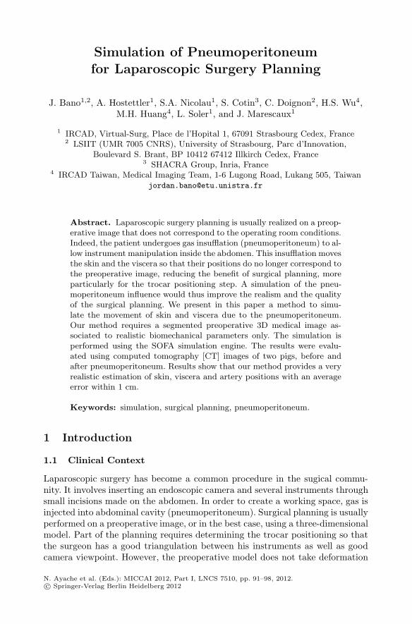
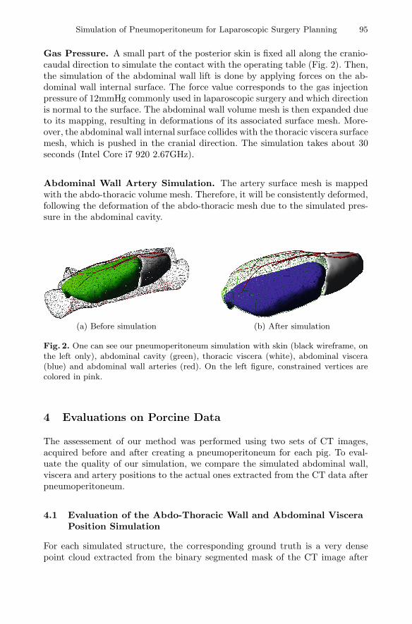
Gas Pressure. A small part of the posterior skin is fixed all along the cranio-caudal direction to simulate the contact with the operating table (Fig. 2). Then,the simulation of the abdominal wall lift is done by applying forces on the ab-dominal wall internal surface. The force value corresponds to the gas injectionpressure of 12mmHg commonly used in laparoscopic surgery and which directionis normal to the surface. The abdominal wall volume mesh is then expanded dueto its mapping, resulting in deformations of its associated surface mesh. More-over, the abdominal wall internal surface collides with the thoracic viscera surfacemesh, which is pushed in the cranial direction. The simulation takes about 30seconds (Intel Core i7 920 2.67GHz).
Abdominal Wall Artery Simulation. The artery surface mesh is mappedwith the abdo-thoracic volume mesh. Therefore, it will be consistently deformed,following the deformation of the abdo-thoracic mesh due to the simulated pres-sure in the abdominal cavity.
(a) Before simulation (b) After simulation
Fig. 2. One can see our pneumoperitoneum simulation with skin (black wireframe, onthe left only), abdominal cavity (green), thoracic viscera (white), abdominal viscera(blue) and abdominal wall arteries (red). On the left figure, constrained vertices arecolored in pink.
4 Evaluations on Porcine Data
The assessement of our method was performed using two sets of CT images,acquired before and after creating a pneumoperitoneum for each pig. To eval-uate the quality of our simulation, we compare the simulated abdominal wall,viscera and artery positions to the actual ones extracted from the CT data afterpneumoperitoneum.
4.1 Evaluation of the Abdo-Thoracic Wall and Abdominal VisceraPosition Simulation
For each simulated structure, the corresponding ground truth is a very densepoint cloud extracted from the binary segmented mask of the CT image after
96 J. Bano et al.
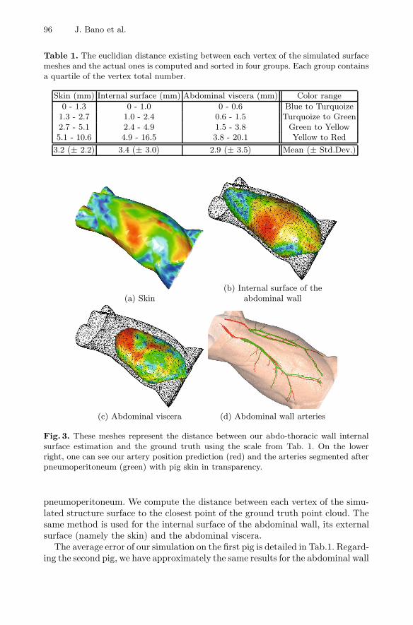
Table 1. The euclidian distance existing between each vertex of the simulated surfacemeshes and the actual ones is computed and sorted in four groups. Each group containsa quartile of the vertex total number.
Skin (mm) Internal surface (mm) Abdominal viscera (mm) Color range
0 - 1.3 0 - 1.0 0 - 0.6 Blue to Turquoize1.3 - 2.7 1.0 - 2.4 0.6 - 1.5 Turquoize to Green2.7 - 5.1 2.4 - 4.9 1.5 - 3.8 Green to Yellow5.1 - 10.6 4.9 - 16.5 3.8 - 20.1 Yellow to Red
3.2 (± 2.2) 3.4 (± 3.0) 2.9 (± 3.5) Mean (± Std.Dev.)
(a) Skin(b) Internal surface of the
abdominal wall
(c) Abdominal viscera (d) Abdominal wall arteries
Fig. 3. These meshes represent the distance between our abdo-thoracic wall internalsurface estimation and the ground truth using the scale from Tab. 1. On the lowerright, one can see our artery position prediction (red) and the arteries segmented afterpneumoperitoneum (green) with pig skin in transparency.
pneumoperitoneum. We compute the distance between each vertex of the simu-lated structure surface to the closest point of the ground truth point cloud. Thesame method is used for the internal surface of the abdominal wall, its externalsurface (namely the skin) and the abdominal viscera.
The average error of our simulation on the first pig is detailed in Tab.1. Regard-ing the second pig, we have approximately the same results for the abdominal wall

Simulation of Pneumoperitoneum for Laparoscopic Surgery Planning 97
(3.8 mm ±3.3 mm for the internal surface and 3.4 mm ±3.5 mm for the skin),but for the abdominal viscera, we obtain a worse result (5.7 mm ±4.9 mm). Alack of realism due to the gas inside viscera distorts the results. Surface error ofthe viscera mainly occurs close to liver since the viscera are constrained by thepelvis. Moreover, reaching a perfect accuracy does not seem crucial for trocarinsertion and the knowledge of abdominal viscera surface is enough. To visuallyassess the error, we display in Fig. 3 the simulated surfaces with a color codedescribed in Tab.1.
4.2 Evaluation of our Artery Position Simulation
Contrary to previous evaluations, we decide to evaluate artery simulation bycomputing the distance between artery bifurcations. Indeed, providing averagedistance between artery surfaces will not highlight translation error along themain direction of arteries. Six bifurcation points have been selected manually insimulated and ground truth arteries, and an average error of 5.1 mm on pig 1and 5.6 mm on pig 2 have been reported.
5 Conclusion
In this paper, we have presented a method to predict abdominal wall and vis-cera positions after pneumoperitoneum using only a preoperative medical imageand realistic biomechanical parameters. Three structures are generated from thepreoperative CT data and are associated to different biomechanical parameters.Then, the pneumoperitoneum is simulated by applying realistic pressure on theabdominal wall (after properly taking gravity into account in our modeling ap-proach). An evaluation has been performed using medical CT acquisitions of twopigs before and after pneumoperitoneum, allowing us to quantitatively measurethe realism of our method. Results show that our method provides very realisticsimulation of skin, abdominal wall and artery, with a reported accuracy below0.5 cm. Although the viscera simulation is less accurate (around 0.6 cm on aver-age), it can still be useful to help surgical planning of laparoscopic procedures.We point out that our model does not depend on empirical values, and thusshould provide reproducible accuracy and realism. In a future work, we plan toimprove viscera simulation by integrating different Young’s moduli to the viscerastructure. We also plan to register our simulated model in the operating roomusing an augmented reality technique to help surgeons during trocar positioning.
References
1. Allard, J., Cotin, S., Faure, F., Bensoussan, P.J., Poyer, F., Duriez, C., Delingette, H.,Grisoni, L., et al.: Sofa-an open source framework for medical simulation. MedicineMeets Virtual Reality 15, 13–18 (2007)
2. Geraci, G., Sciume, C., Pisello, F., Li Volsi, F., Facella, T., Modica, G.: Trocar-related abdominal wall bleeding in 200 patients after laparoscopic cholecistectomy:Personal experience. World Journal of Gastroenterology 12(44), 7165 (2006)

98 J. Bano et al.
3. Kitasaka, T., Mori, K., Hayashi, Y., Suenaga,Y., Hashizume, M., Toriwaki, J.-I.: Vir-tual Pneumoperitoneum for Generating Virtual Laparoscopic Views Based on Volu-metric Deformation. In: Barillot, C., Haynor, D.R., Hellier, P. (eds.) MICCAI 2004.LNCS, vol. 3217, pp. 559–567. Springer, Heidelberg (2004)
4. Lam, A., Kaufman, Y., Khong, S.Y., Liew, A., Ford, S., Condous, G.: Dealingwith complications in laparoscopy. Best Practice & Research Clinical Obstetrics &Gynaecology 23(5), 631–646 (2009)
5. Mori, K., Kito, M., Kitasaka, T., Misawa, K., Fujiwara, M.: Patient-specific la-paroscopic surgery planning system based on virtual pneumoperitoneum technique.International Journal of Computer Assisted Radiology and Surgery 4, S140–S142(2009)
6. Oda, M., Di Qu, J., Nimura, Y., Kitasaka, T., Misawa, K., Mori, K.: Evaluationof deformation accuracy of a virtual pneumoperitoneum method based on clinicaltrials for patient-specific laparoscopic surgery simulator. In: Proceedings of SPIE,vol. 8316, p. 83160G (2012)
7. Saber, A.A., Meslemani, A.M., Davis, R., Pimentel, R.: Safety zones for anteriorabdominal wall entry during laparoscopy: a ct scan mapping of epigastric vessels.Annals of Surgery 239(2), 182 (2004)
8. Samur, E., Sedef, M., Basdogan, C., Avtan, L., Duzgun, O.: A robotic indenter forminimally invasive characterization of soft tissues. International Congress Series,vol. 1281, pp. 713–718. Elsevier (2005)
9. Sanchez-Margallo, F.M., Moyano-Cuevas, J.L., Latorre, R., Maestre, J., Correa, L.,Pagador, J.B., Sanchez-Peralta, L.F., Sanchez-Margallo, J.A., Usan-Gargallo, J.:Anatomical changes due to pneumoperitoneum analyzed by mri: an experimentalstudy in pigs. Surg. Radiol. Anat. 33(5), 389–396 (2011)
10. Shamiyeh, A., Wayand, W.: Laparoscopic cholecystectomy: early and late com-plications and their treatment. Langenbeck’s Archives of Surgery 389(3), 164–171(2004)
11. Soler, L., Marescaux, J.: Patient-specific surgical simulation. World Journal ofSurgery 32(2), 208–212 (2008)
12. Song, C., Alijani, A., Frank, T., Hanna, G.B., Cuschieri, A.: Mechanical propertiesof the human abdominal wall measured in vivo during insufflation for laparoscopicsurgery. Surgical Endoscopy 20(6), 987–990 (2006)
13. Whiteley, J.P.: The solution of inverse non-linear elasticity problems that arisewhen locating breast tumours. Journal of Theoretical Medicine 6(3), 143–149(2005)