Light field photography and microscopy Marc Levoy Computer Science Department Stanford University.
52
Light field photography and microscopy Marc Levoy Computer Science Department Stanford University
-
Upload
kristopher-higgins -
Category
Documents
-
view
222 -
download
2
Transcript of Light field photography and microscopy Marc Levoy Computer Science Department Stanford University.

Light field photography and microscopy
Marc Levoy
Computer Science DepartmentStanford University

Marc Levoy
The light field[Gershun 1936]
Radiance as a function of position and direction
• for general scenes– the “plenoptic function”
– five-dimensional– L ( x, y, z, ) (w/m2sr)
• in free space– four-dimensional
– L ( u, v, s, t )u
v
s
t
two-plane parameterization[Levoy and Hanrahan 1996]

Marc Levoy
Devices for recording light fields
smallbaseline
bigbaseline
• handheld camera [Buehler 2001]
• camera gantry [Stanford 2002]
• array of cameras [Wilburn 2005]
• plenoptic camera [Ng 2005]
• light field microscope [Levoy 2006]
(using geometrical optics)
Marc Levoy
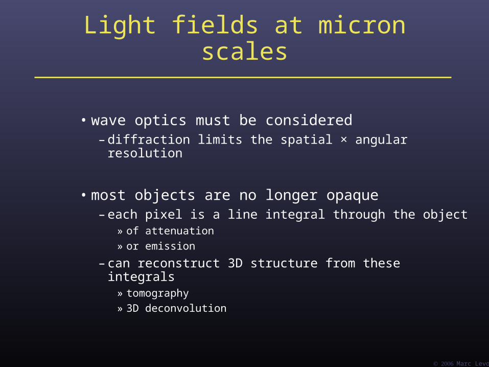
Light fields at micron scales
• wave optics must be considered– diffraction limits the spatial × angular resolution
• most objects are no longer opaque– each pixel is a line integral through the object
» of attenuation
» or emission
– can reconstruct 3D structure from these integrals» tomography
» 3D deconvolution
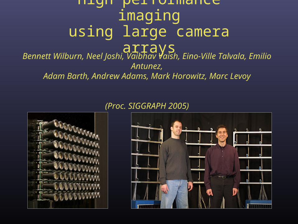
High performance imagingusing large camera arrays
Bennett Wilburn, Neel Joshi, Vaibhav Vaish, Eino-Ville Talvala, Emilio Antunez,Adam Barth, Andrew Adams, Mark Horowitz, Marc Levoy
(Proc. SIGGRAPH 2005)
Marc Levoy
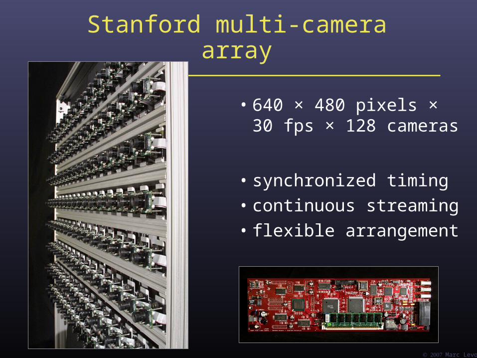
Stanford multi-camera array
• 640 × 480 pixels ×30 fps × 128 cameras
• synchronized timing
• continuous streaming
• flexible arrangement
Marc Levoy
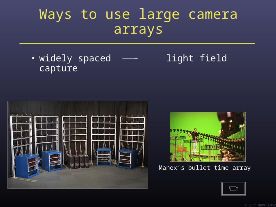
Ways to use large camera arrays
• widely spaced light field capture
Manex’s bullet time array

Marc Levoy
Ways to use large camera arrays
• widely spaced light field capture
• tightly packed high-performance imaging
Marc Levoy
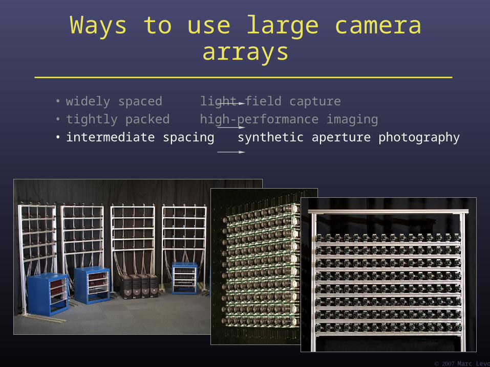
Ways to use large camera arrays
• widely spaced light field capture
• tightly packed high-performance imaging
• intermediate spacing synthetic aperture photography

Marc Levoy
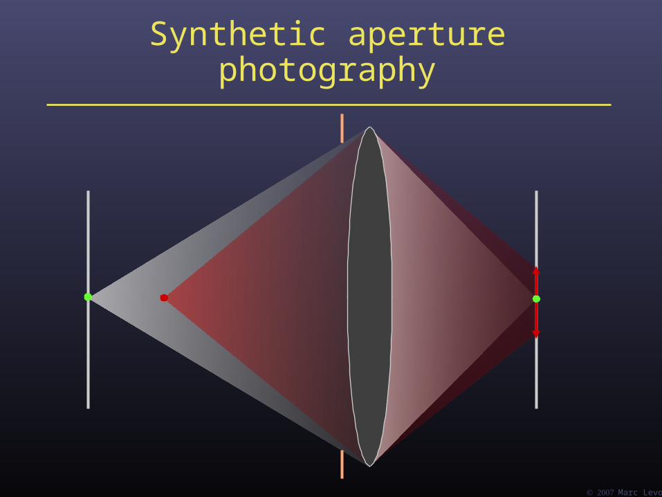
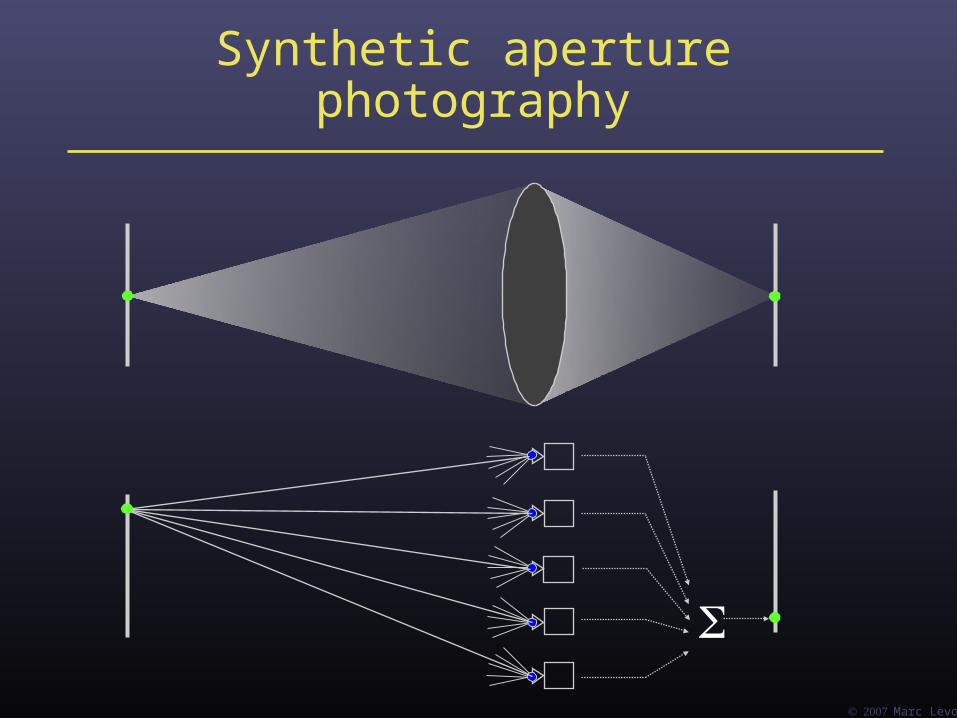
Synthetic aperture photography

Marc Levoy
Synthetic aperture photography
Marc Levoy
Synthetic aperture photography

Marc Levoy
Synthetic aperture photography

Marc Levoy
Synthetic aperture photography
Marc Levoy
Synthetic aperture photography
Marc Levoy
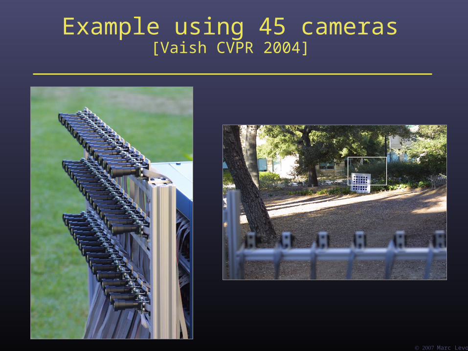
Example using 45 cameras[Vaish CVPR 2004]


Light field photography using a handheld plenoptic camera
Ren Ng, Marc Levoy, Mathieu Brédif,Gene Duval, Mark Horowitz and Pat Hanrahan
(Proc. SIGGRAPH 2005and TR 2005-02)
Marc Levoy
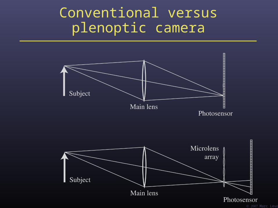
Conventional versus plenoptic camera

Marc Levoy
Conventional versus plenoptic camera
uv-plane st-plane

Prototype camera
4000 × 4000 pixels ÷ 292 × 292 lenses = 14 × 14 pixels per lens
Contax medium format camera Kodak 16-megapixel sensor
Adaptive Optics microlens array 125μ square-sided microlenses

Marc Levoy
Digital refocusing
• refocusing = summing windows extracted from several microlenses
Σ
Σ

Marc Levoy
A digital refocusing theorem
• an f / N light field camera, with P × P pixels under each microlens, can produce views as sharp as an f / (N × P) conventional camera
– or –
• it can produce views with a shallow depth of field ( f / N ) focused anywhere within the depth of field of an f / (N × P) camera
Marc Levoy
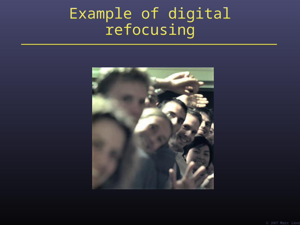
Example of digital refocusing

Marc Levoy
Refocusing portraits
Marc Levoy
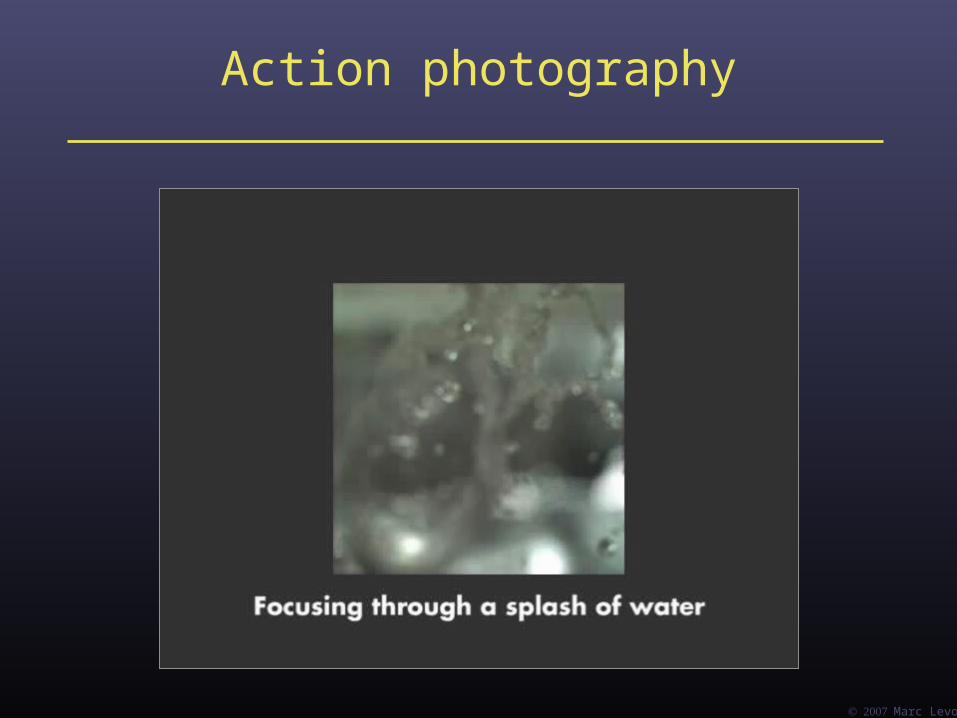
Action photography

Extending the depth of field
conventional photograph,main lens at f / 22
conventional photograph,main lens at f / 4
light field, main lens at f / 4,after all-focus algorithm
[Agarwala 2004]
Marc Levoy
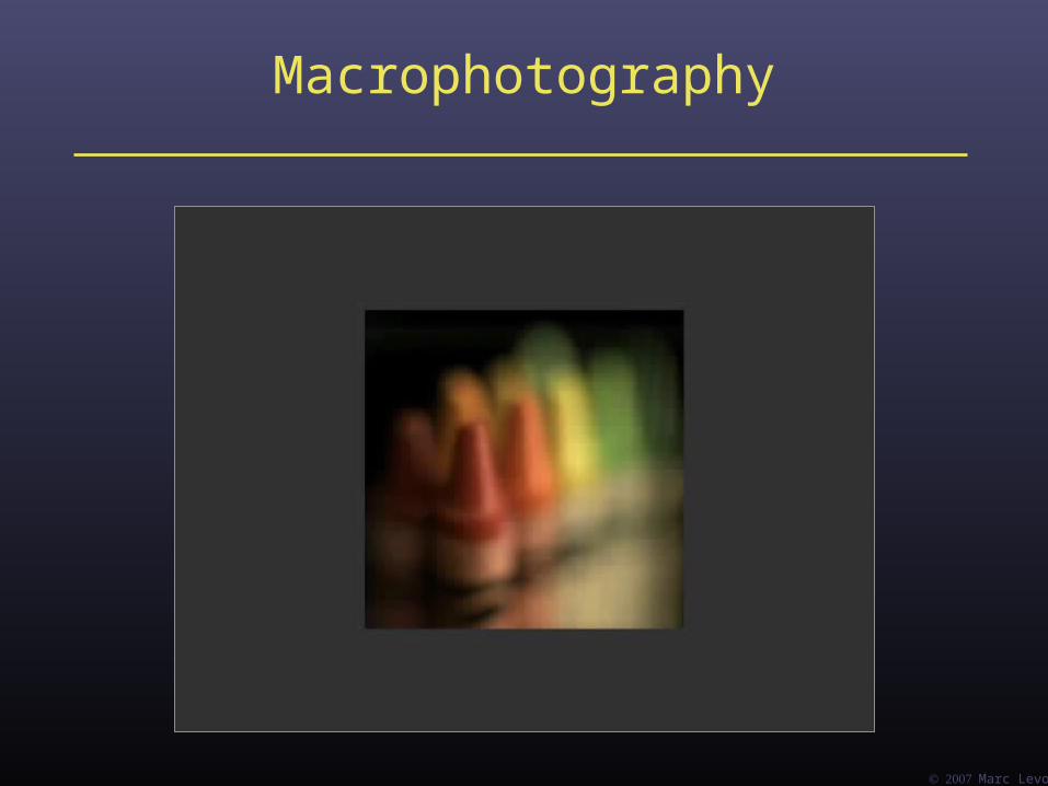
Macrophotography
Marc Levoy
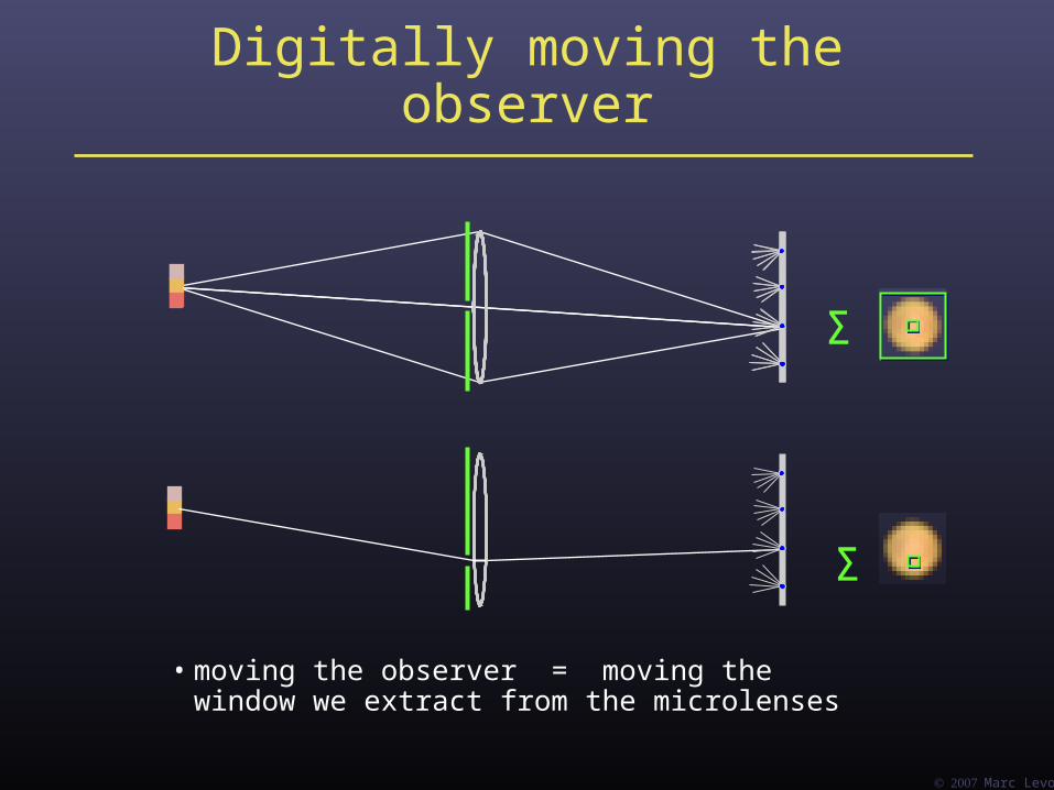
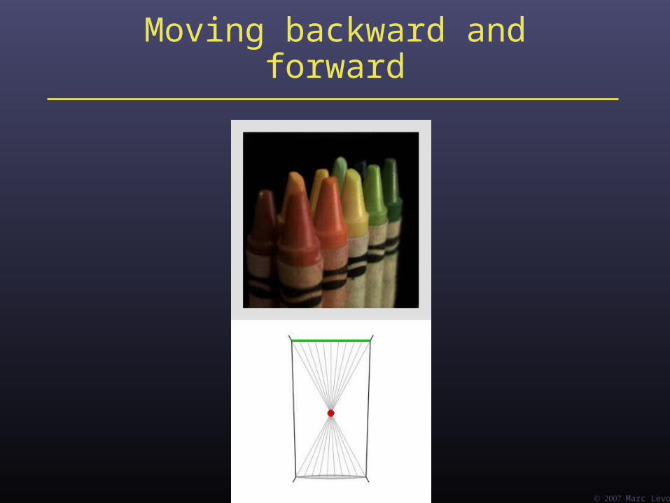
Digitally moving the observer
• moving the observer = moving the window we extract from the microlenses
Σ
Σ

Marc Levoy
Example of moving the observer
Marc Levoy
Moving backward and forward
Marc Levoy
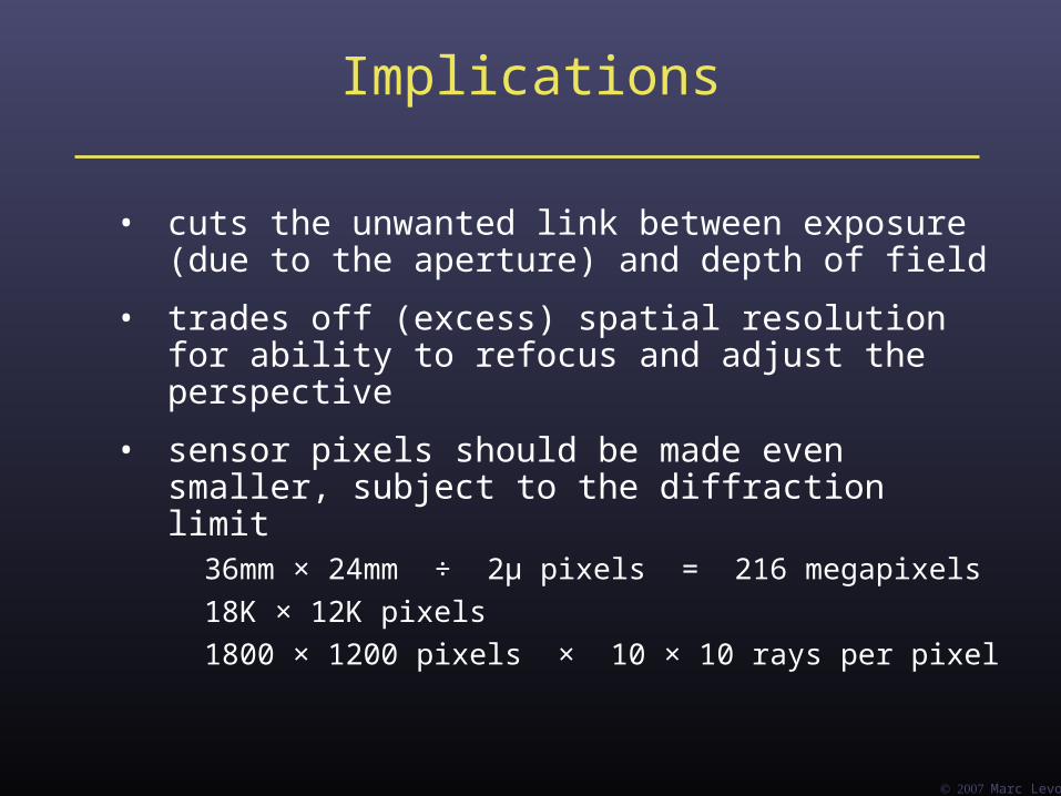
Implications
• cuts the unwanted link between exposure(due to the aperture) and depth of field
• trades off (excess) spatial resolution for ability to refocus and adjust the perspective
• sensor pixels should be made even smaller, subject to the diffraction limit
36mm × 24mm ÷ 2μ pixels = 216 megapixels
18K × 12K pixels
1800 × 1200 pixels × 10 × 10 rays per pixel

Light Field Microscopy
Marc Levoy, Ren Ng, Andrew Adams,Matthew Footer, and Mark Horowitz
(Proc. SIGGRAPH 2006)

Marc Levoy
A traditional microscope
objective
specimen
intermediateimage plane
eyepiece
Marc Levoy
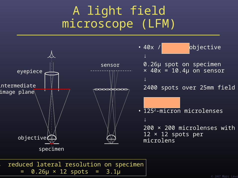
A light field microscope (LFM)
• 40x / 0.95NA objective
↓
0.26μ spot on specimen× 40x = 10.4μ on sensor
↓
2400 spots over 25mm field
• 1252-micron microlenses
↓
200 × 200 microlenses with12 × 12 spots per microlens
objective
specimen
intermediateimage plane
eyepiecesensor
→ reduced lateral resolution on specimen= 0.26μ × 12 spots = 3.1μ

Marc Levoy
A light field microscope (LFM)
sensor

Marc Levoy
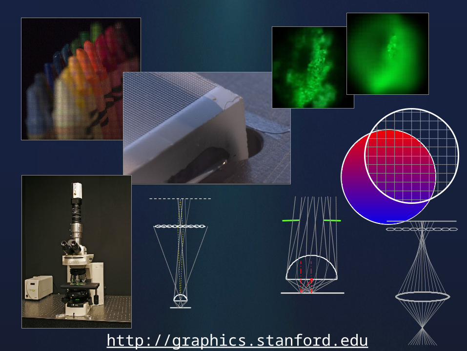
Example light field micrograph
• orange fluorescent crayon
• mercury-arc source + blue dichroic filter
• 16x / 0.5NA (dry) objective
• f/20 microlens array
• 65mm f/2.8 macro lens at 1:1
• Canon 20D digital camera
ordinary microscope light field microscope

Marc Levoy
The geometry of the light fieldin a microscope
• microscopes make orthographic views
• translating the stage in X or Y provides no parallax on the specimen
• out-of-plane features don’t shift position when they come into focus
• front lens element size =aperture width + field width
• PSF for 3D deconvolution microscopy is shift-invariant (i.e. doesn’t change across the field of view)
f
objective lensesare telecentric

Marc Levoy
Panning and focusing
panning sequence focal stack

Marc Levoy
Real-time viewer
Marc Levoy
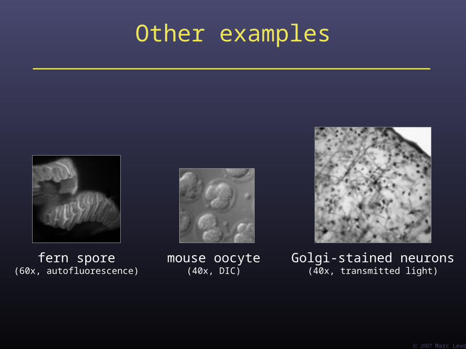
Other examples
fern spore(60x, autofluorescence)
mouse oocyte(40x, DIC)
Golgi-stained neurons(40x, transmitted light)
Marc Levoy
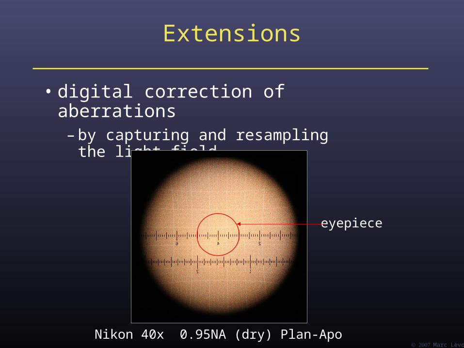
Extensions
• digital correction of aberrations– by capturing and resampling the light field
Nikon 40x 0.95NA (dry) Plan-Apo
eyepiece
Marc Levoy
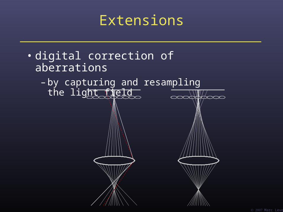
Extensions
• digital correction of aberrations– by capturing and resampling the light field

Marc Levoy
Extensions
• digital correction of aberrations– by capturing and resampling the light field
correcting for aberrations caused by imaging through thick specimens whose index of refraction doesn’t
match that of the immersion medium

Marc Levoy
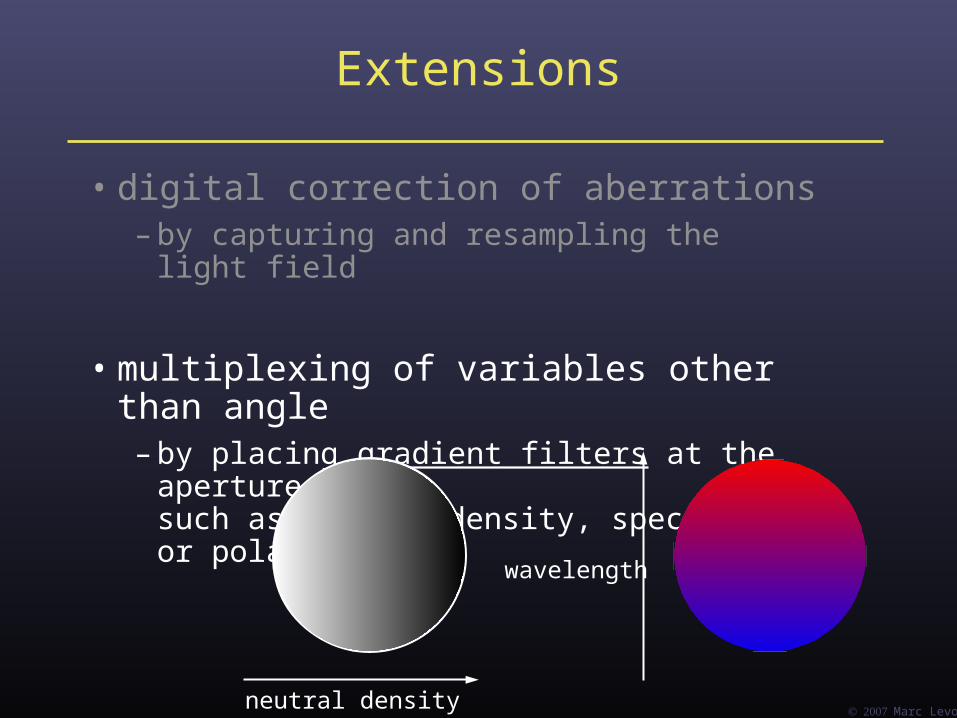
Extensions
• digital correction of aberrations– by capturing and resampling the light field
• multiplexing of variables other than angle– by placing gradient filters at the aperture plane,
such as neutral density, spectral, or polarization
neutral density
Marc Levoy
Extensions
• digital correction of aberrations– by capturing and resampling the light field
• multiplexing of variables other than angle– by placing gradient filters at the aperture plane,
such as neutral density, spectral, or polarization
neutral density
wavelength

Marc Levoy
Extensions
• digital correction of aberrations– by capturing and resampling the light field
• multiplexing of variables other than angle– by placing gradient filters at the aperture plane,
such as neutral density, spectral, or polarization
neutral density
wavelength
... or polarization direction
... or ???
• gives up digital refocusing?

Marc Levoy
Extensions
• digital correction of aberrations– by capturing and resampling the light field
• multiplexing of variables other than angle– by placing gradient filters at the aperture plane,
such as neutral density, spectral, or polarization
• microscope scatterometry– by controlling the incident light field
using a micromirror array + microlens array

Marc Levoy
Programmableincident light field
• light source +micromirror array +microlens array
• 800 × 800 pixels =40 × 40 tiles ×20 × 20 directions
• driven by image from PC graphics card
Marc Levoy
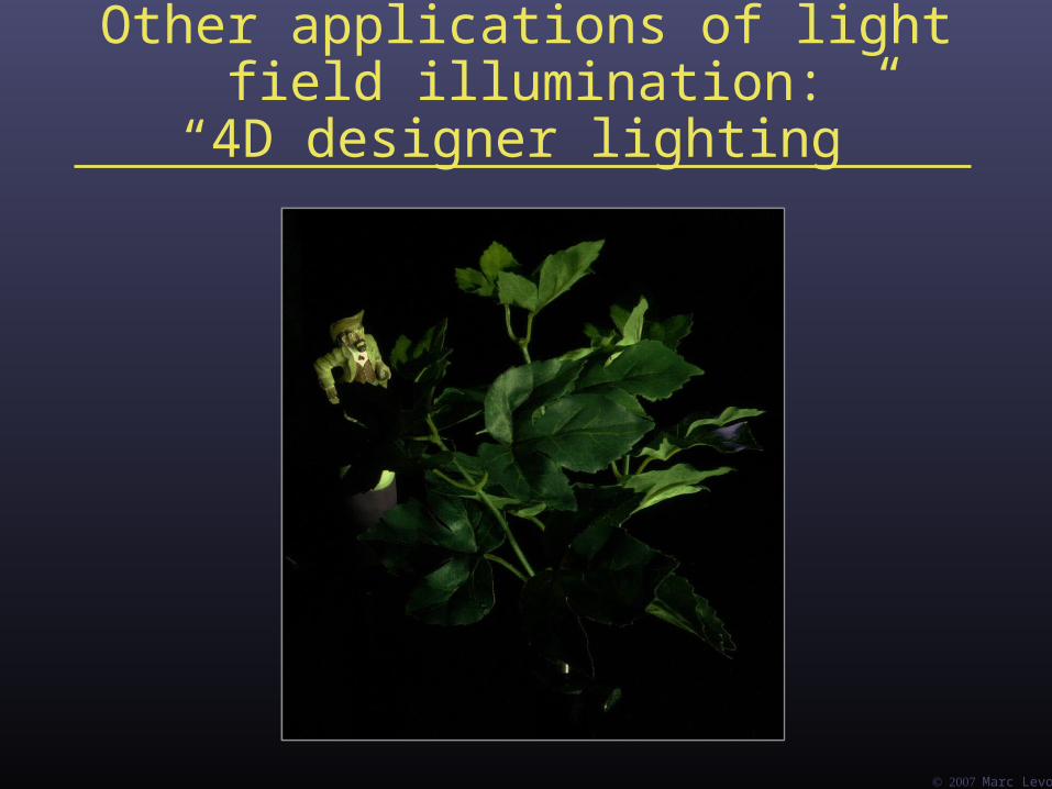
Other applications of light field illumination:“4D designer lighting”
Marc Levoyhttp://graphics.stanford.edu