LESSON ASSIGNMENT LESSON 2 Digestive and Urogenital Systems · MD0959 2-2 LESSON 2 DIGESTIVE AND...
75
MD0959 2-1 LESSON ASSIGNMENT LESSON 2 Digestive and Urogenital Systems TEXT ASSIGNMENT Paragraph 2-1 through 2-24. LESSON OBJECTIVES After completing this lesson, you should be able to select correct answers to questions about: Radiographic and fluoroscopic procedures involving the gastrointestinal tract and its auxiliary organs. Radiographic and fluoroscopic procedures involving the urogenital organs. SUGGESTION After completing the assignment, complete the exercises at the end of this lesson. These exercises will help you to achieve the lesson objectives.
Transcript of LESSON ASSIGNMENT LESSON 2 Digestive and Urogenital Systems · MD0959 2-2 LESSON 2 DIGESTIVE AND...

MD0959 2-1
LESSON ASSIGNMENT LESSON 2 Digestive and Urogenital Systems TEXT ASSIGNMENT Paragraph 2-1 through 2-24. LESSON OBJECTIVES After completing this lesson, you should be able to select correct answers to questions about: Radiographic and fluoroscopic procedures involving the gastrointestinal tract and its auxiliary organs. Radiographic and fluoroscopic procedures involving the urogenital organs. SUGGESTION After completing the assignment, complete the exercises at the end of this lesson. These exercises will help you to achieve the lesson objectives.
MD0959 2-2
LESSON 2
DIGESTIVE AND UROGENITAL SYSTEMS
Section I. THE DIGESTIVE SYSTEM
2-1. GENERAL Examination of the digestive system includes a number of different technical procedures in which various items of equipment and materials are used. The method used will vary in certain details, depending upon the desires of the radiologist. The various components of the digestive system are nearly always examined selectively; for example, the esophagus, the stomach, the duodenum, the colon, and the gallbladder. Examination of each of these parts often constitutes a procedure in itself. In the discussion that follows, each part is considered as a unit. Except under emergency conditions, each of these examinations requires a previous appointment. 2-2. THE ESOPHAGUS The esophagus lies immediately posterior to the trachea. It penetrates the diaphragm and enters the stomach by way of the cardiac orifice. Consider the esophagus as being divided into three portions--cervical, thoracic, and abdominal. Each may use specific radiographic reference points. The cervical portion is located above the upper situs of the mediastinum. The thoracic portion is between the superior aspect of the mediastinum and the diaphragm. The abdominal portion is between the diaphragm and the stomach. a. Preparation of Patient and Scheduling. The study of the esophagus usually entails the combined use of fluoroscopy and radiography. Therefore, for practical reasons, this examination should be scheduled. No special preparation of the patient is required other than that the stomach be fairly empty so that it can accommodate the contrast medium without undue discomfort. b. Preliminary Procedure. Everything should be in complete readiness when the patient reports for the examination. This generally requires the following. (1) A routine setup of the fluoroscopic and radiographic facilities should be accomplished. This includes checking the x-ray unit for correct factors and operational readiness, mounting the footrest or shoulder braces, and checking to make certain that the proper type and numbers of cassettes (including identification material) are readily available for spot-film exposures. Ensure the digital monitor, TV, or videotape is ready for use. At the start of each day, the above equipment should be tested to ensure readiness. (2) Protective aprons and gloves should be laid out for the radiologist and other medical personnel.

MD0959 2-3
(3) The contrast medium should be prepared in accordance with established routine or as prescribed by the radiologist. Some type of barium sulfate preparation is usually used, but if perforation is suspected, an alternate non-barium sulfate radiopaque should be substituted. A thin-water mixture usually contains the contrast medium and water in equal parts by volume. A thick-paste mixture usually contains about six parts contrast medium to one part water. A drinking tube or a spoon should be on hand to administer the mixture to the patient while he is in the recumbent position. c. Fluoroscopic Examination. (1) Upon reporting, the patient is dressed in a suitable gown. (2) The examination procedure should be explained to the patient so that he can cooperate. (3) The patient is instructed (or assisted, if necessary) to take a position between the table and the fluoroscopic apparatus. Whenever possible, the fluoroscopic examination is started with the patient in the erect position. (4) A preliminary screening of the area to be investigated is usually made by the radiologist. (5) At a given signal from the radiologist, the specialist hands the patient a spoonful (or disposable cup, if a thin mixture is called for) of the previously prepared contrast medium. If the patient is in the erect position, the specialist should show him how to hold the cup so that it will not interfere with the free up-and-down movements of the fluoroscopic apparatus. This phase of the procedure is illustrated in figure 2-1. (6) The patient ingests the contrast medium and controls his respiration as directed by the radiologist. The passage and behavior of the contrast medium is observed fluoroscopically and recorded using spot, cine, or other recording media.
Figure 2-1. Patient in position for fluoroscopic examination.
MD0959 2-4
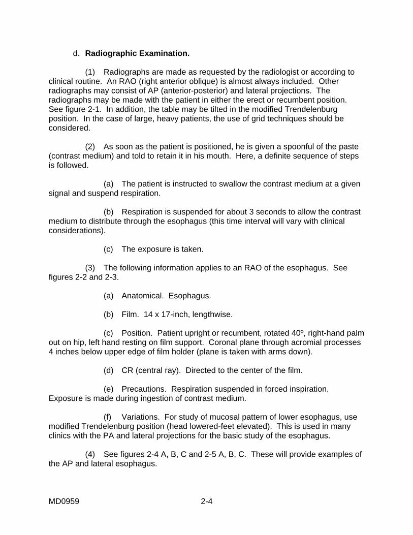
d. Radiographic Examination. (1) Radiographs are made as requested by the radiologist or according to clinical routine. An RAO (right anterior oblique) is almost always included. Other radiographs may consist of AP (anterior-posterior) and lateral projections. The radiographs may be made with the patient in either the erect or recumbent position. See figure 2-1. In addition, the table may be tilted in the modified Trendelenburg position. In the case of large, heavy patients, the use of grid techniques should be considered. (2) As soon as the patient is positioned, he is given a spoonful of the paste (contrast medium) and told to retain it in his mouth. Here, a definite sequence of steps is followed. (a) The patient is instructed to swallow the contrast medium at a given signal and suspend respiration. (b) Respiration is suspended for about 3 seconds to allow the contrast medium to distribute through the esophagus (this time interval will vary with clinical considerations). (c) The exposure is taken. (3) The following information applies to an RAO of the esophagus. See figures 2-2 and 2-3. (a) Anatomical. Esophagus. (b) Film. 14 x 17-inch, lengthwise. (c) Position. Patient upright or recumbent, rotated 40º, right-hand palm out on hip, left hand resting on film support. Coronal plane through acromial processes 4 inches below upper edge of film holder (plane is taken with arms down). (d) CR (central ray). Directed to the center of the film. (e) Precautions. Respiration suspended in forced inspiration. Exposure is made during ingestion of contrast medium. (f) Variations. For study of mucosal pattern of lower esophagus, use modified Trendelenburg position (head lowered-feet elevated). This is used in many clinics with the PA and lateral projections for the basic study of the esophagus. (4) See figures 2-4 A, B, C and 2-5 A, B, C. These will provide examples of the AP and lateral esophagus.

MD0959 2-5
THE ORDER OF PROCEDURE
ESOPHAGUS, RIGHT ANTERIOR OBLIQUE 1. Stuctures best shown. Esophagus between the vertebral column and heart. 2. Remove artifacts. All clothing removed, including undergarments. Remove jewelry in areas of interst. 3. Technical Factors: 14 x 17 in. (35 x 43cm), (LW), Bucky. LM: Corresponding side. 4. Patient/Part position: a. Recumbent or erect. b. Recumbent preferred because of more filling of esophagus (due to gravity factor with erect). c. Rotate 35-40 degrees from a PA with the right anterior body against film holder or table. d. Right arm down; left arm flexed at elbow and by the patient's head. e. Flex left knee, if recumbent. f. Align mid oblique thorax to center of table with top of cassette 2 inches above level of acromion process. 5. Collumination: Use collumination (CF) 10 x 17 LW. 6. CR: Perpendicular to the film holder. 7. SID: 40 " to the bucky. 8. Shielding: Place lead shiel over the patient's pelvic region to shield gonads. 9. Respiration: Suspended respiration while drinking barium.
Figure 2-2. Esophagus, right anterior oblique.

MD0959 2-6
SUGGESTED STARTING TECHNIQUE FOR RAO HOLDER CM KVP* MAS** SID GRID CONE
Cassette
22-28 90 20 40" 8:1 10x17
Cassette
22-28 85 10 72º No LW
Figure 2-3. Radiograph of esophagus, RAO.

MD0959 2-7
Figure 2-4 A. Positioning of AP esopghagus.
Figure 2-4 B. Radiograph. Figure 2-4 C. Diagram.
Figure 2-4. AP esophagus.

MD0959 2-8
Figure 2-5 A. Positioning of lateral esophagus.
Figure 2-5 B. Radiograph. Figure 2-5 C. Diagram.
Figure 2-5. Lateral esophagus.

MD0959 2-9
2-3. THE STOMACH AND DUODENUM (UPPER GI SERIES) The position of the stomach varies with the amount of contents, type of body habitus, mutual pressure of adjacent organs, pathological condition, respiration, emotional state, and whether the subject is in the erect or recumbent position. The position of the duodenum varies in much the same way. a. Preparation of Patient and Scheduling. Except in emergencies or under very unusual circumstances, examinations of the stomach and intestines should be scheduled. All appointments (including cancellations) should be logged in an appointment book or entered in an appropriate calendar chart. (1) Inpatients. The appointment for the examination is made upon receipt of a properly prepared request form from the ward (also called nursing unit). Ward personnel generally prepare the patient for the examination, usually as prescribed by the operating procedures of the installation. In general, the preparation of ingredients is as follows. (a) The patient will not be given a laxative within 24 hours of the examination. (b) The patient should have nothing by mouth for a period of 8-12 hours prior to the time of the examination. (c) The patient is not to drink or eat anything other than the barium mixture he receives in the x-ray department. (2) Outpatients. The procedure is the same as that for inpatients, except that the outpatient is carefully instructed by the x-ray department as to what he must do to prepare for the examination (or is given a prepared form containing the necessary information). b. Preliminary Procedure. (1) A routine setup of the fluoroscopic and radiographic facilities should be accomplished. (a) Single-unit setup. In this plan, a single x-ray unit (radiographic-fluoroscopic) is used for both phases of the examination. When fluoroscopy is completed, the unit is "switched over" and radiography is done. (b) Single-unit, supplemented setup. The operational aspect of this plan is similar to the single-unit setup, except that the radiography is done on a separate x-ray unit located in another room (usually by another x-ray specialist). This plan permits the continuation of fluoroscopy with the same unit from one examination to the next and also expedites the handling and processing of exposed films.

MD0959 2-10
(c) Double-unit setup. Two x-ray units (radiographic-fluoroscopic), located in adjacent rooms, are used. The personnel consist of the radiologist and one, or preferably two, x-ray specialists. Upon completion of fluoroscopy (including spot-film radiography, if indicated), the x-ray unit is changed over and radiography is done. While radiography is being performed on one patient, the examiner may step into the adjacent room and start the fluoroscopic examination of another patient. This process continues, back and forth, until the last examination is completed. The use of the double-unit setup saves excess handling of litter patients, is time saving, and simplifies working conditions. (2) Contrast medium should be prepared in accordance with the established routine as prescribed by the radiologist. Eight to 16 ounces total volume per patient is usually required during fluoroscopic filling. If perforation is suspected, barium is inappropriate and an iodine medium should be substituted. (3) Protective aprons and gloves should be laid out for the radiologist and other interested medical personnel. (4) The radiographic request form containing pertinent information regarding the patient's case should be available to the radiologist at all times. c. Fluoroscopic Examination. (1) Upon reporting, the patient is dressed in a suitable gown. (2) The examination procedure should be explained to the patient so that maximum cooperation may be attained. (3) The patient is directed, or assisted, to take a position between the table and the fluoroscopic apparatus. (4) A preliminary screening of the area under consideration is usually made by the radiologist. The radiologist may request a scout film if the patient’s history indicates prior surgery or recent examinations. (5) At a given signal from the radiologist, the specialist hands the patient a cup of the contrast medium. Just before doing this, he should again stir the mixture. (6) The examination proceeds as the patient ingests the contrast medium and controls his respiration as directed by the radiologist. (7) The radiologist records the fluoroscopic images as necessary.

MD0959 2-11
(8) In some instances, the radiologist will maneuver the patient through various positions under the fluoroscope in order to determine the degree of body angulation and the centering point of the CR for subsequent radiography. This may entail marking the CR-centering site on the body with a skin-marking pencil or measuring the patient on one side to determine the distance that the anterior superior iliac spine (ASIS) should be elevated above the table top during the exposure or both. The radiologist then conveys this information to the specialist, and the required exposures are made. d. Radiographic Examination. The specialist will expose the radiographs according to clinical routine. At times, the radiologist may deviate from the established routine, depending upon his fluoroscopic findings. Figures 2-6 through 2-10 show three common projections. (1) Figures 2-6 and 2-7, PA stomach and the diagram, demonstrate the structures of the stomach and the duodenum. (a) Anatomical. Stomach and portions of duodenum. (b) Film. 14 x 17-inch film. (c) Position. The patient is prone, with median plan perpendicular to the center line of the table. Iliac crests are 5 inches below the center of the film, or as indicated at the time of fluoroscopy. Nonopaque pads may be used under the chest and thighs. (d) Central Ray. Align to center of the film. (e) Respiration. Suspended expiration. (f) Collimation. Full film collimation recommended.

MD0959 2-12
THE ORDER OF PROCEDURE
THE STOMACH AND DUODENUM, PA 1. Structure best shown. Stomach and duodenum with barium in the body and pylorus of stomach. 2. Remove artifacts. All clothing removed, excluding underpants. Remove jewelry in area of interest. 3. Technical Factors: 14 x 17 in. (35x43cm), (LW), Bucky.
LM: Corresponding side.
4. Patient/Part position: Patient prone with arms up beside head, provide pillow. a. Align midsagittal plane to table. b. Insure there is no body rotation. 5. CR: a. Perpendicular to the film holder. b. Iliac crests 5 inches below center of film. 6. Collimation: Use Full Film Collimation. 7. SID: 40-44” to the Bucky. 8. Shielding: Place lead shield over patient’s pelvic region to shield gonads. 9. Respiration: Suspended expiration.
Figure 2-6. Stomach and duodenum, PA.

MD0959 2-13
SUGGESTED STARTING TECHNIQUE HOLDER CM KVP* MAS** SID GRID CONE Cassette
18-22 90 30 40" 8:1 Film
coverage
Radiograph of PA Diagram of the stomach
Figure 2-7, Radiograph of PA (left) and diagram (right) of the stomach demonstrate the parts of the duodenum.
(2) Figure 2-8 and 2-9: stomach and duodenum, RAO. (a) Anatomical. Stomach and parts of the duodenum. (b) Film. 10 x 12-inch lengthwise. (c) Position. Body rotated 40º to 70º midpoint between vertebral column and lateral aspect of the body placed over center of table. Iliac crests at level of the lower film border. (d) CR-UP. Align to the skin marking made surfing fluoroscopy and to the center of the film. (e) Precaution. Suspended respiration. (f) Additional. Grid. (g) Variation. For suspected diaphragmatic hernia, tilt the patient 15º head down and make an exposure with suspended inspiration.

MD0959 2-14
THE ORDER OF PROCEDURE
STOMACH, RIGHT ANTERIOR OBLIQUE 1. Structure best shown. Stomach and C-loop of duodenum. A profile image of the duodenum bulb. 2. Remove artifacts. All clothing removed, excluding underpants. Remove jewelry in area of interest. 3. Technical Factors: 10 x 12 in. (24 x 30 cm), (LW), Bucky.
LM: Corresponding side.
4. Patient/Part position: Recumbent with the body partially rotated into RAO position, provide pillow for head. a. From a prone position rotate 45 degrees with right anterior body against the film holder or table. b. Right arm down; left arm flexed at elbow and by the patient’s head. c. Flex left knee if recumbent. d. Align mid-oblique thorax to center of table with top of cassette 2 inches above level of acromion process. 5. CR: Perpendicular to the film holder. Average body type – CR midway between spinous process and lateral border of abdomen to the center of the cassette. 6. Collimation: Use Full Film Collimation. 7. SID: 40-44” to the bucky. 8. Shielding: Place lead shield over patient’s pelvic region to shield gonads. 9. Respiration: Suspended expiration.
Figure 2-8. Positioning for stomach and duodenum, right anterior oblique.

MD0959 2-15
SUGGESTED STARTING TECHNIQUE HOLDER CM KVP* MAS** SID GRID CONE Cassette
18-22 90 30 40º 8:1 Film
coverage
Radiograph of stomach and duodenum. Diagram of stomach and duodenum.
Figure 2-9. Radiograph and diagram of stomach and duodenum, RAO.
(3) Figures 2-10 and 2-11, lateral stomach, demonstrate the structures of the stomach and the duodenum. (a) Anatomical. Stomach and portions of duodenum. (b) Film. 10 x 12-inch film. (c) Position. The patient is recumbent in a right lateral position with coronal plain perpendicular to the centerline of the table. The bottom of the film is at the iliac crests. Nonopaque pads may be used under the chest and thighs. (d) Central Ray. Align to center of the film. (e) Respiration. Suspended expiration. (f) Collimation. Full film collimation recommended.

MD0959 2-16
Figure 2-10. Positioning for lateral stomach and duodenum.
Radiograph of stomach and duodenum. Diagram of stomach and duodenum.
Figure 2-11. Radiograph and diagram of stomach and duodenum, lateral stomach.

MD0959 2-17
2-4. SMALL INTESTINE (SMALL BOWEL SERIES) In the investigation of the small intestine, the study is usually done by a combination of fluoroscopic and radiographic methods. The preparation of the patient, contrast medium, and management of the facilities are essentially the same as for the examination of the stomach. The radiologist may direct that ice-cold normal saline solution (cold isotonic method) be used as the vehicle for the barium sulfate in place of water, to speed up the examination. The cold solution stimulates peristalsis, causing the barium to pass more rapidly through the gastrointestinal tract. In the double method, a designated quantity of contrast medium is administered to the patient at a specified time prior to fluoroscopy; during fluoroscopy, additional barium is given to the patient (spot-filming may also be done at the time). Radiographs are made at the discretion of the radiologist: for example, a film every 15 minutes for the first hour, then at half-hour intervals, as indicated. Appropriate identification markers should be used for each exposure to indicate the time intervals. 2-5. LARGE INTESTINE (DOUBLE CONTRAST) BARIUM ENEMA a. A method that is widely used for the introduction of the contrast media into the colon is based on a double contrast consisting of barium and air. The liquid component of the contrast media is introduced into the colon by means of gravity. Once the barium has coated the lining of the colon, the barium is mostly drained out of the colon before air is administered. Using the Air-Contrast enema tip and inflator bulb (figure 2-12), air is slowly pumped into the colon either by the radiologist or per his instructions.
Figure 2-12. The rectal tip with catheters for inflation of a retention balloon and an inflator bulb used to inflate the balloon and administer air for double-contrast examinations.

MD0959 2-18
(1) The liquid contrast is now introduced into the colon (under fluoroscopic control) via the disposable enema kit--bag, tubing, and tip--using a gravity to allow the flow of barium. (2) The amount and rate of the flow of the contrast medium into the colon is controlled by means of a clamp on the outlet tubing or by pinching the tubing between the fingers. (3) Posturing of the patient, fluoroscopy, palpation, spot-filming, and radiography are carried out according to the established procedures or the particular needs of the case. (4) When administering air into the colon for double-contrast studies, the procedure is as follows. (a) The outlet tubing is clamped off. (b) The inflator bulb is pumped until the desired amount of air has been administered to the colon (this is determined by the radiologist). (c) Subsequent steps of the examination are carried out according to established procedure.
Figure 2-13. Large bowel, PA (double-contrast study, post-evacuation view).

MD0959 2-19
SUGGESTED STARTING TECHNIQUE
HOLDER *CM KVP MAS SID GRID CONE Cassette 18-22 90 20 40" 8:1 Film coverage
HIGH KILOVOLTAGE TECHNIQUE
HOLDER CM KVP MAS SID GRID CONE Cassette 18-22 125 10 40" 8:1 Film coverage
Figure 2-14. Radiograph of the large bowel, PA or AP with double contrast.

MD0959 2-20
b. In another method (referred to as the "single-stage method"), the air is introduced into the colon at the instant the column of the radiopaque contrast material has advanced to predetermined area in the colon. (1) After patient preparation, ready the necessary facilities for the examination. With the patient in the prone position on the x-ray tilt-table, the radiologist observes the advance of the radiopaque column of contrast material in the colon. This is done under fluoroscopic control. The barium suspension is allowed to run slowly. When the head of the column reaches the splenic flexure, the flow is immediately stopped. (2) The x-ray table is then tilted to the Trendelenburg or modified Trendelenburg position and the patient is rotated to the left. (3) Insufflation of air is begun. The transport of the barium-suspension column is studied fluoroscopically as the air pushes it onward in the colon. The patient is now rolled onto his back and turned to the right as far as directed by the radiologist. This maneuver allows the barium suspension to flow toward the hepatic fissure and enter the ascending and cecal portions of the colon. Spot-films are made as indicated. Usually, as the contrast medium reaches the hepatic fissure, the air input is closely controlled and the tilt-table may be brought to the horizontal or the head-end may be slightly elevated to permit filling of the cecum. Further distribution of the contrast material in the colon is carried out (under fluoroscopic control) by rotating the patient, when necessary, as much as 360º in either direction. (4) After completion of the fluoroscopic phase of the examination, the outlet tubing is clamped off and radiography is done according to established procedure. This procedure may be performed as follows. (a) PA and AP (anterior-posterior) projections are made using a vertical CR with the patient in the horizontal position. (b) PA or AP projections are usually made with the patient in the right and left lateral decubitus positions using a horizontal CR. (c) For optimum results, a minimum of 90 kVp (kilovolt peak) (based on 8 to 1 Potter-Bucky diaphragm) should be used. Grid-front cassettes may be found extremely useful for making the exposure with the patient in the lateral decubitus positions. (d) The AP axial projection is sometimes done during single contrast studies using a 30-40 degree tube angle. It is made according to established procedures.

MD0959 2-21
(e) Other projections include the lateral rectum, sometimes done cross-table with the patient in the prone position, during double contrast examinations. Ensure the central ray is horizontal perpendicular. Posterior oblique projections are also done during single contrast examinations. All projections are done according to established procedures. (f) After necessary radiography has been completed, the rectal tube is withdrawn and the patient is sent to the toilet. c. Another variation of technique is based on the use of a relatively high kVp. Due to the greater penetration of such radiation, it is possible to visualize the intraluminal as well as the circumjacent aspects of the colon. For example, sessile or pedunculated growths, such as polypoid lesions, can be shown just as cholesterol stones are demonstrated within the radiopacified bile in the gallbladder. In general, this method is carried out as follows. (1) Patient preparation is usually the same as for the previously described methods, unless the radiologist prescribes otherwise. (2) The barium sulfate suspension is prepared in the ratio of one part of barium sulfate powder with four to six parts of water by volume, depending upon the preferences of the radiologist and the nature of the particular case under consideration. (3) The barium sulfate suspension is introduced into the colon by gravity. This introduction of the medium is always done under fluoroscopic control. (4) Spot-filming may be carried out by conventional technique or by a relatively high-kilovoltage technique, using from 120 to 140 kVp. (5) Radiography is done after completion of the fluoroscopic part of the examination and before evacuation. The prescribed projections are made with the patient postured according to the instructions of the radiologist. (6) After completion of radiography, the patient is allowed to evacuate, and post-evacuation radiographs are made, if required. 2-6. LARGE INTESTINE (SINGLE CONTRAST) BARIUM ENEMA a. Routine Views of the Barium Enema. (1) PA or AP. (2) RAO and LAO (or RPO and LPO).

MD0959 2-22
(3) Lateral rectum, taken as a cross table ventral lateral decubitus for double contrast exam. (4) AP axial (butterfly position). (5) Bilateral decubitus, taken as part of the double contrast exam. b. Preparation of Patient and Scheduling. (1) Inpatient. Ordinarily, the x-ray department in cooperation with the referring ward accomplishes scheduling. The x-ray department makes the appointment for a special date and time upon receipt of a properly prepared request form from the ward. Ward personnel generally prepare the patient for the examination. Normally, the patient is given a cathartic about 8 to 12 hours before the examination, with the approval of the patient's physician. A laxative is prescribed for the patient in conjunction with a cleansing enema. A simple cleansing enema may be given to the patient about an hour before the examination. In most cases, breakfast is withheld. However, a light breakfast is sometimes allowed. (2) Outpatient. This procedure is essentially the same as for the inpatient. When the appointment is made, the outpatient is given instructions as to pre-examination preparation. Arrangements should be made with a ward or nearby dispensary to provide facilities for the cleansing enema if the patient has no means for accomplishing this himself. c. Preliminary Procedure. (1) A routine setup of the fluoroscopic and radiographic facilities should be accomplished. (2) Contrast medium should be prepared in accordance with established routine or as prescribed by the radiologist. (Note: It is imperative that the specialist made certain that the contrast mixture is prepared exactly as prescribed. The type of examination, single-contrast or double-contrast, will determine the thickness of the suspension.) The contrast medium must be worked into a thoroughly mixed suspension using either an electro-mechanical device or the spoon or paddle method. (a) Step 1. Fill the container with water to about half full. Ordinarily, the water should approximate normal body temperature. (b) Step 2. Add the correct amount of barium, and mix into a homogenous suspension. (3) The following accessory items are required to administer the contrast medium.

MD0959 2-23
NOTE: The type of examination, single-contrast or double-contrast, determines the number of the particular items that will be used.) (a) Enema (irrigator) bag, 2-quart; an apparatus of special designed which actuates the flow of the barium suspension through the tubing by means of gravity. (b) Many medical facilities now use disposable enema bags, tubing, and tips. These are complete disposable units that minimize the chance of transmitting a disease organism from one patient to another. Disposable enema bags are perhaps the most convenient to use of all the barium enema equipment. Most of them come with the barium powder already in the bag. To mix, water is added and the bag is shaken vigorously. Cleanup after the examination is virtually eliminated since the entire kit is discarded after use. Disposable enema kits are illustrated in figure 2-15.
Figure 2-15 A. Disposable enema kit. Figure 2-15 B. Disposable single contrast enema kit.
Figure 2-15. Disposable enema kits. (c) A suitable support for the enema bag. A conventional irrigator (IV) stand may be used. The top of the enema bag should be at an elevation of about 3 feet above the top of the x-ray table. (d) A supply of contamination and latex rubber-free rectal tips--one per patient for each examination session. Disposable rectal tips should be used, whenever possible.
MD0959 2-24
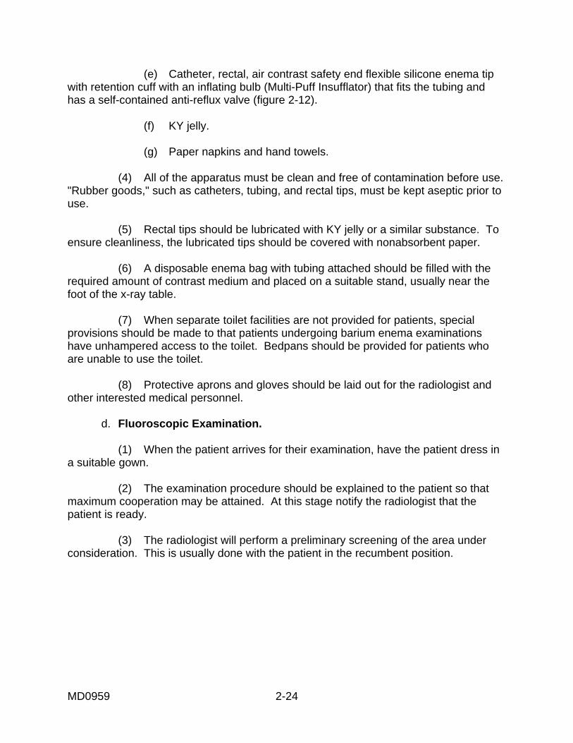
(e) Catheter, rectal, air contrast safety end flexible silicone enema tip with retention cuff with an inflating bulb (Multi-Puff Insufflator) that fits the tubing and has a self-contained anti-reflux valve (figure 2-12). (f) KY jelly. (g) Paper napkins and hand towels. (4) All of the apparatus must be clean and free of contamination before use. "Rubber goods," such as catheters, tubing, and rectal tips, must be kept aseptic prior to use. (5) Rectal tips should be lubricated with KY jelly or a similar substance. To ensure cleanliness, the lubricated tips should be covered with nonabsorbent paper. (6) A disposable enema bag with tubing attached should be filled with the required amount of contrast medium and placed on a suitable stand, usually near the foot of the x-ray table. (7) When separate toilet facilities are not provided for patients, special provisions should be made to that patients undergoing barium enema examinations have unhampered access to the toilet. Bedpans should be provided for patients who are unable to use the toilet. (8) Protective aprons and gloves should be laid out for the radiologist and other interested medical personnel. d. Fluoroscopic Examination. (1) When the patient arrives for their examination, have the patient dress in a suitable gown. (2) The examination procedure should be explained to the patient so that maximum cooperation may be attained. At this stage notify the radiologist that the patient is ready. (3) The radiologist will perform a preliminary screening of the area under consideration. This is usually done with the patient in the recumbent position.

MD0959 2-25
(4) At a given signal from the radiologist, the specialist inserts the rectal tip into the patient's anus. Just before making the insertion, a small amount of contrast medium should be allowed to flow through the tube in order to squeeze out the residual air. The patient is rolled onto his side on the table with his knees flexed. The rectal tip is inserted into the anus with a steady, gradual pressure, exerted anteriorly. When the tip is passed beyond the anus, it should be directed forward at an angle in line with the umbilicus. Due to the extreme sensitivity of the rectal region, care must be exercised in making the insertion. In case a retention catheter is used, caution must be exercised not to distend the inflated bulb excessively. Whenever possible, allow the patient to insert the tip, himself. Be sure to provide appropriate instructions. (5) Upon a signal from the radiologist, the specialist initiates the flow of the contrast mixture and fluoroscopic observation is made. The radiologist will signal the specialist when to interrupt and when to resume the flow of the contrast medium. It is imperative that the specialist respond instantly to these signals. Spot filming may also be done during this phase of the examination. As the patient is maneuvered for changes in position, the specialist should take care to see that the enema bag tubing does not become kinked or accidentally withdrawn. If the tube should become clogged, the obstruction can usually be moved by stripping or "milking" the tube in the direction of flow. If the radiologist desires the filling of the bowel to proceed at a slow rate, the specialist can control the rate of filling by lowering the enema bag or by pinching the tubing between the fingers. Upon completion of "filling" and fluoroscopy, the patient is cautioned to retain the contrast medium until radiography is accomplished. It may be advisable to leave the rectal tip in place until all radiographs are done. This sometimes prevents the patient from prematurely expelling the contrast medium. e. Radiographic Examination. (1) Regardless of the type of setup (single-unit; single-unit, supplemented; or double-unit), it is usually best to do radiography of the barium-filled colon by having the patient remain on the same x-ray table on which fluoroscopy was performed. This lessens the possibility of accidental evacuation. The required radiographs are made in accordance with established routine or as directed by the radiologist. (2) For the single-contrast barium enema, a PA projection of the barium-filled colon (figures 2-16 and 2-17) is obtained. Details are as follows: (a) Anatomical. Colon. (b) Film. 14 x 17-inch, lengthwise. (c) Position. Patient prone, level of iliac crests to the center of the film.

MD0959 2-26
THE ORDER OF PROCEDURE BARIUM ENEMA, AP AND/PA
1. Structure best shown. Entire contrast-filled large intestine. Both PA and AP are generally taken with double contrast study.. 2. Remove artifacts. All clothing removed, including undergarments. Remove jewelry in area of interest. 3. Technical Factors: 14 x 17 in. (35x43 cm), (LW), Bucky. For larger patients, use several CW 14 x 17 in.
LM: Corresponding side.
4. Patient/Part position: Patient prone or supine, with arms up beside head, provide pillow. a. Align midsagittal plane to table. b. Insure there is no body rotation. 5. CR: Perpendicular to the film holder. a. Iliac crests 5 inches center of film. 6. Collimation: Full film coverage. 7. SID: 40-44” to the Bucky. 8. Shielding: Place lead shield over patient’s pelvic region to shield gonads. 9. Respiration: Suspended expiration.
Figure 2-16. Large Intestine, barium enema, PA (pre-evacuation).
MD0959 2-27
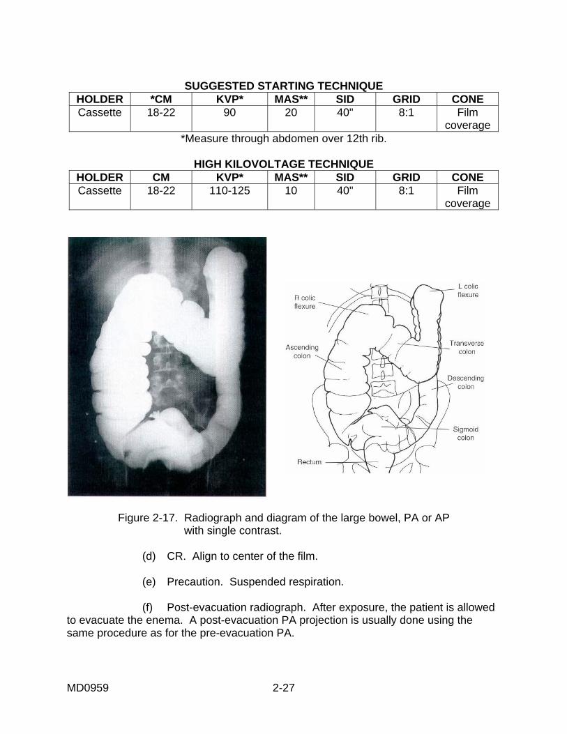
SUGGESTED STARTING TECHNIQUE
HOLDER *CM KVP* MAS** SID GRID CONE Cassette
18-22 90 20 40" 8:1 Film
coverage *Measure through abdomen over 12th rib.
HIGH KILOVOLTAGE TECHNIQUE
HOLDER CM KVP* MAS** SID GRID CONE Cassette
18-22 110-125 10 40" 8:1 Film
coverage
Figure 2-17. Radiograph and diagram of the large bowel, PA or AP
with single contrast. (d) CR. Align to center of the film. (e) Precaution. Suspended respiration. (f) Post-evacuation radiograph. After exposure, the patient is allowed to evacuate the enema. A post-evacuation PA projection is usually done using the same procedure as for the pre-evacuation PA.

MD0959 2-28
(3) A special projection of the sigmoid colon (LPO) is sometimes done. Details are as follows: (a) Anatomical. Sigmoid colon overlying the sacrum and left wing. (b) Film. 14 x 17. (c) Position. Patient supine. Left hip down, right hip and trunk rotated up to 30º to 60º (LPO-lateral posterior oblique) as indicated by fluoroscopy. (d) CR. Align to center of film. Place area outlined fluoroscopically over the center of the film. (e) Precaution. Suspended respiration. (f) Additional. Grid. (4) A special projection of the rectum (LAT) is also required at times (figures 2-18 and 2-19). Details are as follows: (a) Anatomical. Rectal ampulla, rectum, sigmoid, and a portion of the descending colon. (b) Film. 10 x 12-inch. (c) Position. Patient is left lateral recumbent position. Knees partially flexed for support with a point 2 inches anterior post skin surface over the centerline of table to upper border of film. (d) Align. CR to center of film. (e) Precaution. Respiration suspended. Patient should be in true lateral position and will sometimes require buttressing. (f) Additional. Grid. (5) The double-contrast (air contrast) barium enema involves the simultaneous use of two types of contrast media--radiopaque in the form of residual barium adhering to the mucosa; and radiolucent, or air, which is introduced by means of an insufflator. In general, the procedure is as follows. (a) Fluoroscopy and radiography are accomplished as for single-contrast study. That is, the patient is screened, the colon is filled with contrast medium, and exposures are made of the filled colon. The patient is then instructed to evacuate as rapidly as possible (20-30 seconds).

MD0959 2-29
THE ORDER OF PROCEDURE
BARIUM ENEMA, LATERAL RECTUM 1. Structure best shown. Rectum and sigmoid colon anterior to the sacrum. 2. Remove artifacts. All clothing removed, including undergarments. Remove jewelry in area of interest. 3. Technical Factors: 10x12 in. (24x30 cm), (LW), Bucky. For larger patients, use several CW 14 x 17 in.
LM: Corresponding side.
4. Patient/Part position: a. Recumbent in true lateral position, work quickly. b. Center mid-axillary plane to centerline of table. c. Knees and hips partially flexed. d. Center patient and cassette to CR. NOTE: Ventral decub lat. rectum (Alternate projection with double contrast exam, this allows the air to fill the rectum), cross table. 5. CR: Vertical Perpendicular to 1” distal to level of ASIS, centered to mid-axillary plane (midway between ASIS and posterior sacrum). 6. Collimation: To outer film borders. 7. SID: 40-44” to the Bucky. 8. Respiration: Expose at full expiration.
Figure 2-18. Lateral rectum, barium enema.

MD0959 2-30
SUGGESTED STARTING TECHNIQUE HOLDER *CM KVP* MAS** SID GRID CONE Cassette
28-34 90 220 40º 8:1 Film
coverage * Measure through the plane of the iliac crests.
Figure 2-19. Radiograph of lateral rectum.
(b) Immediately after evacuation, the patient is recalled for fluoroscopy. Then, air is introduced into the colon by means of a colonic insufflator. This is done under fluoroscopic control. (c) Routine radiographs are usually obtained in both the prone and supine positions, because the opaque medium may tend to collect and "puddle" due to the influence of gravity. Radiographs made with a horizontal CR and the patient in the right lateral decubitus position and the left lateral decubitus position are often obtained. Stereoscopic films may be made if indicated. (d) The specialist must accomplish the necessary radiography as rapidly as possible since the retention of a considerable volume of air in the colon may cause distress to the patient.

MD0959 2-31
(e) If a retention catheter is used, it should first be deflated and then withdrawn immediately upon completion of radiography to enable the patient to expel the air from the colon. (f) Examination of the rectum is necessary during the barium enema in both the single and double contrast studies. Figure 2-19, demonstrates the Lateral Rectum in the single contrast study. During double contrast studies with air injected, the lateral rectum is viewed in the decubitus position with a ventral cross-table lateral projection. (g) The patient is directed to a toilet and instructed to evacuate. After evacuation, a post-evacuation radiograph is made. 2-7. GALLBLADDER a. General. Radiographic studies of the Gallbladder are rarely done. These studies have been replaced with other modalities such as ultrasound. (1) Radiographic visualization of the gallbladder is done by cholecystography. Radiographic investigation of the biliary tract is by cholangiography. For these procedures, it is necessary to convey a contrast medium to the gallbladder along with the bile. (2) Bile is manufactured by the polyhedral cells of the liver which extract the necessary constituents from the circulating blood. The gallbladder's ability to concentrate the bile makes it possible for a sufficient amount of the cholecystopaque to collect within the gallbladder to permit radiographic visualization. After oral administration, the contrast medium, if in pill form, disintegrates in the stomach. (a) Most of the contrast medium is absorbed in the small bowel and conveyed to the liver via the portal vein. (b) As the contrast medium moves throughout the liver, it becomes associated with the liver cells and is secreted with the bile. (c) As the bile containing the contrast medium passes along the ducts, some of it is discharged into the duodenum and some of it backs up into the gallbladder where concentration occurs. (d) The elimination of the contrast medium from the body is dependent upon various factors such as the type of contrast medium and the nature and degree of dysfunction related to the digestive system. Normally, some of the contrast medium is not absorbed, but is eliminated via the colon. The kidneys eliminate the part that is not removed from the blood as it passes through the liver.
MD0959 2-32
(e) As the gallbladder discharges the bile containing the contrast medium into the small bowel after the ingestion of fatty meal, the medium is reabsorbed and conveyed to the liver and secreted again. This cycle continues until the contrast medium is completely eliminated from the body via the kidneys and colon. b. Preparation of Patient and Scheduling. Patient preparation and scheduling are in accordance with the established clinical routine. In general, the preparation of the patient may be as follows. (1) On the day before the examination (before ingestion of the contrast medium), a PA projection of the abdomen (11x14) may be done. This is a survey or scout film. (2) The patient is not allowed to eat fats after the noon meal the day before the examination. (3) About 12 hours prior to the examination, the patient ingests the contrast medium according to the manufacturer's instructions. Telepaque is usually used with the average dose being 6 tablets. (4) After taking the pills, the patient should not be allowed to eat or drink anything until the time of the examination. (5) To make certain that none of the contrast medium has been lost, the patient should be instructed to report any vomiting or bowel movements. (6) If the initial films show no stones, the patient is given a fatty meal to promote good gallbladder contraction. An additional film is usually done 30 minutes to one hour after the fatty meal. Sometimes commercially prepared compounds or mixtures may be used in place of the fatty meal. NOTE: Before giving a fatty meal, consult the radiologist. A fatty meal should not be given to patients if stones are seen in the initial films). This precaution is necessary because the "emptying" of the gallbladder caused by the fatty meal may release one or more stones into the biliary ducts causing obstruction. (7) The procedure may vary since every radiologist has his preferred method. For example, some radiologists may request that the patient be given two teaspoonfuls of paregoric one-half hour after the ingestion of cholecystopaque or that an enema be administered one hour prior to radiography.

MD0959 2-33
(8) For an examination of the biliary ducts (by the intravenous injection of Cholografin), the referring ward schedules and provides the preparation of the patient. Usually, the patient does not eat anything after 1800 hours on the day prior to the examination. On the morning of the examination, the patient is given a sensitivity test. If the test is negative and there are no contraindications, 20 to 40 cc of the cholecystopaque is injected intravenously by the radiologist. This examination is done in the operating room using sterile techniques. 2-8. CHOLECYSTOGRAPHY Cholecystography is a radiographic procedure for the demonstration of the gallbladder after making the bile radiopaque by means of a contrast medium, which may be administered either orally or intravenously. a. Oral Method. (1) The patient is prepared in accordance with the prescribed pre-examination procedure. (2) The contrast medium is administered according to prescribed procedures. (3) Upon reporting to the x-ray department, the patient is dressed in a suitable gown. He should be questioned as to how he carried out each step of the pre-examination procedure and whether he experienced any vomiting or diarrhea. If any vomiting or diarrhea occurred, the x-ray specialist should report it to the radiologist. (4) The number and type of projections made depends upon prescribed procedures and the suspected pathology. Usually, a survey film is made to determine the position of the gallbladder, the correctness of the exposure factors, the presence and extent of gas or unabsorbed contrast medium in the bowels, and evidence of outstanding pathology. Details of the scout film are described below and illustrated in figures 20 and 2-21. (a) Anatomical. General localization of the gallbladder. (b) Film. 14x17-inch film, and lengthwise. (Some radiologists prefer a 10x12-inch film.) (c) Position. The patient is prone with arms alongside the body. He is positioned as for a PA abdomen exposure with the crest of the ilium 3 inches below the center of the film. (d) CR. The CR is aligned to the center of the film. (e) Precaution. Suspended expiration straight.

MD0959 2-34
Figure 2-20. Gallbladder, PA Projection, “SCOUT”.
Figure 2-21. Radiograph of a gallbladder.
MD0959 2-35
(5) Other films may include the following. (a) PA prone projection using an 8 x 10-inch film and a tightly restricted cone field. (b) LAO using a tightly restricted cone field (6 inches) and an 8 to 10-inch film. This radiograph may be done with the patient recumbent (figure 2-22) or erect. This position tends to displace the transverse processes of the vertebrae away from the gallbladder. The body is rotated 20º to 30º. The degree of body rotation necessary for optimum demonstration of the gallbladder varies according to body habitus and position; for example, thin subjects generally require greater rotation than stout subjects. Varying degrees of rotation may be necessary to differentiate gallstones from kidney stones or calcified bodies in the mesenteric structure. (c) Right lateral projection with the patient in the recumbent position, utilizing an 8 by 10-inch film and a restricted cone field. (d) PA projection with the patient on his right side (Kirklin) using an 8 by 10-inch film, restricted cone field, and vertical Bucky diaphragm or grid-cassette. (e) RPO (right posterior oblique) using an 8-inch x 10-inch film and a restricted cone field. This radiograph may also be done with the patient in either the recumbent or erect position. (6) Radiography of the gallbladder with the patient in the erect position may be done by the using essentially the same relationships with reference to part, film plane, and alignment of the CR. The erect position will cause the bile laden with cholecystopaque to stratify into fluid levels according to the degree of concentration and relative specific gravity. The gallstones, which are lighter than certain layers of the bile, will float and, upon floating together, form a "density layer" which renders them radiographically demonstrable. Small, but heavier-than-bile, stones will tend to gravitate to, and collect in, the fundus portion of the gallbladder. In addition, the gallbladder tends to shift downward, backward, and towards the midline. Therefore, when the patient is first x-rayed in the recumbent position and then in the erect position, some modification of the CR alignment is necessary for the latter position (approximately 1 1/2 to 2 1/2 inches lower).

MD0959 2-36
Figure 2-22. Left anterior oblique position for radiography of the gallbladder. NOTE: Body habitus variation: Hypersthenic(broad), gallbladder is more horizontal, 2 in. higher and more lateral; Asthenic(thin), gallbladder is more vertical, 2 in. lower, near the midline of body.

MD0959 2-37
(7) Special methods for radiography of the gallbladder may include the use of spot-film or tomography. (a) Spot-filming. Following exposure and processing of the survey film, the patient is positioned under the fluoroscope, and the radiopaque gallbladder is localized. Various spot-film exposures may be obtained with the patient in either the recumbent or the erect position. (b) Tomography. Though still used, has mostly been replaced with computerized tomography and ultrasound. After accomplishing the routine survey film, the radiologist specifies the number of layers or "cuts" to the spot-filmed and also the level at which each is to be made. By the use of tomographic technique, it is possible to avoid troublesome gas pockets and loculi or, at least, to lessen their adverse effects. Also, under certain conditions, gallstones casting doubtful densities when produced by conventional radiography can be more readily distinguished. (c) Ultrasound. A preferred method of visualizing the gallbladder is the ultrasound. This reduces the patient’s exposure to ionizing radiation. (8) The cholecystographic series is usually terminated with the final film begin taken 1/2 to 1 hour after ingestion of a "fatty meal" that is given to the patient immediately after satisfactory demonstration of the gallbladder. b. Intravenous Method. (1) The preparation of the patient and the radiographic procedure are essentially the same as for the oral method. The patient is given nothing by mouth the night before. The difference lies in the contrast medium used, the time at which it is introduced, and the method of introduction. (2) A cholecystopaque, such as cholografin sodium, is injected into the vein of the arm. (3) Radiographic examination is made in accordance with established routine--usually about 4 hours after injection. Additional films may be taken at subsequent intervals. (4) A fatty meal is given to the patient if this has been ordered by the radiologist. 2-9. CHOLANGIOGRAPHY Cholangiography is a procedure for the demonstration of the biliary tract after the introduction of a contrast medium. The contrast medium may be introduced by either of two methods: direct and intravenous.

MD0959 2-38
a. Direct Method. The direct method embraces two types of procedures: operative (immediate) and postoperative (delayed). (1) Immediate or operative cholangiography. (a) This procedure is carried out in the operating room. The surgeon aspirates the bile in the ducts and injects a contrast medium such as Hypaque or Hippuran into the duct. (b) A mobile x-ray unit, a portable high-speed grid cassette, and an adequate supply of loaded cassettes, are used. The cassette tray with handle is first loaded with a cassette; then it is positioned under the patient before the surgery is started. (c) The cassette tray handle is graduated with a centimeter scale. A scout film is made to check positioning and the handle is marked, this enables the technician to reposition the cassette tray during surgery. All movement of the cassette tray is done at the head end of the table. Since the operative site is sterile-draped when the tube is moved into position, the x-ray specialist must ask the surgeon to point out the exact site for directing the CR. (d) It is necessary to use as short an exposure time as possible, especially when other than spinal anesthesia is used. If spinal anesthesia is given, the patient should be instructed to suspend respiration during the exposure of the film. If the patient cannot respond, ask the anesthesiologist to suspend the patient's respirations during the exposure. (e) Since the exposure must be made at a critical time during injection of the contrast medium, the specialist should ask the surgeon to give him a signal so that he can expose the film at the proper time. (f) Before exposing the film, the specialist should direct his attention to the surgical site to make certain that none of the surgical instruments overlie or obscure the area to be x-rayed. This is extremely important. Failure to observe this precaution may necessitate re-exposure and delay the surgical procedure. (g) Exposed films should be processed immediately for reading. Additional films are made at the request of the surgeon. (h) The entire procedure must be carried out under aseptic conditions. Ensure that you pay attention to all sterile fields and patient drapes, taking care not to contaminate those areas. (i) A representative cholangiogram made during surgery is shown in figure 2-23. A sketch to aid in positioning the patient is shown as figure 2-24.

MD0959 2-39
Figure 2-23. A representative cholangiogram made during surgery.
Figure 2-24. Drawing showing structures in a cholangiogram.

MD0959 2-40
(2) Delayed cholangiography. This examination usually follows the removal of the gallbladder and is normally performed in the x-ray department. Following surgery, a T-tube is left in place within the biliary tract for continuous drainage. Utilizing the same type of contrast medium as for immediate cholangiography and eliminating the need of anesthesia, the material is injected through the T-tube into the biliary tract by the radiologist. The radiologist will also decide the type and the number of radiographs to be made. b. Intravenous Method. (1) Usually, the patient does not eat or drink anything after 6 p.m. on the day prior to the examination. (2) On the morning of the examination, the patient is given a sensitivity test. If there are no contraindications, the radiologist slowly injects intravenously 40 cc of cholangiopaque (Cholografin). (3) Ten minutes after the injection, the first film is made. Meanwhile, observe the patient for any reaction. (4) RPO radiographs are casually made to prevent superimposition of the common bile duct over the spine. Body rotation is 15 to 20 degrees. (5) The initial film is processed immediately and is read by the radiologist. This film also provides the specialist a means of checking for the proper positioning of the patient and the corrections of the exposure factors. (6) Subsequent films are exposed at 10-minute intervals for the first hour and at 20-minute intervals for the second hour. In each instance, the film is processed immediately and read by the radiologist. (7) Ordinarily, this completes the examination. However, under certain conditions, tomographic variations of positioning may be used.

MD0959 2-41
Section II. THE UROGENITAL SYSTEM 2-10. INTRODUCTION a. The urogenital system is described in subcourse MD0956. b. Urography is the radiographic study of the functional and structural aspects of the parts of the urinary tract after they have been rendered radiopaque. This term embraces two methods of examining the kidneys utilizing a contrast medium--excretory urography and retrograde urography. Excretory urography is a functional examination and retrograde urography is a structural examination. In either case, specific patient preparation is required. In retrograde urography, the contrast medium is introduced directly into the renal pelvis via the ureter using special catheters. In excretory urography, the contrast medium is usually injected intravenously and passes quickly into the urine; however, it can be introduced intramuscularly, subcutaneously, or orally, depending upon clinical contingencies. Excretory urography is commonly referred to as intravenous pyelography and retrograde urography is commonly referred to as retrograde pyelography. The films thus obtained are called pyelograms. c. Terms applied to other urographic examinations and the organs to which they pertain are given below. (1) Ureterography--ureters. (2) Cystography--urinary bladder. (3) Urethrography--urethra. (4) Prostatography--prostate. (5) Epididymography--epididymis. (6) Vesiculography--seminal vesicles. (7) Hypertrography or uterography--uterus. (8) Hysterosalpingography or uterosalpinography--uterus and oviducts (fallopian tubes). (9) Nephrography--kidney tubules.

MD0959 2-42
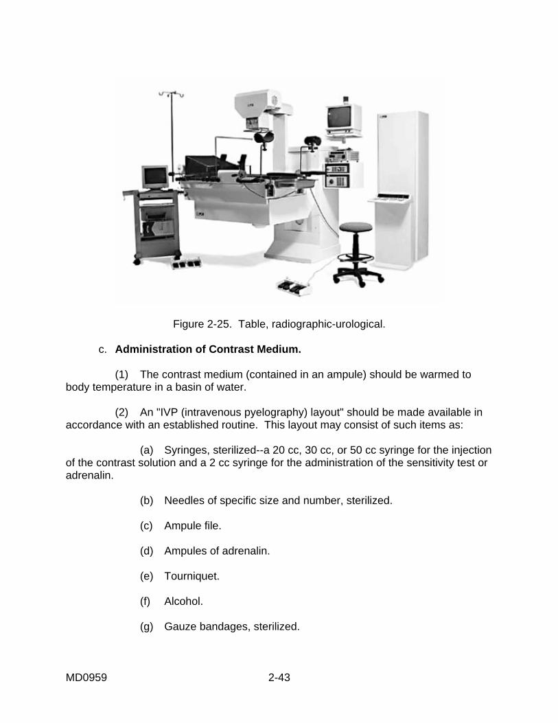
2-11. INTRAVENOUS PYELOGRAPHY a. Patient Preparation and Scheduling. Preparation and scheduling of the patient depends upon local policies and may include the following. (1) Requests for this examination should be properly filled and by the referring medical authority and submitted to the radiology service. (2) In some hospitals, intravenous pyelography is the responsibility of the urology section of the surgical service. (3) Radiopaque pills or tablets should not be given to the patient prior to the examination. (4) The gastrointestinal tract should be empty; the patient should be given a cathartic at least 12 hours prior to the examination. (5) The patient should not have any fluids or foods by mouth after 10 p.m. on the evening before the examination. If it is necessary to dehydrate the patient, a longer period of abstention from fluids may be prescribed. (6) The patient should void just prior to the examination. b. Equipment. (1) Radiographic-urological table. A special radiographic-urological table, readily tiltable, is the most practical and convenient apparatus for urographic examinations. Ordinarily, this unit is equipped with a Potter-Bucky diaphragm, leg and shoulder rests, and immobilization band. It should be used in combination with an x-ray tube and generator of sufficient capacity to minimize the adverse effects of motion (blurring) by permitting the utilization of short exposure times. For the structural arrangement of this unit, see figure 2-25. It is often located in the urological service or in an operating room suite where accessory items of equipment, contrast media, and medical items are conveniently stored. The urological suite often has its own darkroom. This setup is especially desirable when the urological service has a large patient population that suffers from urological diseases. (2) Conventional x-ray table unit. This unit should be equipped with a Potter-Bucky diaphragm, foot and shoulder rests, and immobilization band, and should be tiltable. The tube and generator should be of ample capacity to allow for short exposure times.
MD0959 2-43
Figure 2-25. Table, radiographic-urological.
c. Administration of Contrast Medium. (1) The contrast medium (contained in an ampule) should be warmed to body temperature in a basin of water. (2) An "IVP (intravenous pyelography) layout" should be made available in accordance with an established routine. This layout may consist of such items as: (a) Syringes, sterilized--a 20 cc, 30 cc, or 50 cc syringe for the injection of the contrast solution and a 2 cc syringe for the administration of the sensitivity test or adrenalin. (b) Needles of specific size and number, sterilized. (c) Ampule file. (d) Ampules of adrenalin. (e) Tourniquet. (f) Alcohol. (g) Gauze bandages, sterilized.

MD0959 2-44
(h) Hand towels, sterile. (i) Small water basin, sterilized. (3) Either the radiographic-urological table or the conventional x-ray table may be used. The patient is placed on the table in the supine position with the midline of the body aligned to the center of the table (figures 2-26 and 2-27).
Figure 2-26. Patient in position for urography.
Figure 2-27. Abdomen, AP (K.U.B.).

MD0959 2-45
(4) Ordinarily, the patient is given a minute quantity of the contrast medium (orally, ophthalmically, subcutaneously, or intravenously) to determine sensitivity. (5) A K.U.B. (kidneys, ureters, bladder) film should be made before the injection of the contrast medium. This film should be processed immediately and given to the radiologist or other responsible medical authority for examination. It will show whether or not the gastrointestinal tract has been properly cleared and demonstrate the presence of any calculi or any outstanding pathology. It also serves as a means of checking for correctness of technique factors and accuracy of positioning. (6) After preliminary testing, a sterile aqueous contrast solution containing from 35 to 50 percent iodine compound (for example, Conray, Renografin, or Hypaque) is injected slowly into one of the antecubital veins at the elbow. THE INJECTION IS ALWAYS GIVEN BY THE RADIOLOGIST OR ANOTHER PHYSICIAN. NOTE: Non-ionic contrasts are the media of choice, resulting in fewer severe reactions. d. Radiography. (1) In general, the positioning of the patient (figure 2-27) and the technique factors are the same as for the routine K.U.B. procedure. (2) Films are exposed at specified intervals. Exposure time should as short as practicable to minimize motion and increase detail. Unless otherwise specified, exposure is made upon suspension of respiration at the end of exhalation. Exposures are made according to clinical routine. The following sequence is an example: (a) 1st film: 05 minutes after injection. (b) 2nd film: 10 minutes after injection. (c) 3rd film: 10 minutes after injection. (d) 4th film: 20 minutes after injection. NOTE: At least one film (normally, the last) is made with the patient in the upright position. For upright projections, the normal exposure should be increased. (3) In addition to the above film, it is sometimes necessary to take one or more of the following: (a) Posterior obliques right and/or left. (b) Laterals right and/or left.

MD0959 2-46
(c) Post-micturition film of the urinary bladder. (d) Tomograms are rarely done, but some departments still have the equipment and perform studies. (e) When computerized tomography is available, CT studies of the kidneys are done while the patient still has contrast in their system. (4) In addition to the regular identification data, lead number markers are used to indicate the time-interval at which each film was exposed. The time-interval marker should be so placed that it is easily distinguished on the processed film. Also, be sure to identify any upright film, if any are taken, with an arrow or other appropriate marker. (5) Unless otherwise specified, as soon as each pyelogram is exposed, it is immediately processed and presented to the radiologist for reading. (6) To induce urine stasis for the purpose of obtaining the best filling and thereby greater concentration of the contrast medium in the renal pelvis and calyces, one of several methods may be used. The two methods most commonly used are presented below. NOTE: Urine stasis is induced only at the request of the responsible medical authority. (a) Gravitational method. The patient is placed in the modified Trendelenburg (20º to 40º) position, which induces urine stasis with the aid of gravity. (b) Compression method. A radiopaque compression device, such as a suitably shaped block of balsa wood or an inflatable bag, is placed on the lower part of the patient's abdomen and the proper amount of pressure is applied by an immobilization band. See figure 2-28. Compression is usually applied before the start of the injection and released prior to the exposure of the last film. Ureterograms may be obtained while the ureters are filled with the contrast agent by exposing a film immediately after compression is released.

MD0959 2-47
Figure 2-28. Patient has compression method applied.
(7) If subsequent pyelograms are to be taken, great care should be exercised to match all films with respect to position, SID, radiographic contrast, and density. This is necessary for satisfactory comparison with the results of prior urographic examinations. A representative intravenous pyelogram is shown in figure 2-29. A labeled tracing of the pyelogram is shown in figure 2-30.
Figure 2-29. Intravenous pyelogram. Contrast medium in kidneys and ureters.

MD0959 2-48
Figure 2-30. Sketch of kidney and ureter area. e. Precautions. (1) There is always the possibility of the patient having adverse reactions following injection of the contrast medium. For this reason, the following precautions must be observed: (a) AT NO TIME DURING THE EXAMINATION WILL THE PATIENT BE LEFT UNATTENDED. (b) Should the patient show such signs as hives, sweating, pallor, dyspnea, severe vomiting, restlessness, or rapid pulse, competent medical aid will be summoned immediately. (c) The specialist should take care to not alarm or suggest symptoms to the patient. (d) Treatment material must be available and ready for instant use in case an emergency should arise. An anaphylactic shock and emergency resuscitation layout should be kept in complete readiness.

MD0959 2-49
2-12. RETROGRADE PYELOGRAPHY a. Patient Preparation and Scheduling. Patient preparation and scheduling for pyelography is similar to that for intravenous pyelography, except for the following. (1) Fluid intake (water) is not restricted; instead, unless otherwise ordered, fluid intake is as much as the patient will tolerate. (2) Sedation is administered as ordered. b. Equipment. (1) The radiograph-urological table unit (see figure 2-25). (2) Cystoscope of the type especially suited for this examination. (3) A special layout consisting of sterile and nonsterile accessory supplies, drugs, and apparatus. The content and arrangement of this layout will vary with the case and the preferences of the urologist. A representative layout may include such items as the following: (a) Catheters: ureteral, x-ray, 4 to 14 Fr. (French); urethral, 14 to 24 Fr. (b) Syringes: bulb type, with assorted nozzles. Luer 2, 5, and 10 cc. (c) Specimen bottles, culture tubes, and medicine glasses. (d) Towels, drapes, sheets, leggings, gauze, cotton, and gloves. (e) Tourniquet, large and small water basins, and small trays. (f) Drugs, antiseptic solutions, and contrast solutions. c. Procedure. (1) The patient is placed on the radiographic-urological table. (2) The patient's lower extremities are sheathed in leggings especially designed for this purpose. (3) The urologist introduces the cystoscope via the urethra and makes a preliminary examination of the urinary bladder. He then passes the urethral catheters through the cystoscope into the ureters as far as the renal pelves.

MD0959 2-50
(4) A film is usually made to check the position of the catheter(s) and to check patient position and technical factors. (5) This film is immediately processed and presented to the radiologist or the urologist for reading. (6) At this stage of the procedure, the urologist will, in most instances, retract the cystoscope, leaving the catheters in place. (7) A contrast solution is introduced into the renal pelves and calyces through the respective catheters by means of syringes. This procedure is accomplished by the urologist. (8) At this point, the following routine is usually carried out: (a) At a signal from the urologist, the x-ray specialist exposes the pyelogram (figure 2-31). For this exposure, the patient is instructed to suspend respiration at the end of exhalation.
Figure 2-31. Retrograde pyelogram showing the urethral catheter in place and
the distribution of the contrast medium in the kidney and ureter.

MD0959 2-51
(b) The patient is allowed to breathe normally while the film in the Potter-Bucky tray is quickly changed. (c) The urologist withdraws the ureteral catheters and simultaneously maintains pressure on the syringes to express the amount of contrast solution required for maximum filling of the ureters. (d) At a signal from the urologist, the ureterographic exposure is made. The timing of the exposure is regulated so that the film will record the incidence of maximum filling of the ureters. Exposure time should be as short as practical. Such films are termed ureterograms. (9) There are several variations of the above routine. For example, in some instances, the kidney on one side is filled and the pyelographic film is exposed. Then the same procedure is repeated for the kidney on the opposite side. The head of the table is often elevated 35º to 45º for the ureterogram to demonstrate any kinking of the ureters and to determine any downward displacement of the kidneys. On occasion, the "split-exposure" technique may be used to differentiate between a ureteral calculus and a calculus-like density superimposed with the density pattern cast by one or both of the ureters. For this technique, the first exposure is made with the x-ray tube positioned as for normal centering and the second exposure is made with the tube displaced laterally 1 1/2 to 3 inches. One half of the normal exposure time is used for the first exposure, and two-thirds of the normal exposure time is used for the second exposure. Only the tube is moved during this procedure, the position of the patient and the film remains constant. (10) In actual practice, the number and kind of films exposed depends upon the patient's symptoms. The x-ray specialist should be prepared to take such views as posterior-obliques, or laterals. 2-13. CYSTOGRAPHY AND PNEUMOCYSTOGRAPHY a. General. Cystography is the radiographic study of the urinary bladder after the introduction of a radiopaque contrast medium. The films thus obtained are cystograms. When a radiolucent contrast medium (such as air) is used, the examination is termed pneumocystography and the film is a pneumocystogram. b. Patient Preparation and Scheduling. (1) Unless specific orders are given, patient preparation and scheduling are the same as for pyelography. (2) The patient is instructed to void before being placed on the x-ray table.

MD0959 2-52
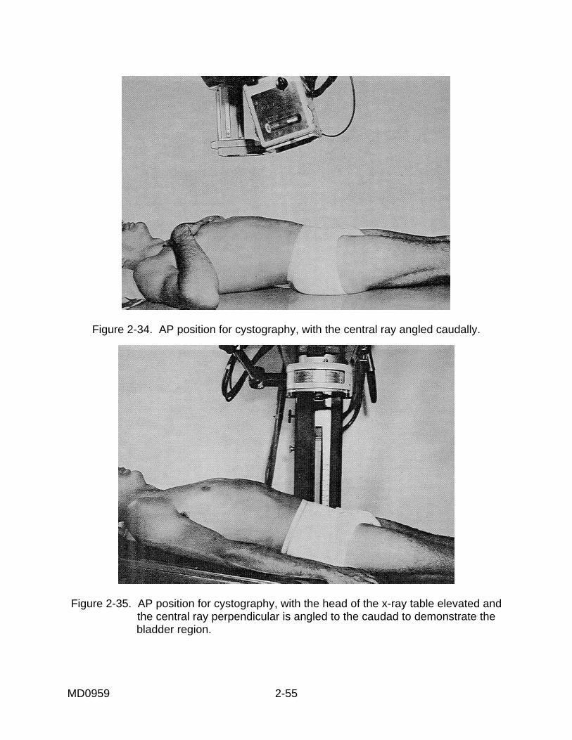
c. Equipment. (1) The conventional x-ray unit equipped with a Potter-Bucky diaphragm and a tilting mechanism is preferred. (2) When cystoscopy is required, the radiographic-urological table unit may provide certain advantages. (3) A cystoscopic layout that suits the needs of the given case should be readily available. Ordinarily, this layout is made up and supplied. d. Procedure. (1) The patient is placed on the table in the supine position with the median plane of the body centered to the midline of the table. A 10 x 12-inch film is placed lengthwise in the Potter-Bucky film tray and it is centered 1 inch above the symphysis pubis on the median plan of the body. The CR (central ray) is projected perpendicularly and directed to the center of the film. A preliminary film of the bladder region is made upon request of the examiner. (2) To clear away any gas in the rectum, a rectal tube may be inserted. The tube must be removed before the exposures are made. (3) The urologist introduces a contrast medium, such as Cystokon, into the bladder through a urethral catheter in an amount sufficient to distend the bladder (200 to 300 cc). To retain the contrast solution in the bladder during radiography, the catheter should be clamped. (4) Cystograms are exposed with the patient's respiration suspended at the end of exhalation. The first exposure is an AP projection of the bladder region. Usually, right and left posterior-obliques (45º to 60º body rotation) (figure 2-32) are made following the AP projection. Additional exposures may include lateral or stereoscopic projections in the prone or supine positions. A representative cystogram is shown in figure 2-33. (5) In some cases, the AP projection is taken with CR angled 15º to 25º caudad from the vertical relationship (figure 2-34). Similar results may be obtained with a perpendicular CR and the head of the table elevated 15º to 25º (figure 2-35).

MD0959 2-53
Figure 2-32. Right posterior-oblique position for cystography. Note relationships of part, film, and central ray.

MD0959 2-54
Figure 2-33. A representative cystogram.
.
MD0959 2-55
Figure 2-34. AP position for cystography, with the central ray angled caudally.
Figure 2-35. AP position for cystography, with the head of the x-ray table elevated and the central ray perpendicular is angled to the caudad to demonstrate the bladder region.
MD0959 2-56
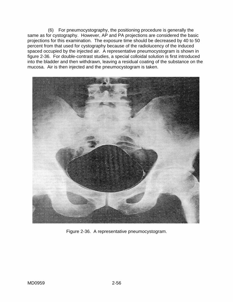
(6) For pneumocystography, the positioning procedure is generally the same as for cystography. However, AP and PA projections are considered the basic projections for this examination. The exposure time should be decreased by 40 to 50 percent from that used for cystography because of the radiolucency of the induced spaced occupied by the injected air. A representative pneumocystogram is shown in figure 2-36. For double-contrast studies, a special colloidal solution is first introduced into the bladder and then withdrawn, leaving a residual coating of the substance on the mucosa. Air is then injected and the pneumocystogram is taken.
Figure 2-36. A representative pneumocystogram.

MD0959 2-57
2-14. NEPHROGRAPHY a. General. Nephrography is a special procedure for the radiographic demonstration of the parenchymal structures of the kidneys during their radiopacification by means of a contrast medium. b. Patient Preparation and Scheduling. Patient preparation and scheduling are essentially the same as for intravenous pyelography. However, in some cases, a test is administered for the calculation time (for example, arm-to-tongue decholin) to determine the optimum intervals of time and the sequence to be used in exposing the films following the injection of the contrast medium. c. Introduction of Contrast Medium and Radiography. (1) The patient is placed in the supine position with the midline of the body to the center of the x-ray table unit. A 10x12-inch or a 14x17-inch film is placed in the Potter-Bucky diaphragm film tray and centered at the level of the 2d lumbar vertebra. The CR is directed to the center of the film. (2) The pertinent aspects of the procedure should be explained to the patient, and he should be warned about the sensations he is likely to experience during the procedure (such as hot flashes, gagging, and nausea). The necessity for controlling respiratory or bodily movements during the exposure of the films should be stressed. (3) A preliminary film is exposed, developed, and immediately presented to the examiner for reading. This film will serve as a check for correctness of positioning, technique factors, and adequacy of patient preparation. (4) A 12-gauge injection needle (for example, Robb-Steinberg type) is inserted into an antecubital vein of the arm. A wide-bore syringe that has been previously filled with the contrasting medium (consisting of one of the sterile aqueous solutions and containing 70 to 75 percent iodine compound such as Hypaque or Renografin) is attached to the needle. (5) With all participants alerted, technique factors selected, and x-ray apparatus in readiness, the examiner gives the "ready" signal and starts injecting the contrast medium (40 to 50 cc). As the last of the contrast solution leaves the syringe, the examiner calls out, "Now," at which time the x-ray specialist (or an assistant) starts a stopwatch or audibly counts the passage of seconds on any available timepiece which has a second hand.

MD0959 2-58
(6) The first film is exposed at a predetermined interval following the injection of the contrast solution. This interval usually ranges from 8 to 20 seconds, depending upon the circulating time. Subsequent films are exposed at predetermined intervals. If a conventional x-ray unit is being used, the subsequent exposures should be made as rapidly as possible, especially during the critical filling phase or when there is maximal concentration of the contrast material in the cortical and medullary regions of the kidneys. The patient is instructed to hold in his breath and exposure is made on full inspiration. If a rapid-sequence serializing apparatus is used, two or more (only when indicated by the examiner) exposures may be made while the patient continues to hold his breath. Usually, exposure of films is discontinued approximately 35 seconds following the completion of the injection. (7) The identification marker should be placed on each of the cassettes prior to beginning the examination. (8) All films should be developed as quickly as possible and presented to the examiner for reading before the patient is moved off the x-ray table. (9) Nephrograms may sometimes result as a side-effect. They are apt to occur during such special procedures as angiocardiography or aortography, wherein relatively large amounts of contrast solution containing a high percentage of iodine are introduced at a relatively rapid rate. (10) A representative nephrogram is shown in figure 2-37.
Figure 2-37. A representative nephrogram.

MD0959 2-59
2-15. NEPHROTOMOGRAPHY a. General. Nephrotomography is a special procedure for the sectional representation of the parenchymal structures of the kidneys by the use of tomography during radiopacification following the intravenous administration of a contrast medium. This technique helps to obtain a clearer image of the renal parenchyma by blurring out the image details of underlying and overlying structures. b. Patient Preparation and Scheduling. Unless otherwise indicated, patient preparation and scheduling for nephrotomography is essentially the same as for nephrography. c. Procedure. (1) Two AP radiographs of the abdominal region are obtained, one by use of conventional radiographic technique, the other by tomography. The two films are processed immediately and presented to the examiner for reading. They serve as control films and as a means for checking the correctness of the technique factors. (2) Any necessary adjustments in regard to positioning of the patient are made, the tomographic fulcrum is set at the correct elevation, and the contrast medium is introduced. Usually, the introduction of the contrast medium is done as for nephrography. The numbers of exposures to be made during radiopacification of the renal parenchyma, as well as the exact exposure time-intervals to be used, are predetermined by the examiner according to circulation time. (3) All films should be developed immediately after exposure and presented to the examiner for reading prior to releasing the patient. 2-16. URETHROGRAPHY a. General. Urethrograms are accomplished by radiography of the urethra after filling with a suitable contrast medium. b. Patient Preparation. No special preparation of the patient is required, but the patient is requested to void just prior to examination. c. Equipment. (1) Either a conventional x-ray unit equipped with a Potter-Bucky diaphragm or a radiographic-urological table unit. (2) Sandbags or cellulose positioning blocks for supporting and immobilizing the patient in the required position. (3) Routine layout for administration of contrast media.

MD0959 2-60
d. Procedure. (1) The urologist irrigates the urinary bladder. (2) A preliminary film (or films) of the urogenital tract is exposed as ordered. (3) The patient is placed on the x-ray table in the posterior-oblique position with the body rotated 35º to 40º, either to the right. For male patients, the penis is extended along the medial aspect of the thigh nearest the film. The region of the symphysis pubis is centered to the midline of the table. The knee nearest the table surface is moderately flexed. The leg farthest from the table surface is extended and moved slightly backwards. Sandbags are placed to secure and immobilize the position. See figure 2-38.
Figure 2-38. The right posterior oblique position for urethrography.
(4) A 10 x 12-inch film is placed lengthwise in the Bucky tray with the mid-length aligned at the level of the interior border of the symphysis pubis. (5) Proper collimation is used. (6) The CR is projected perpendicularly and directed to the center of the film. (7) The urologist introduces a contrast medium (viscous solution) into the urethra. (8) Exposure is made at a signal from the urologist. Routine pelvic technique factors are appropriate. Respiration is suspended during exposure.

MD0959 2-61
(9) The foregoing procedure may be varied by taking AP or PA projections of the filled urethra using screen or cardboard (non-Bucky) technique. For these radiographs, the patient's legs are parted, the penis is positioned to the central portion of the film, and the CR is angled about 12 degrees caudad from the vertical relationship. Routine digit technique factors are appropriate. (10) A representative urethrogram is shown in figure 2-39
Figure 2-39. A representative urethrogram.
2-17. HYSTEROSALPINGOGRAPHY (UTEROSALPINGOGRAPHY) a. General. This procedure deals with radiographic and/or fluoroscopic examination of specified structures of the female reproductive system utilizing a contrast medium. The radiography is hysterosalpingogram. The specified technique will depend upon the nature of existing pathology, the condition of the patient, the special types of apparatus, and the preferences of the examiner. The general principles of the procedure are presented herein primarily for guidance. b. Patient Preparation and Scheduling. (1) A properly filled out request form is submitted to the x-ray department by the referring medical authority. (2) Examination is usually scheduled 1 to 8 days after menstruation or at a time determined by the gynecologist or radiologist.

MD0959 2-62
(3) The patient's lower bowels should be cleared of gas and fecal material, as ordered by the responsible medical authority. (4) The patient is requested to void just prior to the examination. c. Procedure. (1) The patient is placed in the supine position with the median plane of the body aligned to the midline of the combination radiographic-fluoroscopic table. If fluoroscopy is not to be done, the standard radiographic-urological table unit may be preferred. (2) A 10 x 12-inch film is placed lengthwise in the Potter-Bucky film tray with the mid-length of the film aligned to the level of the ASIS. The CR is projected perpendicularly and directed to the center of the film. Collimation should be used to confirm the field of irradiation to the most practical limits. (3) A preliminary film is exposed as ordered. Routine pelvic factors will serve for basic exposure technique. (4) The gynecologist or the radiologist introduces a vaginal speculum. (5) A cannula is inserted into the cervical canal of the uterus. (6) The gynecologist injects from 3 to 10 cc radiopaque contrast material (usually Salpix) containing from 35 to 40 percent iodine compound. In some instances, the course of the contrast medium is observed fluoroscopically. Spot-filming is done by the radiologist, when necessary. (7) Hysterosalpingograms are exposed by the specialist in a given signal from the gynecologist. Respiration is suspended for the exposure. (8) Additional films are taken as ordered. For example, these may include one or more of the following: (a) AP projection with or without caudal angulation of the CR. (b) PA projections with or without cephalad angulation of the CR. (c) Obliques, anterior or posterior (right and/or left). (d) Decubitus--AP, PA, or lateral projections. (e) Video taping the fluoroscopy during injection.

MD0959 2-63
(f) Pelvic inlet views. (g) Digital recordings. (9) Sometimes a pelvic pneumoperitoneum exposure may be performed to demonstrate the external contours of the uterus and adjacent structures. For this procedure, approximately 500 cc of carbon dioxide or oxygen is introduced into the peritoneal cavity. The gynecologist introduces the radiolucent contrast medium or radiologist while the patient is in the Trendelenburg position. For radiography, the patient is turned in the prone position, and the table is tilted 45º to 50º to bring the hips above the level of the head (figure 2-40). The CR is projected vertically and directed through the region of the rectum to the center of the film. The exposure mAs should be reduced by approximately one-half of the normal. Other radiographs are taken as ordered.
Figure 2-40. Patient positioned for pelvic pneumoperitoneum.

MD0959 2-64
Section III. THE DECUBITUS POSITIONS 2-18. ESSENTIALS OF THE POSITIONS In any radiographic decubitus position, two conditions coexist: a. The patient must be lying down. b. The central ray (CR) must be horizontal. 2-19. DESCRIPTION a. There are four decubitus positions--dorsal, ventral, right lateral, and left lateral (figure 2-41). These names signify the surface of the body upon which the patient is resting in a recumbent position. In the dorsal decubitus position, the patient is supine. In the ventral decubitus position, the patient is supine. In the right lateral decubitus position, the patient is lying on his right side. In the left lateral decubitus position, the patient is lying on this left side. b. Using a horizontal CR, it is possible to secure two different projections for each decubitus position. The lateral projections (right and left) can be accomplished with the patient in either the left lateral decubitus or right lateral decubitus position. c. If a “PA projection of a left lateral decubitus position” is requested, the projection should be made as the patient lies on his left side. The vertical film holder should be supported at the anterior aspect of the patient's body; the horizontal CR should enter the posterior surface and exit at the anterior surface. For a left lateral projection of a dorsal decubitus position, the patient should be in a supine position. The vertical film holder should be supported at the left side of the patient; the horizontal CR should enter the right side and exit from the left. Lateral projections of dorsal or ventral decubitus positions of the thorax or abdomen may also be referred to as "transthoracic" (transabdominal)." 2-20. CLINICAL USE Decubitus positions are used for two main reasons: a. To effect a shift of fluid, air, contrast media, or anatomical part so that additional diagnostic information can be obtained in a radiograph. b. To make desired radiographs that would ordinarily be accomplished by rotating a part and using a perpendicular CR, but cannot be performed in this instance because of the condition of the patient.

Figure 2-41. Decubitus position.
MD0959 2-65

MD0959 2-66
2-21. CONSIDERATIONS It is desirable to see both sides of the patient's body on the film in any given projection of a decubitus position. The affected side MUST be shown with no cut-off. Accordingly, a firm pad, folded blanket or folded sheets should be placed between the table surface and the patient to elevate the dependent side of the patient. If the condition of the patient or other factors makes this elevation inadvisable, the Bucky tray, partially out, may be used as a support for the vertical film holder and a standard wheeled hospital litter with pad used as a table. Allow the patient to remain in position approximately five minutes so the free air rises and/or free fluid moves downward. 2-22. TECHNIQUE a. The essential position, the reference point for placement of the part, and the technique factors used for a decubitus position should be in keeping with those used for a normal projection of the same area. If an AP projection of the abdomen in a left lateral decubitus position is requested, the crest of the ilia should be centered and the horizontal CR should be directed to the center of the film. In the examination of those parts calling for a grid or Bucky, a portable Bucky or grid cassette should be used. b. A vertical x-ray table with Bucky used in combination with the standard wheeled hospital litter with pad is an effective combination for decubitus positions. It permits easy handling of the patient and equipment and is usually comfortable for the patient. If a patient is brought to the x-ray department on the wheeled hospital litter, it is often possible to accomplish all necessary film exposures without moving him to the x-ray table and with a radiographic quality close to that possible with table-positioning. This possibility should be kept in mind not only for decubitus positions, but also for emergency and hard-to-work with patients. 2-23. IDENTIFICATION a. The identification data are usually placed along the lateral border of the cassette corresponding to the side of the body that is uppermost. The letter marked (R or L) indicates the side nearest the film in lateral projections and indicates the side with which it is placed in AP (anteroposterior) or PA projections. In addition to the regular identification data, the following facts should be included: (1) The type of decubitus position. (2) The designation of the projection. (3) The identity of the surface that is up. b. These added data identify the film unquestionably as a decubitus-type position, making the details on the film more readily recognizable to any film interpreter.
MD0959 2-67
2-24. CLINICAL APPLICATIONS There are many clinical applications of decubitus positions, although not called by that name. PA right lateral decubitus is a great aid in demonstrations of stones and moving the gall bladder away from the spine in cholecystography. Right and left lateral decubitus positions are used in air contrast barium enemas. In addition, they can demonstrate free air and fluid in chest and abdominal x-rays to rule out damage to thoracic and abdominal structures following trauma, especially, when the patient is unable to stand or sit up.
Continue with Exercises
Return to Table of Contents

MD0959 2-68
EXERCISES, LESSON 2 INSTRUCTIONS: Answer the following exercises by marking the lettered response that best answers the question or best completes incomplete statement. After you have completed all the exercises, turn to "Solutions to Exercises" at the end of the lesson and check your answers. For each exercise answered incorrectly, reread the material referenced with the solution. 1. Fluoroscopic examination of the esophagus is normally started with the patient in what position? a. Prone. b. Supine. c. Erect. d. Laterally recumbent. 2. A thick-paste barium sulfate mixture is often used in the radiographic study of the: a. Esophagus. b. Small intestine. c. Stomach. d. Large intestine. 3. Radiography of the esophagus almost always includes what position? a. Right anterior oblique. b. Caldwell. c. Lateral decubitus. d. Supine decubitus.

MD0959 2-69
4. What is the essential patient preparation order for radiographic examination of the stomach and duodenum? a. Cleansing enema 1-2 hours before appointment. b. Nothing by mouth for a period of 8-12 hours prior to appointment. c. Six telepaque tablets 10-14 hours before appointment. d. Laxative 4-6 hours prior to appointment. 5. Ice-cold normal saline solution may be used in place of water as a vehicle for the contrast medium in radiographic studies of the: a. Kidneys. b. Small intestines. c. Aorta. d. Gallbladder. 6. When the sigmoid colon is of special interest, a projection may be made with the patient supine, left hip down, right hip and trunk rotated upward __________ to __________ degrees. a. 10, 20. b. 20, 50. c. 30, 60. d. 40, 75. 7. To inflate the retention cuff on the double-contrast rectal enema tip, which of the following is used? a. Air pump. b. Blood pressure bulb. c. Multi-Puff Insufflator. d. 60 cc syringe.

MD0959 2-70
8. High-kilovoltage spot-filming of the barium-filled colon utilizes what kilovoltage level? a. 90-110. b. 120-140. c. 210-220. d. 400 plus. 9. What position should be used for scout films of the gallbladder? a. PA. b. Left anterior oblique. c. Right anterior oblique. d. Kirklin. 10. While a gallbladder scout film is usually large, a tightly restricted cone field and a(n) __________ film are usually employed for subsequent projections. a. 6x8-inch. b. 8x10-inch. c. 10x12-inch. d. 12x14-inch. 11. Cholangiography is a radiographic study of the: a. Gallbladder. b. Liver. c. Kidneys. d. Biliary tract.

MD0959 2-71
12. During intravenous cholecystography, a contrast media is injected into circulatory system to demonstrate the: a. Liver. b. Pancreas. c. Spleen. d. Gallbladder. 13. What procedure is the basic functional study of the urinary system? a. Instrumental urography. b. Retrograde pyelography. c. Intravenous pyelography. d. Retrograde urography. 14. In which of these procedures would adrenalin most likely be ready for prompt use in case of patient reaction to a contrast medium? a. Intravenous pyelography. b. Retrograde pyelography. c. Small bowel series. d. Large bowel series. 15. There is a "split-exposure" technique used in pyelography to differentiate between a ureteral calculus and a calculus-like density. In this procedure, what element does the specialist change between the two exposures? a. The film. b. The position of the film holder. c. The position of the patient. d. The position of the x-ray tube.

MD0959 2-72
16. Where is the centering point for an AP view of the urinary bladder? a. One inch above symphysis pubis. b. Crest of ilium. c. Midway between ASIS and crest. d. Fifth lumbar vertebra. 17. Suppose that the exposure time for making a cystogram of a certain patient is 6 seconds. Approximately, what would be the exposure time to make a pneumo-cystogram of the same patient? a. 3-3.5 seconds. b. 6 seconds. c. 12-14 seconds. d. 24-30 seconds. 18. In routine nephrography, the contrast medium is administered by what route? a. Intravenous. b. Catheter. c. Oral. d. Intramuscular. 19. Nephrograms may sometimes result as a side-effect in which of these procedures? a. Cholecystography. b. Operative cholangiography. c. Angiocardiography. d. Retrograde pyelography.

MD0959 2-73
20. A urethrogram is exposed with the film centered at the interior border of the: a. Eleventh rib. b. Anterior-superior iliac spine. c. First lumbar vertebra. d. Symphysis pubis. 21. To take a hysterosalpingogram, the film is centered at the: a. Symphysis pubis. b. Symphysis menti. c. ASIS. d. Coccygeal tip. 22. Approximately what quantity of carbon dioxide or oxygen is introduced for a pelvic pneumoperitongeogram? a. 2 cc. b. 30 cc. c. 100 cc. d. 500 cc. 23. In making a pneumoperitongeogram, how would technique factors be changed from the technique used in normal radiographs of the same region? a. Increase the kilovoltage. b. Decrease the SID. c. Decrease the mAs. d. Increase the SID.
Check Your Answers on Next Page

MD0959 2-74
SOLUTION TO EXERCISES: LESSON 2 1. c (para 2-2c(3)) 2. a (para 2-2b(3)) 3. a (para 2-2d(1)) 4. b (para 2-3a(1)) 5. b (para 2-4) 6. c (para 2-63(3)(c)) 7. c (para 2-6c(3)(e)) 8. b (para 2-5c(4)) 9. a (para 2-7b(1)) 10. b (para 2-8a(5)(a)) 11. d (para 2-9) 12. d (para 2-8) 13. c (para 2-10b) 14. a (para 2-11e(1)(d)) 15. d (para 2-12c(9)) 16. a (paras 2-13d(1), (4)) 17. a (para 2-13d(6)) 18. a (paras 2-14b, c(4)) 19. c (para 2-14c(9)) 20. d (para 2-16d(4))

MD0959 2-75
21. c (para 2-17c(2)) 22. d (para 2-17c(9)) 23. c (para 2-17c(9))
Return to Table of Contents