Laparoscopic Nephrectomy for a Nonfunctioning … · Laparoscopic Nephrectomy for a Nonfunctioning...
3
Laparoscopic Nephrectomy for a Nonfunctioning Pelvic Kidney in Preparation for Renal Transplantation Paul M. Milhoua, MD, Abraham Knoll, MD, Philip T. Koi, MD, David M. Hoenig, MD, Reza Ghavamian, MD ABSTRACT Pelvic kidneys pose a problem for any planned surgical intervention given their anomalous blood supply. Al- though minimally invasive approaches have been de- scribed for the management of benign conditions, only a handful of reports have described the use of laparoscopy for removal of ectopic or fused kidneys. We describe the laparoscopic removal of a symptomatic pelvic kidney in a patient before renal transplantation. Key Words: Pelvic kidney, Laparoscopy, Transplantation. INTRODUCTION Renal pelvic ectopia is a relatively rare occurrence, with a reported incidence of 1 in 3000 autopsies. 1 Although no significant difference exists in the incidence among the sexes, a slightly higher propensity is present for the left side over the right. The majority of patients remain asymp- tomatic, with most ectopic kidneys discovered as inciden- tal findings during radiological evaluation. However, sur- gical intervention is often required for those patients who suffer from recurrent infection, symptomatic ureteropelvic junction obstruction, calculi, or chronic pain. Laparoscopic and laparoscopic-assisted approaches to pelvic kidneys and those for fused, crossed renal ectopia have previously been described for the treatment of cal- culi and for the removal of nonfunctioning kidneys. 2–8 Herein, we describe our technique for the removal of a symptomatic, pelvic kidney in a patient with end-stage renal disease (ESRD) before a living-related renal trans- plant. CASE REPORT A 58-year-old-man with ESRD was referred by the renal transplantation service for removal of a symptomatic, pel- vic right kidney before undergoing a planned living-re- lated renal transplantation (LRRT). The patient had been undergoing hemodialysis for 2 years and had a longstand- ing history of vague lower abdominal pain secondary to nephrolithiasis within the pelvic kidney and recurrent urinary tract infections. Physical examination revealed a soft abdomen with a palpable mass in the right lower quadrant. Contrast-enhanced computed tomography (CT) of the abdomen and pelvis with 3-dimensional (3-D) re- constructions revealed the left kidney to be within its normal anatomical location and a sizable pelvic right kid- ney (85.52 cm) with mild hydronephrosis containing a 2.5-cm calculus (Figure 1). Further imaging revealed the right renal artery to arise from the right common iliac artery, with the right renal vein draining into the left common iliac vein. Several smaller accessory veins were also demonstrated to drain into the right internal iliac vein. The patient underwent a laparoscopic pelvic nephrec- tomy via a 3-port approach (Figure 2). At the discretion Department of Urology, Montefiore Medical Center/Albert Einstein College of Medicine, Bronx, New York, USA (all authors). Address reprint requests to: Reza Ghavamian, MD, Associate Professor Urology, Montefiore Medical Center, Department of Urology, 3400 Bainbridge Ave, Fifth Floor, Bronx, NY 10467, USA. E-mail: [email protected] © 2006 by JSLS, Journal of the Society of Laparoendoscopic Surgeons. Published by the Society of Laparoendoscopic Surgeons, Inc. JSLS (2006)10:538 –540 538 CASE REPORT
Transcript of Laparoscopic Nephrectomy for a Nonfunctioning … · Laparoscopic Nephrectomy for a Nonfunctioning...
Laparoscopic Nephrectomy for aNonfunctioning Pelvic Kidney in Preparation for
Renal TransplantationPaul M. Milhoua, MD, Abraham Knoll, MD, Philip T. Koi, MD,
David M. Hoenig, MD, Reza Ghavamian, MD
ABSTRACT
Pelvic kidneys pose a problem for any planned surgicalintervention given their anomalous blood supply. Al-though minimally invasive approaches have been de-scribed for the management of benign conditions, only ahandful of reports have described the use of laparoscopyfor removal of ectopic or fused kidneys. We describe thelaparoscopic removal of a symptomatic pelvic kidney in apatient before renal transplantation.
Key Words: Pelvic kidney, Laparoscopy, Transplantation.
INTRODUCTION
Renal pelvic ectopia is a relatively rare occurrence, with areported incidence of 1 in 3000 autopsies.1 Although nosignificant difference exists in the incidence among thesexes, a slightly higher propensity is present for the leftside over the right. The majority of patients remain asymp-tomatic, with most ectopic kidneys discovered as inciden-tal findings during radiological evaluation. However, sur-gical intervention is often required for those patients whosuffer from recurrent infection, symptomatic ureteropelvicjunction obstruction, calculi, or chronic pain.
Laparoscopic and laparoscopic-assisted approaches topelvic kidneys and those for fused, crossed renal ectopiahave previously been described for the treatment of cal-culi and for the removal of nonfunctioning kidneys.2–8
Herein, we describe our technique for the removal of asymptomatic, pelvic kidney in a patient with end-stagerenal disease (ESRD) before a living-related renal trans-plant.
CASE REPORT
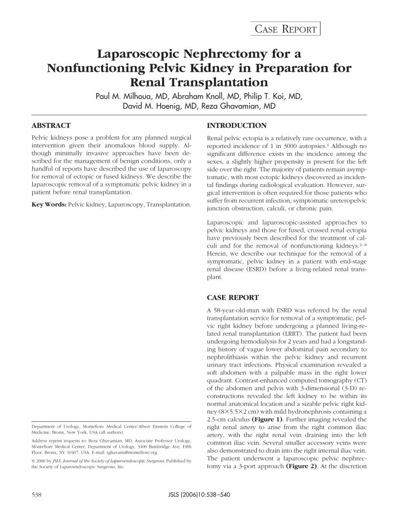
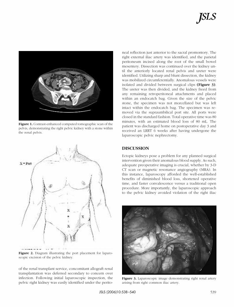
A 58-year-old-man with ESRD was referred by the renaltransplantation service for removal of a symptomatic, pel-vic right kidney before undergoing a planned living-re-lated renal transplantation (LRRT). The patient had beenundergoing hemodialysis for 2 years and had a longstand-ing history of vague lower abdominal pain secondary tonephrolithiasis within the pelvic kidney and recurrenturinary tract infections. Physical examination revealed asoft abdomen with a palpable mass in the right lowerquadrant. Contrast-enhanced computed tomography (CT)of the abdomen and pelvis with 3-dimensional (3-D) re-constructions revealed the left kidney to be within itsnormal anatomical location and a sizable pelvic right kid-ney (8�5.5�2 cm) with mild hydronephrosis containing a2.5-cm calculus (Figure 1). Further imaging revealed theright renal artery to arise from the right common iliacartery, with the right renal vein draining into the leftcommon iliac vein. Several smaller accessory veins werealso demonstrated to drain into the right internal iliac vein.The patient underwent a laparoscopic pelvic nephrec-tomy via a 3-port approach (Figure 2). At the discretion
Department of Urology, Montefiore Medical Center/Albert Einstein College ofMedicine, Bronx, New York, USA (all authors).
Address reprint requests to: Reza Ghavamian, MD, Associate Professor Urology,Montefiore Medical Center, Department of Urology, 3400 Bainbridge Ave, FifthFloor, Bronx, NY 10467, USA. E-mail: [email protected]
© 2006 by JSLS, Journal of the Society of Laparoendoscopic Surgeons. Published bythe Society of Laparoendoscopic Surgeons, Inc.
JSLS (2006)10:538–540538
CASE REPORT
of the renal transplant service, concomitant allograft renaltransplantation was deferred secondary to concern overinfection. Following initial laparoscopic inspection, thepelvic right kidney was easily identified under the perito-
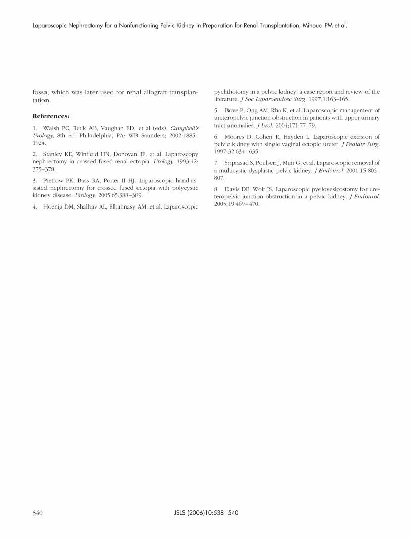
neal reflection just anterior to the sacral promontory. Theright external iliac artery was identified, and the parietalperitoneum incised along the root of the small bowelmesentery. Dissection was continued over the kidney un-til the anteriorly located renal pelvis and ureter wereidentified. Utilizing sharp and blunt dissection, the kidneywas mobilized circumferentially. Anomalous vessels wereisolated and divided between surgical clips (Figure 3).The ureter was then divided, and the kidney freed fromany remaining retroperitoneal attachments and placedwithin an endocatch bag. Given the size of the pelvicstone, the specimen was not morcellated but was leftintact within the endocatch bag. The specimen was re-moved via the supraumbilical port site. All ports wereclosed in the standard fashion. Total operative time was 80minutes, with an estimated blood loss of 80 mL. Thepatient was discharged home on postoperative day 3 andreceived an LRRT 6 weeks after having undergone thelaparoscopic pelvic nephrectomy.
DISCUSSION
Ectopic kidneys pose a problem for any planned surgicalintervention given their anomalous blood supply. As such,adequate preoperative imaging is crucial, whether by 3-DCT scan or magnetic resonance angiography (MRA). Inthis instance, laparoscopy afforded the well-establishedbenefits of diminished blood loss, shortened operativetime, and faster convalescence versus a traditional openprocedure. More importantly, the laparoscopic approachto the pelvic kidney avoided violation of the right iliac
Figure 1. Contrast-enhanced computed tomographic scan of thepelvis, demonstrating the right pelvic kidney with a stone withinthe renal pelvis.
Figure 2. Diagram illustrating the port placement for laparo-scopic excision of the pelvic kidney.
Figure 3. Laparoscopic image demonstrating right renal arteryarising from right common iliac artery.
JSLS (2006)10:538–540 539
fossa, which was later used for renal allograft transplan-tation.
References:
1. Walsh PC, Retik AB, Vaughan ED, et al (eds). Campbell’sUrology, 8th ed. Philadelphia, PA: WB Saunders; 2002;1885–1924.
2. Stanley KE, Winfield HN, Donovan JF, et al. Laparoscopynephrectomy in crossed fused renal ectopia. Urology. 1993;42:375–378.
3. Pietrow PK, Bass RA, Porter II HJ. Laparoscopic hand-as-sisted nephrectomy for crossed fused ectopia with polycystickidney disease. Urology. 2005;65:388–389.
4. Hoenig DM, Shalhav AL, Elbahnasy AM, et al. Laparoscopic
pyelithotomy in a pelvic kidney: a case report and review of theliterature. J Soc Laparoendosc Surg. 1997;1:163–165.
5. Bove P, Ong AM, Rha K, et al. Laparoscopic management ofureteropelvic junction obstruction in patients with upper urinarytract anomalies. J Urol. 2004;171:77–79.
6. Moores D, Cohen R, Hayden L. Laparoscopic excision ofpelvic kidney with single vaginal ectopic ureter. J Pediatr Surg.1997;32:634–635.
7. Sriprasad S, Poulsen J, Muir G, et al. Laparoscopic removal ofa multicystic dysplastic pelvic kidney. J Endourol. 2001;15:805–807.
8. Davis DE, Wolf JS. Laparoscopic pyelovesicostomy for ure-teropelvic junction obstruction in a pelvic kidney. J Endourol.2005;19:469–470.
Laparoscopic Nephrectomy for a Nonfunctioning Pelvic Kidney in Preparation for Renal Transplantation, Mihoua PM et al.
JSLS (2006)10:538–540540