KERATODERMIA BLENNORRHAGICA*sti.bmj.com/content/sextrans/27/3/143.full.pdf · purulent urethral...
7
KERATODERMIA BLENNORRHAGICA* REPORT OF A CASE AND A SUGGESTION CONCERNING ITS NATURE BY G. AUCKLAND Manchester The clinical condition known as Reiter's disease is of interest to venereologists, dermatologists, ophthalmologists, bacteriologists, and physicians. Some doubt exists regarding its cause though many theories have been put forward to explain it. Harkness (1950) gives a detailed account of the condition and reviews the literature thoroughly. He advocates separating the disease into two distinct types, firstly those of dysenteric origin (like Reiter's original case) to be called " dysenteric arthritis ", and, secondly, those of venereal origin to be called " the non-gonococcal syndrome ". He later admits, however, that both types are probably due to the same virus or pleuropneumonia- like organism, the portal of entry being the diseased mucous membrane of either the large intestine or the urethra. It should also be remembered that many workers consider that the disease bears no relation to sexual intercourse and is not connected with venereal disease. The following case history, if the patient's story is believed, would support this view, but the main purpose of this report is to suggest a possible explanation (not new) for the skin lesions (kerato- dermia blennorrhagica) in Reiter's disease. Case Report Mr. N. J. S., aged 39, was first seen on September 13, 1950, when he attended a V.D. clinic. A tall, pale man, he limped into the consulting room complaining of a urethral discharge and pain in the ankles and feet. History.-This was the fourth attack of this condition; the first had occurred in 1934 and had commenced with a urethral discharge, followed a week later by a rash confined to the soles and dorsum of the feet. Three weeks later swelling and pain developed in his knees, wrists, and elbows. He was admitted to hospital and recovery took place in 7 weeks. The second attack, * Received for publication April 18, 1951. in 1938, was similar in all respects to the first but less severe and the patient recovered in 3 weeks. The third attack developed in 1942 whilst serving in the army. He was stationed in the U.K. and had never been abroad. The outbreak was similar in severity to the second and was identical in type to both previous attacks. The present outbreak, his fourth, had commenced in August, 1950, with a urethral discharge, followed by the skin eruption and joint pains exactly as before, but the rash spread to involve his arms, trunk, and scalp. He described this attack as more severe than any of the previous ones. There was an additional symptom of a sensation of numbness in the legs and feet. Close questioning elicited a further symptom, which he stated had been present for 2 or 3 weeks before each attack and which disappeared as soon as the signs appeared. This consisted of an uncontrollable hunger. He stated that he awake once or twice every night and was obliged to " raid the larder " to satisfy his ravenous appetite. Combined with the -hunger was a sense of well-being and physical fitness which was unusual in him as, although rarely confined to bed or off work, he was by nature quiet and indolent. He denied any previous illne3ses except colds. There was no family history of skin disease and between attacks he had no skin lesions of any kind. He had never suffered from gonorrhoea and had never complained of eye symptoms. He was married in 1931, but separated from his wife in 1933, and then had a regular consort until he was re-united with his wife in 1936, since when he had lived a normal married life. He denied extra-marital risk except for the period 1933-36. Examination Genito-Urinary Traci.-A profuse, greyish-white, muco- purulent urethral discharge. The glans penis was congested and bright red in colour. There was no purulent balanitis (Fig. 1, overleaf). Joints.-Visible swelling of the metacarpo-phalangeal joints of the index and middle fingers of the right hand, right knee joint, and both ankle joints. The circumfer- ence of the right knee was 14k in. compared with the left knee which was 13 in. The affected joints and feet were painful when moved and during weight bearing. 143 copyright. on 15 July 2018 by guest. Protected by http://sti.bmj.com/ Br J Vener Dis: first published as 10.1136/sti.27.3.143 on 1 September 1951. Downloaded from
Transcript of KERATODERMIA BLENNORRHAGICA*sti.bmj.com/content/sextrans/27/3/143.full.pdf · purulent urethral...
KERATODERMIA BLENNORRHAGICA*REPORT OF A CASE AND A SUGGESTION CONCERNING ITS NATURE
BY
G. AUCKLANDManchester
The clinical condition known as Reiter's diseaseis of interest to venereologists, dermatologists,ophthalmologists, bacteriologists, and physicians.Some doubt exists regarding its cause though manytheories have been put forward to explain it.Harkness (1950) gives a detailed account of thecondition and reviews the literature thoroughly.He advocates separating the disease into twodistinct types, firstly those of dysenteric origin (likeReiter's original case) to be called " dysentericarthritis ", and, secondly, those of venereal originto be called " the non-gonococcal syndrome ".He later admits, however, that both types areprobably due to the same virus or pleuropneumonia-like organism, the portal of entry being the diseasedmucous membrane of either the large intestine orthe urethra. It should also be remembered thatmany workers consider that the disease bears norelation to sexual intercourse and is not connectedwith venereal disease.The following case history, if the patient's story
is believed, would support this view, but the mainpurpose of this report is to suggest a possibleexplanation (not new) for the skin lesions (kerato-dermia blennorrhagica) in Reiter's disease.
Case ReportMr. N. J. S., aged 39, was first seen on September 13,
1950, when he attended a V.D. clinic. A tall, paleman, he limped into the consulting room complainingof a urethral discharge and pain in the ankles and feet.
History.-This was the fourth attack of this condition;the first had occurred in 1934 and had commenced witha urethral discharge, followed a week later by a rashconfined to the soles and dorsum of the feet. Threeweeks later swelling and pain developed in his knees,wrists, and elbows. He was admitted to hospital andrecovery took place in 7 weeks. The second attack,
* Received for publication April 18, 1951.
in 1938, was similar in all respects to the first but lesssevere and the patient recovered in 3 weeks. The thirdattack developed in 1942 whilst serving in the army.He was stationed in the U.K. and had never beenabroad. The outbreak was similar in severity to thesecond and was identical in type to both previousattacks.The present outbreak, his fourth, had commenced in
August, 1950, with a urethral discharge, followed bythe skin eruption and joint pains exactly as before, butthe rash spread to involve his arms, trunk, and scalp.He described this attack as more severe than any of theprevious ones. There was an additional symptom of asensation of numbness in the legs and feet. Closequestioning elicited a further symptom, which he statedhad been present for 2 or 3 weeks before each attackand which disappeared as soon as the signs appeared.This consisted of an uncontrollable hunger. He statedthat he awake once or twice every night and was obligedto " raid the larder " to satisfy his ravenous appetite.Combined with the -hunger was a sense of well-beingand physical fitness which was unusual in him as,although rarely confined to bed or off work, he was bynature quiet and indolent. He denied any previousillne3ses except colds. There was no family history ofskin disease and between attacks he had no skin lesionsof any kind. He had never suffered from gonorrhoeaand had never complained of eye symptoms.He was married in 1931, but separated from his wife
in 1933, and then had a regular consort until he wasre-united with his wife in 1936, since when he had liveda normal married life. He denied extra-marital riskexcept for the period 1933-36.
ExaminationGenito-Urinary Traci.-A profuse, greyish-white, muco-
purulent urethral discharge. The glans penis wascongested and bright red in colour. There was nopurulent balanitis (Fig. 1, overleaf).
Joints.-Visible swelling of the metacarpo-phalangealjoints of the index and middle fingers of the right hand,right knee joint, and both ankle joints. The circumfer-ence of the right knee was 14k in. compared with theleft knee which was 13 in. The affected joints and feetwere painful when moved and during weight bearing.
143
copyright. on 15 July 2018 by guest. P
rotected byhttp://sti.bm
j.com/
Br J V
ener Dis: first published as 10.1136/sti.27.3.143 on 1 S
eptember 1951. D
ownloaded from

BRITISH JOURNAL OF VENEREAL DISEASES
FIG. 1.- Scattered guttate lesions on trunk,groins, and left leg. Non-suppurativebalanitis is also shown.
FIG. 2.-Many guttate lesions on buttocks andback, especially in lumbar region. Scalyplaques of psoriasis appear on the extensorsurfaces of the elbows.
144
copyright. on 15 July 2018 by guest. P
rotected byhttp://sti.bm
j.com/
Br J V
ener Dis: first published as 10.1136/sti.27.3.143 on 1 S
eptember 1951. D
ownloaded from
KERATODERMIA BLENNORRHAGICA
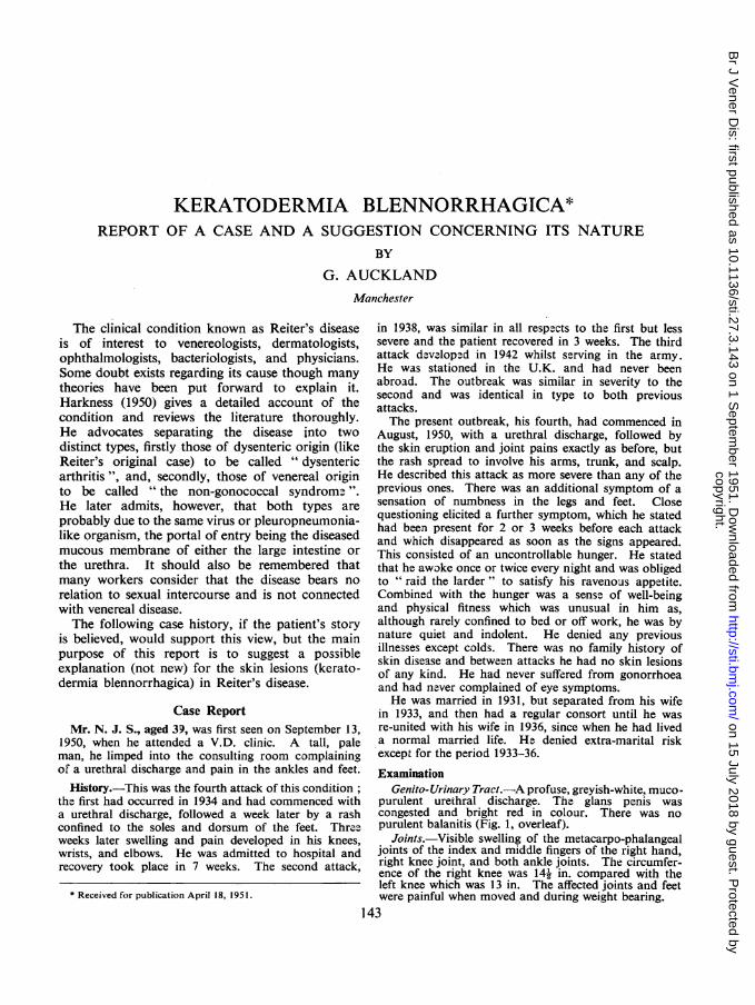
"W. ~~~~~~~thethenar eminences, were several red
papules and nodules, many of which wereThree or four similar lesions were presenton the backs of the hands. The finger
.... $ vnails were thickened at their free edgei and for some distance proximallv wereli :.< .,.:.' j '.< ,l:e F.A' , T ;raised from the nail bed and pitted.1 ~~~~~~~~~~~~Periungually and subungually the skin was
red and scaly with some desquamation.The appearance was that of "pustular"psoriasis (Fig. 4).
Groins.-Many closely set, scaly, guttatelesions were present in the groins and pubicarea, while on the perineum and scrotum
3% Fe._thelesions had become confluent andmoist.
FIG. 3. Palmar lesions showing keratotic papules. Note also scaly,heaped-up condition of peri-ungual and sub-'ungual areas (thumbs).
SkiniScalp.-Scattered throughout the scalp
were several discrete, red, scaly plaqueindistinguishable from psoriasis capitis.Trunk.-On the chest wall, abdomen,
and back were many small red spots, somemacular. some papular, and some coveredwith silvery scales resembling guttate'psoriasis.Arms.-On the extensor surface of both
elbows were plaques identical with psoriasis ---...vulgaris (Fig. 2). A few guttate lesionswere present on the wrists.Hands and Fingers.-On the palms, flexor
surfaces of the fingers, and especially on , a
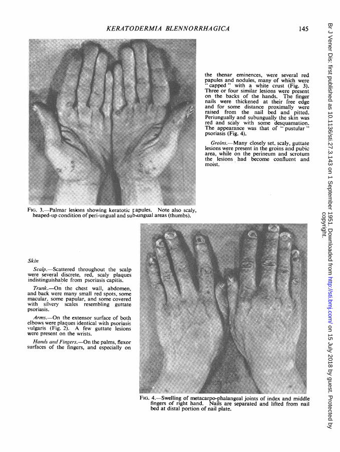
FIG. 4. Swelling of metacarpo-phalangeal joints of index and middlefingers of right hand. Nails are separated and lifted from nailbed at distal portion of nail plate.
145
copyright. on 15 July 2018 by guest. P
rotected byhttp://sti.bm
j.com/
Br J V
ener Dis: first published as 10.1136/sti.27.3.143 on 1 S
eptember 1951. D
ownloaded from

BRITISH JOURNAL OF VENEREAL DISEASES
On the plantar surface of the feet, heels,and metatarsal heads there was markedhyperkeratosis through which could beseen many dark red papules surroundedby a white halo. Localized areas ofhyperkeratosis of a nodular variety werepresent on the insteps (Fig. 6). Theresemblance to "pustular" and " ru-poidal " psoriasis was striking.Eyes.-No abnormality was found.
Treatment.-He was admitted to hospitalthe next day and treated by rest in bed.No active therapy was given, except fora mild salicylic acid ointment for the moreresistant psoriasiform skin lesions.
FIG. 6.-Classical keratodermiablennorrhagica with peeling andexfoliation commencing at base
of toes on right foot.
FIG. 5.-Moist patches on dorsumof feet with marked digital, inter-digital, and peri-ungual scaliness.
Legs, Feet, and Ankles.-On the outerside of the left leg and knee were severalpsoriasis-like lesions. On the inner side ofthe left ankle, the dorsum of both feet, andseveral toes were clearly defined discoidpatches. Some were isolated, others soclosely set that they merged, forming aserpiginous outline. They were red in colourand surrounded by a lip of underminedthickened skin similar to a ruptured bulla.The surface was dry with an attempt at scaleformation, except on one or two of thesmallest (and youngest) lesions where thesurface was still moist. The nails, subun-gual, and periungual areas showed changessimilar to those on the fingers (Fig. 5).
cy.R,.2 .. --:-Xr,- .. @,1.:,,.tg< .0. .. ,. :.i* .- . ...... a |::6 -
.. .. |*. .:: . . : '* . : . : . : :.: '. . . :: . .. :: :.l.: U.! .. S |
*: ::. :.:.::*:: ::: : .!. :.: : :.::. :.. ....; . : .... .... ............................. ...* ....... .... $ ^ S ............................. z
..S: . : '1.... .. i.i.s
i ... 3
*:t:t@g: :X
146
VV
copyright. on 15 July 2018 by guest. P
rotected byhttp://sti.bm
j.com/
Br J V
ener Dis: first published as 10.1136/sti.27.3.143 on 1 S
eptember 1951. D
ownloaded from

KERATODERMIA BLENNORRHAGICA;t- -z .>--.-.-
~~~ I~I-
W U eA.:v.AA . E~~4&~~~~
WAO-~~~~~~~~-
FIG. 7. " Vesicular stage showing intact stratum corneum and parakeratosis. Acanthosis is present withmigration of leucocytes into the epidermis. A leucocytic infiltration is also present in the upper partof the corium.
Progress.-At first he was seen daily and rapid improve-ment was noticed at each visit. The urethral dischargedisappeared first, then the joint swellings, and finallythe skin lesions. The guttate-type spots faded first andthe psoriasis vulgaris lesions (elbows) last. All signsand symptoms had gone by October 19, 1950, when theillness had lasted for 8 weeks. The skin lesions formedthe most striking part of the syndrome, the youngestlesion was always a vesicle but differed from an ordinaryvesicle by the presence of a red papule beneath. Thevesicles soon ruptured leaving the red papule with amoist surface; those on the trunk then became dry andslowly faded; those on the arms, legs, and scalp becameflat and covered with white scales identical with psoriasis;those on the palms and soles became markedly hyper-keratotic.
InvestigationsBiopsy specimens were taken of three isolated
skin lesions from the dorsum of the left foot andboth wrists. All specimens were of approximatelythe same size and were removed whole. It washoped to show the three distinct stages of the evolu-tion of the cutaneous manifestations; vesicular,papular, and psoriasiform. Histological sectionsshowed quite clearly that the three stages did, infact, exist.c
(1) Vesicular Stage.-The stratum comeum is intactand forms a covering to a mass consisting of layers ofparakeratotic horn cells, coagulated exudate, andenmeshed leucocytes. The rete cells are oedematous,and leucocytes are present in the inter-cellular spaces.There is acanthosis. A leucocytic infiltration is presentin the papillary layer of the dermis. The blood vesselsare dilated (Fig. 7).
(2) Papular Stage.-The stratum corneum has dis-appeared. Layers of parakeratotic horn cells rest onthe rete from which the stratum granulosum is absent.There is a marked degree of acanthosis and papillaryoedema. The degree of leucocytic infiltration is less(Fig. 8, overleaf).
(3) Psoriasiform Stage.-The mass of parakeratotichorn cells appears to have pressed the rete into thecorium, forming a flat, plaque-like lesion instead of araised papule. Acanthosis is still present (Fig. 9, over-leaf).
Urethral smears taken on several occasions failedto reveal the gonococcus. Specimens of the urethraldischarge and scrapings from the moist skin lesionswere searched for L. organisms without success.The patient's temperature varied between 98.20
and 100l40F.
147
copyright. on 15 July 2018 by guest. P
rotected byhttp://sti.bm
j.com/
Br J V
ener Dis: first published as 10.1136/sti.27.3.143 on 1 S
eptember 1951. D
ownloaded from

BRITISH JOURNAL* OF VENEREAL DISEASES
FIG. 8.-" Papular " stage showing absence of stratum corneum. The squama crust rests on acanthoticinterpapillary processes.
A full blood count taken on September 15, 1950,showed
Haemoglobin .. .. 102 per cent. (Haldane)Red blood cells .. .. 4,700,000 per c.mm.Colour index .. .. 1-08Total white cells .. 7,800 per c.mm.Polymorphs .. 74 per cent.Lymphocytes .. 19 per cent.Monocytes .. .. 3 per cent.Eosinophiles .. .. 4 per cent.
The Wassermann reaction and gonococcal com-plement-fixation test were negative.The erythrocyte sedimentation rate was 78 mm.
in the first hour and 94 mm. in the second hour.A skiagram of the left hand on September 20,
1950, showed no abnormality.
DiscussionThere can be little doubt that the diagnosis of
Reiter's disease with keratodermia blennorrhagicais the correct one despite the absence of conjuncti-vitis. The abacterial urethritis, polyarthritis, andbalanitis are sufficient evidence alone, and the skineruption confirmed the opinion. Adamson (1920)suggested that keratodermia blennorrhagica was aform of psoriasis, and summarized his conclusionsas follows:
(1) there are cases of arthropathic psoriasis in which thelesions on the palms and soles strikingly recall those ofkeratodermia blennorrhagica;
(2) in many cases of keratodermia blennorrhagicathere are eruptions on the trunk and limbs indistinguish-able from psoriasis;
(3) there are cases in which it is difficult to make adefinite diagnosis between arthropathic psoriasis andkeratodermia blennorhagica;
(4) there is a close similarity between the histopath-ology of the two diseases.He then suggested that if the two conditions are
identical then psoriasis may be caused by a micro-organism more or less closely related to thegonococcus. This view was put forward at a timewhen most workers considered that the gonococcuswas usually responsible for keratodermia blennor-rhagica. For instance, Lees and Percival (1931)state that " clinical evidence points definitely to thecutaneous eruption being a toxic manifestation dueto a gonococcal infection . . .". The majority ofworkers now would probably agree with Harkness(1950) that a virus is the more likely infective agentthough there is often a concomitant gonococcalinfection as well. Dermatologists are still unableto agree on the cause of psoriasis, but they often
148
UT
Vy
copyright. on 15 July 2018 by guest. P
rotected byhttp://sti.bm
j.com/
Br J V
ener Dis: first published as 10.1136/sti.27.3.143 on 1 S
eptember 1951. D
ownloaded from

KERATODERMIA BLENNORRHA GICA
4'~ ~ ~ 4
t S *--;0
FIG. 9.-' Psoriasiform stage. The squama crust, consisting of coagulated exudate, collections ofdegenerated leucocytes, and parakeratotic cells, appears to have pressed the interpapilary processes(rete pegs) into the corium.
observe the sudden and widespread exacerbationsof the condition which occurs when the suffererdevelops a bacterial infection of the throat, sinuses,lungs, or skin. It is not unusual to find that apatient's first attack of psoriasis occurs during orshortly after one of the infectious exanthemata, asevere boil, an attack of tonsillitis or sinusitis,influenza, bronchitis or impetigo. Subsequent out-breaks of psoriasis may then occur only when thepatient again succumbs to a re-infection, or he maybe left with a persistent chronic psoriasis which onlyspreads to distant parts of the body, like palmsand soles, when a re-infection supervenes. It mustnot be thought that psoriasis is entirely due toinfection; it is more probable that some constitu-tional or unknown factor is responsible for a personbeing a potential psoriatic, but an exciting super-added factor is required, acting like a triggermechanism, before clinical manifestations appear.This provoking factor may be mental or physicalshock, worry, trauma, or infection.The histopathological differences in the two
diseases are so slight that they could be accountedfor by differences in the stage of development ofthe lesions examined, not by any fundamental
diagnostic detail. Personally I do not believe thatany differences exist. May it not be then, thatkeratodermia blennorrhagica is psoriasis, provokedby the stimulus of a virus infection; the infectionbeing at the same time responsible for the urethritis,arthritis, and conjunctivitis of Reiter's disease ?
ConclusionIt would seem desirable to substitute the diagnosis
of " Reiter's disease with psoriasis " for the term"keratodermia blennorrhagica ".
SummaryA case of Reiter's disease is reported. A sug-
gestion is made that the cutaneous manifestationsin such cases are in fact psoriasis.
I wish to thank Dr. P. B. Mumford and Dr. R. Leesfor their encouragement, and Mr. J. Hodkinson andMr. P. Morewood for the photographs and microscopicsections.
REFERENCESAdamson, H. G. (1920). Brit. J. Derm. Syph., 32, 183.Harkness, A. H. (1950). " Non-gonococcal Urethritis."
Livingstone, Edinburgh.Lees, D., and Percival, G. H. (1931). Lancet, 2, 1116.
149
copyright. on 15 July 2018 by guest. P
rotected byhttp://sti.bm
j.com/
Br J V
ener Dis: first published as 10.1136/sti.27.3.143 on 1 S
eptember 1951. D
ownloaded from