Kent Academic Repository - Texas A&M University
64
Kent Academic Repository Full text document (pdf) Copyright & reuse Content in the Kent Academic Repository is made available for research purposes. Unless otherwise stated all content is protected by copyright and in the absence of an open licence (eg Creative Commons), permissions for further reuse of content should be sought from the publisher, author or other copyright holder. Versions of research The version in the Kent Academic Repository may differ from the final published version. Users are advised to check http://kar.kent.ac.uk for the status of the paper. Users should always cite the published version of record. Enquiries For any further enquiries regarding the licence status of this document, please contact: [email protected] If you believe this document infringes copyright then please contact the KAR admin team with the take-down information provided at http://kar.kent.ac.uk/contact.html Citation for published version Hawks, John and Elliot, Marina and Schmid, Peter and Churchill, Steven E and de Ruiter, Darryl J and Roberts, Eric M and Hilbert-Wolf, Hannah and Garvin, Heather M and Williams, Scott A and Delezene, Lucas K and Feuerriegel, Elen M and Randolph-Quinney, Patrick and Kivell, Tracy L. and Laird, Myra F and Tawane, Gaokgatlhe and DeSilva, Jeremy M and Bailey, Shara DOI https://doi.org/10.7554/eLife.24232 Link to record in KAR http://kar.kent.ac.uk/61664/ Document Version Publisher pdf
Transcript of Kent Academic Repository - Texas A&M University

Kent Academic RepositoryFull text document (pdf)
Copyright & reuse
Content in the Kent Academic Repository is made available for research purposes. Unless otherwise stated all
content is protected by copyright and in the absence of an open licence (eg Creative Commons), permissions
for further reuse of content should be sought from the publisher, author or other copyright holder.
Versions of research
The version in the Kent Academic Repository may differ from the final published version.
Users are advised to check http://kar.kent.ac.uk for the status of the paper. Users should always cite the
published version of record.
Enquiries
For any further enquiries regarding the licence status of this document, please contact:
If you believe this document infringes copyright then please contact the KAR admin team with the take-down
information provided at http://kar.kent.ac.uk/contact.html
Citation for published version
Hawks, John and Elliot, Marina and Schmid, Peter and Churchill, Steven E and de Ruiter, DarrylJ and Roberts, Eric M and Hilbert-Wolf, Hannah and Garvin, Heather M and Williams, ScottA and Delezene, Lucas K and Feuerriegel, Elen M and Randolph-Quinney, Patrick and Kivell,Tracy L. and Laird, Myra F and Tawane, Gaokgatlhe and DeSilva, Jeremy M and Bailey, Shara
DOI
https://doi.org/10.7554/eLife.24232
Link to record in KAR
http://kar.kent.ac.uk/61664/
Document Version
Publisher pdf

*For correspondence: Lee.
Competing interests: The
authors declare that no
competing interests exist.
Funding: See page 57
Received: 12 December 2016
Accepted: 18 April 2017
Published: 09 May 2017
Reviewing editor: George H
Perry, Pennsylvania State
University, United States
Copyright Hawks et al. This
article is distributed under the
terms of the Creative Commons
Attribution License, which
permits unrestricted use and
redistribution provided that the
original author and source are
credited.
New fossil remains of Homo naledi fromthe Lesedi Chamber, South AfricaJohn Hawks1,2, Marina Elliott1, Peter Schmid1,3, Steven E Churchill1,4,Darryl J de Ruiter1,5, Eric M Roberts6, Hannah Hilbert-Wolf6,Heather M Garvin1,7,8, Scott A Williams1,9,10, Lucas K Delezene1,11,Elen M Feuerriegel1,12, Patrick Randolph-Quinney1,13,14, Tracy L Kivell1,15,16,Myra F Laird1,17, Gaokgatlhe Tawane1, Jeremy M DeSilva1,18, Shara E Bailey9,10,Juliet K Brophy1,19, Marc R Meyer20, Matthew M Skinner1,15,16,Matthew W Tocheri21,22, Caroline VanSickle1,2,23, Christopher S Walker1,4,24,Timothy L Campbell5, Brian Kuhn25, Ashley Kruger1,26, Steven Tucker1,Alia Gurtov1,2, Nompumelelo Hlophe1, Rick Hunter1, Hannah Morris1,27,Becca Peixotto1,28, Maropeng Ramalepa1, Dirk van Rooyen1,Mathabela Tsikoane1, Pedro Boshoff1, Paul HGM Dirks6, Lee R Berger1*
1Evolutionary Studies Institute, University of the Witwatersrand, Wits, South Africa;2Department of Anthropology, University of Wisconsin, Madison, United States;3Anthropological Institute and Museum, University of Zurich, Winterthurerstr,Zurich, Switzerland; 4Department of Evolutionary Anthropology, Duke University,Durham, United States; 5Department of Anthropology, Texas A&M University,College Station, United States; 6Geosciences, College of Science and Engineering,James Cook University, Townsville, Australia; 7Department of Anthropology/Archaeology, Mercyhurst University, Erie, United States; 8Department of AppliedForensic Sciences, Mercyhurst University, Erie, United States; 9Center for the Studyof Human Origins, Department of Anthropology, New York University, New York,United States; 10New York Consortium in Evolutionary Primatology, New York,United States; 11Department of Anthropology, University of Arkansas, Fayetteville,United States; 12Department of Anthropology, University of Washington, Seattle,United States; 13School of Anatomical Sciences, University of the WitwatersrandMedical School, Johannesburg, South Africa; 14School of Forensic and AppliedSciences, University of Central Lancashire, Preston, United Kingdom; 15School ofAnthropology and Conservation, University of Kent, Canterbury, United Kingdom;16Department of Human Evolution, Max Planck Institute for EvolutionaryAnthropology, Leipzig, Germany; 17Department of Organismal Biology andAnatomy, University of Chicago, Chicago, United States; 18Department ofAnthropology, Dartmouth College, Hanover, United States; 19Department ofGeography and Anthropology, Louisiana State University, Baton Rouge, UnitedStates; 20Department of Anthropology, Chaffey College, Rancho Cucamonga,United States; 21Department of Anthropology, Lakehead University, Thunder Bay,Canada; 22Human Origins Program, Department of Anthropology, National Museumof Natural History, Smithsonian Institution, Washington, United States;23Department of Anthropology, Bryn Mawr College, Bryn Mawr, United States;24Department of Molecular Biomedical Sciences, College of Veterinary Medicine,North Carolina State University, Raleigh, United States; 25Department of Geology,University of Johannesburg, Johannesburg, South Africa; 26School of Geosciences,University of the Witwatersrand, Johannesburg, South Africa; 27Department of
Hawks et al. eLife 2017;6:e24232. DOI: 10.7554/eLife.24232 1 of 63
RESEARCH ARTICLE

Forestry and Natural Resources, University of Georgia, Athens, United States;28Department of Anthropology, American University, Washington, United States
Abstract The Rising Star cave system has produced abundant fossil hominin remains within the
Dinaledi Chamber, representing a minimum of 15 individuals attributed to Homo naledi. Further
exploration led to the discovery of hominin material, now comprising 131 hominin specimens,
within a second chamber, the Lesedi Chamber. The Lesedi Chamber is far separated from the
Dinaledi Chamber within the Rising Star cave system, and represents a second depositional context
for hominin remains. In each of three collection areas within the Lesedi Chamber, diagnostic
skeletal material allows a clear attribution to H. naledi. Both adult and immature material is present.
The hominin remains represent at least three individuals based upon duplication of elements, but
more individuals are likely present based upon the spatial context. The most significant specimen is
the near-complete cranium of a large individual, designated LES1, with an endocranial volume of
approximately 610 ml and associated postcranial remains. The Lesedi Chamber skeletal sample
extends our knowledge of the morphology and variation of H. naledi, and evidence of H. naledi
from both recovery localities shows a consistent pattern of differentiation from other hominin
species.
DOI: 10.7554/eLife.24232.001
IntroductionThe Rising Star cave system (26˚10130 0 S; 27˚4204300 E, Figure 1) in the Cradle of Humankind World
Heritage Site, Gauteng Province, South Africa, is known for the discovery in 2013 of more than
1,550 fossils representing a novel hominin species, Homo naledi (Berger et al., 2015; Dirks et al.,
2015). These remains, representing at least 15 individuals of various ages at death, were recovered
from a deep chamber (30 m below ground surface), named the Dinaledi Chamber.
Additional fossil hominin material was subsequently discovered in the Lesedi Chamber of the
cave system in November 2013 by Rick Hunter and Steven Tucker. The deposition of sediment and
skeletal remains in the Lesedi Chamber has no direct geological connection to the Dinaledi Cham-
ber. In the time following the first discovery of hominin material in the Lesedi Chamber, excavators
have recovered 131 hominin specimens within three discrete collection areas. The sedimentary con-
text of the three collection areas is broadly similar, but we have not yet established whether the fos-
sil material resulted from a single depositional episode or from multiple distinct events.
We approached the hominin skeletal remains from the Lesedi Chamber with the aim of identifying
elements, assessing the number of individuals represented by the material, and determining the tax-
onomic identity of the sample. Preliminary examination of the hominin remains suggested that they
are morphologically consistent with H. naledi. To test this hypothesis, we carried out systematic com-
parisons, employing the taxonomic diagnosis of this species (Berger et al., 2015) and focusing upon
those characters that distinguish H. naledi from other hominin taxa. We also present essential con-
textual information to place the specimens within the Lesedi Chamber and provide descriptions of
the hominin specimens, focusing upon those features that contribute to the taxonomic diagnosis of
the sample. All identifiable hominin fragments, including those that do not present information use-
ful to taxonomic diagnosis, are listed in Table 1.
Results
Name of the chamberFollowing the University of the Witwatersrand’s fossil-numbering system (Zipfel and Berger, 2009),
this second H. naledi locality has been designated U.W. 102. The chamber itself has been named the
Lesedi Chamber, a word meaning ‘light’ in Setswana. By contrast, the Dinaledi Chamber was num-
bered site U.W. 101. Excavations in the Lesedi Chamber have been carried out in three areas, desig-
nated U.W. 102a, U.W. 102b, and U.W. 102c.
Hawks et al. eLife 2017;6:e24232. DOI: 10.7554/eLife.24232 2 of 63
Research article Genomics and Evolutionary Biology

Location of the Lesedi ChamberThe Lesedi Chamber is in the central sector of the Rising Star system (Figure 2), at a depth of ~30 m
from the surface directly above the chamber. All measurements reported here are approximate. The
first fossil deposit to be recognized (U.W. 102a) is located just off the southwest corner of the
North-South Fracture Passage, a northern arm of the Lesedi Chamber. This fossil deposit is approxi-
mately 60 m NNE in a straight line from the Dinaledi Chamber. There is no straight-line route
between the Dinaledi and Lesedi Chambers, and the shortest traversable route between the two
areas is almost 145 m. There are currently four access routes from the surface to the Lesedi Cham-
ber. The most accessible of these currently follows an 86 m downward-sloping path with several nar-
row passages and short climbs, but only one squeeze and no significant crawls. This has been the
main access route for excavators. The other three routes are each substantially more challenging.
Location of skeletal material within the Lesedi ChamberIn addition to the first fossil deposit to be recognized in the chamber, two additional concentrations
of skeletal material have been identified to date (Figure 3), and we have designated these as areas
102a, 102b, and 102c. We began investigating each of these areas because team members noticed
hominin fossil material exposed on sediment surfaces. The discovery of 102a by Rick Hunter and Ste-
ven Tucker led to the initial scientific investigation of the chamber; discoveries of both 102b and
eLife digest Species of ancient humans and the extinct relatives of our ancestors are typically
described from a limited number of fossils. However, this was not the case with Homo naledi. More
than 1500 fossils representing at least 15 individuals of this species were unearthed from the Rising
Star cave system in South Africa between 2013 and 2014. Found deep underground in the Dinaledi
Chamber, the H. naledi fossils are the largest collection of a single species of an ancient human-
relative discovered in Africa.
After the discovery was reported, a number of questions still remained. These questions
included: why were so many fossils from a single species found at the one site, and how did they
come to rest so far into the cave system? Possible explanations such as H. naledi living in the cave or
being washed in by a flood were considered but ruled out. Instead, the evidence was largely
consistent with intact bodies being deliberately disposed of in the cave and then decomposing.
Now, Hawks et al. – who include many of the researchers who were involved in the discovery of
H. naledi – report that yet more H. naledi fossils have been unearthed from a second chamber in the
Rising Star cave system, the Lesedi Chamber. The chamber is 30 meters below the surface and there
is no direct route between it and the Dinaledi Chamber. Again, the evidence is most consistent with
the bodies arriving intact into the chamber, and there were no signs that the remains had been
exposed to the surface environment.
Also like the Dinaledi Chamber, no remains of other ancient humans or their relatives were found
in the Lesedi Chamber. In total, 133 fossils of H. naledi have been found in this second chamber
representing at least three individuals: two adults and a juvenile. However, and as Hawks et al. point
out, only a small volume of the chamber has been excavated so far, and so there are likely more
fossils still to be found.
The fossils in the Lesedi Chamber are similar to those found before but include intact examples
of bones, like the collarbone, that were previously known only from fragments. Perhaps the most
impressive among the new fossils is a relatively complete skull that is part of a partial skeleton. The
skull could have housed a brain that was 9% larger than the maximum estimate calculated from the
previous H. naledi fossils.
Though these new fossils provide us with yet more information about H. naledi, some questions
still remain unanswered – the material from the Lesedi Chamber is undated, for example. However,
a related study by Dirks et al. does give an estimate for the age of the fossils from the Dinaledi
Chamber, while Berger et al. provide an explanation for why this date might be much younger than
was previously predicted.
DOI: 10.7554/eLife.24232.002
Hawks et al. eLife 2017;6:e24232. DOI: 10.7554/eLife.24232 3 of 63
Research article Genomics and Evolutionary Biology

102c were made by Hannah Hilbert-Wolf during the course of geological sampling of the chamber.
These three areas do not represent a systematic sampling of the chamber’s contents and we have
excavated only a very small sediment volume, less than 200 L (<0.2 m3) in total from all three areas.
The chamber contains a much greater volume of sediment and we do not know what density of fossil
bone it may contain beyond our samples.
U.W. 102a is located at the entrance of a 20–50-cm-wide blind tunnel, which is 1.8 m long in total.
The blind tunnel leads off of the southwest corner of the North-South Fracture Passage (Figure 3).
Fossil material was exposed on the surface within this blind tunnel at the time of discovery. We have
excavated the proximal 1.5 m of this blind tunnel, which has a tapering width of less than 50 cm in
our excavation unit. The depth of excavation in this area is a maximum of 40 cm. The deposit in this
area is a weakly stratified, unlithified mud-clast breccia. Most hominin material has been recovered
from an approximately 10-cm-thick horizon of fine-grained mud-clast breccia, beneath a surface
layer of ~2 cm of lighter brown-colored mudstone. This deposit is the source of at least some of the
sediments that slope from the blind tunnel into the Antechamber. Fossil material attributed to 102a
has also been recovered from the surface within the North-South Fracture Passage.
U.W. 102b is a sediment deposit on a horizontal chert shelf 80 cm above the cave floor along the
western wall of the Antechamber. It is also dominated by unlithified mud-clast breccia. The 102b
deposit is located ~3.8 m to the south and 1.8 m below the 102a deposit. After the discovery of
hominin fossil material on the surface here, we undertook limited excavations, with a total volume
of ~20 L.
U.W. 102c is a small unlithified sediment deposit within an irregular dissolution cavity on the north
wall of the east–west-running Cake-Icing Fracture. This deposit is 1.3 m above the current cave floor.
It is 11.6 m from U.W. 102a, and 0.3 m below the level of the 102a fossils. We have excavated this
small sediment pocket in its entirely, with a total volume of approximately 2 L.
Geological work to characterize the Lesedi Chamber depositional history is underway. The stratig-
raphy is complex, with some hominin and faunal material concentrated in deposits of poorly consoli-
dated mud-clast breccia, generally similar to the facies in the Dinaledi Chamber (Dirks et al., 2015).
Notably, the fossil material in the Lesedi Chamber is concentrated in minor side fractures, dissolution
cavities, or on chert shelves well above the current chamber floor. Our working hypothesis is that
the chamber once held a greater volume of sediment than is present today, and when sediment
eroded from the chamber, erosional remnants remained in protected fractures, wall cavities, and on
chert shelves along the chamber walls. This and other indications of reworking of the deposits make
it uncertain how much of the hominin assemblage may remain in its primary depositional context.
Figure 1. Geographical location of the Rising Star cave in the Cradle of Humankind UNESCO World Heritage Site.
DOI: 10.7554/eLife.24232.003
Hawks et al. eLife 2017;6:e24232. DOI: 10.7554/eLife.24232 4 of 63
Research article Genomics and Evolutionary Biology

Table 1. Hominin fossil material from the Lesedi Chamber. All diagnostic hominin specimens are listed, with attribution to element.
Specimens that have been refitted are not listed separately. Most Locality 102a cranial fragments are presumed to be part of LES1 and
are not listed separately.
Specimen number Element Notes
LOCALITY 102a
LES1 cranium constituted of 57 specimens, not listed separately
U.W. 102a-001 proximal right femur
U.W. 102a-002 proximal right humerus
U.W. 102a-003 proximal left femur
U.W. 102a-004 distal left femur
U.W. 102a-010 right scapula fragment acromion
U.W. 102a-013 humeral head fragments
U.W. 102a-015 right proximal ulna
U.W. 102a-018 long bone fragment immature
U.W. 102a-019 partial rib
U.W. 102a-020 right ulna fragment
U.W. 102a-021 right clavicle
U.W. 102a-025 right radius shaft fragment
U.W. 102a-028 right fourth metacarpal
U.W. 102a-036 T10 vertebra
U.W. 102a-039 rib fragments
U.W. 102a-040 long bone shaft fragment
U.W. 102a-117 right scaphoid
U.W. 102a-138 right ilium fragments immature
U.W. 102a-139 L5 vertebra fragments
U.W. 102a-148 sternum fragment
U.W. 102a-151 T11 vertebra
U.W. 102a-152 rib fragments
U.W. 102a-154 T12 and L1 vertebrae found in articulation
U.W. 102a-155 mid-thoracic vertebral body
U.W. 102a-171 atlas fragment
U.W. 102a-172 atlas fragment
U.W. 102a-189 rib fragment
U.W. 102a-195 rib fragment
U.W. 102a-206 left clavicle fragment
U.W. 102a-207 rib fragment
U.W. 102a-210 sacral element immature, possibly S1
U.W. 102a-231 rib fragment
U.W. 102a-232 rib fragment
U.W. 102a-236 humerus head fragment
U.W. 102a-239 left clavicle fragment
U.W. 102a-247 right scapula fragment coracoid process
U.W. 102a-250 right first rib
U.W. 102a-252 rib fragment
U.W. 102a-256 left scapula fragment portion of body, spine, and acromion
U.W. 102a-257 left proximal humerus
Table 1 continued on next page
Hawks et al. eLife 2017;6:e24232. DOI: 10.7554/eLife.24232 5 of 63
Research article Genomics and Evolutionary Biology

Hominin material from 102aHominin material from the 102a area includes 118 identifiable specimens (Table 1; Figure 4). Fifty-
seven of these are cranial and dental specimens that either refit directly or are morphologically com-
patible with a nearly complete fossil cranium, designated LES1 (Figure 5). Hominin postcranial
remains from locality 102a include 61 identified specimens that represent a minimum of 31 postcra-
nial elements, not counting ribs. These include a minimum of two partial femora, two partial humeri,
one complete clavicle and two clavicular fragments, two partial ulnae, several fragments of
scapula and radius, many rib fragments, a near-complete first rib, a partial sternum, four hand and
wrist elements, an immature ilium and sacrum fragment, and a partial thoracic and lumbar vertebral
column. Every anatomical region of the skeleton is represented with the notable exceptions of tibia,
fibula and pedal remains.
Table 1 continued
Specimen number Element Notes
U.W. 102a-279 left scapula fragment partial glenoid fossa
U.W. 102a-280 rib fragment
U.W. 102a-300 vertebral fragment
U.W. 102a-306 L4 vertebra body
U.W. 102a-322 L2 vertebra body
U.W. 102a-337 vertebral fragment neural arch
U.W. 102a-348 right pubic ramus fragment
U.W. 102a-349 vertebral fragment neural arch
U.W. 102a-358 rib fragments
U.W. 102a-360 vertebral fragment
U.W. 102a-455 ulna shaft fragment
U.W. 102a-456 ulna shaft fragment
U.W. 102a-470 rib fragments
U.W. 102a-471 right distal radius fragment
U.W. 102a-474 long bone fragment immature
U.W. 102a-476 right capitate
U.W. 102a-477 partial right lunate
U.W. 102a-479 rib fragment
LOCALITY 102b
U.W. 102b-178 LI2
U.W. 102b-437 rdm2
U.W. 102b-438 right mandibular corpus fragment immature, RP4 in crypt
U.W. 102b-502 cranial fragments
U.W. 102b-503 RP4 crown
U.W. 102b-506 cranial fragment
U.W. 102b-507 cranial fragment
U.W. 102b-509 cranial fragment
U.W. 102b-511 LC1 crown
U.W. 102b-514 cranial fragment
U.W. 102b-515 LI2
U.W. 102b-516 cranial fragment
LOCALITY 102c
U.W. 102 c-589 left mandibular fragment LM1 and LM2 in place
DOI: 10.7554/eLife.24232.004
Hawks et al. eLife 2017;6:e24232. DOI: 10.7554/eLife.24232 6 of 63
Research article Genomics and Evolutionary Biology
LES1The LES1 cranium is fragmented but is represented by most of the vault and part of the face (Fig-
ure 5). To date, we have successfully refitted the near-complete mandible, the near-complete right
maxilla, a partial palate and a partial left maxillary dental row, and a partial vault including the near-
complete frontal, left and right nasal and left lacrimal bones, near-complete left parietal and tempo-
ral, partial right parietal, and a portion of left occipital. LES1 has a complete adult dentition except
for the crowns of the lower left central and lateral incisors. The face is reconstructed from the partial
right maxillary bone, including the frontal process, which refits to the right nasal bone and frontal.
Figure 2. Location of the Lesedi Chamber (U.W.102) in the Rising Star system (red circle). The Dinaledi Chamber
(U.W. 101) is marked by a yellow circle, while three surface entrances into the system are marked by blue circles.
DOI: 10.7554/eLife.24232.005
Hawks et al. eLife 2017;6:e24232. DOI: 10.7554/eLife.24232 7 of 63
Research article Genomics and Evolutionary Biology

Figure 3. Schematic of the Lesedi Chamber, showing the three hominin-bearing collection areas: U.W.102a, 102b,
and 102c.
DOI: 10.7554/eLife.24232.006
Hawks et al. eLife 2017;6:e24232. DOI: 10.7554/eLife.24232 8 of 63
Research article Genomics and Evolutionary Biology

Figure 4. Skeletal material from locality 102a provisionally assigned to the LES1 skeleton. The adult cranial
material from 102a all belongs to a single cranium; most of the adult postcranial material probably belongs to the
same individual. The adult cranial and postcranial material is shown here, except for the U.W. 102a-001 femur. The
possibility that the femora represent two adult individuals makes it unclear which femur may be attributable to the
skeleton; for the purposes of illustration, the U.W. 102a-003/U.W. 102a-004 femur is included in this photograph.
DOI: 10.7554/eLife.24232.007
Hawks et al. eLife 2017;6:e24232. DOI: 10.7554/eLife.24232 9 of 63
Research article Genomics and Evolutionary Biology

Figure 5. LES1 cranium. Clockwise from upper left: three-quarter, frontal, superior and left lateral views. Fragments of the right temporal, the parietal
and the occipital have also been recovered (not pictured), but without conjoins to the reconstructed vault or face. Scale bar = 5 cm.
DOI: 10.7554/eLife.24232.008
Hawks et al. eLife 2017;6:e24232. DOI: 10.7554/eLife.24232 10 of 63
Research article Genomics and Evolutionary Biology

The left mandibular ramus is well-enough preserved to allow a rough estimation of the condyle posi-
tion, enabling an approximation of the midsagittal contour of the face (Figure 5).
All additional cranial fragments in the present 102a collection are non-duplicative with this refit-
ted vault and face, and where they represent the opposite side of the vault, they match in morpho-
logical detail. However, many of the fragments lack clear refits with the existing vault or maxillary
portions. Further physical reconstruction of the cranium will await fragments that may emerge from
excavation in the future. The refitted vault, with the application of virtual mirror reconstruction, is
sufficient to allow an estimate of endocranial volume of approximately 610 ml (Figure 6).
Most of the features of the LES1 vault are characteristic of H. naledi from the Dinaledi Chamber
(Supplementary file 1; Figure 7). The LES1 vault is relatively short anteroposteriorly, without the
elongation and sharp occipital angulation found in H. erectus. LES1 exhibits mild frontal and parietal
bossing, similar to H. naledi DH3. Other features on the vault that are consistent with H. naledi
include prelambdoidal flattening, limited postorbital constriction, widely spaced temporal lines, a
continuous supraorbital torus with a supratoral sulcus, an occipital torus, and a marked angular torus.
In the temporal region, LES1 has an anteroinferiorly oriented root of the zygomatic process of the
temporal, a medially positioned mandibular fossa, a small and obliquely oriented external auditory
meatus, a projecting Eustachian process, a small vaginal process, a weak crista petrosa, a triangular-
shaped mastoid process, and a small suprameatal spine. Each of these traits characterizes the Dina-
ledi H. naledi sample (Berger et al., 2015; Laird et al., 2017). Some of these traits occur individually
in other species, including H. erectus, H. habilis, H. rudolfensis, and Australopithecus sediba, but
they have never been found in combination except in H. naledi (Figure 7).
The maxilla and mandible of LES1 are also consistent with the Dinaledi H. naledi sample
(Supplementary file 1; Figures 8, 9, 10 and 11). The maxilla has a mediolaterally flattened subnasal
region, a parabolic dental arcade, and an anteriorly shallow palate. The mandible of LES1 has a grac-
ile mandibular corpus, a vertical mandibular symphysis with weak mentum osseum, a steeply inclined
lingual alveolar plane, weak inferior and absent superior transverse tori, continuous and deeply exca-
vated anterior and posterior subalveolar fossae, mental foramina positioned above mid-corpus
height, well defined ectoangular and endoangular tuberosities, and a root of the ascending ramus
that originates at the mesial border of the M2. Again, many of these traits can be found individually
in other hominin species, but in combination, they are uniquely found in H. naledi.
The teeth exhibit moderate occlusal wear on the second and third molars, trending toward near-
complete dentine exposure on the occlusal surfaces of first molars and substantial removal of occlu-
sal detail of the anterior dentition. The dental morphology of LES1 is entirely consistent with the
Figure 6. Digital reconstruction of endocranial volume in LES1. The refitted calvaria was mirrored and filled,
resulting in a volume estimate of 610 ml. Scale sphere = 10 mm.
DOI: 10.7554/eLife.24232.009
Hawks et al. eLife 2017;6:e24232. DOI: 10.7554/eLife.24232 11 of 63
Research article Genomics and Evolutionary Biology

Figure 7. Frontal and vault morphology in H. naledi compared to that in other hominin species. Several of the
crania pictured here are similar to H. naledi in endocranial volume, including Sts 5, MH1, KNM-ER 1813, and
D2282, representing four different species. However, these skulls contrast strongly in other features. H. erectus is
highly variable in size, as illustrated here by D2282 from Dmanisi, Georgia, one of the smallest and earliest H.
erectus crania, and the L2 cranium from Zhoukoudian, China, one of the largest and latest H. erectus specimens.
Figure 7 continued on next page
Hawks et al. eLife 2017;6:e24232. DOI: 10.7554/eLife.24232 12 of 63
Research article Genomics and Evolutionary Biology

Dinaledi sample of H. naledi (Figures 10 and 12). The mesiodistal and buccolingual (or labiolingual)
crown dimensions of all the LES1 teeth fall within the range of the Dinaledi dental sample, except
Figure 7 continued
The relatively early KNM-ER 3733 has a size and endocranial volume close to the mean for H. erectus. Cranial
remains that are attributed to H. erectus share a combination of anatomical features despite their diversity in size.
Many such features of H. erectus are also shared with H. naledi, H. habilis, or Au. sediba, and notably, the
differences in the frontal and vault between KNM-ER 1813 (H. habilis) and KNM-ER 1470 (H. rudolfensis) are mostly
features that the smaller KNM-ER 1813 shares with H. naledi, H. erectus, and Au. sediba. The H. naledi skulls share
some aspects of frontal morphology with Au. sediba, H. habilis and H. erectus that are not found in Au. africanus
or H. rudolfensis, including frontal bossing and a supratoral sulcus. Two additional traits of the H. naledi anterior
vault are shared with Au. sediba and H. erectus:slight postorbital construction and a posterior position of the
temporal crest on the supraorbital torus. More posteriorly on the vault, H. naledi further shares an angular torus
with H. erectus, and some individuals also have sagittal keeling. Both of these traits are also present in some
archaic humans. Some H. naledi crania, such as DH3, are substantially smaller than any H. erectus cranium, and the
small size and thin vault bone of even the largest H. naledi skull, LES1, are outliers compared to H. erectus,
matched only by some Dmanisi crania. The facial morphology of H. naledi is more distinct from those of H. erectus
and H. habilis. The nasal bones of LES1 do not project markedly anteriorly, although like many specimens of H.
erectus, LES1 has a projecting nasal spine. LES1 has a relatively flat lower face, with a transversely concave clivus
and incisors that project only slightly past the canines. This morphology is similar but less extreme than that found
in KNM-ER 1470 of H. rudolfensis, and is not shared with the other species pictured here. H. naledi has several
distinctive features of the temporal bone that are absent from or found in only a few specimens of the other
species pictured, including a laterally inflated mastoid process (comparable to some specimens of Au. afarensis), a
weak or absent crista petrosa (comparable to Au. afarensis), and a small external auditory meatus (comparable to
KNM-WT 40000 of Kenyanthropus platyops [Leakey et al., 2001]). In this illustration, KNM-ER 1813, KNM-ER 1470,
KNM-ER 3733, and ZKD L2 are represented by casts. Because these images are in a nonstandard orientation, scale
is approximate.
DOI: 10.7554/eLife.24232.010
Figure 8. LES1 mandible compared to the DH1 holotype mandible of H. naledi. In each pair, LES1 is on the left and DH1 on the right. Top left:
anterior view. Top right: occlusal view. Bottom left: left lateral view. Bottom right: posterior view. Scale bar = 2 cm.
DOI: 10.7554/eLife.24232.011
Hawks et al. eLife 2017;6:e24232. DOI: 10.7554/eLife.24232 13 of 63
Research article Genomics and Evolutionary Biology

for those teeth where interproximal wear has clearly reduced the mesiodistal dimension (Table 2;
Figure 13). The P3 crowns are worn, but they are roughly symmetrical about their mesiodistal axis in
occlusal view; they are fully bicuspid and multirooted, with a smaller circular mesiobuccal root and
larger, more platelike, distal root. This configuration is repeated throughout the Dinaledi dental
assemblage. The shared overall P3 morphology of LES1 and the Dinaledi sample is distinctive in H.
naledi and not observed in other species of hominins (Figure 12; Berger et al., 2015). The P3 and
P4 are both three-rooted, with two ovoid roots present buccally and a larger ovoid root present lin-
gually. The roots are not widely splayed as in some other multi-rooted hominins, and especially for
the P4, the buccal roots are closely packed in buccal view. This root configuration is seen in the H.
naledi type specimen, U.W. 101–1277. The mandibular canine crowns have asymmetrically placed
crown shoulders, with the mesial more apically placed than the distal. Further, the distal shoulder is
formed by an accessory cuspule. These features are strongly distinctive in H. naledi (Berger et al.,
2015), with only a few specimens of H. erectus approaching this canine configuration. None of the
molars exhibit any evidence of supernumerary cusps, and cingular features, such as the protostylid
and Carabelli’s feature, are either absent or weakly developed and are expressed independently of
the grooves of the crown. The molar size gradient in the LES1 mandible is M1 < M2 < M3 as in the
Dinaledi Chamber sample of H. naledi (Figure 10). The Dinaledi Chamber includes no maxillary den-
tition with all three molars in place, but U.W. 101–1269 is a LM3 that exhibits a mesial interproximal
facet that matches the distal facet of the LM2 present in the U.W. 101–1277 (DH1) maxilla. If these
specimens do represent a single individual, then the maxillary molar gradient for this specimen
would be M1 < M3 < M2, which is also seen in the LES1 maxilla. In total, these dental features are
within the known range for H. naledi in every instance and distinguish LES1 clearly from all other
hominin species.
The LES1 cranium does exhibit some traits that differ from comparable examples in the Dinaledi
Chamber. The cranium is slightly larger overall, with an estimated endocranial volume of approxi-
mately 610 ml, and this larger size is reflected in the external vault measurements. Previously, the
largest known H. naledi endocranium was DH1 at approximately 560 ml (Berger et al., 2015). LES1
contrasts in morphological features with the small DH3 cranium in ways that have been observed
Figure 9. Comparison of LES1 maxilla to the DH1 holotype maxilla of H. naledi. In each pair, LES1 is on the left and DH1 on the right. Top left: anterior
view. Top right: right (LES1) and left (DH1) lateral view. Bottom: occlusal view. Scale bar = 2 cm.
DOI: 10.7554/eLife.24232.012
Hawks et al. eLife 2017;6:e24232. DOI: 10.7554/eLife.24232 14 of 63
Research article Genomics and Evolutionary Biology

when comparing male individuals with female individuals in other hominin species. The supraorbital
torus of LES1 is more pronounced than that in the small DH3 individual. LES1 has a stronger supra-
mastoid/suprameatal crest, a larger mastoid process, and a more marked pterygoid insertion
when compared to the U.W. 101–361 mandible. Although LES1 is outside of the endocranial volume
range of specimens presently attributed to H. naledi, the larger size and more robust features of
LES1 are consistent with the combination of cranial and mandibular characters in H. naledi.
Figure 10. Mandibular and dental anatomy in H. naledi compared to other species of Homo. Right demi-mandibles attributed to H. rudolfensis, H.
habilis, H. naledi, H. erectus, and H. sapiens are pictured. All mandibles are aligned using the line marking the distal edge of the first molar. Each of the
six horizontal lines corresponds to the edges of teeth in the DH1 mandible, the holotype specimen of H. naledi, with corresponding teeth labeled to
the left. Using these lines, it is apparent which specimens have longer premolars and first molars, and which have longer second and third molars
compared to DH1. The dentition of the LES1 mandible has been affected by interproximal wear, resulting in shorter mesiodistal measurements.
Mandibular morphology and dental proportions vary slightly among most species of Homo, particularly in comparison with the large differences in
dental proportions among species of Australopithecus and Paranthropus. Still, H. naledi is clearly distinguishable from other species of Homo
(Berger et al., 2015; Laird et al., 2017). Fossils of H. rudolfensis, H. habilis, and H. erectus differ from H. naledi in the proportions of different parts of
the postcanine tooth row and in features of the mandibular corpus. H. erectus. While fossils attributed to H. erectus vary in dental proportions, the
early African and Georgian fossil specimens (here represented by KNM-ER 992, D211 and D2600) have larger first molars than H. naledi, comparable
premolar sizes, and highly variable second and third molar sizes. The mandibles attributed to H. erectus mostly have greater corpus height than H.
naledi mandibles and are highly diverse in corpus breadth, symphyseal thickness, and robusticity. Many have a strong post-incisive planum, most
obvious in D2600 (shown). All three also differ from H. naledi in the crown complexity of their molars and premolar morphology, as illustrated in more
detail in Figure 12. Some specialists would attribute these three mandibles of H. erectus to three different species. H. habilis. The two Olduvai
mandibles of H. habilis are themselves quite different from each other in size. Both have similar dental proportions to H. naledi with bigger teeth across
the postcanine dentition. Tobias (1967) viewed O.H. 13 as being similar to H. erectus and described it as an ‘evolved H. habilis’. Its occlusal
morphology and dental proportions do resemble KNM-ER 992 (Wood, 1991), although the mandibular corpus is thinner and shallower, with a curved
base in lateral profile. A strong post-incisive planum is evident in both mandibles. H. rudolfensis. The KNM-ER 1802 and Uraha (UR 501) mandibles
have often been attributed to H. rudolfensis, although both attributions may be doubtful (Leakey et al., 2012). However, both lack any special
similarities with contemporary australopiths and represent a megadont early Homo morphology with corpus size and robusticity much greater than
those of H. naledi. Au. sediba. Molar sizes in the MH2 mandible are around 1 mm larger than the average for H. naledi, but the proportions are very
similar to those of H. naledi, and like H. naledi, MH2 has a weak post-incisive planum and a small symphysis area. H. sapiens. The modern human
mandible shown here, from a recent South African individual, has similar first molar size to the H. naledi mandibles, but much smaller premolars and
second and third molars. The crown complexity in this individual, which is not unusual for African population samples, is substantially greater than
evidenced in H. naledi. The mandibular corpus is smaller and much less robust than H. naledi. KNM-ER 1802, UR 501, O.H. 13, O.H. 7, and KNM-ER 992
are illustrated here with casts; the remainder are original specimens. The left side of O.H. 7 is shown here mirrored.
DOI: 10.7554/eLife.24232.013
Hawks et al. eLife 2017;6:e24232. DOI: 10.7554/eLife.24232 15 of 63
Research article Genomics and Evolutionary Biology

Figure 11. Comparison of H. naledi mandibles to other hominin species, from lateral view. The DH1 holotype
mandible and the LES1 mandible of H. naledi have a moderately deep mandibular corpus compared to other
species of Homo; the LES1 mandible has a slightly greater corpus height anteriorly (at P3) than posteriorly (at M2).
LES1 has rather a high coronoid process; the height of the condyle was probably lower than this. The mental
foramen is at the midpoint or slightly higher in both H. naledi mandibles, and in both, the symphysis is nearly
Figure 11 continued on next page
Hawks et al. eLife 2017;6:e24232. DOI: 10.7554/eLife.24232 16 of 63
Research article Genomics and Evolutionary Biology

Comparative cranial anatomyThe comparative anatomy of the H. naledi cranial remains from the Dinaledi Chamber was presented
in detail by Laird et al. (2017), and morphometric comparative analyses of that collection and
of other hominin samples were presented by Schroeder et al., 2017. Additionally, Rightmire et al.,
2017 addressed the morphological features of H. naledi in comparison with the Dmanisi fossil crania,
in particular the robust D4500 cranium. The anatomy of the LES1 cranium reinforces the conclusions
of those studies in most respects (Supplementary file 1, 2; Figure 7).
Crania of H. naledi are most similar in cranial vault shape to other Homo or Homo-like australo-
pith crania with small endocranial volume, including D2700, D2280, MH1, KNM-ER 1470, and KNM-
ER 1813 (Schroeder et al., 2017). These shape similarities do not reflect small size alone: for exam-
ple, H. naledi cranial material is quite distinct in shape from LB1, and the small DH3 calvaria of H.
naledi is also notably different from KNM-ER 42700 in shape analyses (Schroeder et al., 2017).
Additional differences between H. naledi and other small specimens of Homo are evident among
the morphological characters of the skull (Laird et al., 2017). Compared to specimens attributed to
H. erectus, H. habilis or H. rudolfensis, the temporal bone of H. naledi exhibits a small external audi-
tory meatus, a shallow and relatively narrow mandibular fossa, a small postglenoid process, and a
laterally inflated mastoid process. The features of the occiput that distinguish H. naledi from H. erec-
tus (Laird et al., 2017) are not part of the preserved sections of LES1. However, the maxilla of LES1
is better preserved than DH1, and like the latter specimen, presents a transversely flat nasoalveolar
clivus, similar to H. rudolfensis but not H. erectus or H. habilis, a shallow anterior palate, unlike H.
erectus or H. habilis, and an anteriorly projecting anterior nasal spine, comparable to H. erectus but
not present in H. habilis or H. rudolfensis. The LES1 cranium is similar to the Dinaledi H. naledi sam-
ple in its morphological differences from the H. floresiensis LB1 cranium (Berger et al., 2015). All H.
naledi crania are estimated to have been larger than LB1 in endocranial volume, while LES1 and the
other H. naledi cranial remains lack the reduced cranial height, marked frontal keel, canine juga and
anterior pillars of the LB1 cranium. The cranial anatomiesof H. naledi and LES1 share a unique set of
traits that otherwise distinguish Homo species from each other.
The LES1 mandible shares very similar overall dimensions, shape, and morphological features
with DH1 from the Dinaledi Chamber (Supplementary file 1; Figures 8, 10 and 11). Comparative
analysis of overall mandibular shape places H. naledi closer to australopith mandibles such as Sts 36
and Sts 52b than to any specimens of H. habilis, H. rudolfensis, or H. erectus, despite the large range
of shape variation among H. erectus mandibular specimens (Schroeder et al., 2017). Dmanisi man-
dibular specimens, including D2735 and D2600, are different from the DH1 mandible despite the
similarity in vault shape between D2700 and D2280 and H. naledi DH3, and these mandibular differ-
ences are likewise reflected in the LES1 mandible. The morphological features of the LES1 mandible
align it clearly with DH1 and other partial H. naledi mandibles from the Dinaledi Chamber, setting it
apart from other species of Homo, including those with similar-sized molars (Figures 10 and
12). Unlike the H. floresiensis mandibles LB1 and LB2, the mandibular symphyses of LES1 and DH1
have steeply inclined posterior faces that lack any post-incisive planum or superior transverse torus.
The mandibular molar gradient of H. naledi and the morphology of the mandibular premolars also
Figure 11 continued
vertical. These features vary substantially within Homo. Modern humans (bottom) typically have a chin, but
otherwise vary substantially in corpus height, whether the base of the corpus is parallel with the alveolar portion or
with the occlusal surfaces of the teeth. Here that variability is illustrated with two modern human mandibles of
male individuals, one from island Melanesia (left), and one from southern Africa (right). H. erectus exhibits very
extensive variation in corpus height and thickness. D2600 (shown) is extremely thick and robust, but is not an
outlier; other H. erectus mandibles approach or equal its corpus dimensions. The position of the mental foramen
also varies, as does the relative anterior versus posterior corpus height and the symphyseal profile, from more
sloping to near vertical (as illustrated by KNM-ER 992, although this specimen is damaged at the symphysis). MH2
(Au. sediba) has comparable corpus height and robusticity to the H. naledi mandibles, with a more sloping
symphysis. O.H. 13 is a more gracile mandible than the H. naledi specimens in many respects. It has a curved base
and a sloping symphysis. The more complete left side of LES1 is shown here and mirrored for comparison to other
specimens. KNM-ER 992 and O.H. 13 are represented here by casts.
DOI: 10.7554/eLife.24232.014
Hawks et al. eLife 2017;6:e24232. DOI: 10.7554/eLife.24232 17 of 63
Research article Genomics and Evolutionary Biology

distinguish it from H. floresiensis (Brown and Maeda, 2009; Kaifu et al., 2011, 2015). The symphy-
seal morphology of H. naledi likewise distinguishes LES1 and DH1 from mandibular remains attrib-
uted to H. habilis and H. rudolfensis, such as OH 7, OH 13, KNM-ER 60000 and KNM-ER 1802.
ClaviculaeU.W. 102a-021 is a nearly complete right clavicle, missing only the articular surface of the sternal
end, where trabecular bone is exposed over the entire articular area, including a small bit of the
anterior surface (Figure 14). The shaft is broken into two pieces near the midshaft but the two
pieces conjoin cleanly. There is also a small bit of damage to the acromial end. On the posterior sur-
face, the medial part of the crest for the conoid tubercle is broken off. The specimen exhibits a dark
Figure 12. Occlusal view of H. naledi mandibular teeth compared to those of other hominins. Teeth from the canine to the third molar are shown, if
present, in the orientation in which they are found within the mandible. All individuals are aligned vertically by the distal margin of the first molar.
Mandibles from the Lesedi Chamber, U.W. 102 c-589 and LES1 are shown next to DH1 and U.W. 101–377 (Berger et al., 2015). The mandibles
illustrated from H. erectus have relatively little occlusal wear, so their morphology can be seen more clearly than that of worn mandibles. The immature
U.W. 101–377 (H. naledi) is comparable in developmental age and wear to O.H. 7 (H. habilis), as well as to D2735 and KNM-WT 15000 (H. erectus).
When compared to H. habilis, H. erectus, and australopiths, H. naledi is notable for its relatively small first molars, its relatively small canines, and its
lack of supernumerary cusps and crenulation on the molars. The complexity of molar cusp and groove patterns is especially evident in the
chronologically early H. erectus specimens from Africa and Georgia shown here. For example, the unworn M2 of the immature U.W. 101–377 mandible
of H. naledi has a relatively simple crown anatomy with very little wrinkling or crenulation. By comparison, the M2 of D2735, D211, and KNM-WT 15000,
all with minimal occlusal wear, show extensive crenulation and supernumerary cusps. Canine size and molar crown complexity vary substantially among
modern human populations, but the southern African individual illustrated on the right is not atypical for its population, and has greater molar crown
complexity and larger canine dimensions than any of the H. naledi mandibular dentitions. The morphology of the third premolar varies extensively
among these hominin species and within H. erectus. The H. naledi P3 anatomy can be seen clearly in the immature U.W. 101–377 individual. It is
characterized by roughly equally prominent lingual and buccal cusps and an expanded talonid. In H. naledi, this tooth is broadly similar in morphology
and size to the P4. This configuration of the P3 is not present in the other species, with only KNM-WT 15000 exhibiting some expansion of the lingual
cusp in what remains an asymmetrical and rounded P3. A.L. 400–1, O.H. 7 and KNM-WT 15000 are represented by casts; The left dentition of U.W. 102
c-589 and O.H. 7 have been mirrored to compare to right mandibles. Images have been scaled by measured first molar dimensions.
DOI: 10.7554/eLife.24232.015
Hawks et al. eLife 2017;6:e24232. DOI: 10.7554/eLife.24232 18 of 63
Research article Genomics and Evolutionary Biology

Table 2. Dental measurements for Lesedi Chamber specimens.
Specimen Mesiodistal diameter Buccolingual (or labiolingual) diameter
U.W. 102b-437 ldm2 10.7 8.7
U.W. 102b-503 RP4 8.4 10.9
U.W. 102b-515 LI2 6.8 6.5†
U.W. 102b-178 LI2 5.6 5.9
U.W. 102b-511 LC1 6.8 6.8†
U.W. 102 c-589 LM1 11.4 10.6
U.W. 102 c-589 LM2 13.1 11.3
LES1 maxillary
RI1 7.6* 6.9
RI2 6.8* 7.0
RC1 7.5 8.7
RP3 8.1 10.8
RP4 8.1 11.3
RM1 10.6* 11.8
RM2 11.7 12.7
RM3 11.4 12.7
LI1 7.4* 6.9
LI2 6.1* 6.8
LC1 7.4 8.7
LP3 8.0 10.9
LP4 8.1 11.3
LM1 10.7* 11.9
LM2 12.1 12.8
LM3 11.4 13.6
LES1 mandibular
RI1 5.8* 6.3
RI2 5.4* 6.1
RC1 7.1 7.7
RP3 8.4 9.3
RP4 8.2 9.1
RM1 10.8* 10.6
RM2 12.3 11.5
RM3 13.3 11.7
LC1 7.8 7.5 †
LP3 8.4 9.3
LP4 8.2 9.1
LM1 11.2* 10.6
LM2 12.3 11.5
LM3 13.3 11.7
*Denotes measurements where the tooth is extremely worn, and mesiodistal diameter reported here has not been
corrected for the degree of wear.†Denotes instances where we report a minimum value for labiolingual measurements because the crown is not
complete or is broken.
DOI: 10.7554/eLife.24232.017
Hawks et al. eLife 2017;6:e24232. DOI: 10.7554/eLife.24232 19 of 63
Research article Genomics and Evolutionary Biology

Figure 13. Metric comparisons of the Lesedi Chamber dental material. H. naledi is clearly differentiated in first molar and canine dimensions from
other species with broadly similar cranial and dental morphology, including Au. sediba, H. habilis, H. rudolfensis, and early H. erectus samples from
Africa and Georgia. The material from the Lesedi Chamber is within the range of or similar to H. naledi in these dimensions and well differentiated from
the other samples. Top left: mandibular first molar dimensions. Top right: maxillary first molar dimensions. Bottom left: mandibular canine dimensions.
Bottom right: maxillary canine dimensions. The LES1 first molars and maxillary canines have a substantial degree of interproximal wear, and the values
plotted here are not corrected for this wear, which shortened the mesiodistal dimension by as much as a millimeter. The values plotted here should
thus be regarded as minimum values. The H. erectus sample here includes specimens from the Lake Turkana area, Konso, Tighenif (Ternifine), Thomas
Quarry, and Dmanisi; Asian H. erectus specimens are omitted. Attributions of H. habilis and H. rudolfensis specimens are indicated in the Materials and
methods.
DOI: 10.7554/eLife.24232.016
Hawks et al. eLife 2017;6:e24232. DOI: 10.7554/eLife.24232 20 of 63
Research article Genomics and Evolutionary Biology

surface coating on the anterior aspect of the sternal half and patchy areas of black staining on its
acromial half. There are fine hairline longitudinal cracks on much of the acromial half of the bone.
U.W. 102a-206 is a ca. 41-mm-long fragment of left clavicular shaft, preserving the midshaft
region (based on anatomical comparisons with U.W. 102a-021). The shaft anteroposterior (AP) and
superoinferior (SI) dimensions are slightly smaller than those of the right side clavicle at this position.
The fragment compares favorably to U.W. 102a-021 in overall size, curvature, and shaft morphology.
U.W. 102a-239 is the acromial end of a left clavicle, including the lateral 51.5 mm, preserving the
conoid tubercle (but not its medial crest) and the articular surface for the acromion of the scapula.
This is slightly larger than the acromial end of U.W. 102a-021, but otherwise fairly similar in
morphology.
Comparative clavicular anatomyThe clavicular anatomy of H. naledi is comparable to that present in Au. sediba (Churchill et al.,
2013) and H. habilis (Oxnard, 1969; Ohman, 1986; Voisin, 2001), suggesting a superiorly posi-
tioned shoulder (Feuerriegel et al., 2017). The U.W. 102a-021 and U.W. 102a-206 claviculae are
Figure 14. U.W. 102a-021 right clavicle from the Lesedi Chamber. Left, from top: superior, anterior, inferior, posterior views. Right, from top:
medial and lateral views. Scale bar = 2 cm.
DOI: 10.7554/eLife.24232.018
Hawks et al. eLife 2017;6:e24232. DOI: 10.7554/eLife.24232 21 of 63
Research article Genomics and Evolutionary Biology

consistent with the morphology noted in the clavicular material from the Dinaledi Chamber. As with
clavicular specimens from U.W. 101, the overall appearance of the clavicle is smooth (non-rugose),
with only a few weakly developed entheses. The deltoid crest is present as a mildly rugose line on
the anterior surface of the lateral curvature. The conoid tubercle appears well-developed and forms
a posteriorly projecting flange, producing a pronounced border to a deep subclavian sulcus. How-
ever, unlike some specimens from the Dinaledi Chamber (such as U.W. 101–258), the conoid is not
centrally positioned on the shaft, but rather occurs on the posterior margin.
HumerusU.W. 102a-002 is a proximal shaft fragment of a right humerus (Figure 15). The head and greater
tubercle are missing, as is all but the very distal base of the lesser tubercle. From this metaphyseal
region, the fragment preserves approximately 50–60% of the shaft, with a total fragment length of
85 mm. U.W. 102a-013 includes two fragments identified as humeral head, each with some articular
subchondral bone, which may derive from the same element as U.W. 102a-002. They appear to be
consistent in curvatures of the articular surface with U.W. 102a-257. The specimen is mostly coated
with a brown to dark-brown mineral patina, the surface is unweathered with only slight surface
removal on the distal end of the anterior surface. The breaks, both proximal and distal, are sharp.
U.W. 102a-257 is a fragment of left humerus, including the head and proximal shaft, and is
largely complete from head to just around midshaft (Figure 16). There is corrosion to the superior
aspect of the proximal articular surface (which precludes an accurate measurement of the superoin-
ferior diameter of the head), and to the articular margin of the superolateral head. The surface of
the specimen is otherwise very well preserved with no signs of weathering. A dark-brown to black
patina covers much of the posterior surface of the shaft, wrapping around to the anterior surface on
the most distal part. The proximal 40 mm or so of the lateral crest of the deltoid tuberosity is pres-
ent. U.W. 102a-257 is consistent with the morphology and size of U.W. 102a-002, and they may rep-
resent left and right humeri of the same individual.
Figure 15. U.W. 102a-002 right humerus fragment. From left: posterior, medial, anterior and lateral views. Right, from top: Scale bar = 5 cm.
DOI: 10.7554/eLife.24232.019
Hawks et al. eLife 2017;6:e24232. DOI: 10.7554/eLife.24232 22 of 63
Research article Genomics and Evolutionary Biology

The proximal humerus fragments from the Lesedi Chamber have morphology consistent with the
Dinaledi Chamber collection of H. naledi, both in the size of the head and in the shaft diameter. In
both assemblages, the bicipital groove appears deep and narrow, and the lesser tubercle is projec-
ting. The most distinctive aspect of the humerus material of H. naledi in comparison with other homi-
nin species is the very low humeral torsion angle in the adult U.W. 101–283 humerus, in which the
head faces nearly directly posteriorly (Feuerriegel et al., 2017). This aspect cannot be assessed
directly in the fragments from the Lesedi Chamber.
UlnaU.W. 102a-015 is a right proximal ulna, on which much of the trochlear notch and olecranon
process are preserved in addition to the proximal half of the diaphysis (Figure 17). There is erosion
to the anterior tips and margins of the coronoid and olecranon process. The surface of the shaft is
well-preserved and exhibits very slight hairline longitudinal cracks. The break to the distal end is
sharp and cleanly transverse. The olecranon process is mediolaterally narrow and the trochlear notch
appears to have opened anterosuperiorly, as in modern humans but not Neandertals. While there is
only one fragmentary (and probably immature) proximal ulna from the Dinaledi Chamber, U.W.
102a-015 generally compares well with U.W. 101–560 in terms of morphology and gracility.
Hand and wristFour adult hand and wrist elements have been recovered from the 102a locality (Figure 18). U.W.
102a-028 is a right fourth metacarpal (RMc4), with a small base and a relatively radioulnarly broad
Figure 16. U.W. 102a-257 left proximal humerus fragment. From left: posterior, medial, anterior and lateral views. Top: proximal view. Bottom: distal
view. Scale bar = 5 cm.
DOI: 10.7554/eLife.24232.020
Hawks et al. eLife 2017;6:e24232. DOI: 10.7554/eLife.24232 23 of 63
Research article Genomics and Evolutionary Biology

head (Figure 15). The metacarpal shaft shows substantial curvature and is relatively robust for its
length, although it still falls within the upper range of variation found in modern humans (Figure 16).
U.W. 102a-117 is a complete right scaphoid; U.W. 102a-476 is a complete right capitate; and U.W.
102a-477 is a partial right lunate. The scaphoid, lunate, and capitate are consistent in size and
appear to match each other when placed in anatomical articulation; the RMc4 is likewise a good
match in size, with a lateral base matching in dorsopalmar contour the base of the capitate. Thus,
these four bones may represent the right hand of one individual.
These four bones are qualitatively similar in overall shape to that described for H. naledi, but they
are absolutely larger in most of their overall dimensions (Kivell et al., 2015). The lunate is missing a
large portion of its articular surface for the radius and adjacent areas, precluding quantitative com-
parisons of its morphology. A canonical variates analysis of scaphoid and capitate comparative mor-
phology in African apes and hominins demonstrates that the Dinaledi and Lesedi scaphoids and
capitates fall together within a distinct space relative to other fossil and extant hominids. Along the
first canonical axis, Dinaledi and Lesedi wrist remains cluster with modern humans and Neandertals
because they all share derived features relative to those of African apes (Figure 19;
Supplementary file 3). For instance, the scaphoid’s trapezium facet extends further onto the
Figure 17. U.W. 102a-015 ulna fragment. From left: anterior, medial, posterior and lateral views. Right from top: proximal and distal views. Scale
bar = 2 cm.
DOI: 10.7554/eLife.24232.021
Hawks et al. eLife 2017;6:e24232. DOI: 10.7554/eLife.24232 24 of 63
Research article Genomics and Evolutionary Biology
tubercle, and together, the trapezium and trapezoid facets are relatively large, as in modern humans
and Neandertals (Supplementary file 3). The Dinaledi and Lesedi scaphoid and capitate morphol-
ogy are distinguished from those of modern humans and Neandertals on the second canonical axis
because the Mc2 facet orientation in H. naledi is roughly intermediate between that of modern
humans and Neandertals on the one hand and that of African apes on the other. In this respect, the
H. naledi capitates are more similar to those of H. floresiensis and several australopiths.
VertebraeSeven vertebrae have been recovered in the 102a assemblage, all from the lower thoracic and lum-
bar region of the spine. These vertebrae are roughly equivalent in preservation. The thin cortical
bone of the vertebral bodies is eroded in large patches on these elements with exposure of underly-
ing trabeculae. They have minimal surface staining or patination, and where the vertebral arches are
present, the cortical surface is well-preserved.
U.W. 102a-036 is a largely complete antepenultimate thoracic vertebra, inferred as T10, with lim-
ited erosion to the anterior surface of the vertebral body and some damage distally on the trans-
verse processes, particularly on the right side, and to the posterior end of the spinous process
(Figure 20). The vertebral body is ovoid to kidney-shaped and the ring apophyses are relatively
thick, covering approximately three-quarters of the vertebral body surface. The spinal canal is ovoid
in shape and about one-third the size of the vertebral body. Facets for the tenth rib are posteriorly
positioned, almost entirely on the pedicles. The pedicles themselves are transversely thick, as are
the transverse processes, which are strongly posteriorly oriented. Together, the transverse processes
and pedicles form nearly continuous, robust lateral structures for anchoring epaxial muscles and
for transmitting forces to and from the vertebral body, respectively.
U.W. 102a-151 is a nearly complete penultimate thoracic vertebra, inferred as T11, with some
erosion and loss to the left side of the vertebral body and missing the left-side transverse process
(Figure 21). Portions of the superior vertebral body surface are eroded away, revealing trabeculae.
Figure 18. U.W. 102a-028 right fourth metacarpal. From left: dorsal, ulnar, palmar and radial views. Right from top: distal and proximal views. Scale
bar = 1 cm.
DOI: 10.7554/eLife.24232.022
Hawks et al. eLife 2017;6:e24232. DOI: 10.7554/eLife.24232 25 of 63
Research article Genomics and Evolutionary Biology

The superior articular facets are planiform and posteriorly oriented. The inferior articular facets are
asymmetrical – the right side is curved and anterolaterally oriented, whereas the left side is planiform
and oriented anteriorly on the coronal plane, as in the transitional vertebra. Costal facets are large,
extending from the posterior aspect of the body inferiorly and posteriorly onto the pedicle. The ver-
tebral body is kidney- to heart-shaped and the spinal canal is ovoid, with a slightly triangular shape.
The spinous process is relatively long and relatively horizontal in its orientation, with its major axis
deflecting inferiorly at an angle of approximately 20˚ from the surface of the superior vertebral
body.
U.W. 102a-154a is a nearly complete last thoracic vertebra, inferred as T12. The right inferior
articular facet, distal spinous process, and anterior aspect of the inferior vertebral body are broken
away. The anterior portion of the body is eroded on the right side, as are the lateral aspects of the
superior vertebral body. The superior articular facets are asymmetrical, matching the inferior articular
Figure 19. Quantitative comparisons of hand and wrist material from the Lesedi Chamber. Left: ratios of fourth metacarpal dimensions in H. naledi
compared to those in other hominin and great ape samples. Right: canonical variates analysis of capitate and scaphoid morphology in humans,
chimpanzees, gorillas, and fossil hominins. H. naledi from the Dinaledi Chamber occupies a unique position in scaphoid and capitate joint
configurations, which is closely matched by the capitate and scaphoid from the Lesedi Chamber. In this analysis, no a priori groups are assumed; we
also examined the scenario in which Homo naledi and other fossil specimens are included as a priori groups and the results are essentially identical.
DOI: 10.7554/eLife.24232.023
Hawks et al. eLife 2017;6:e24232. DOI: 10.7554/eLife.24232 26 of 63
Research article Genomics and Evolutionary Biology

facets of the superjacent vertebra (U.W. 102a-151): the left superior articular facet is planiform and
posteriorly oriented on the coronal plane, whereas the right superior articular facet is curved and
posterolaterally oriented. The right superior articular facet is comparatively diminutive in size, partic-
ularly in transverse dimension. The vertebral body is kidney- to heart-shaped and transversely wide.
The costal facets are positioned at the body-pedicle border but are eroded on both sides; thus, their
morphology cannot be fully appreciated. The pedicles themselves are anteroposteriorly short and
contribute to a wide, ovoid spinal canal.
U.W. 102a-154b, U.W. 102a-322, and U.W. 102a-306 are vertebral bodies associated with little
or no vertebral arch structures. U.W. 102a-139 is a lumbar vertebra preserving most aspects of the
vertebral body and neural arch, but it is broken into five pieces that refit reasonably well, although
the spinous process is missing. None of the bodies or preserved aspects of pedicles bear costal fac-
ets. U.W. 102a-154b nicely articulates with U.W. 102a-154a superiorly and U.W. 102a-322 inferiorly,
and U.W. 102a-306 and U.W. 102a-139 articulate with each other; however, U.W. 102a-322 and U.
W. 102a-306 do not articulate. The lumbar transverse processes of U.W. 102a-139 are anteroposter-
iorly wide, emerging anteriorly from the posterior aspect of the vertebral body along the pedicles
and posteriorly to the bases of the superior articular processes. Its body is clearly posteriorly
wedged in lateral view. Together, these features indicate that U.W. 102a-139 is the last lumbar ver-
tebra. Therefore, the likely seriation is as follows: U.W. 102a-154b is L1, U.W. 102a-322 is L2, U.W.
102a-306 is L4, U.W. 102a-139 is L5, and L3 is missing.
Comparative vertebral anatomyThe U.W. 102a-036 T10 and U.W. 102a-151 T11 vertebrae are directly comparable to the near-com-
plete U.W. 101–855 T10 and U.W. 101–1733 T11 vertebrae from the Dinaledi Chamber. The Dinaledi
Figure 20. U.W. 102a-036 vertebra, T10. Clockwise from top left: posterior, superior, inferior, left, right, and anterior views. Scale bar = 2 cm.
DOI: 10.7554/eLife.24232.024
Hawks et al. eLife 2017;6:e24232. DOI: 10.7554/eLife.24232 27 of 63
Research article Genomics and Evolutionary Biology

and Lesedi Chamber pairs are comparable in size, but the Lesedi vertebrae clearly belong to a
larger, more robust (presumed male) individual. Although the Dinaledi transverse processes are bro-
ken at their bases, the preserved aspects are strongly posteriorly oriented, albeit to a lesser degree
than those from the Lesedi Chamber (Figure 22). The lower thoracic transverse processes of Au.
afarensis, Au. africanus, and Au. sediba possess more laterally oriented transverse processes. Only
SKX-41692, a presumed P. robustus T10, possesses similarly posteriorly oriented transverse pro-
cesses among australopiths. However, its relatively large vertebral body, small spinal canal, and over-
all shape contrast with the lower thoracic transverse processes of H. naledi (Williams et al., 2017).
The combination of a relatively large vertebral body and spinal canal is present in both the Dinaledi
and the Lesedi Chamber T10 vertebrae, but not inAustralopithecus and Paranthropus specimens.
The Dinaledi T11 bears planiform articular facets superiorly and inferiorly and is therefore not the
transitional vertebra. In the Lesedi Chamber vertebral column, the change in articular facet orienta-
tion occurs asymmetrically across the T11 and T12 vertebrae, as occurs in <4% of modern humans
(Williams et al., 2017). In all known Australopithecus and H. erectus specimens, the transitional ver-
tebra is T11 (Haeusler et al., 2002, 2011; Williams et al., 2013; Meyer et al., 2015).
CostaeU.W. 102a-250 is a nearly complete right first rib, with erosion and breakage to the head, tubercle,
lateral border and distal end. The neck is flattened in its superior-inferior dimension and descends in
the vertebro-inferior direction. The tubercle and the posterior angle coincide. The facet of the articu-
lar tubercle was damaged post-mortem.
Figure 21. U.W. 102a-151 vertebra, T11. Clockwise from top left: posterior, superior, inferior, left, right, and anterior views. Scale bar = 2 cm.
DOI: 10.7554/eLife.24232.025
Hawks et al. eLife 2017;6:e24232. DOI: 10.7554/eLife.24232 28 of 63
Research article Genomics and Evolutionary Biology

Two partial first ribs (U.W. 101–083 and U.W. 101–621) of H. naledi are preserved in the Dinaledi
Chamber hominin sample, but neither rib has its head nor enough of the shaft preserved to allow
accurate estimation of curvature (Williams et al., 2017). The angulation and shape of these frag-
ments appears comparable to those of MH2 Au. sediba and A.L. 288–1 Au. afarensis. U.W. 102a-
250 is more complete than the Dinaledi first rib fragments, and is similar in morphology in the over-
lapping regions. This rib is slightly more curved than the Sterkfontein first rib, StW 670
(Tawan et al., 2016). The anatomy of U.W. 102a-250 is entirely compatible with attribution to H.
naledi, although the bone is also similar in morphology and size to known australopith first ribs.
Thirteen additional specimens from 102a are partial ribs or rib fragments, none are identifiable to
element and none present anatomical information that is useful for testing the taxonomic affiliation
of the sample.
Figure 22. Vertebral transverse process orientation. H. naledi is distinctive when compared to many other hominin species in having T10 and T11
vertebral transverse processes oriented with a relatively low angle. Left: U.W. 102a-036 compared to U.W. 101–855 from the Dinaledi Chamber (top),
and U.W. 102a-151 compared to U.W. 101–1733 (bottom). All of these vertebrae have transverse processes oriented more posteriorly than those of
most other hominins, U.W. 102a-036 is the most extreme. Right: charts showing the comparative orientation of transverse processes in humans, living
great apes, and fossil hominins. For the T10 (top), the U.W. 102a-036 value (labeled ‘Lesedi’) is lower than that for any other hominins, while the
Dinaledi T10 is similar to the Neandertal value and extremely low compared to that for modern humans. The T11 (bottom) shows a similar but less
extreme pattern.
DOI: 10.7554/eLife.24232.026
Hawks et al. eLife 2017;6:e24232. DOI: 10.7554/eLife.24232 29 of 63
Research article Genomics and Evolutionary Biology

Ossa coxaeU.W. 102a-138 (Figure 23) is a fragmentary right ilium of an immature individual (as evident by the
presence of triradiate cartilage, by an unfused apophysis at the anterior inferior iliac spine, and by
very small overall size). The fragment is very light, with thin cortical bone, and is eroded around mar-
gins of the acetabular portion. The iliac blade is mostly missing, but the auricular surface, greater sci-
atic notch, acetabulosacral buttress, and anterior margin of the iliac blade are present. Despite the
thin and fragile nature of this element, the surface is well-preserved.
The adult pelvic material of H. naledi from the Dinaledi Chamber is notable in combining an Au.
afarensis-like degree of iliac flare, a weak and anteriorly placed iliac pillar, and a narrow tuberoace-
tabular sulcus on the ischium (Berger et al., 2015; VanSickle et al., personal communication). U.W.
102a-138 represents the most complete immature ilium fragment of H. naledi found to date, and its
morphology is comparable to that of the juvenile U.W. 101–486 ilium fragment, and thus consistent
with the morphology seen in H. naledi. It lacks the diagnostic characters that could differentiate it
clearly from ilium fragments from other hominin species, as the iliac blade and iliac pillar are both
poorly preserved. The lack of an accompanying ischial fragment precludes an evaluation of tuberoa-
cetabular sulcus morphology in the 102a material.
FemoraU.W. 102a-001 is a proximal right femur, in which much of the head and neck, and the proximal sub-
trochanteric shaft are preserved (Figure 24). The head is badly eroded, especially anteriorly, and
Figure 23. U.W. 102a-138 immature right os coxa fragment. The medial view is at the center. Clockwise from top: superior, lateral, inferior and anterior
views. The unfused triradiate suture is notable.
DOI: 10.7554/eLife.24232.027
Hawks et al. eLife 2017;6:e24232. DOI: 10.7554/eLife.24232 30 of 63
Research article Genomics and Evolutionary Biology

only a few small patches of subchondral articular bone are preserved on the posterior aspect. The
posterior side of the neck is fairly well preserved from the head all the way to the lesser trochanter,
which is planed off, with only the base remaining. The anterior side of the neck is missing. Trabecular
bone is exposed from the anterior head all the way to the lateral surface at the base of the greater
trochanter. The greater trochanter is missing entirely, save for a small bit of its distal lateral surface.
The surface overall is marred by areas of post-depositional damage, including a number of trans-
verse scratches on the shaft.
U.W. 102a-003 is a left femoral shaft fragment, from the lesser trochanter proximally to about
midshaft (Figure 25). Only the base of the lesser trochanter remains. The head and neck are not
present.
U.W. 102a-004 is a fragment of left distal femur, preserved from roughly midshaft to the distal
subchondral bone surface of the intercondylar notch (Figure 26). Both condyles are missing. The
shaft has surficial markings similar to those present on U.W. 102a-001. This fragment is morphologi-
cally compatible with U.W. 102a-003 in shaft diameter and cross-section, and the two specimens
exhibit no morphological overlap, suggesting that they may represent the same femur.
U.W. 102a-003 and U.W. 102a-004 may conjoin with each other. Both fragments are morphologi-
cally compatible in shaft diameter and cross-section, and at the broken distal end of U.W. 102a-003
and at the proximal end of U.W. 102a-004, a small part of the circumference of the shaft (approxi-
mately 10 mm in total) on the posterolateral side appears to provide a refit. However, the edges of
this apparent break are abraded, reducing the certainty of the association. Joining the bones at this
point, U.W. 102a-003 and U.W. 102a-004 preserve 321 mm of a femoral shaft. Using the similarly
sized U.W. 101–002 to represent the missing proximal end and KNM-ER 1481 to represent the distal
end (Figure 27), we preliminarily estimate the femoral length of this individual to be ~375 mm.
U.W. 102a-001 (right proximal) and U.W. 102a-003 (left proximal) are similar in
size. They preserve an overlapping area of anatomy from just above the lesser trochanter down to
around the midshaft area. However, despite their similarity in size, the two contrast in several
Figure 24. U.W. 102a-001 proximal femoral fragment. From left: posterior, medial, anterior and lateral views. Right from top: proximal and distal views.
Scale bar = 5 cm.
DOI: 10.7554/eLife.24232.028
Hawks et al. eLife 2017;6:e24232. DOI: 10.7554/eLife.24232 31 of 63
Research article Genomics and Evolutionary Biology

anatomical details. U.W. 102a-001 is more platymeric in the subtrochanteric area, with a greater
mediolateral (ML) breadth than U.W. 102a-003. The lesser trochanter is abraded in both specimens,
but the morphology of the inferior and medial aspects of it appear different in the two bones. U.W.
102a-001 has a shallow sloping border to the lesser trochanter medially, and the inferior aspect tails
off into a broad, less marked line leading to a very slight linea aspera by midshaft. By contrast, U.W.
102a-003 has a steep medial aspect to the lesser trochanter, and it tails into a sharply defined crest
that broadens around 15 mm down the shaft into a rugose, double crest, which narrows by midshaft
into a strong linea aspera. The insertion for m. gluteus maximus is prominent and rugose in both
femora but in U.W. 102a-001, the rugosity extends further down the shaft. Overall, the asymmetry of
these two bones would be very unusual in the left and right femora of a single individual. We accept
this provisionally as evidence for a second adult individual in the 102a assemblage.
Comparative femoral anatomyThe femoral morphology of H. naledi is a mosaic of features seen in Australopithecus species such as
Au. afarensis, Au. africanus and Au. sediba, and features otherwise known in Homo, including Plio-
Pleistocene H. erectus and fossils attributed to Homo sp. indet., such as KNM-ER 1472 and KNM-ER
1481 (Figure 28; Marchi et al., 2017). The femoral remains from the 102a locality share this mosaic
of features, including: a marked linea aspera, with a weak pilaster in U.W. 102a-003; a strong muscle
insertion for m. gluteus maximus; a platymeric shaft just inferior to the lesser trochanter; a long and
anteroposteriorly flattened femoral neck; and an AP expanded femoral midshaft. The Dinaledi
remains of H. naledi have markedly anteverted femoral necks. Damage to the femoral neck of the U.
W. 102a-001 specimen limits the accuracy of an estimate of anteversion, but at 115 degrees, this
Figure 25. U.W. 102a-003 left proximal femur fragment. From left: posterior, medial, anterior and lateral views. Right from top: proximal and distal
views. Scale bar = 5 cm.
DOI: 10.7554/eLife.24232.029
Hawks et al. eLife 2017;6:e24232. DOI: 10.7554/eLife.24232 32 of 63
Research article Genomics and Evolutionary Biology

estimate is within the range for the Dinaledi specimens. The most distinctive feature of the H. naledi
proximal femur is the presence of two mediolaterally oriented pillars on the superior femur neck,
separated by a medially positioned shallow and vascularized groove where m. obturator internus
and gemelli insert (Marchi et al., 2017). This configuration is not seen in other hominin species. The
U.W. 102a-001 femoral neck is damaged superiorly, precluding a clear assessment of whether two
distinct pillars were present. What is preserved posteriorly demonstrates the presence of a medially
positioned vascularized groove, but not of the inferior pillar present in the Dinaledi femora. With this
exception, the U.W. 102a-001, U.W. 102a-003 and U.W. 102a-004 femoral fragments are entirely
consistent with the morphology known for H. naledi, and the combination of features in the U.W.
102a-001 proximal femur is not consistent with known fossil examples attributed to other species of
Homo or Australopithecus.
Individuals represented in 102aThe hominin material from 102a appears to represent a minimum of two adult individuals and one
immature individual. The inference of two adults is based upon the morphological incongruence of
the left (U.W. 102a-003) and right (U.W. 102a-001) femoral elements (discussed above). Still, no adult
element is clearly duplicated in the collection. The U.W. 102a-138 ilium, along with an immature
sacrum fragment and two immature long bone fragments not described here, demonstrates the
presence of at least one immature individual.
The lack of duplication of elements suggests that much (but not all) of the adult material may rep-
resent a single individual skeleton, which parsimoniously would also include the LES1 cranium. We
Figure 26. U.W. 102a-004 left distal femur fragment. From left: posterior, medial, anterior and lateral views. Right from top: proximal and distal views.
Scale bar = 5 cm.
DOI: 10.7554/eLife.24232.030
Hawks et al. eLife 2017;6:e24232. DOI: 10.7554/eLife.24232 33 of 63
Research article Genomics and Evolutionary Biology

accept this hypothesis provisionally. All elements in the current 102a collection were recovered from
within an excavation area less than 50 cm x 70 cm, and 40 cm deep. Two vertebrae (U.W. 102a-154a
and U.W. 102a-154b) were in articulation in situ. The articular morphology and sizes of seven verte-
brae suggest strongly that they represent a single individual; the remainder of these elements were
recovered in close physical proximity but not in articulation. All fragments attributed to the LES1 cra-
nium were likewise recovered from a small area. The hand and wrist material is consistent with a sin-
gle right hand on the basis of articular morphology. None of the other elements lend themselves to
an evaluation of articular compatibility, but they are consistent in size. We consider it unlikely that
the number of elements in the current collection would be recovered from a commingled assem-
blage consisting of substantial parts of multiple skeletons without also introducing duplicate
elements.
This raises the problem of what seems to be a mismatch of the U.W. 102a-001 and U.W. 102a-
003 femora. These two are similar in size, and the difference in their shaft dimensions is not greater
than that found in 95% of a large sample of paired left and right human femora. However, they are
different in muscle attachments and diaphyseal morphology, to the extent that would represent
unusual asymmetry in a single individual. The data do not allow us to discard the hypothesis that
one of the femora represents a second adult individual in 102a, albeit an individual of similar body
size. The descriptive and measurement data do not indicate which (if either) of these femora may
belong to the individual represented by LES1, nor which (if any) of the other postcranial elements
are associated with either femur. On the basis of the non-duplication of elements, it seems likely
that if there is a second individual, this second individual is represented by only a small number of
elements, possibly just the femur. The two humerus specimens, U.W. 102a-002 and U.W. 102a-257,
differ slightly in shaft diameter where it can be compared, but no other morphological differences
are apparent on the preserved fragments, and a slight degree of upper limb asymmetry is not
Figure 27. Length estimation of femur based on U.W. 102a-003 and U.W. 102a-004. Two specimens were used to estimate the missing proximal and
distal ends of the femur. Top: U.W. 101–215 is a distal femur fragment that presents a similar morphology to the U.W. 102a-004 distal femur, while
preserving the distal articular surface. Middle: U.W. 102a-004 and U.W. 102a-003 conjoined, in posterior view. Bottom: U.W. 102a-001 is comparable in
size with U.W. 102a-003, and while the morphology of the muscle markings is different, the alignment of the lesser trochanters gives a good basis for
estimating the proximal extent of the bone. The length estimate is 375 mm.
DOI: 10.7554/eLife.24232.031
Hawks et al. eLife 2017;6:e24232. DOI: 10.7554/eLife.24232 34 of 63
Research article Genomics and Evolutionary Biology

Figure 28. Comparison of H. naledi femora to those attributed to early Homo. Top: roximal femora attributed to Au. afarensis, Homo sp., H. naledi,
and H. erectus, all shown in posterior view. The femora have been aligned by matching the inferior point on the lesser trochanter to the horizontal line
on the figure, and all are shown at approximately the same shaft angle. Many of the H. naledi femora have notably thin shafts, although the largest
shown here, U.W. 101–1475, is greater in shaft thickness than the complete KNM-ER 1480, KNM-ER 1472, or D4167 femora. The specimens here
attributed to ‘Homo sp.’ were surface finds without associated cranial material. Their anatomy has been considered consistent with Homo, although
Figure 28 continued on next page
Hawks et al. eLife 2017;6:e24232. DOI: 10.7554/eLife.24232 35 of 63
Research article Genomics and Evolutionary Biology
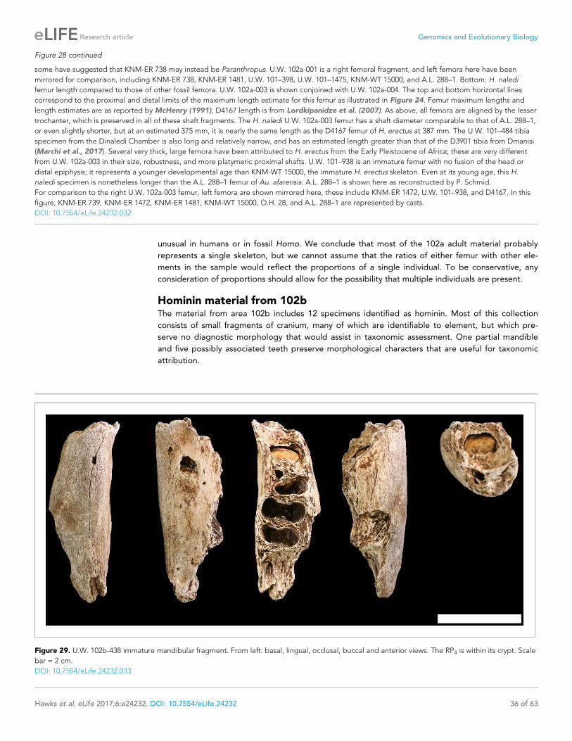
unusual in humans or in fossil Homo. We conclude that most of the 102a adult material probably
represents a single skeleton, but we cannot assume that the ratios of either femur with other ele-
ments in the sample would reflect the proportions of a single individual. To be conservative, any
consideration of proportions should allow for the possibility that multiple individuals are present.
Hominin material from 102bThe material from area 102b includes 12 specimens identified as hominin. Most of this collection
consists of small fragments of cranium, many of which are identifiable to element, but which pre-
serve no diagnostic morphology that would assist in taxonomic assessment. One partial mandible
and five possibly associated teeth preserve morphological characters that are useful for taxonomic
attribution.
Figure 28 continued
some have suggested that KNM-ER 738 may instead be Paranthropus. U.W. 102a-001 is a right femoral fragment, and left femora here have been
mirrored for comparison, including KNM-ER 738, KNM-ER 1481, U.W. 101–398, U.W. 101–1475, KNM-WT 15000, and A.L. 288–1. Bottom: H. naledi
femur length compared to those of other fossil femora. U.W. 102a-003 is shown conjoined with U.W. 102a-004. The top and bottom horizontal lines
correspond to the proximal and distal limits of the maximum length estimate for this femur as illustrated in Figure 24. Femur maximum lengths and
length estimates are as reported by McHenry (1991), D4167 length is from Lordkipanidze et al. (2007). As above, all femora are aligned by the lesser
trochanter, which is preserved in all of these shaft fragments. The H. naledi U.W. 102a-003 femur has a shaft diameter comparable to that of A.L. 288–1,
or even slightly shorter, but at an estimated 375 mm, it is nearly the same length as the D4167 femur of H. erectus at 387 mm. The U.W. 101–484 tibia
specimen from the Dinaledi Chamber is also long and relatively narrow, and has an estimated length greater than that of the D3901 tibia from Dmanisi
(Marchi et al., 2017). Several very thick, large femora have been attributed to H. erectus from the Early Pleistocene of Africa; these are very different
from U.W. 102a-003 in their size, robustness, and more platymeric proximal shafts. U.W. 101–938 is an immature femur with no fusion of the head or
distal epiphysis; it represents a younger developmental age than KNM-WT 15000, the immature H. erectus skeleton. Even at its young age, this H.
naledi specimen is nonetheless longer than the A.L. 288–1 femur of Au. afarensis. A.L. 288–1 is shown here as reconstructed by P. Schmid.
For comparison to the right U.W. 102a-003 femur, left femora are shown mirrored here, these include KNM-ER 1472, U.W. 101–938, and D4167. In this
figure, KNM-ER 739, KNM-ER 1472, KNM-ER 1481, KNM-WT 15000, O.H. 28, and A.L. 288–1 are represented by casts.
DOI: 10.7554/eLife.24232.032
Figure 29. U.W. 102b-438 immature mandibular fragment. From left: basal, lingual, occlusal, buccal and anterior views. The RP4 is within its crypt. Scale
bar = 2 cm.
DOI: 10.7554/eLife.24232.033
Hawks et al. eLife 2017;6:e24232. DOI: 10.7554/eLife.24232 36 of 63
Research article Genomics and Evolutionary Biology

U.W. 102b-438 is a fragment of right mandibular corpus from an immature individual (Figure 29).
The alveoli for the RM1 are present; the alveoli for the rdm2 are also present and reveal the crypt
with RP4 crown intact within. The fragment is broken anteriorly at the crypt for RP3 and posteriorly
at the crypt for RM2, neither permanent tooth is present. The preserved corpus height at the mid-
point of rdm2 is 14.9 mm; this is perhaps 1–2 mm less than the true value because of the erosion of
the alveolar bone surface. Corpus breadth at the anterior edge of M1 is 15.2 mm; total length of the
fragment as preserved is 43.5 mm.
Several teeth excavated from 102b are compatible with the same approximate developmental
stage as the U.W. 102b-438 mandibular fragment, and were recovered in close proximity to each
other and to the mandible; no elements are duplicated. We hypothesize that these
fragments represent the same individual, at least until the recovery of additional material makes us
reassess this possible association. U.W. 102b-437 is the complete crown and two nearly complete
roots of a ldm2 with moderate occlusal wear, including dentine exposure at the centra of cusps, and
roots that appear to have begun to resorb at the tips. U.W. 102b-503 is a RP4 crown, nearly com-
plete but not erupted. U.W. 102b-511 is a LC1 crown, nearly complete with no occlusal wear. U.W.
102b-515 is a LI2 that is nearly crown complete and unerupted. U.W. 102b-178 is a broken but
apparently unworn probable RI2 crown that was recovered separately from these other teeth, which
may also represent this individual.
The teeth all fall within the size range of equivalent elements from the Dinaledi Chamber sample.
The U.W. 102b-438 RP4 crown is partially obscured within its crypt and not complete, to the minimal
extent it is visible at this time, it is consistent with H. naledi mandibular P4 morphology. The U.W.
102b-503 maxillary P4 is likewise consistent with comparable Dinaledi examples, although this tooth
differs little among several species of Homo, including modern humans. Like the incisors of LES1, U.
W. 102b-515 lacks prominent cervico-incisal crown curvature, has no prominent crests on its lingual
surface, and is not shoveled.
The U.W. 102b-437 ldm2 is buccolingually narrow and mesiodistally long, with an ovo-rectangular
shape. Five well-developed cusps are present in a Y-fissure pattern, and the talonid is wider than the
trigonid. Compared to the two lower dm2 specimens from the Dinaledi chamber, the metaconid and
protoconid are relatively small, although it is not clear whether or not this is an artifact of mesial
wear. Although the mesial aspect is worn, it is clear that the mesial marginal ridge was thick; it forms
the distal border of a fissure-like anterior fovea. A thick distal marginal ridge is also present and it
borders a fissure-like posterior fovea. Both the mesio- and disto-buccal grooves are deep and are
associated with a wide V-shaped furrow. No protostylid is present. The mesial and distal roots each
have a buccal and lingual canal connected by a dentin plate. The crown morphology is nearly identi-
cal to that of the two analogous teeth from the Dinaledi chamber (U.W. 101–655 and U.W. 101–
1686), and supports their taxonomic designation to H. naledi. Although few have been found, the
lower dm2s of H. erectus s.l. are relatively longer and more rectangular than those of H. naledi. In
addition, the substantially wider talonid compared to the trigonid of both the 102 tooth and the H.
naledi lower dm2s differentiates these dm2s from those of most other Homo.
The most important of the 102b teeth for morphological assessment is the LC1. The U.W. 102b-
511 crown preserves its occlusal morphology and replicates with better detail the same morphology
observed in the LES1 mandibular canines (Figure 30). It has asymmetrically placed crown shoulders,
with the mesial shoulder more apically placed than the distal. Further, the distal shoulder is formed
by an accessory cuspule and the mesial crest is shorter and more convex than the vertically disposed
distal crest. These features are identical to those found in the Dinaledi permanent mandibular can-
ines and are among the defining features of H. naledi. The asymmetrical shoulders of the canine
crown and distal accessory cuspule are found to some degree in a number of specimens attributed
to H. erectus, H. rudolfensis, H. habilis and Australopithecus, but the small canine and incisor sizes
are inconsistent with attribution to any of these other taxa. On the basis of these observations, the
immature dentition from 102b is identifiable as H. naledi.
All of the cranial fragments recovered from 102b are compatible with an immature developmental
stage, and may come from the same individual as U.W. 102b-438. The minimum number of
individuals (MNI) in 102b is therefore one individual at present. Without more information about the
depositional history of the 102b locality in comparison with that of 102a, we cannot say for sure
whether any of the specimens collected at this locality may represent the same immature individual
that is represented by the pelvic fragments in 102a. The evidence does not currently exclude the
Hawks et al. eLife 2017;6:e24232. DOI: 10.7554/eLife.24232 37 of 63
Research article Genomics and Evolutionary Biology

hypothesis that the U.W. 102a-138 ilium and other immature pelvic fragments may represent the
same individual as the U.W. 102b-438 mandible. The 102a and 102b areas are separated by approxi-
mately 3 m, and there is a possibility that slumping of material might have brought the remains of a
single individual into both areas. Further work to establish the life-history stage of the immature
remains may help to resolve this question.
Hominin material from 102cThe 102c deposit is a very small (~2 L) volume of sediment enclosed within a dissolution cavity in the
cave wall. Only one morphologically identifiable hominin specimen has been recovered from this
area. U.W. 102c-589 is a fragment of left mandibular corpus with worn LM1 and LM2 in situ (Fig-
ure 31). These teeth have less occlusal wear than those of the LES1 mandible, but are morphologi-
cally very similar. The mandibular corpus is broken irregularly at the alveoli and the base of the
mandible is not present. Anteriorly, it is broken at the mesial M1 root; posteriorly, it is broken at the
mesial alveolus for the M3. The root of the ascending ramus is preserved and becomes independent
Figure 30. U.W. 102b-511 left mandibular canine crown from locality 102b. Top row, from left: occlusal, lingual, distal, labial and mesial views. Bottom
row: U.W. 102b-511 (left), compared to U.W. 101–1126 (middle) and U.W. 101–985 (right) relatively unworn left mandibular canines from the Dinaledi
Chamber. All three teeth share a distinctive morphology, which is also present in the other Dinaledi mandibular canines, that includes an asymmetrical
crown, higher mesial shoulder, and distal accessory cuspule. Scale bar = 1 cm.
DOI: 10.7554/eLife.24232.034
Hawks et al. eLife 2017;6:e24232. DOI: 10.7554/eLife.24232 38 of 63
Research article Genomics and Evolutionary Biology

at approximately the midpoint of M2. Corpus breadth at M2 is approximately the anatomical value
at 19.8 mm; the broken corpus does not permit this measurement elsewhere.
The morphology of the mandible and tooth crowns is similar to that of the mandibular remains of
H. naledi from the Dinaledi Chamber. The size and preserved morphology of the mandibular corpus
and root of the ascending ramus are very similar to that of the DH1 holotype of H. naledi. The mor-
phology and wear stage of the U.W. 102 c-589 LM1are nearly identical to those of the U.W. 101–297
RM1 crown from the Dinaledi sample, and the LM2 is very similar in morphology to the U.W. 101–
284 LM2 from Dinaledi (Figure 31). Neither of the U.W. 102 c-589 teeth exhibit supernumerary
cusps, and both have simple crowns with a Y-5 cusp pattern without crenulation or complexity, simi-
lar to the Dinaledi teeth. The areas represented by the cusps appear similar to those in the Dinaledi
molars, as do the crown heights. The relatively small size of the mandible and molars rule out attribu-
tion to Au. africanus or P. robustus, and the crown morphology rules out attribution to H. erectus or
H. habilis. This mandible is entirely consistent with H. naledi.
The U.W. 102 c-589 mandible clearly duplicates the LES1 mandible, thereby demonstrating the
presence of a second adult individual in the Lesedi Chamber as a whole. Nevertheless, it is more dif-
ficult to determine whether the U.W. 102 c-589 individual also contributed a femur to 102a, account-
ing for the evidence of two adult individuals in that deposit. The 102c deposit is separated by a
distance of approximately 12 m from the 102a assemblage. While this distance does not rule out the
occurrence of the remains of a single individual in both locations, the context does not provide any
reason for an assumption that they are the same.
Figure 31. U.W. 102 c-589 mandibular fragment. Top row, from left: lingual view, buccal view and occlusal view. Top right: two H. naledi teeth from the
Dinaledi Chamber that present comparable occlusal morphology and wear to the U.W. 102 c-589 teeth: U.W. 101–297 RM1 (top, reversed to represent
left side) and U.W. 101–284 LM2 (bottom). Bottom row, from left: U.W. 102 c-589 posterior view, anterior view and basal view. Scale bar = 2 cm.
DOI: 10.7554/eLife.24232.035
Hawks et al. eLife 2017;6:e24232. DOI: 10.7554/eLife.24232 39 of 63
Research article Genomics and Evolutionary Biology

TaphonomyThe general preservation of the Lesedi Chamber hominin material resembles that of the skeletal
assemblage from the Dinaledi Chamber (Dirks et al., 2015). The Lesedi skeletal material has a sur-
face coloration that ranges from light grey to red-brown, and internal structures or cortex at fresh
break points are colored pale buff to off-white, contrasting with unbroken adjacent surfaces. The
remains present no evidence of calcite crystal formation. Specific comments on preservation have
been included with the specimen descriptions above, and taphonomic observations on each speci-
men are summarized in Supplementary file 5.
The Lesedi skeletal assemblage exhibits varying degrees of post-mortem damage. Most speci-
mens are fragmented or broken to some degree, with areas of cortical bone removal or abrasion
(Supplementary file 5). All fractures observed in the assemblage are consistent with post-mortem
(dry bone) failure (Supplementary file 5), and there are no spiral or incomplete fractures indicative
of green or wet bone (L’Abbe et al., 2015; Symes et al., 2014). Fractures in the assemblage include
transverse or right-angled breaks on the shafts of long bones, showing block-comminution between
major breaks and step-fractures following the longitudinal grain of the long bone, which are distinct
from cracking or crazing resulting from weathering (Symes et al., 2014).
The Lesedi remains display no evidence of sub-aerial weathering processes (sensu Behren-
smeyer, 1978; see also Lyman and Fox, 1989 and Junod and Pokines, 2014, indicating that the
dry bones were not subject to surface processes before deposition in the cave environment. Sub-
aerial weathering is primarily characterized by both cracking and delamination. Within the Lesedi
assemblage, there is some evidence of surface cracking, but it generally does not penetrate deep
into the cortex. No indications of the secondary features of weathering, such as delamination, deep
patination, bleaching or cortical exfoliation, were observed in any of the bone fragments from
Lesedi, suggesting that the bones were not affected by surface exposure (Supplementary file 5). A
comparison with human bone derived from forensic or sealed archaeological contexts (Figure 32)
suggests that the cracking and fracture patterns observed in the Lesedi assemblage can be
explained most parsimoniously by the effects of the burial environment (Pokines, 2014), and were
exacerbated in specimens where fluctuations in moisture content caused swelling and shrinkage of
the cortical structure (Conard et al., 2008; Dirks et al., 2016; Dirks et al., 2015).
The assemblage presents no evidence of high-energy fluvial transport, such as smoothing and
rounding, polish, frosting, cortex thinning, or aperture formation (Bassett and Manhein, 2002; Beh-
rensmeyer, 1988; Evans, 2014; Nawrocki et al., 1997). We have found no traces of carnivore or
scavenger modification, with an absence of bone cylinders, tooth scores or traces of gastric corro-
sion (Blumenschine et al., 1996; Haynes, 1983; Hill, 1976; Pickering et al., 2004; Thompson and
Lee-Gorishti, 2007). In addition, the profiles of damaged diaphyses show no evidence of end gnaw-
ing, scalloping or flaking (Blumenschine et al., 1996; Lyman, 1994; Wood, 1991). In some cases,
missing areas of epiphyses display patterns of localized cortical destruction, which may be consistent
with a process referred to as ‘coffin wear’ (Rogers, 2005; Schultz, 2012; Schultz et al., 2003)
whereby bones come into direct contact with an underlying substrate following soft tissue decompo-
sition; this may lead to the loss of cortex or of portions of elements that are in contact with the sub-
strate. This sub-surface process may completely remove processes or condyles from major elements
such as the femur or humerus, leaving behind flattened or sheared areas of bone (Rogers, 2005;
Pokines and Baker, 2014).
As with the Dinaledi material, some of the Lesedi Chamber remains are stained by iron oxides
and manganese oxy-hydroxide (Dirks et al., 2015; Randolph-Quinney et al., 2016). This staining
primarily occurs as diffuse spots or mats of mineral, some of which cover and encapsulate the frac-
tured ends of long bones, indicating a complex post-mortem history of mineral deposition. However,
we have observed no mineral tide marks upon the Lesedi Chamber skeletal material which, unlike
material from the Dinaledi Chamber, provides us with no information about the former position of
skeletal remains relative to the sediment–air interface. Some of the Lesedi material exhibits minor
pitting, striations, grooves, scratches, or gouges. Striations and grooves are consistent with abrasion
marks, as defined by D’Errico and Villa (1997) and Fisher (1995). Some of the elements have a
palimpsest of taphonomic traces, with mineral deposition preceding invasive surface modification or
fracturing (Figure 32). The pitting that is present on the Lesedi material shows that the modifying
agents (abiotic or biotic) penetrated already-altered bone, suggesting that damage occurred on
Hawks et al. eLife 2017;6:e24232. DOI: 10.7554/eLife.24232 40 of 63
Research article Genomics and Evolutionary Biology

LES1
modern
forensic
burial
U.W. 101a-010
Figure 32. Surface cracking and pitting in Lesedi Chamber material. Top: right corpus and ramus of LES1
mandible prior to reconstruction, showing patterns of cracking consistent with the effects of sediment loading and
wetting during the burial process and during skeletal decomposition. This pattern of taphonomic alteration is
superficially similar to sub-aerial weathering processes, but is independent of surface exposure and occurs in both
deep- and shallow-buried deposits. Middle: forensic known-history case for comparison. This specimen was
Figure 32 continued on next page
Hawks et al. eLife 2017;6:e24232. DOI: 10.7554/eLife.24232 41 of 63
Research article Genomics and Evolutionary Biology

bone surfaces that were already covered in coatings of manganese and iron oxide (Randolph-
Quinney et al., 2016). Generally, the pattern of pitting and stripping is similar in composition and
gross morphology to patterns observed in the Dinaledi Chamber assemblage, which were attributed
to gastropod and other invertebrate activity (Dirks et al., 2015, 2016; Randolph-Quinney et al.,
2016), although these must be studied in more detail to be certain.
Overall, the gross taphonomic signature of the hominin remains in the Lesedi Chamber appears
consistent with sub-surface deposition with limited post-depositional dispersal. Particularly in the U.
W. 102a locality, this is evidenced by the presence of articulated remains, the proximity of cranial
and postcranial remains, and the recovery of small elements such as teeth and carpals. The bone sur-
face condition, as well as the absence of other markers of transport and secondary modification, are
generally consistent with sub-surface skeletal decomposition (Carter and Tibbett, 2008;
Carter et al., 2007). At present, we find no supporting evidence for sub-aerial weathering or post-
mortem exposure outside of the cave environment (Hill, 1976; Junod and Pokines, 2014;
Lyman and Fox, 1989; Tappen and Peske, 1970). Nor are there signs of carnivore or scavenger
modification (Pokines, 2013), water transport (Behrensmeyer, 1988; Evans, 2013), or peri-mortem
trauma such as may be expected in a natural death-trap scenario (L’Abbe et al., 2015; Symes et al.,
2014).
Faunal materialIn addition to the hominin skeletal material, some faunal remains have been recovered from the
Lesedi Chamber. Over 80 faunal elements or fragments were collected from U.W. 102a. U.W. 102b
yielded 23 specimens of fauna, while a single rodent tooth was recovered from U.W. 102c. In-depth
analyses of the faunal material are ongoing, and comprehensive descriptions are in preparation. We
provide here a preliminary list of the taxa identified for reference (Table 3).
The Lesedi faunal assemblage includes micromammal, small to mid-size mammal, and non-mam-
malian remains. With respect to the micromammals (prey body mass <500 g), 5 genera of rodents
and 1 genus of shrew were identified out of 28 craniodental specimens (21 MNI). There are also
potentially two additional murine genera and one soricid genus present in the assemblage, although
lower-level identification is not possible at this time. Interestingly, all of the non-hominin fauna are
of relatively small species. The largest mammalian specimens come from dental material attributed
to Canis aff. C. familiaris. The size range of this material is outside the range of modern C. mesome-
las but the morphology is definitively not Lycaon (Hartstone-Rose et al., 2010; Wayne, 1986). The
felid material is also small, falling in the size range of the African wildcat. None of these individuals is
likely to have exceeded 10–15 kg (Kingdon, 2015) and the rest of the assemblage consists almost
entirely of animals smaller than 3 kg, including four specimens from the family Herpestidae. Aside
from a single lagomorph specimen, the macro-mammalian material comes exclusively from the order
Carnivora, a situation that is unusual in the fossil record (Werdelin and Sanders, 2010).
We do not presently know whether some or all of these faunal remains may be contemporaneous
with any of the hominin fossil material. Faunal remains have been recovered both on the surface and
also from within sediments near hominin remains. However, the Lesedi Chamber is not a completely
isolated environment, and sediment deposits are currently eroding from their original depositional
contexts, with evidence for slumping and reworking in the chamber. Therefore, we cannot yet com-
ment on the relative timing of deposition of the hominin and faunal material. Additional tests, includ-
ing attempts to date both hominin and faunal elements directly, will help us answer these questions
Figure 32 continued
recovered from a deep known-history inhumation. The fleshed body was deposited at a depth of 1.5 metres and
recovered after a 30-year burial period. Note the similarities in surface texture, and superficial cracking, which
follows the biomechanical stress lines (grain) of the bone. Bottom: U.W. 102a-010 acromial fragment showing
surface modification with cortical pitting and punctate marks. Note that many of these marks (circled in yellow)
penetrate pre-existing layers of manganese oxy-hydroxide deposited on the bone surface; the damage was
therefore produced inside the Lesedi Chamber on bones that were already covered in coatings of manganese
mineral.
DOI: 10.7554/eLife.24232.036
Hawks et al. eLife 2017;6:e24232. DOI: 10.7554/eLife.24232 42 of 63
Research article Genomics and Evolutionary Biology

and to relate the faunal material to the chronological and environmental context of H. naledi in the
Lesedi Chamber.
DiscussionThe hominin material from all three excavation contexts in the Lesedi Chamber can be clearly attrib-
uted to H. naledi, and no diagnostic material in this chamber represents any other hominin taxon.
This confirms that a second chamber within the Rising Star cave system, in a distinct depositional
context isolated from the Dinaledi Chamber, also holds the skeletal remains of H. naledi.
As in the Dinaledi Chamber, both adult and immature individuals are commingled in the Lesedi
Chamber. When the fossils recovered from the 102a, 102b, and 102c localities are considered in
aggregate, duplication of elements and age-at-death indicators combine to produce a MNI of three:
two adults and one immature individual. However, the presence of material in three spatially sepa-
rated localities within the chamber leads us to hypothesize that the current collection more
probably represents additional individuals. The adult mandibular fragment in 102c is separated from
the adult femora in 102a by more than 10 m, and the presence of this fragment in 102c, with an
opening 1.3 m above the present cave floor, suggests that the sedimentary deposits may once have
been much more voluminous. In light of the situation, we view it as likely that the 102c mandibular
fragment represents an additional adult individual beyond what may be two individuals in 102a. The
102b deposit is separated by less distance from 102a, and the data do not yet provide a clear
answer as to whether or not the juvenile remains in both areas could represent a single individual.
Considering the spatial context, we hypothesize that four individuals may be represented: two adults
Table 3. Mammal species recorded in the Lesedi Chamber. Several specimens from the classes Aves, Amphibia and Reptilia were also
recovered, but individual counts and taxonomic identifications are pending further examination.
Class Order Family Subfamily Genus/species MNI NISP
Mammalia Lagomorpha Leporidae 1 1
Soricomorpha Soricidae 3 3
Crocidurinae Crocidura 1 1
Rodentia 1 1
Bathyergidae Bathyerginae 1 1
Muridae 1 1
Otomyinae Otomys 1 1
Murinae 5 7
Mus 1 2
Nesomyidae Mystromyinae Mystromys 1 1
Dendromurinae 3 4
Steatomys 3 6
Carnivora Felidae Felis aff. F. sylvestris 1 1
Felis cf. sylvestris 1 6
Felis sp. 1 2
Herpestidae cf. Mungos 1 1
cf. Herpestidae 1 3
Canidae Canis aff. C. familiaris 1 14
Canis cf. mesomelas 1 3
Canis sp. 1 21
Vulpes cf. chama 1 3
cf. Vulpes 1 6
Vulpes sp. 1 5
DOI: 10.7554/eLife.24232.037
Hawks et al. eLife 2017;6:e24232. DOI: 10.7554/eLife.24232 43 of 63
Research article Genomics and Evolutionary Biology

and one immature individual in 102a and 102b; and one adult in 102c. Further excavation may test
this hypothesis.
The Lesedi Chamber sample extends our knowledge of H. naledi in several ways. The complete
U.W. 102a-021 clavicle confirms the morphological assessment of fragmentary clavicles from the
Dinaledi Chamber (Feuerriegel et al., 2017). The U.W. 102a-036 and U.W. 102a-151 T10 and T11
vertebrae duplicate the anatomy observed in the Dinaledi material (Williams et al., 2017), while
additional vertebrae add information on the lower vertebral column. The U.W. 102a-250 first rib is
more complete than any example from the Dinaledi Chamber, but presents similar anatomy
(Williams et al., 2017). U.W. 102b-438 and its associated teeth present an additional immature man-
dibular specimen that adds to the developmental series of mandibles and immature dentitions from
the Dinaledi Chamber. Comparative study of these elements is underway.
The relative completeness of the LES1 cranium allows us to examine cranial length and the nasal
aperture for the first time in H. naledi, in part confirming the interpretation of partial crania and iso-
lated cranial elements from the Dinaledi Chamber. This specimen shows a relatively short and tall
cranium and a moderate-sized nasal aperture. This specimen expands the range of endocranial vol-
ume (ECV) known for H. naledi, which now extends from approximately 460 ml to approximately 610
ml. The maximum value for H. naledi is now somewhat above the maximum ECV observed for aus-
tralopith species, including Au. afarensis, Au. africanus, P. robustus, and P. boisei (Table 4; Fig-
ure 33). No crania attributed to H. habilis, H. rudolfensis, or H. erectus have ECVs as small as
the 460 ml estimated for DH3, but the larger H. naledi specimens do overlap with the smaller end of
the reported values for H. habilis and H. erectus. The single specimen of H. floresiensis with an esti-
mate of endocranial volume, LB1, is smaller than any specimen of H. naledi. The addition of the
LES1 cranium now brings the range of observed ECVs in H. naledi into overlap with two specimens
of H. erectus (D2700 and D4500 from Dmanisi; Table 4, Figure 33).
The hominin material from the Lesedi Chamber remains undated. The Dinaledi Chamber hominin
material appears to have been deposited sometime between 236 ka and 335 ka (Dirks et al., 2017).
The Dinaledi hominin sample is morphologically very uniform, and variations in many metric charac-
ters in that sample is less than those in local populations of modern humans (Berger et al., 2015).
Such morphological uniformity suggests that the Dinaledi Chamber does not represent successive
biologically diverse populations sampled over tens of thousands of years or longer, known as ‘time-
averaging’, but instead may sample a single biological population. The Lesedi Chamber H. naledi
sample adds very little to the morphological variability of the Dinaledi Chamber sample, and most
measurements of cranial, dental, and postcranial elements fall within the existing range known from
that chamber. The nonmetric traits observed on cranial and postcranial elements from the Lesedi
Chamber are nearly all duplicated in the Dinaledi Chamber sample (Supplementary file 1; Fig-
ures 34 and 35). It is therefore reasonable to hypothesize that the Lesedi Chamber may sample the
same biological population as the Dinaledi Chamber. This hypothesis will be tested with further geo-
logical information, including attempts to apply direct dating to the hominin material directly. If the
two hominin assemblages represent diverse geological ages, or if either assemblage was deposited
over a geologically long time interval, they would demonstrate a remarkable stasis of H. naledi met-
ric and morphological characters across time.
The emphasis of this study has been to test the hypothesis that the newly discovered Lesedi skel-
etal material represents H. naledi. Based upon this diagnosis, the morphology of H. naledi (including
the Lesedi material) across the skeleton has implications for its relationship to other Plio-Pleistocene
hominins (Figures 34 and 35). Most of the anatomical features across the skeleton of H. naledi are
known from multiple specimens, many of which have now been replicated in two assemblages (Fig-
ure 35). Phylogenetic analysis of the Dinaledi hominin assemblage has shown that the mosaic of fea-
tures in the H. naledi cranium and dentition does not provide unambiguous or clear evidence of
where the species should be placed in the phylogeny of Homo (Figure 34; Dembo et al., 2016). We
briefly consider here how the overall pattern of morphological and temporal evidence relates to the
possible affinities of H. naledi with other hominins.
Modern and archaic humansThe Dinaledi Chamber assemblage of H. naledi represents individuals who lived at roughly the same
time as skeletal remains attributed to early modern or near-modern humans in some parts of Africa
(Figure 36; Berger et al., 2017). No derived features of the H. naledi skeleton require a close or
Hawks et al. eLife 2017;6:e24232. DOI: 10.7554/eLife.24232 44 of 63
Research article Genomics and Evolutionary Biology

Table 4. Endocranial volume of LES1 compared to key specimens of other hominin species. Estimates
from Asfaw et al. (1999), Kubo et al. (2013), Holloway et al., (2014), Lee and Wolpoff (2003),
Kimbel (2004); Berger et al. (2010), (2015), Lordkipanidze et al. (2006) and Lordkipanidze et al.
(2013).
Specimen Cranial capacity in cc
Au. afarensis Mean 444
AL 162–28 400
AL 288–1 387
AL 333–45 485
AL 333–105 400
AL 444–2 550
Au. africanus Mean 455
MLD 1 510
MLD 37/38 425
Sts 5 485
Sts 19 436
Sts 60 400
Sts 71 428
StW 505 505
Au. sediba MH1 420
H. floresiensis LB1 426
H. naledi Mean 513
DH1 560
DH3 465
LES1 610
H. habilis Mean 616
KNM-ER 1805 582
KNM-ER 1813 509
O.H. 7 729
O.H. 13 650
O.H. 16 638
O.H. 24 590
H. rudolfensis Mean 789
KNM-ER 1470 752
KNM-ER 1590 825
KNM-ER 3732 750
H. erectus Mean 917
BOU-VP-2/66 995
D2280 730
D2282 650
D2700 601
D3444 625
D4500 546
KNM-ER 3733 848
KNM-ER 3883 804
KNM-ER 42700 691
Table 4 continued on next page
Hawks et al. eLife 2017;6:e24232. DOI: 10.7554/eLife.24232 45 of 63
Research article Genomics and Evolutionary Biology

exclusive relationship with modern humans. Overall, H. naledi resembles more primitive species of
Homo such as H. erectus, H. habilis, or H. rudolfensis much more than it resembles archaic or mod-
ern humans (Berger et al., 2015; Laird et al., 2017; Marchi et al., 2017; Feuerriegel et al., 2017;
Table 4 continued
Specimen Cranial capacity in cc
KNM-WT 15000 900
O.H. 9 1,067
O.H. 12 727
Sangiran 2 813
Sangiran 4 908
Sangiran 17 1,004
Zhoukoudian DI 915
Zhoukoudian LI 1,025
Zhoukoudian LII 1,015
Zhoukoudian LIII 1,030
Ngandong 1 1,172
Ngandong 5 1,251
Ngandong 6 1,013
Ngandong 9 1,135
Ngandong 10 1,231
Ngandong 11 1,090
Trinil 940
DOI: 10.7554/eLife.24232.039
Figure 33. Endocranial volumes of hominin species. With the addition of LES1 to the sample, the range of
endocranial volume in H. naledi is extended slightly beyond the range represented in the Dinaledi Chamber. This
range overlaps with two specimens of H. erectus, and LES1 is larger than the largest Au. africanus or Au. afarensis
specimens. Data and sources are listed in Table 4.
DOI: 10.7554/eLife.24232.038
Hawks et al. eLife 2017;6:e24232. DOI: 10.7554/eLife.24232 46 of 63
Research article Genomics and Evolutionary Biology

postorbital constriction
frontal bossing
postbregmatic depression
prelambdoidal !attening
temporal crest position on
supraorbital torus
angular torus
supramastoid crest
developmentangulation of root of the
zygomatic
eustachian process of
tympaniclateral in!ation of
mastoid process
postglenoid process size
vaginal process size
mastoid process size
occipital torus
external occipital
protuberance
supratoral sulcus
supraorbital contour
canine fossa
nasoalveolar clivus
contourcanine jugum
development
post-incisive planum
hollowing above/
behind mental foramen
anterior palate depth
Figure 34. Selected cranial trait observations in H. naledi and other species. A subset of observations of cranial traits reported in Supplementary file
1 that vary among species attributed to Homo. This list omits traits that are present in only one species of Homo or for which nearly all species exhibit
more than one state. Here, the character state observed in H. naledi, including both Dinaledi Chamber and Lesedi Chamber material, is reported
on the left. Character states for other species are reported in terms of whether they share the same state as H. naledi. Some traits that are shared with
H. naledi may be interpreted as shared derived traits, based on their absence from Au. afarensis and Au. africanus, but some species of Homo also
Figure 34 continued on next page
Hawks et al. eLife 2017;6:e24232. DOI: 10.7554/eLife.24232 47 of 63
Research article Genomics and Evolutionary Biology

Williams et al., 2016; Schroeder et al., 2017). H. naledi does, however, possess a number of
derived features that are otherwise known only from modern humans and Neandertals
(Supplementary file 1; Figures 34 and 35; Berger et al., 2015; Dembo et al., 2016; Kivell et al.,
2015; Harcourt-Smith et al., 2015; Williams et al., 2016). Some of these derived features, including
features of the wrist, cannot be assessed in H. erectus because no fossils of the relevant bones exist
for this species (Kivell et al., 2015). But others, including features of the cranium and dentition, raise
at least the possibility that H. naledi may be a sister to H. antecessor or to a clade including H. ante-
cessor with other archaic and modern humans (Dembo et al., 2016). It is also conceivable that,
rather than indicating a recent branching of H. naledi from an archaic human lineage, such derived
similarities may have resulted from introgressive hybridization between H. naledi and other hominin
lineages (Berger et al., 2017), although testing this hypothesis will likely require genetic data.
Homo floresiensisThe late Middle Pleistocene geological age of H. naledi, when considered together with its small
endocranial volume, prompt comparisons with H. floresiensis. Skeletal remains of H. floresiensis are
known from ~100–60-ka-old sediments at Liang Bua, Flores (Brown et al., 2004; Sutikna et al.,
2016), and possibly also in Mata Menge, Flores sediments from the early Middle Pleistocene
(van den Bergh et al., 2016; Brumm et al., 2016). The features shared by H. naledi and H. floresien-
sis are nearly all inferred to be primitive features shared with australopiths and/or in some cases with
early Homo (Figures 35 and 36). For most features of the skeleton where H. naledi exhibits derived
morphology, including aspects of the foot and hand, H. floresiensis exhibits morphology thought to
be primitive within hominins (Harcourt-Smith et al., 2015; Kivell et al., 2015; Tocheri et al., 2007;
Jungers et al., 2009a; Orr et al., 2013). All known H. floresiensis long bones are small, reflecting
small stature and mass (Jungers et al., 2009b; Larson et al., 2009; Grabowski et al., 2015), and
none approach the much larger size, particularly the taller stature, manifested in adult specimens of
H. naledi. The mandibles of H. floresiensis are relatively robust, even for their small size,(Brown and
Maeda, 2009; Daegling et al., 2014) and in this sense, are comparable to the robust mandibles of
H. naledi, though the two species differ in symphyseal morphology (Laird et al., 2017). The denti-
tion of H. floresiensis displays a unique combination of primitive and derived characters (Kaifu et al.,
2015), which are different from the combination found in H. naledi. Homo floresiensis canines and
premolars display a combination of features that are primitive for hominins (Kaifu et al., 2015), while
H. naledi mandibular canines and third premolars have a uniquely derived form. The H. floresiensis
molars exhibit derived proportions and some other features similar to those of modern humans,
which may reflect the extremely shortened molar crowns, particularly of the first molars (Kaifu et al.,
2015). By contrast, H. naledi has primitive proportions of the molars and occlusal morphology that
is broadly primitive within Homo, although simplified in complexity. Shape analyses also place the
cranium of H. floresiensis (LB1) far from any H. naledi cranial specimen (Schroeder et al., 2017).
These differences in anatomy may reflect a distant phylogenetic relationship between the two spe-
cies, although these data do not clearly resolve where on the Homo phylogeny either species should
be placed. Some phylogenetic evidence suggests that the H. floresiensis branch preceded the node
linking H. erectus and modern humans (Argue et al., 2009; Dembo et al., 2015). Cranial and dental
evidence does not resolve whether H. naledi may also have branched early in the evolution of
Homo, or whether it may instead be a sister taxon to modern and archaic humans (Dembo et al.,
2016). No data that are available at present answer whether either or both of these lineages may
have retained small EVC from an ancestry among the earliest members of Homo, or whether their
smaller brain sizes may have evolved secondarily from larger-brained ancestors. Together, they
Figure 34 continued
share primitive traits with H. naledi that may also be found in the australopiths. The pattern of shared morphological characters among these species
and H. naledi is complex, and there is no consistent anatomical grouping of the characters shared with any given species. H. naledi shares more
characters with H. erectus and H. habilis than with other species, and behind them, with Au. sediba and archaic and modern humans. H. erectus is
variable for a large number of traits also found in H. naledi. The fact that variability is noted in H. erectus, Neandertals and modern humans for these
traits is partially a function of the large samples available for these groups. It is probable that a larger sample of other species would likewise
encompass greater variability. Any phylogenetic tree of these species would reveal a high degree of homoplasy for these cranial and mandibular traits.
DOI: 10.7554/eLife.24232.040
Hawks et al. eLife 2017;6:e24232. DOI: 10.7554/eLife.24232 48 of 63
Research article Genomics and Evolutionary Biology
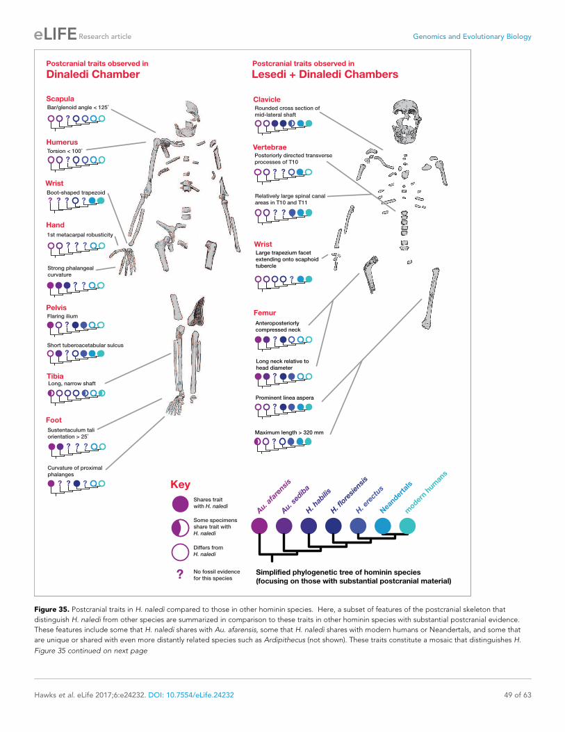
Figure 35. Postcranial traits in H. naledi compared to those in other hominin species. Here, a subset of features of the postcranial skeleton that
distinguish H. naledi from other species are summarized in comparison to these traits in other hominin species with substantial postcranial evidence.
These features include some that H. naledi shares with Au. afarensis, some that H. naledi shares with modern humans or Neandertals, and some that
are unique or shared with even more distantly related species such as Ardipithecus (not shown). These traits constitute a mosaic that distinguishes H.
Figure 35 continued on next page
Hawks et al. eLife 2017;6:e24232. DOI: 10.7554/eLife.24232 49 of 63
Research article Genomics and Evolutionary Biology

establish that diverse hominin lineages with varying brain and body sizes existed during the Middle
Pleistocene, suggesting the influence of ecological factors that promoted diversity during the Pleis-
tocene evolution of humans and great apes (Berger et al., 2017; Tocheri et al., 2011,
2016; Dunn et al., 2014).
Homo erectusH. naledi shares many derived cranial and dental characters with H. erectus (Figures 34 and
36; Dembo et al., 2016; Laird et al., 2017), and Dmanisi specimens of H. erectus are among the
closest in multivariate shape to H. naledi crania (Schroeder et al., 2017). H. naledi overlaps with H.
erectus in long bone lengths, inferred stature, and estimated body mass (Figure 28; Berger et al.,
2015). To the extent that the H. naledi postcranial skeleton is different from material attributed to
H. erectus, it is mostly because H. naledi manifests primitive traits that are not otherwise seen in
Homo, or because H. naledi manifests traits not otherwise seen in hominins (Figure 35). H. naledi
also shares several derived postcranial features with archaic and modern humans, but a lack of post-
cranial evidence for these parts of the H. erectus skeleton has made it impossible to determine
whether H. erectus may also have shared these derived features (Figure 35). With respect to the cra-
nium and dentition, nearly all of the nonmetric traits shared by H. naledi and H. erectus are also
shared with Au. sediba, H. habilis, or both (Supplementary file 1; Figure 34). H. naledi and H. erec-
tus share at least three derived nonmetric features of the skull that are not also found in Au. sediba
or H. habilis: an angular torus, sagittal keeling, and an anteriorly projecting nasal spine. But each of
these three traits can also be found in some archaic or modern humans. H. naledi also possesses
some cranial and dental traits not seen in any specimens of H. erectus; these include both primitive
traits shared with Au. afarensis, Au. africanus or Au. sediba and derived traits shared either with H.
rudolfensis or with archaic and modern humans. Even though H. naledi crania are most similar in
shape to the smallest H. erectus crania from Dmanisi (Schroeder et al., 2017), they are distinct from
the Dmanisi sample in numerous aspects of cranial, dental, and postcranial morphology
(Berger et al., 2015; Laird et al., 2017; Marchi et al., 2017; Feuerriegel et al., 2017;
Rightmire et al., 2017), and Bayesian analysis of cranial and dental morphology provides strong evi-
dence against the hypothesis of a sister taxon relationship for these samples (Dembo et al., 2016).
In summary, no traits link H. naledi exclusively or specifically with H. erectus, and many traits distin-
guish the two. The evidence is not sufficient to say whether H. naledi may have evolved from an ear-
lier population that resembled H. erectus, or whether earlier fossils representing the H. naledi
lineage may already have been found and until now attributed to H. erectus (Berger et al., 2017).
Australopithecus sediba, Homo habilis, and Homo rudolfensisFossil samples from Malapa, Olduvai Gorge, and the Lake Turkana area have been attributed to line-
ages often thought to represent some of the earliest species of Homo or their near relatives among
the australopiths. H. naledi shares many derived cranial and mandibular features with Au. sediba and
H. habilis, although most of these are also shared with H. erectus (Supplementary file 1; Figures 34
and 35). Compared to these other species, H. naledi shares many fewer cranial and dental features
with H. rudolfensis, except for its relatively flat and squared nasoalveolar clivus. Phylogenetic analysis
does not reject the hypothesis of a sister taxon relationship between H. naledi and Au. sediba or H.
habilis (Dembo et al., 2016). However, in comparison to both Au. sediba and H. habilis, H. naledi is
derived and similar to modern humans and Neandertals in several aspects of hand and wrist mor-
phology (Kivell et al., 2015). It is also derived in its foot morphology in comparison to both MH2 of
Au. sediba and the O.H. 8 foot usually attributed to H. habilis (Harcourt-Smith et al., 2015). H.
naledi also lacks the derived configuration of the Au. sediba ilium (Figure 35). H. naledi further
shares a number of derived cranial characters with H. erectus and with archaic or modern humans as
discussed above (Supplementary file 1, Figure 34). Whatever the relationship of these species, their
Figure 35 continued
naledi clearly from other species of Homo. Many of the traits that are notable in the Dinaledi Chamber sample are also represented in the Lesedi
Chamber material.
DOI: 10.7554/eLife.24232.041
Hawks et al. eLife 2017;6:e24232. DOI: 10.7554/eLife.24232 50 of 63
Research article Genomics and Evolutionary Biology

Figure 36. Lateral cranial comparison of H. naledi crania to crania of other hominin species. H. naledi crania, DH1, LES1, and DH3 are in the center
row. All crania are oriented as near as possible to the Frankfort plane, delineated by the light gray lines in the background of the figure. Compared to
other hominin genera, including Australopithecus and Paranthropus, fossil Homo is often recognized by cranial and dental features such as a more
vertical face profile, a reduced postcanine dentition, larger endocranial volume, a higher frontal, and a true supraorbital torus. Au. africanus (Sts 5, top
left) represents the ancestral hominin condition lacking these traits. The other crania in the top two rows vary substantially in these features. LB1 has a
Figure 36 continued on next page
Hawks et al. eLife 2017;6:e24232. DOI: 10.7554/eLife.24232 51 of 63
Research article Genomics and Evolutionary Biology

evolution must have involved homoplasy of many features and the evidence does not yet make
it clear how they are connected. If any one of these species were a possible ancestor of H. naledi,
the branch leading to the Dinaledi sample of H. naledi would approach 1.5 Ma or longer in evolu-
tionary time. With abundant opportunity for adaptive and nonadaptive evolution of the H. naledi
lineage over this time, it would be very hard to distinguish a long branch connecting it to Au. sediba
or H. habilis from a somewhat longer branch connecting it to the very base of Homo.
ConclusionsThe excavation of the Lesedi Chamber has added to our knowledge of the biology of H. naledi and
has confirmed the presence of this species in a second depositional context. The skeletal material
described here derives from a very small and limited excavation, and the total sediment volume of
the chamber has not yet been sampled sufficiently to estimate the abundance of hominin-bearing
deposits or the relationship of faunal and hominin species. Further resolution of how the material
was originally deposited must await more detailed sedimentological analysis and more excavation
work.
The relative completeness of the morphological evidence from H. naledi has not resolved its phy-
logenetic placement within the genus Homo (Dembo et al., 2016). The morphological evidence
from the Lesedi Chamber hominin material reinforces the observations made on the basis of the
Dinaledi hominin sample. In particular, the more complete LES1 cranium and the more fragmented
cranial remains from multiple individuals already known for H. naledi share an almost identical pat-
tern of derived features with other hominin species (Laird et al., 2017). The discovery of H. naledi
within the Dinaledi Chamber documented a pattern of anatomy that had not been anticipated by
anthropologists on the basis of earlier fossil discoveries. Learning how H. naledi connects to other
fossil evidence of human origins may require more unexpected discoveries.
Materials and methods
ExcavationFormal excavations began in the Lesedi Chamber in May 2014, after initial surface and ex situ mate-
rial had been recovered. Like the Dinaledi Chamber, the access route to the Lesedi Chamber is very
restricted. Additionally, the excavation area available in the Lesedi Chamber is considerably more
confined than that in the Dinaledi Chamber. As a result, protocols followed those used in the Dina-
ledi Chamber (Dirks et al., 2015), with a few exceptions. Specifically, attempts to use some of the
Figure 36 continued
vertical face, reduced dentition, and high, rounded frontal, but has comparatively small endocranial volume. KNM-ER 1470 has a large volume, a high
frontal, and a more vertical face profile, but also is inferred to have a large postcanine dentition and has no true supraorbital torus. MH1 (Au. sediba)
has a small volume, but shares features with Homo that include the less sloping face profile, a supraorbital torus, and reduced postcanine dentition. O.
H. 24 has a low, sloping frontal, and a concave facial profile, but a true supraorbital torus and reduced postcanine teeth. This variability among
species that are interpreted as ‘primitive’ Homo, such as H. habilis and H. floresiensis, and Homo-like australopiths makes it difficult to delineate the
genus Homo (Wood and Collard, 1999; Dembo et al., 2016). H. erectus is also highly variable. It includes several crania with endocranial volumes
below 700 ml, including KNM-ER 42700 and D2282, but also many larger crania, here represented by Sangiran 17 and Zhoukoudian L2 (ZKD L2).
Specimens attributed to H. erectus tend to share a series of traits first noted in Asian H. erectus samples, including a long, low cranial profile, thick
cranial bone, sagittal keeling, prominent supraorbital, angular, and occipital tori, a sharply angled occiput, and a postbregmatic depression. The
smallest H. erectus crania share most of these features, with a low cranial profile, angled occiput and postbregmatic depression visible here in D2282.
But these features do vary substantially and are less evident in the immature KNM-ER 42700. The H. naledi crania are similar to KNM-ER 1470 in having
a transversely flat clivus contour, but all are smaller, with a much smaller palate and with very different frontal morphology. Like O.H. 24 and KNM-ER
1813, the H. naledi crania have relatively thin cranial bone and a thin and projecting supraorbital torus. But H. naledi manifests a different clivus shape,
a projecting nasal spine, a greater cranial height, sagittal keeling and an angular torus. The H. naledi crania bear little resemblance to LB1, differing in
face profile, size, and their larger postcanine dentitions. Known African Homo specimens from the later Middle Pleistocene other than H. naledi, such as
the Kabwe skull (pictured), contrast strongly with H. naledi in cranial size and morphology. The Omo 2 skull, one of the earliest known modern human
crania at approximately 196,000 years (McDougall et al., 2005), is vastly larger and very different from any H. naledi specimen, despite being near the
same geological age. In this figure, O.H. 24, KNM-ER 1470, LB1, KNM-ER 42700, ZKD L2, and Omo 2 are represented by casts. Images have been
adjusted to a common scale by maximum cranial length, or by glabella-bregma length where maximum length is not available. Photos of Sangiran
17 and D2282 are courtesy of Milford Wolpoff.
DOI: 10.7554/eLife.24232.042
Hawks et al. eLife 2017;6:e24232. DOI: 10.7554/eLife.24232 52 of 63
Research article Genomics and Evolutionary Biology

surface-scanning methods applied in the Dinaledi Chamber (Kruger et al., 2016) failed due to the
limited working distances of the scanner, the convoluted surfaces, and the poor contrast of the sur-
rounding dolomite. However, high-resolution laser scan data have been acquired for the Lesedi
Chamber and will form the basis of future spatial work. Consequently, documentation of the excava-
tions and provenience of the material was conducted using traditional archaeological methods,
including written descriptions, photography and drawings. Excavations were conducted in tandem
with geological mapping and with sedimentologic and taphonomic analyses of the site.
For excavations in U.W. 102a, the area was divided into 20–40 cm sections that were based on
the contours of the sloped surface and the width of the tunnel at that point. Sections were exca-
vated in 2–5 cm levels. In some areas where few specimens were being recovered, levels were
expanded to 10 cm.
In the U.W. 102b area, excavations began on the surface of the sediment deposit in which the
first material was recovered, and proceeded in 2–5 cm levels until the chert layer on which the sedi-
ment had accumulated was reached. When this area was exhausted, excavations moved along the
chert shelf, removing sediments from the surface, to the chert, in 20 cm horizontal sections. A sedi-
ment pocket above the fossil deposit (on Chert 3) was also excavated to determine whether material
had trickled down from above. Preliminary excavations were also begun on the antechamber floor
below the primary fossil deposit.
Owing to the confines of the dolomite recess in which the hominin material was found, U.W.102c
was not divided into sections. However, it was also excavated in 2–5 cm levels, from the top of the
sediment accumulation to the chert shelf at its base.
For all three areas, all sediments were collected for every section and level separately. Each bag
of back dirt was labeled, removed from the cave and dry screened to recover small elements, frag-
ments and microfaunal remains. All sediments were retained and are currently stored at the Univer-
sity of the Witwatersrand’s Evolutionary Studies Institute.
Comparative samplesTaxonomic identification of the Lesedi Chamber hominin material depended on a series of system-
atic comparisons to other hominin species. Many of these observations were carried out on the origi-
nal fossil specimens by the authors; some have relied upon observations taken from research-grade
casts of fossil specimens available at the University of the Witwatersrand and elsewhere, while in
some cases, observations were only available from the literature. The fossil comparative samples
employed in this study are essentially the same as those described in Berger et al. (2015), with the
addition of the Dinaledi Chamber sample of H. naledi (Berger et al., 2015; Kivell et al., 2015;
Marchi et al., 2017; Feuerriegel et al., 2017; Laird et al., 2017; Williams et al., 2017; Zipfel and
Berger, 2009). The gross morphological appearance and dental dimensions of the remains immedi-
ately made it clear that they were inconsistent with Paranthropus, and with Australopithecus afaren-
sis, Australopithecus africanus, or Australopithecus garhi, and so we focused our comparisons upon
Homo and Australopithecus sediba. The composition of samples of other hominin species is as fol-
lows, largely repeated from Berger et al. (2015).
Australopithecus sediba. The partial skeletons MH1 and MH2 from Malapa, South Africa were
included in this study on the basis of examination of the original specimens by the authors.
Homo habilis. Samples from Olduvai Gorge, East Lake Turkana, the Omo Shungura sequence,
Hadar, and Sterkfontein were considered within the hypodigm of H. habilis for this study. Original
Olduvai Gorge and East Lake Turkana fossils were examined first-hand, whereas for the Omo and
Hadar materials, we relied on our original observations on casts and originals and published reports
(Tobias, 1991; Boaz et al., 1977; Kimbel et al., 1997). As in the initial announcement of H. naledi
(Berger et al., 2015), in this paper we adopt a conservative approach that follows a more conven-
tional hypodigm, thereby encompassing a maximum amount of variation in this taxon; for a more
detailed discussion of the probable hypodigms of early Homo species, see de Ruiter et al., 2017.
We therefore include the following fossils in the hypodigm of H. habilis: A.L. 666–1, KNM-ER 1478,
KNM-ER 1501, KNM-ER 1502, KNM-ER 1805, KNM-ER 1813, KNM-ER 3735, O.H. 4, O.H. 6, O.H. 7,
O.H. 8, O.H. 13, O.H. 15, O.H. 16, O.H. 21, O.H. 24, O.H. 27, O.H. 31, O.H. 35, O.H. 37, O.H. 39,
O.H. 42, O.H. 44, O.H. 45, O.H. 62, OMO-L894-1, and Stw 53.
Homo rudolfensis. Samples from Olduvai Gorge, East Lake Turkana, and Lake Malawi were con-
sidered as part of the hypodigm of H. rudolfensis for this study. The East Lake Turkana fossils
Hawks et al. eLife 2017;6:e24232. DOI: 10.7554/eLife.24232 53 of 63
Research article Genomics and Evolutionary Biology

available prior to 2010 were examined first-hand, while for the Olduvai and Lake Malawi fossils and
for KNM-ER 60000, 62000, and 62003, we relied on original observations of fossils and casts as well
as published reports (Blumenschine et al., 2003; Schrenk et al., 1993; Leakey et al., 2012). As
above, and in the initial announcement of H. naledi (Berger et al., 2015), in this paper we adopt a
conservative approach that follows a more conventional hypodigm, thereby encompassing a maxi-
mum amount of variation in this taxon; for a more detailed discussion of the probable hypodigms of
early Homo species, see de Ruiter et al. (2017). We include the following fossils in the hypodigm of
H. rudolfensis: KNM-ER 819, KNM-ER 1470, KNM-ER 1482, KNM-ER 1483, KNM-ER 1590, KNM-ER
1801, KNM-ER 1802, KNM-ER 3732, KNM-ER 3891, KNM-ER 60000, KNM-ER 62000, KNM-ER
62003, O.H. 65, and UR 501.
Homo erectus. Samples from Buia, Chemeron, Daka, Dmanisi, East and West Lake Turkana,
Gona, Hexian, Konso, Mojokerto, Olduvai Gorge, Sangiran, Swartkrans, Trinil, and Zhoukoudian
were included in the hypodigm of H. erectus for the purposes of this study. South African material is
of special interest in this comparison because of the geographic proximity, and because of the diffi-
culty of clearly identifying Homo specimens within the large fossil sample from Swartkrans. In partic-
ular, the following specimens from Swartkrans are considered to represent H. erectus: SK 15, SK
18a, SK 27, SK 43, SK 45, SK 68, SK 847, SK 878, SK 2635, SKW 3114, SKX 257/258, SKX 267/2671,
SKX 268, SKX 269, SKX 334, SKX 339, SKX 610, SKX 1756, SKX 2354, SKX 2355, SKX 2356, and SKX
21204. We considered ‘Homo ergaster’ (and also ‘Homo aff. erectus’ from Wood, 1991) to be syno-
nyms of Homo erectus for this study; Turkana Basin specimens that are attributed to H. erectus thus
include KNM-ER 730, KNM-ER 820, KNM-ER 992, KNM-ER 1808, KNM-ER 3733, KNM-ER 3883,
KNM-ER 42700, KNM-WT 15000. Olduvai specimens include O.H. 9, O.H. 12 and O.H. 28. Original
fossil materials from Chemeron, Lake Turkana, Swartkrans, Trinil, and Dmanisi were examined first-
hand by the authors, whereas the remainder were based on casts and published reports
(Abbate et al., 1998; Gilbert and Asfaw, 2008; Wood, 1991; Weidenreich, 1943; Suwa et al.,
2007; Anton, 2003; Rightmire et al., 2006, 2017; Martinon-Torres et al., 2008 ; Spoor et al.,
2007).
A large number of postcranial specimens have been collected from the Turkana Basin and appear
consistent with the anatomical range otherwise found in Homo, and inconsistent with known samples
of Australopithecus and Paranthropus from elsewhere. These include KNM-ER 1472, KNM-ER 1481,
KNM-ER 3228, KNM-ER 737, KNM-ER 5881 (Ward et al., 2015), and others.
Specimens from the latest Lower Pleistocene and Middle Pleistocene of Europe and Africa that
cannot be attributed to H. erectus were also included in the comparisons in this study. These include
fossils that have been attributed to H. heidelbergensis, H. rhodesiensis, ‘archaic H. sapiens’ or
‘evolved H. erectus’ by a variety of other authors. Specimens include KNM-ES 11693, Arago 2,
Arago 13, Arago 21, Atapuerca 1, Atapuerca 2, Atapuerca 4, Atapuerca 5, Atapuerca 6, Cave of
Hearths, Ceprano, SAM-PQ-EH1, Kabwe, Mauer, Ndutu, Sale, Petralona, Reilingen-Schwetzingen,
and Steinheim. We also included Neandertal samples from Krapina, Vindija, La Chapelle-aux-Saints,
La Ferrassie, Monte Circeo, Saccopastore, and Feldhofer.
Homo floresiensis. The hypodigm of H. floresiensis used in this study includes specimens from
Liang Bua, Flores, as described by Brown et al. (2004), Brown and Maeda (2009), Tocheri et al.
(2007), Orr et al. (2013), Jungers et al. (2009a, 2009b), Larson et al. (2009), Morwood et al.
(2005), Falk et al., 2005, Kaifu et al. (2011), and Kubo et al. (2013).
Endocranial volume estimationThe LES1 ECV was estimated virtually using 3D surface scans of the reconstructed partial cranium.
These methods were similar to those performed on the previously published Dinaledi H. naledi cra-
nia (Berger et al., 2015). The LES1 partial cranium was scanned using a NextEngine 3D Scanner and
associated ScanStudio HD Pro software. Two 360-degree scans were collected using 16 divisions
and 1000 dpi with the cranium in different orientations in order to capture the maximum ectocranial
and endocranial surfaces. The two 360-degree scans were then merged and exported as a .ply file.
The .ply file was opened in GeoMagic Studio, converted to a point cloud, and re-wrapped to mini-
mize the number of edges in the model (created from the individual surface scans). The LES1 virtual
model was then duplicated and mirrored, and the mirror-image (right portion) was aligned with the
original model (left portion) using both the Manual and Global Registration functions. In doing this,
the program uses an iterative process to find the greatest congruency between the region of overlap
Hawks et al. eLife 2017;6:e24232. DOI: 10.7554/eLife.24232 54 of 63
Research article Genomics and Evolutionary Biology

in the two models, minimizing deviations. Convergence was detected after 20 iterations, with an
average deviation of 0.47 mm and a standard deviation of 0.43 mm. Some deviation is expected
given the natural anatomical asymmetry and refitting of the fragments. A deviation map illustrates
that areas with the highest deviation were fairly localized (Figure 37). We inspected the mirror
image in comparison to the preserved but not refitted fragments that represent portions of the right
parietal and right temporal bones of LES1, finding that they are similar in size and curvature. The
LES1 original images and mirror-image were then merged.
The endocranial surface was then isolated by manually selecting the surfaces and creating a new
model (Figure 38). Fragment edges were deleted and small holes in the model were filled using the
‘Fill by Curvature’ function. Gaps in the model remained in the posterior parietal, occipital, and cra-
nial base regions. In order to compute a volume, the model must be completely closed; thus, these
regions were carefully filled using either the ‘Fill by Curvature’ or ‘Flat Filling’ functions depending
on the regions. This procedure was carefully monitored and the approximated surfaces were modi-
fied as necessary to ensure that they were congruent with the endocranial surfaces present. The vol-
ume of the closed endocranial model was then computed in GeoMagic Studio.
Previous research suggests that using a model cranial base from another hominin taxon does not
significantly alter virtual estimate results (Berger et al., 2015); however, given the uniformity in the
cranial base reconstruction of the endocranial model presented here (e.g., the lack of consideration
for sella turcica, etc.), the current estimate may be a slight overestimate. Holloway (personal commu-
nication) found that by manually constructing a cranial base, ECV estimates for the previously pub-
lished DH1/DH2 and DH3/DH4 H. naledi composite endocasts decreased by 5 ml (0.9% and 1.1%,
respectively).
TaphonomyThe hominin assemblage from the Lesedi Chamber was analysed using the taphonomic protocols
and methods applied to the U.W. 101 Dinaledi fossils, and detailed in Dirks et al. (2015): 22–24 and
32–33. Specimens were viewed macroscopically, and fragments >50 mm diameter were imaged on
all surfaces using a Canon 70D DSLR fitted with a Canon EF-S 60mm f2.8 macro lens and ring-flash.
Criteria for scoring taphonomic observations are detailed in Supplementary file 5. Additional tapho-
nomic analyses are underway and will be described in future publications.
Figure 37. LES1 model congruency. Congruency between the original LES1 3D scan and an aligned mirror-image. Scale bar = 2 cm. (A) Frontal view of
a 3D scan of the original LES1 specimen (brown) aligned with the mirror-image (grey), illustrating congruency between overlapping regions. (B)
Deviation map of the internal view of the frontal region. Deviation scale bar in mm.
DOI: 10.7554/eLife.24232.043
Hawks et al. eLife 2017;6:e24232. DOI: 10.7554/eLife.24232 55 of 63
Research article Genomics and Evolutionary Biology

Faunal identificationsThe taxonomic classification of the micromammal remains follows that of Wilson and Reeder, 2005,
using published descriptions and images (Avery, 2007; Coetzee, 1972; Davis, 1965; De Graaff,
1981; Meester and Setzer, 1971; Reppening, 1967; Skinner and Chimimba, 2005), as well as
comparisons with a photographic database of southern African rodents from various museum collec-
tions. As genus is the lowest taxonomic level at which most material can be identified accurately
(Reed, 2005, (2007); Reed and Geraads, 2012), specimens were identified to this level or higher.
Macrovertebrate remains were also identified using published reference guides (Walker, 1985) and
visual comparisons with mammalian collections at the University of the Witwatersrand. Wherever
possible, specimens were attributed to species. In cases where this was not possible, the next high-
est taxonomic level was used. Identification of the non-mammalian remains was limited to the level
of class or order, pending more detailed analyses.
Access to materialAll fossil material from the Lesedi Chamber is available for study by researchers upon application to
the Evolutionary Studies Institute at the University of the Witwatersrand where the material is
curated. Three-dimensional surface renderings and other digital data are available from the Morpho-
Source digital repository (http://morphosource.org).
AcknowledgementsThe authors would like to thank the many funding agencies that supported various aspects of this
work. In particular, the authors would like to thank the National Geographic Society, the South Afri-
can National Research Foundation and the Gauteng Provincial Government for particularly
Figure 38. LES1 endocast reconstruction. Virtual reconstruction of LES1 endocast for endocranial volume estimation. Scale bar = 3 cm. (A) Oblique
posterior view illustrating missing portions in the endocranial reconstruction that were filled to close the model for volume estimation. (B-E) Left lateral
(B), superior (C), anterior (D), and inferior (E) views of the completed endocranial model. (F) Right lateral view of the completed endocranial model
within the surrounding cranium.
DOI: 10.7554/eLife.24232.044
Hawks et al. eLife 2017;6:e24232. DOI: 10.7554/eLife.24232 56 of 63
Research article Genomics and Evolutionary Biology
significant funding of the discovery, recovery and analysis of this material. Other funding agencies
include and the Palaeontological Scientific Trust, the Texas A&M College of Liberal Arts Seed Grant
Program, the Lyda Hill Foundation, the Wisconsin Alumni Research Foundation, the Vilas Trust, and
the Fulbright Scholar Program. Further support was provided by ARC (DP140104282: PHGMD, ER,
HHW). We wish to thank the Jacobs Family, and later the Lee R. Berger Foundation for Exploration,
for access to the site, and the South African Heritage Resource Agency and Cradle of Humankind
UNESCO World Heritage Site Management Authority for issuing the various permits required for
this work, including the excavation permit (PermitID: 952). We would also like to thank the University
of the Witwatersrand and the Evolutionary Studies Institute, as well as the South African National
Centre of Excellence in PalaeoSciences, for curating the material and for hosting the authors while
they were studying the material.
Additional information
Funding
Funder Grant reference number Author
Wisconsin Alumni ResearchFoundation
John Hawks
Vilas Trust John Hawks
Texas A and M College ofLiberal Arts
Seed Grant Program Darryl J de Ruiter
National Geographic Society Lee R Berger
National Research Foundation Lee R Berger
Gauteng Provincial Govern-ment
Lee R Berger
Palaeontological ScientificTrust
Lee R Berger
Lyda Hill Foundation Lee R Berger
Fulbright Scholar Program John Hawks
The funders had no role in study design, data collection and interpretation, or the decision tosubmit the work for publication.
Author contributions
JH, assisted in project conception and administration, developed methodology, supervised analyses,
investigated morphological and contextual information, carried out statistical analyses, curated data,
created visualizations, wrote the original draft and edited the paper; ME, supervised fieldwork in the
Lesedi Chamber, assisted in project administration, recovered and conserved hominin fossil remains,
carried out analysis of contextual and morphological information, and curated data; PS, led the con-
servation and reconstruction of hominin fossil remains, investigated morphological data, and created
visualizations; SEC, DJdR, investigated morphological information, assisted in project conception,
supervised analyses, carried out statistical analyses, and wrote parts of the manuscript; EMR, carried
out fieldwork, investigated the stratigraphic and geological context, created visualizations, and
wrote parts of the manuscript; HH-W, carried out fieldwork, investigated the stratigraphy and geo-
logical context, assisted in recovery of hominin material, and wrote parts of the manuscript; HMG,
investigated the endocast morphology, carried out virtual reconstruction, created visualizations, and
wrote parts of the manuscript; SAW, investigated vertebral and costal material, carried out statistical
analyses, and wrote part of the manuscript; LKD, SEB, JKB, MMS, investigated dental material, car-
ried out formal analyses, and wrote part of the manuscript; EMF, investigated upper limb material,
carried out formal analyses, and wrote part of the manuscript; PR-Q, investigated taphonomic
aspects of the sample, carried out formal analyses, created visualizations, and wrote part of the man-
uscript; TLK, MWT, investigated hand and wrist material, carried out formal analyses, and wrote part
of the manuscript; MFL, investigated cranial material, carried out formal analyses, and wrote part of
the manuscript; GT, investigated costal material, carried out formal analyses, and wrote part of the
Hawks et al. eLife 2017;6:e24232. DOI: 10.7554/eLife.24232 57 of 63
Research article Genomics and Evolutionary Biology

manuscript; JMD, CSW, investigated lower limb material, carried out formal analyses, and wrote
part of the manuscript; MRM, investigated vertebral material, carried out formal analyses, and wrote
part of the manuscript; CV, investigated pelvic material, carried out formal analyses, and wrote part
of the manuscript; TLC, BK, investigated faunal material, carried out formal analyses, and wrote part
of the manuscript; AK, analyzed spatial data and support for fieldwork; ST, NH, RH, HM, BP, MR,
DvR, MT, carried out fieldwork in the Lesedi Chamber, investigated contextual data and assisted in
design of excavation methodology; AG, carried out fieldwork in the Lesedi Chamber, recovered and
recorded hominin fossil material; PB, carried out fieldwork in the Lesedi Chamber, investigated con-
textual data and assisted in design of excavation methodology; PHGMD, assisted with the concep-
tion of the project, developed methodology, investigated stratigraphic and contextual information,
supervised contextual analyses, assisted in creating visualizations and wrote part of the manuscript;
LRB, led the conception and administration of the project, developed field and laboratory methodol-
ogy, carried out formal analyses, investigated morphological and contextual information, wrote parts
of the manuscript
Author ORCIDs
John Hawks, http://orcid.org/0000-0003-3187-3755
Paul HGM Dirks, http://orcid.org/0000-0002-1582-1405
Lee R Berger, http://orcid.org/0000-0002-0367-7629
Additional filesSupplementary files. Supplementary file 1. Traits of the LES1 cranium in comparison to H. naledi and other hominin
species.
DOI: 10.7554/eLife.24232.045
. Supplementary file 2. Cranial and mandibular measurements.
DOI: 10.7554/eLife.24232.046
. Supplementary file 3. Postcranial measurements.
DOI: 10.7554/eLife.24232.047
. Supplementary file 4. Canonical variates analysis of carpal morphology.
DOI: 10.7554/eLife.24232.048
. Supplementary file 5. Taphonomic observations by specimen from the Lesedi Chamber.
DOI: 10.7554/eLife.24232.049
ReferencesAbbate E, Albianelli A, Azzaroli A, Benvenuti M, Tesfamariam B, Bruni P, Cipriani N, Clarke RJ, Ficcarelli G,Macchiarelli R, Napoleone G, Papini M, Rook L, Sagri M, Tecle TM, Torre D, Villa I. 1998. A one-million-year-oldHomo cranium from the Danakil (Afar) Depression of Eritrea. Nature 393:458–460. doi: 10.1038/30954, PMID:9623999
Anton SC. 2003. Natural history of Homo erectus. American Journal of Physical Anthropology Suppl 37:126.doi: 10.1002/ajpa.10399, PMID: 14666536
Argue D, Morwood MJ, Sutikna T, Jatmiko, Saptomo EW. 2009. Homo floresiensis: a cladistic analysis. Journal ofHuman Evolution 57:623–639. doi: 10.1016/j.jhevol.2009.05.002, PMID: 19628252
Asfaw B, White T, Lovejoy O, Latimer B, Simpson S, Suwa G. 1999. Australopithecus garhi: a new species of earlyhominid from Ethiopia. Science 284:629–635. doi: 10.1126/science.284.5414.629, PMID: 10213683
Avery DM. 2007. Pleistocene micromammals from Wonderwerk Cave, South Africa: practical issues. Journal ofArchaeological Science 34:613–625. doi: 10.1016/j.jas.2006.07.001
Bassett HE, Manhein MH. 2002. Fluvial transport of human remains in the lower Mississippi River. Journal ofForensic Sciences 47:15448J–15724. doi: 10.1520/JFS15448J, PMID: 12136979
Behrensmeyer AK. 1978. Taphonomic and ecologic information from bone weathering. Paleobiology 4:150–162.doi: 10.1017/S0094837300005820
Behrensmeyer AK. 1988. Vertebrate preservation in fluvial channels. Palaeogeography, Palaeoclimatology,Palaeoecology 63:183–199. doi: 10.1016/0031-0182(88)90096-X
Berger LR, de Ruiter DJ, Churchill SE, Schmid P, Carlson KJ, Dirks PH, Kibii JM. 2010. Australopithecus sediba: anew species of Homo-like australopith from South Africa. Science 328:195–204. doi: 10.1126/science.1184944,PMID: 20378811
Hawks et al. eLife 2017;6:e24232. DOI: 10.7554/eLife.24232 58 of 63
Research article Genomics and Evolutionary Biology
Berger LR, Hawks J, de Ruiter DJ, Churchill SE, Schmid P, Delezene LK, Kivell TL, Garvin HM, Williams SA,DeSilva JM, Skinner MM, Musiba CM, Cameron N, Holliday TW, Harcourt-Smith W, Ackermann RR, Bastir M,Bogin B, Bolter D, Brophy J, et al. 2015. Homo naledi , a new species of the genus Homo from the DinalediChamber, South Africa. eLife 4:e09560. doi: 10.7554/eLife.09560
Berger LR, Hawks J, Dirks P, Elliott M, Roberts EM. 2017. Homo naledi and pleistocene hominin evolution inSubequatorial Africa. eLife 6:e24234. doi: 10.7554/eLife.24234
Bermudez de Castro JM, Martinon-Torres M, Sier MJ, Martın-Frances L. 2014. On the variability of the dmanisimandibles. PLoS One 9:e88212. doi: 10.1371/journal.pone.0088212, PMID: 24586309
Blumenschine RJ, Marean CW, Capaldo SD. 1996. Blind tests of inter-analyst correspondence and accuracy inthe identification of cut marks, percussion marks, and carnivore tooth marks on bone surfaces. Journal ofArchaeological Science 23:493–507. doi: 10.1006/jasc.1996.0047
Blumenschine RJ, Peters CR, Masao FT, Clarke RJ, Deino AL, Hay RL, Swisher CC, Stanistreet IG, Ashley GM,McHenry LJ, Sikes NE, Van Der Merwe NJ, Tactikos JC, Cushing AE, Deocampo DM, Njau JK, Ebert JI. 2003.Late Pliocene Homo and hominid land use from Western Olduvai Gorge, Tanzania. Science 299:1217–1221.doi: 10.1126/science.1075374, PMID: 12595689
Boaz NT, Howell FC, Clark Howell F. 1977. A gracile hominid cranium from upper member G of the Shunguraformation, Ethiopia. American Journal of Physical Anthropology 46:93–108. doi: 10.1002/ajpa.1330460113,PMID: 402081
Brown P, Maeda T. 2009. Liang Bua Homo floresiensis mandibles and mandibular teeth: a contribution to thecomparative morphology of a new hominin species. Journal of Human Evolution 57:571–596. doi: 10.1016/j.jhevol.2009.06.002, PMID: 19589559
Brown P, Sutikna T, Morwood MJ, Soejono RP, Jatmiko, Saptomo EW, Due RA. 2004. A new small-bodiedhominin from the late pleistocene of Flores, Indonesia. Nature 431:1055–1061. doi: 10.1038/nature02999,PMID: 15514638
Brumm A, van den Bergh GD, Storey M, Kurniawan I, Alloway BV, Setiawan R, Setiyabudi E, Grun R, Moore MW,Yurnaldi D, Puspaningrum MR, Wibowo UP, Insani H, Sutisna I, Westgate JA, Pearce NJ, Duval M, Meijer HJ,Aziz F, Sutikna T, et al. 2016. Age and context of the oldest known hominin fossils from Flores. Nature 534:249–253. doi: 10.1038/nature17663, PMID: 27279222
Carter DO, Tibbett M. 2008. Cadaver decomposition and soil: processes. In: Tibbett M, Carter DO (Eds). SoilAnalysis in Forensic Taphonomy: Chemical and Biological Effects of Buried Human Remains. Florida: CRC Press.p. 29–51.
Carter DO, Yellowlees D, Tibbett M. 2007. Cadaver decomposition in terrestrial ecosystems. DieNaturwissenschaften 94:12–24. doi: 10.1007/s00114-006-0159-1, PMID: 17091303
Churchill SE, Holliday TW, Carlson KJ, Jashashvili T, Macias ME, Mathews S, Sparling TL, Schmid P, de Ruiter DJ,Berger LR. 2013. The upper limb of australopithecus sediba. Science 340:1233477. doi: 10.1126/science.1233477, PMID: 23580536
Coetzee C. 1972. The Identification of Southern African Small Mammal Remains in Owl Pellets. Cimbebasia. p.53–62.
Conard NJ, Walker SJ, Kandel AW. 2008. How heating and cooling and wetting and drying can destroy densefaunal elements and lead to differential preservation. Palaeogeography, Palaeoclimatology, Palaeoecology266:236–245. doi: 10.1016/j.palaeo.2008.03.036
D’Errico F, Villa P. 1997. Holes and grooves: the contribution of microscopy and taphonomy to the problem ofart origins. Journal of Human Evolution 33:1–31. doi: 10.1006/jhev.1997.0141, PMID: 9236076
Daegling DJ, Patel BA, Jungers WL. 2014. Geometric properties and comparative biomechanics of Homofloresiensis mandibles. Journal of Human Evolution 68:36–46. doi: 10.1016/j.jhevol.2014.01.001, PMID: 24560803
Davis DHS. 1965. Classification problems of African Muridae. Zoologica Africana 1:121–145. doi: 10.1080/00445096.1965.11447305
De Graaff G. 1981. The Rodents of Southern Africa: Notes on Their Identification, Distribution, Ecology, andTaxonomy. Butterworth-Heinemann.
de Ruiter DJ, Churchill SE, Hawks J, Berger LR. 2017. Late australopiths and the emergence of Homo. AnnualReview of Anthropology 46. doi: 10.1146/annurev-anthro-102116-041734
Dembo M, Matzke NJ, Mooers AØ, Collard M. 2015. Bayesian analysis of a morphological supermatrix shedslight on controversial fossil hominin relationships. Proceedings. Biological Sciences 282:20150943. doi: 10.1098/rspb.2015.0943, PMID: 26202999
Dembo M, Radovcic D, Garvin HM, Laird MF, Schroeder L, Scott JE, Brophy J, Ackermann RR, Musiba CM, deRuiter DJ, Mooers AØ, Collard M. 2016. The evolutionary relationships and age of Homo naledi: an assessmentusing dated bayesian phylogenetic methods. Journal of Human Evolution 97:17–26. doi: 10.1016/j.jhevol.2016.04.008, PMID: 27457542
Dirks P, Roberts EM, Hilbert-Wolf H, Kramers JD, Hawks J, Dosseto A, Duval M, ElliottM EM, Grun R, HellstromJ, Herries AIR, Joannes-Boyau R, Placzek CJ, Robbins J, Spandler C, Wiersma J, Woodhead J, Berger LR. 2017.The age of Homo naledi and associated sediments in the rising star cave, South Africa. eLife 6:e24231. doi: 10.7554/eLife.24231
Dirks PH, Berger LR, Hawks J, Randolph-Quinney PS, Backwell LR, Roberts EM. 2016. Comment on "Deliberatebody disposal by hominins in the Dinaledi Chamber, Cradle of Humankind, South Africa?" [J. Hum. Evol. 96(2016) 145-148]. Journal of Human Evolution 96:149–153. doi: 10.1016/j.jhevol.2016.04.007, PMID: 27289545
Hawks et al. eLife 2017;6:e24232. DOI: 10.7554/eLife.24232 59 of 63
Research article Genomics and Evolutionary Biology

Dirks PH, Berger LR, Roberts EM, Kramers JD, Hawks J, Randolph-Quinney PS, Elliott M, Musiba CM, ChurchillSE, de Ruiter DJ, Schmid P, Backwell LR, Belyanin GA, Boshoff P, Hunter KL, Feuerriegel EM, Gurtov A,Harrison JG, Hunter R, Kruger A, et al. 2015. Geological and taphonomic context for the new hominin speciesHomo naledi from the Dinaledi Chamber, South Africa. eLife 4:e09561. doi: 10.7554/eLife.09561,PMID: 26354289
Dunn RH, Tocheri MW, Orr CM, Jungers WL. 2014. Ecological divergence and talar morphology in gorillas.American Journal of Physical Anthropology 153:526–541. doi: 10.1002/ajpa.22451, PMID: 24374860
Evans T. 2013. Fluvial Taphonomy. In: Manual of Forensic Taphonomy. CRC Press. p. 115–142.Evans T. 2014. Fluvial taphonomy. In: Pokines J. T, Symes S. A (Eds). Manual of Forensic Taphonomy. BocaRaton, Florida: CRC Press. p. 115–142.
Falk D, Hildebolt C, Smith K, Morwood MJ, Sutikna T, Brown P, Jatmiko, Saptomo EW, Brunsden B, Prior F.2005. The brain of LB1, Homo floresiensis. Science 308:242–245. doi: 10.1126/science.1109727, PMID: 15749690
Feuerriegel EM, Green DJ, Walker CS, Schmid P, Hawks J, Berger LR, Churchill SE. 2017. The upper limb ofHomo naledi. Journal of Human Evolution 104:155–173. doi: 10.1016/j.jhevol.2016.09.013, PMID: 27839696
Fisher JW. 1995. Bone surface modifications in zooarchaeology. Journal of Archaeological Method and Theory 2:7–68. doi: 10.1007/BF02228434
Gilbert WH, Asfaw B. 2008. Homo Erectus: Pleistocene Evidence From the Middle Awash, Ethiopia. 1st Edn.Univ of California Press.
Grabowski M, Hatala KG, Jungers WL, Richmond BG. 2015. Body mass estimates of hominin fossils and theevolution of human body size. Journal of Human Evolution 85:75–93. doi: 10.1016/j.jhevol.2015.05.005,PMID: 26094042
Haeusler M, Martelli SA, Boeni T. 2002. Vertebrae numbers of the early hominid lumbar spine. Journal of HumanEvolution 43:621–643. doi: 10.1006/jhev.2002.0595, PMID: 12457852
Haeusler M, Schiess R, Boeni T. 2011. New vertebral and rib material point to modern bauplan of theNariokotome Homo erectus skeleton. Journal of Human Evolution 61:575–582. doi: 10.1016/j.jhevol.2011.07.004, PMID: 21868059
Harcourt-Smith WE, Throckmorton Z, Congdon KA, Zipfel B, Deane AS, Drapeau MS, Churchill SE, Berger LR,DeSilva JM. 2015. The foot of Homo naledi. Nature Communications 6:8432. doi: 10.1038/ncomms9432,PMID: 26439101
Hartstone-Rose A, Werdelin L, De Ruiter DJ, Berger LR, Churchill SE. 2010. The Plio-Pleistocene ancestor of wilddogs, Lycaon sekowei n. sp. Journal of Paleontology 84:299–308. doi: 10.1666/09-124.1
Haynes G. 1983. A guide for differentiating mammalian carnivore taxa responsible for gnaw damage toherbivore limb bones. Paleobiology 9:164–172. doi: 10.1017/S0094837300007545
Hill A. 1976. On Carnivore and Weathering damage to bone. Current Anthropology 17:335–336. doi: 10.1086/201732
Holloway RL, Broadfield DC, Carlson KJ. 2014. New high-resolution computed tomography data of the Taungpartial cranium and endocast and their bearing on metopism and hominin brain evolution. PNAS 111:13022–13027. doi: 10.1073/pnas.1402905111, PMID: 25157138
Jungers WL, Harcourt-Smith WE, Wunderlich RE, Tocheri MW, Larson SG, Sutikna T, Due RA, Morwood MJ.2009a. The foot of Homo floresiensis. Nature 459:81–84. doi: 10.1038/nature07989, PMID: 19424155
Jungers WL, Larson SG, Harcourt-Smith W, Morwood MJ, Sutikna T, Due Awe R, Djubiantono T. 2009b.Descriptions of the lower limb skeleton of Homo floresiensis. Journal of Human Evolution 57:538–554. doi: 10.1016/j.jhevol.2008.08.014, PMID: 19062072
Junod CA, Pokines JT. 2014. Subaerial weathering. In: Pokines J. T, Symes S. A (Eds). Manual of ForensicTaphonomy. Boca Raton, Florida: CRC Press. p. 287–314.
Kaifu Y, Baba H, Sutikna T, Morwood MJ, Kubo D, Saptomo EW, Jatmiko, Awe RD, Djubiantono T. 2011.Craniofacial morphology of Homo floresiensis: description, taxonomic affinities, and evolutionary implication.Journal of Human Evolution 61:644–682. doi: 10.1016/j.jhevol.2011.08.008, PMID: 22036083
Kaifu Y, Kono RT, Sutikna T, Saptomo EW, Jatmiko, Due Awe R, Wahyu Saptomo E, Awe D. 2015. Unique dentalmorphology of Homo floresiensis and its Evolutionary implications. PLoS One 10:e0141614. doi: 10.1371/journal.pone.0141614, PMID: 26624612
Kimbel WH, Johanson DC, Rak Y. 1997. Systematic assessment of a maxilla of Homo from Hadar, Ethiopia.American Journal of Physical Anthropology 103:235–262. doi: 10.1002/(SICI)1096-8644(199706)103:2<235::AID-AJPA8>3.0.CO;2-S, PMID: 9209580
Kimbel WH. 2004. The Skull of Australopithecus Afarensis. Oxford: Oxford University Press.Kingdon J. 2015. The Kingdon Field Guide to African Mammals. Bloomsbury Publishing.Kivell TL, Deane AS, Tocheri MW, Orr CM, Schmid P, Hawks J, Berger LR, Churchill SE. 2015. The hand of Homonaledi. Nature Communications 6:8431. doi: 10.1038/ncomms9431, PMID: 26441219
Kruger A, Randolph-Quinney P, Elliott M, Evolutionary Studies Institute, School of Geosciences, University of theWitwatersrand, Johannesburg, South Africa. 2016. Multimodal spatial mapping and visualisation of dinaledichamber and rising star cave. South African Journal of Science 112:1–11. doi: 10.17159/sajs.2016/20160032
Kubo D, Kono RT, Kaifu Y. 2013. Brain size of Homo floresiensis and its evolutionary implications. Proceedings ofthe Royal Society B: Biological Sciences 280:20130338. doi: 10.1098/rspb.2013.0338
L’Abbe EN, Symes SA, Pokines JT, Cabo LL, Stull KE, Kuo S, Raymond DE, Randolph-Quinney PS, Berger LR.2015. Evidence of fatal skeletal injuries on malapa hominins 1 and 2. Scientific Reports 5:15120. doi: 10.1038/srep15120, PMID: 26459912
Hawks et al. eLife 2017;6:e24232. DOI: 10.7554/eLife.24232 60 of 63
Research article Genomics and Evolutionary Biology

Laird MF, Schroeder L, Garvin HM, Scott JE, Dembo M, Radovcic D, Musiba CM, Ackermann RR, Schmid P,Hawks J, Berger LR, de Ruiter DJ. 2017. The skull of Homo naledi. Journal of Human Evolution 104:100–123.doi: 10.1016/j.jhevol.2016.09.009, PMID: 27855982
Larson SG, Jungers WL, Tocheri MW, Orr CM, Morwood MJ, Sutikna T, Awe RD, Djubiantono T. 2009.Descriptions of the upper limb skeleton of Homo floresiensis. Journal of Human Evolution 57:555–570. doi: 10.1016/j.jhevol.2008.06.007, PMID: 19056103
Leakey MG, Spoor F, Brown FH, Gathogo PN, Kiarie C, Leakey LN, McDougall I. 2001. New hominin genus fromeastern Africa shows diverse middle Pliocene lineages. Nature 410:433–440. doi: 10.1038/35068500,PMID: 11260704
Leakey MG, Spoor F, Dean MC, Feibel CS, Anton SC, Kiarie C, Leakey LN. 2012. New fossils from Koobi Fora inNorthern Kenya confirm taxonomic diversity in early Homo. Nature 488:201–204. doi: 10.1038/nature11322,PMID: 22874966
Lee S-H, Wolpoff MH. 2003. The pattern of evolution in Pleistocene human brain size. Paleobiology 29:186–196.doi: 10.1017/S0094837300018054
Lordkipanidze D, Jashashvili T, Vekua A, Ponce de Leon MS, Zollikofer CP, Rightmire GP, Pontzer H, Ferring R,Oms O, Tappen M, Bukhsianidze M, Agusti J, Kahlke R, Kiladze G, Martinez-Navarro B, Mouskhelishvili A,Nioradze M, Rook L. 2007. Postcranial evidence from early Homo from Dmanisi, Georgia. Nature 449:305–310.doi: 10.1038/nature06134, PMID: 17882214
Lordkipanidze D, Ponce de Leon MS, Margvelashvili A, Rak Y, Rightmire GP, Vekua A, Zollikofer CP. 2013. Acomplete skull from Dmanisi, Georgia, and the evolutionary biology of early Homo. Science 342:326–331.doi: 10.1126/science.1238484, PMID: 24136960
Lordkipanidze D, Vekua A, Ferring R, Rightmire GP, Zollikofer CP, Ponce de Leon MS, Agusti J, Kiladze G,Mouskhelishvili A, Nioradze M, Tappen M. 2006. A fourth hominin skull from Dmanisi, Georgia. The AnatomicalRecord. Part A, Discoveries in Molecular, Cellular, and Evolutionary Biology 288:1146–1157. doi: 10.1002/ar.a.20379, PMID: 17031841
Lyman RL, Fox GL. 1989. A critical evaluation of bone weathering as an indication of bone assemblageformation. Journal of Archaeological Science 16:293–317. doi: 10.1016/0305-4403(89)90007-1
Lyman RL. 1994. Vertebrate Taphonomy. Cambridge: Cambridge University Press.Marchi D, Walker CS, Wei P, Holliday TW, Churchill SE, Berger LR, DeSilva JM. 2017. The thigh and leg of Homonaledi. Journal of Human Evolution 104:174–204. doi: 10.1016/j.jhevol.2016.09.005, PMID: 27855981
Martinon-Torres M, Bermudez de Castro JM, Gomez-Robles A, Margvelashvili A, Prado L, Lordkipanidze D,Vekua A. 2008. Dental remains from Dmanisi (Republic of Georgia): morphological analysis and comparativestudy. Journal of Human Evolution 55:249–273. doi: 10.1016/j.jhevol.2007.12.008, PMID: 18486183
McDougall I, Brown FH, Fleagle JG. 2005. Stratigraphic placement and age of modern humans from Kibish,Ethiopia. Nature 433:733–736. doi: 10.1038/nature03258, PMID: 15716951
McHenry HM. 1991. Femoral lengths and stature in Plio-Pleistocene hominids. American Journal of PhysicalAnthropology 85:149–158. doi: 10.1002/ajpa.1330850204, PMID: 1882979
Meester J, Setzer HW. 1971. The Mammals of Africa. an Identification Manual. Washington, USA: SmithsonianInstitution Press, 1111 North Capitol Street.
Meyer MR, Williams SA, Smith MP, Sawyer GJ. 2015. Lucy’s back: Reassessment of fossils associated with the A.L. 288-1 vertebral column. Journal of Human Evolution 85:174–180. doi: 10.1016/j.jhevol.2015.05.007,PMID: 26058822
Morwood MJ, Brown P, Jatmiko, Sutikna T, Saptomo EW, Westaway KE, Due RA, Roberts RG, Maeda T, WasistoS, Djubiantono T. 2005. Further evidence for small-bodied hominins from the late pleistocene of Flores,Indonesia. Nature 437:1012–1017. doi: 10.1038/nature04022, PMID: 16229067
Nawrocki SP, Pless JE, Hawley DA, Wagner SA. 1997. Fluvial transport of human crania. In: HaglundW. D, Sorg M.H (Eds). Forensic Taphonomy: The Postmortem Fate of Human Remains. London: CRC Press. p. 529–552.
Ohman JC. 1986. The first rib of hominoids. American Journal of Physical Anthropology 70:209–229. doi: 10.1002/ajpa.1330700208, PMID: 3090892
Orr CM, Tocheri MW, Burnett SE, Awe RD, Saptomo EW, Sutikna T, Jatmiko, Wasisto S, Morwood MJ, JungersWL. 2013. New wrist bones of Homo floresiensis from Liang Bua (Flores, Indonesia). Journal of HumanEvolution 64:109–129. doi: 10.1016/j.jhevol.2012.10.003, PMID: 23290261
Oxnard CE. 1969. Evolution of the human shoulder: some possible pathways. American Journal of PhysicalAnthropology 30:319–331. doi: 10.1002/ajpa.1330300302, PMID: 5791011
Pickering TR, Clarke RJ, Moggi-Cecchi J. 2004. Role of carnivores in the accumulation of the SterkfonteinMember 4 hominid assemblage: a taphonomic reassessment of the complete hominid fossil sample (1936-1999). American Journal of Physical Anthropology 125:1–15. doi: 10.1002/ajpa.10278, PMID: 15293327
Pokines J. 2013. Faunal Dispersal, Reconcentration, and Gnawing Damage to Bone in Terrestrial Environments.In: Manual of Forensic Taphonomy. CRC Press. p. 201–248. doi: 10.1201/b15424-10
Pokines JT, Baker JE. 2014. Effects of burial environment on osseous remains. In: Pokines J. T, Symes S. A (Eds).Manual of Forensic Taphonomy. Boca Raton, Florida: CRC Press.
Pokines JT. 2014. Faunal dispersal, reconcentration, and gnawing damage to bone in terrestrial environments. In:Pokines J. T, Symes S. A (Eds). Manual of Forensic Taphonomy. Boca Raton, Florida: CRC Press. p. 201–248.
Randolph-Quinney PS, Backwell LR, Berger LR, Hawks J, Dirks PHGM, Roberts EM, Nhauro G, Kramers J,Department of Geology, University of Johannesburg, Johannesburg, South Africa. 2016. Response toThackeray (2016) – The possibility of lichen growth on bones of Homo naledi: were they exposed to light?South African Journal of Science 112:a0177. doi: 10.17159/sajs.2016/a0177
Hawks et al. eLife 2017;6:e24232. DOI: 10.7554/eLife.24232 61 of 63
Research article Genomics and Evolutionary Biology

Reed DN, Geraads D. 2012. Evidence for a late pliocene faunal transition based on a new rodent assemblagefrom oldowan locality Hadar A.L. 894, Afar Region, Ethiopia. Journal of Human Evolution 62:328–337. doi: 10.1016/j.jhevol.2011.02.013, PMID: 21514622
Reed DN. 2005. Taphonomic implications of roosting behavior and trophic habits in two species of African owl.Journal of Archaeological Science 32:1669–1676. doi: 10.1016/j.jas.2005.05.007
Reed DN. 2007. Serengeti micromammals and their implications for Olduvai paleoenvironments. In: HomininEnvironments in the East African Pliocene: An Assessment of the Faunal Evidence. Springer. p. 217–255.
Reppening C. 1967. Subfamilies and Genera of the Soricidae. Classification, Historical Zoogeography, andTemporal Correlation of the Shrews. Vol. 565. United States: Government Printing Office. p. 1–74.
Rightmire GP, Lordkipanidze D, Vekua A. 2006. Anatomical descriptions, comparative studies and evolutionarysignificance of the hominin skulls from Dmanisi, Republic of Georgia. Journal of Human Evolution 50:115.doi: 10.1016/j.jhevol.2005.07.009, PMID: 16271745
Rightmire GP, Ponce de Leon MS, Lordkipanidze D, Margvelashvili A, Zollikofer CP. 2017. Skull 5 from Dmanisi:descriptive anatomy, comparative studies, and evolutionary significance. Journal of Human Evolution 104:50–79. doi: 10.1016/j.jhevol.2017.01.005, PMID: 28317556
Rogers TL. 2005. Recognition of cemetery remains in a forensic context. Journal of Forensic Sciences 50:1–7.doi: 10.1520/JFS2003389, PMID: 15830990
Schrenk F, Bromage TG, Betzler CG, Ring U, Juwayeyi YM. 1993. Oldest Homo and pliocene biogeography ofthe Malawi rift. Nature 365:833–836. doi: 10.1038/365833a0, PMID: 8413666
Schroeder L, Scott JE, Garvin HM, Laird MF, Dembo M, Radovcic D, Berger LR, de Ruiter DJ, Ackermann RR.2017. Skull diversity in the Homo lineage and the relative position of Homo naledi. Journal of Human Evolution104:124–135. doi: 10.1016/j.jhevol.2016.09.014, PMID: 27836166
Schultz J, Williamson M, Nawrocki S, Falsetti A, Warren M. 2003. A taphonomic profile to aid in the recognitionof human remains from historic and/or cemetery contexts. Florida Anthropologist 56:141–147.
Schultz J. 2012. Determining the forensic significance of skeletal remains. In: Dirkmaat D (Ed). A Companion toForensic Anthropology. Chichester: Wiley-Blackwell. p. 66–84.
Skinner JD, Chimimba CT. 2005. The Mammals of the Southern African Sub-Region. Cambridge University Press.Spoor F, Leakey MG, Gathogo PN, Brown FH, Anton SC, McDougall I, Kiarie C, Manthi FK, Leakey LN. 2007.Implications of new early Homo fossils from Ileret, east of Lake Turkana, Kenya. Nature 448:688–691. doi: 10.1038/nature05986, PMID: 17687323
Sutikna T, Tocheri MW, Morwood MJ, Saptomo EW, Jatmiko, Awe RD, Wasisto S, Westaway KE, Aubert M, Li B,Zhao JX, Storey M, Alloway BV, Morley MW, Meijer HJ, van den Bergh GD, Grun R, Dosseto A, Brumm A,Jungers WL, et al. 2016. Revised stratigraphy and chronology for Homo floresiensis at Liang Bua in Indonesia.Nature 532:366–369. doi: 10.1038/nature17179, PMID: 27027286
Suwa GEN, Asfaw B, Haile-Selassie Y, White TIM, Katoh S, Woldegabriel G, Hart WK, Nakaya H, Beyene Y. 2007.Early pleistocene Homo erectus fossils from Konso, Southern Ethiopia. Anthropological Science 115:133–151.doi: 10.1537/ase.061203
Symes S, L’Abbe E, Stull K, La Croix M, Pokines J. 2014. Taphonomy and the timing of bone fractures in traumaanalysis. In: Pokines J, Symes S (Eds). Manual of Forensic Taphonomy. Boca Raton, Florida: CRC Press. p. 341–365.
Tappen NC, Peske GR. 1970. Weathering cracks and Split-Line patterns in archaeological bone. AmericanAntiquity 35:383–386. doi: 10.2307/278350
Tawan G, Garcıa-Martınez D, Eyre J, Bastir M, Berger L, Schmid P, Nalla S, Williams SA, New York Consortium inEvolutionary Primatology, New York, New York, USA. 2016. A hominin first rib discovered at the SterkfonteinCaves, South Africa. South African Journal of Science 112. doi: 10.17159/sajs.2016/20150278
Thompson JC, Lee-Gorishti Y. 2007. Carnivore bone portion choice and surface modification on modernexperimental boiled bone assemblages. Journal of Taphonomy 5:121–135.
Tobias PV. 1967. Pleistocene deposits and new fossil localities in Kenya, part II: the chemeron temporal. Nature215:479–480. doi: 10.1038/215479a0
Tobias PV. 1991. Olduvai Gorge Volume 4: The Skulls, Endocasts and Teeth of Homo Habilis. Cambridge:Cambridge University Press.
Tocheri MW, Dommain R, McFarlin SC, Burnett SE, Troy Case D, Orr CM, Roach NT, Villmoare B, Eriksen AB,Kalthoff DC, Senck S, Assefa Z, Groves CP, Jungers WL. 2016. The evolutionary origin and population history ofthe grauer Gorilla. American Journal of Physical Anthropology 159:S4–S18. doi: 10.1002/ajpa.22900, PMID: 26808111
Tocheri MW, Orr CM, Larson SG, Sutikna T, Jatmiko, Saptomo EW, Due RA, Djubiantono T, Morwood MJ,Jungers WL. 2007. The primitive wrist of Homo floresiensis and its implications for hominin evolution. Science317:1743–1745. doi: 10.1126/science.1147143, PMID: 17885135
Tocheri MW, Solhan CR, Orr CM, Femiani J, Frohlich B, Groves CP, Harcourt-Smith WE, Richmond BG, ShoelsonB, Jungers WL. 2011. Ecological divergence and medial cuneiform morphology in gorillas. Journal of HumanEvolution 60:171–184. doi: 10.1016/j.jhevol.2010.09.002, PMID: 21093014
van den Bergh GD, Kaifu Y, Kurniawan I, Kono RT, Brumm A, Setiyabudi E, Aziz F, Morwood MJ. 2016. Homofloresiensis-like fossils from the early Middle Pleistocene of Flores. Nature 534:245–248. doi: 10.1038/nature17999, PMID: 27279221
Voisin J-L. 2001. Evolution De La morphologie claviculaire au sein Du Genre Homo. consequences architecturaleset fonctionnelles sur la ceinture scapulaire. L’Anthropologie 105:449–468. doi: 10.1016/S0003-5521(01)80048-0
Hawks et al. eLife 2017;6:e24232. DOI: 10.7554/eLife.24232 62 of 63
Research article Genomics and Evolutionary Biology

Walker R. 1985. A Guide to Post-Cranial Bones of East African Animals: Mrs Walker’s Bone Book. Norwich,United Kingdom: Hylochoerus Press.
Ward CV, Feibel CS, Hammond AS, Leakey LN, Moffett EA, Plavcan JM, Skinner MM, Spoor F, Leakey MG.2015. Associated ilium and femur from Koobi Fora, Kenya, and postcranial diversity in early Homo. Journal ofHuman Evolution 81:48–67. doi: 10.1016/j.jhevol.2015.01.005, PMID: 25747316
Wayne RK. 1986. Cranial morphology of domestic and Wild Canids: the influence of development onmorphological Change. Evolution 40:243–261. doi: 10.2307/2408805
Weidenreich F. 1943. The skull of Sinanthropus. In: Palaeontologia Sinica. p. 1–485.Werdelin L, Sanders WJ. 2010. Cenozoic Mammals of Africa. University of California Press.Williams SA, Garcıa-Martınez D, Bastir M, Meyer MR, Nalla S, Hawks J, Schmid P, Churchill SE, Berger LR. 2017.The vertebrae and ribs of Homo naledi. Journal of Human Evolution 104:136–154. doi: 10.1016/j.jhevol.2016.11.003, PMID: 28094004
Williams SA, Middleton ER, Villamil CI, Shattuck MR. 2016. Vertebral numbers and human evolution. AmericanJournal of Physical Anthropology 159:19–36. doi: 10.1002/ajpa.22901, PMID: 26808105
Williams SA, Ostrofsky KR, Frater N, Churchill SE, Schmid P, Berger LR. 2013. The vertebral column ofaustralopithecus sediba. Science 340:1232996. doi: 10.1126/science.1232996, PMID: 23580532
Wilson DE, Reeder DM. 2005. Mammal Species of the World: A Taxonomic and Geographic Reference. JHUPress.
Wood B, Collard M. 1999. The human genus. Science 284:65–71. doi: 10.1126/science.284.5411.65,PMID: 10102822
Wood BA. 1991. Koobi Fora Research Project IV: Hominid Cranial Remains From Koobi Fora. Oxford: Clarendon.Zipfel B, Berger LR. 2009. New Cenozoic fossil-bearing site abbreviations for collections of the University of theWitwatersrand. Palaeontologica Africana 44:77–81.
Hawks et al. eLife 2017;6:e24232. DOI: 10.7554/eLife.24232 63 of 63
Research article Genomics and Evolutionary Biology