Jihad Elkareh, Sankaridrug M. Periyasamy, Amjad … · Olga V. Fedorova,3 M. Bashar Kahaleh,1...
9
doi:10.1152/ajprenal.90710.2008 296:1219-1226, 2009. First published Mar 4, 2009; Am J Physiol Renal Physiol Xie, Deepak Malhotra, Dennis K. Watson, Alexei Y. Bagrov and Joseph I. Shapiro Gupta, Larisa Fedorova, Jiang Liu, Olga V. Fedorova, M. Bashar Kahaleh, Zijian Jeremy Schroeder, Vanamala Raju, Imad M. Hariri, Nasser El-Okdi, Shalini Jihad Elkareh, Sankaridrug M. Periyasamy, Amjad Shidyak, Sandeep Vetteth, You might find this additional information useful... 22 articles, 13 of which you can access free at: This article cites http://ajprenal.physiology.org/cgi/content/full/296/5/F1219#BIBL including high-resolution figures, can be found at: Updated information and services http://ajprenal.physiology.org/cgi/content/full/296/5/F1219 can be found at: AJP - Renal Physiology about Additional material and information http://www.the-aps.org/publications/ajprenal This information is current as of July 17, 2009 . http://www.the-aps.org/. American Physiological Society. ISSN: 0363-6127, ESSN: 1522-1466. Visit our website at (monthly) by the American Physiological Society, 9650 Rockville Pike, Bethesda MD 20814-3991. Copyright © 2005 by the respective cells and vasculature, as well as to the control of body fluid volume and composition. It is published 12 times a year publishes original manuscripts on a broad range of subjects relating to the kidney, urinary tract, and their AJP - Renal Physiology on July 17, 2009 ajprenal.physiology.org Downloaded from
Transcript of Jihad Elkareh, Sankaridrug M. Periyasamy, Amjad … · Olga V. Fedorova,3 M. Bashar Kahaleh,1...
doi:10.1152/ajprenal.90710.2008 296:1219-1226, 2009. First published Mar 4, 2009;Am J Physiol Renal Physiol
Xie, Deepak Malhotra, Dennis K. Watson, Alexei Y. Bagrov and Joseph I. Shapiro Gupta, Larisa Fedorova, Jiang Liu, Olga V. Fedorova, M. Bashar Kahaleh, ZijianJeremy Schroeder, Vanamala Raju, Imad M. Hariri, Nasser El-Okdi, Shalini Jihad Elkareh, Sankaridrug M. Periyasamy, Amjad Shidyak, Sandeep Vetteth,
You might find this additional information useful...
22 articles, 13 of which you can access free at: This article cites http://ajprenal.physiology.org/cgi/content/full/296/5/F1219#BIBL
including high-resolution figures, can be found at: Updated information and services http://ajprenal.physiology.org/cgi/content/full/296/5/F1219
can be found at: AJP - Renal Physiologyabout Additional material and information http://www.the-aps.org/publications/ajprenal
This information is current as of July 17, 2009 .
http://www.the-aps.org/.American Physiological Society. ISSN: 0363-6127, ESSN: 1522-1466. Visit our website at (monthly) by the American Physiological Society, 9650 Rockville Pike, Bethesda MD 20814-3991. Copyright © 2005 by therespective cells and vasculature, as well as to the control of body fluid volume and composition. It is published 12 times a year
publishes original manuscripts on a broad range of subjects relating to the kidney, urinary tract, and theirAJP - Renal Physiology
on July 17, 2009 ajprenal.physiology.org
Dow
nloaded from

Marinobufagenin induces increases in procollagen expression in a processinvolving protein kinase C and Fli-1: implications for uremic cardiomyopathy
Jihad Elkareh,1 Sankaridrug M. Periyasamy,1 Amjad Shidyak,1 Sandeep Vetteth,1 Jeremy Schroeder,1
Vanamala Raju,1 Imad M. Hariri,1 Nasser El-Okdi,1 Shalini Gupta,1 Larisa Fedorova,1 Jiang Liu,1
Olga V. Fedorova,3 M. Bashar Kahaleh,1 Zijian Xie,1 Deepak Malhotra,1 Dennis K. Watson,2
Alexei Y. Bagrov,3 and Joseph I. Shapiro1
1Departments of Medicine and Pharmacology, University of Toledo College of Medicine, Toledo, Ohio; 2Departmentof Pathology and Laboratory Medicine and Hollings Cancer Center, Medical University of South Carolina, Charleston,South Carolina; and 3Laboratory of Cardiovascular Science, National Institute on Aging, Baltimore, Maryland
Submitted 26 November 2008; accepted in final form 23 January 2009
Elkareh J, Periyasamy SM, Shidyak A, Vetteth S, SchroederJ, Raju V, Hariri IM, El-Okdi N, Gupta S, Fedorova L, Liu J,Fedorova OV, Kahaleh MB, Xie Z, Malhotra D, Watson DK,Bagrov AY, Shapiro JI. Marinobufagenin induces increases inprocollagen expression in a process involving protein kinase C andFli-1: implications for uremic cardiomyopathy. Am J Physiol RenalPhysiol 296: F1219 –F1226, 2009. First published March 4, 2009;doi:10.1152/ajprenal.90710.2008.—The cardiotonic steroid mari-nobufagenin (MBG) has been implicated in the pathogenesis ofexperimental uremic cardiomyopathy, which is characterized by pro-gressive cardiac fibrosis. We examined whether the transcriptionfactor Friend leukemia integration-1 (Fli-1) might be involved in thisprocess. Fli-1-knockdown mice demonstrated greater cardiac colla-gen-1 expression and fibrosis compared with wild-type mice; bothdeveloped increased cardiac collagen expression and fibrosis after 5/6nephrectomy. There was a strong inverse relationship between theexpressions of Fli-1 and procollagen in primary culture of rat cardiacand human dermal fibroblasts as well as a cell line derived from renalfibroblasts and MBG-induced decreases in nuclear Fli-1 as well asincreases in procollagen-1 expression in these cells. Transfection of aFli-1 expression vector prevented increased procollagen-1 expressionfrom MBG. MBG exposure induced a rapid translocation of the�-isoform of protein kinase C (PKC�) to the nucleus. This transloca-tion was prevented by pharmacological inhibition of phospholipase C,and MBG-induced increases in procollagen-1 expression were pre-vented with a PKC�- but not a PKC�-specific inhibitor. Finally,immunoprecipitation studies strongly suggest that MBG inducedphosphorylation of Fli-1. We feel these data support a causal rela-tionship with MBG-induced translocation of PKC�, which results inphosphorylation of as well as decreases in nuclear Fli-1 expression,which, in turn, leads to increases in collagen production. Should thesefindings be confirmed, we speculate that this pathway may representa therapeutic target for uremic cardiomyopathy as well as otherconditions associated with excessive fibrosis.
renal failure; cardiotonic steroids; fibrosis
WE HAVE IDENTIFIED that the cardiotonic steroid marinobufage-nin (MBG) is responsible for many of the clinical features ofexperimental uremic cardiomyopathy and that cardiotonic ste-roids in general directly stimulate cardiac fibroblasts to pro-duce increased amounts of collagen (5, 10, 12). Somewhatsurprisingly, we did not observe increases in transforminggrowth factor (TGF)-� or Smad proteins either in vivo or
in vitro with this process, although we did find that a TGF-�antagonist, SB-431542, blocked stimulation of collagen pro-duction from cardiotonic steroids (5). Because of these obser-vations, we chose to examine other mechanisms by whichcardiotonic steroids might stimulate collagen production.
Recently, the transcription factor Friend leukemia integra-tion-1 (Fli-1), which belongs to the ETS family, has beenidentified as a negative regulator of collagen synthesis indermal fibroblasts (3, 14, 20). It has also been identified thatstimulation of protein kinase C (PKC), specifically the �-iso-form, can phosphorylate Fli-1 and stimulate collagen synthesis(9). Because it has been shown that cardiotonic steroids mayinduce increases in PKC activity through signaling through theNa-K-ATPase (13), we proposed to examine whether thispathway could explain our findings.
MATERIALS AND METHODS
Materials. MBG (�99% pure) was isolated from the venom ofBufo marinus as described previously (6). U-73122 (a PLC inhibitor)was purchased from Cayman Chemical (Ann Arbor, MI). Rottlerin (aPKC� inhibitor), GF-109203X (a PKC� inhibitor), and proteaseinhibitors were obtained from Sigma-Aldrich, (St. Louis, MO). Anti-type I collagen antibody was purchased from Southern Biotech(Birmingham, AL). We used two sources of anti-Fli-1 antibody, bothof which were polyclonal. One that was used in the samples derivedfrom in vitro studies was purchased from Santa Cruz Biotechnology(Santa Cruz, CA), whereas a rabbit anti-Fli-1 antibody that wassupplied by one of the authors (D. K. Watson) was used for theWestern blots derived from cardiac tissue (22). Anti-PKC antibodieswere purchased from BD Biosciences (San Jose, CA). Anti-phospho-serine and anti-phosphothreonine antibodies were obtained from Cal-biochem (San Diego, CA). [32P]orthophosphoric acid was purchasedfrom Perkin Elmer (Waltham, MA). Normal human dermal fibroblastswere obtained from Cambrex Bioscience (Walkersville, MD), and ratrenal fibroblasts were purchased from American Type Culture Col-lection (Manassas, VA). A pSG5 plasmid expressing Fli-1 gene wasemployed as previously described by one of the authors (M. B.Kahaleh) (20).
Animal studies. All animal experimentation described in this articlewas conducted in accordance with the National Institutes of Health(NIH) Guide for the Care and Use of Laboratory Animals underprotocols approved by the University of Toledo Institutional AnimalCare and Use Committee. Male mice (B6; 129SvEv-Fli-1tm1) weigh-ing between 25 and 30 g were generated at the University of Toledoafter a breeding pair were obtained from the animal colony at theMedical University of South Carolina and used in this study (21).Systolic blood pressure was measured in conscious animals by thetail-cuff method as previously described (11). The animals were then
Address for reprint requests and other correspondence: J. I. Shapiro, MailStop #1186 Health Science Campus, Univ. of Toledo College of Medicine,3000 Arlington Ave., Toledo, OH 43614-2598 (e-mail: [email protected]).
Am J Physiol Renal Physiol 296: F1219–F1226, 2009.First published March 4, 2009; doi:10.1152/ajprenal.90710.2008.
http://www.ajprenal.org F1219
on July 17, 2009 ajprenal.physiology.org
Dow
nloaded from
subjected to either sham surgery or partial nephrectomy (PNx) as wehave also previously described (11). After surgery, systolic bloodpressure was monitored weekly until the animals were killed. At theend of 4 wk, mice were anesthetized with pentobarbital sodium (50mg/kg ip). Blood was collected for measurement of plasma MBGconcentration. The animal’s heart was then removed, weighed, andused for histological (trichrome staining and morphometric analysis)and biochemical analysis as we have previously reported (5, 10–12).Western blot analysis was performed on tissue homogenates preparedfrom different groups as described below.
Cell culture. Adult rat cardiac fibroblasts were isolated as previ-ously described by Brilla et al. (2), with modifications as previouslydescribed by us (5). Cardiac, renal, and human dermal fibroblasts weregrown to confluence in DMEM with 15% FBS and starved for 18–24h in a medium containing 1% FBS before treatment. Cardiac, renal,and human dermal fibroblasts were used up to passages 2, 10, and 7,respectively. Protein contents of various preparations were determinedwith the Bio-Rad protein assay method. U-73122, a PLC inhibitor,was used at a concentration of 10 �M in the medium (7). Rottlerin, a
PKC inhibitor specific for PKC�, and GF-109203X, a PKC inhibitorspecific for PKC�, were used at a concentration of 4 �M (9).
Generation of stable transfectants. Renal fibroblasts were grown to80% confluence, and they were transfected with either pSG5 Fli-1gene or pSG5 vector and pTargeT vector carrying neomycin-resistantgene (Promega, Madison, WI). The transfection was carried out at aratio of 10 parts of pSG5-Fli-1 to 1 part of pTargeT vector with thetransfection reagent FuGENE 6 (Roche, Nutley, NJ) according tothe manufacturer’s protocol. Individual antibiotic-resistant colonieswere selected and expanded in medium containing 400 �g/ml G418.The cells transfected with the pSG5-Fli-1 and pSG5 (control) geneswere analyzed for the expression of procollagen determined on wholecell lysates and the nuclear expression of the Fli-1 protein.
Western blot analysis. Cell lysates were prepared by washing thecells twice with ice-cold phosphate-buffered saline (PBS) and incu-bating them on ice for 20 min with lysis buffer containing 10 mMTris �HCl, pH 7.5, 0.5% Nonidet P-40, and protease and phosphataseinhibitors. Cell lysates were collected, vortexed for 30 s, and usedimmediately or stored at �80°C. For procollagen detection, cell
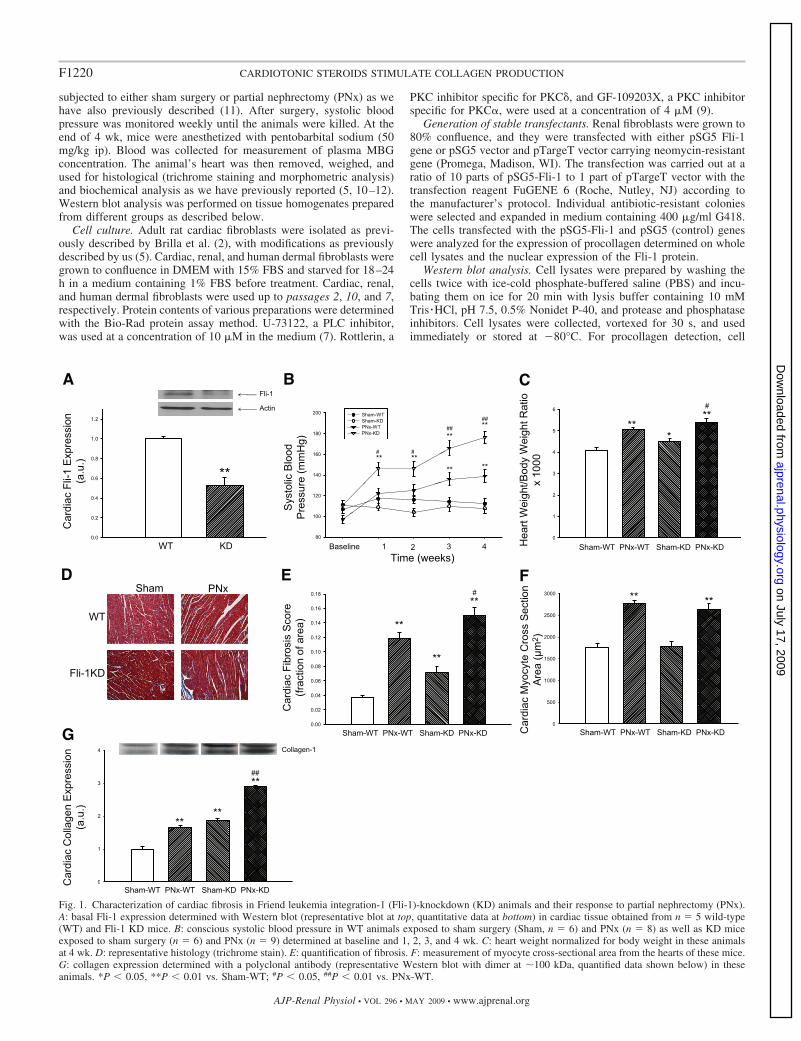
Fig. 1. Characterization of cardiac fibrosis in Friend leukemia integration-1 (Fli-1)-knockdown (KD) animals and their response to partial nephrectomy (PNx).A: basal Fli-1 expression determined with Western blot (representative blot at top, quantitative data at bottom) in cardiac tissue obtained from n � 5 wild-type(WT) and Fli-1 KD mice. B: conscious systolic blood pressure in WT animals exposed to sham surgery (Sham, n � 6) and PNx (n � 8) as well as KD miceexposed to sham surgery (n � 6) and PNx (n � 9) determined at baseline and 1, 2, 3, and 4 wk. C: heart weight normalized for body weight in these animalsat 4 wk. D: representative histology (trichrome stain). E: quantification of fibrosis. F: measurement of myocyte cross-sectional area from the hearts of these mice.G: collagen expression determined with a polyclonal antibody (representative Western blot with dimer at �100 kDa, quantified data shown below) in theseanimals. *P 0.05, **P 0.01 vs. Sham-WT; #P 0.05, ##P 0.01 vs. PNx-WT.
F1220 CARDIOTONIC STEROIDS STIMULATE COLLAGEN PRODUCTION
AJP-Renal Physiol • VOL 296 • MAY 2009 • www.ajprenal.org
on July 17, 2009 ajprenal.physiology.org
Dow
nloaded from

lysates or tissue homogenates were dissolved in loading buffer andproteins (10 �g/lane) were separated by SDS-PAGE using 4–15%Tris �HCl precast ready gels (Bio-Rad, Hercules, CA). For Fli-1detection, a large 10% gel was used and the protein loading wasbetween 80 and 200 �g/lane for nuclear extract or 50 �g/lane fortissue homogenates. After separation, proteins were transferred ontopolyvinylidene difluoride (PVDF) membranes. Membranes wereblocked with 5% nonfat dry milk (Bio-Rad) in Tris-buffered salinesupplemented with 0.05% Tween 20 (TBS-T) at room temperature for2 h and then incubated with primary antibody in blocking buffer at4°C overnight. After being washed in TBS-T, membranes wereincubated for 2 h at room temperature with horseradish peroxidase-conjugated secondary antibody in blocking buffer. After being washedin TBS, membranes were developed with ECL or ECL plus (Amer-sham Biosciences, Piscataway, NJ). For loading controls, tubulin oractin was probed; for both, the primary antibody was diluted 1/3,000and the secondary antibody (goat anti-mouse, Santa Cruz) was diluted1/2,000. The images captured on X-ray film were scanned withCanoScan Li DE 60 from Canon and quantified by using Image J
version 1.37V software (NIH). The quantified signals were in thelinear range of our detection system.
Immunostaining and confocal microscopy. Cells were fixed withcold absolute methanol or 4% paraformaldehyde in PBS, permeabil-ized in permeabilization buffer (PBS with 0.3% Triton X-100 and0.1% BSA) for 15 min, and blocked with GSDB buffer [20 mMsodium phosphate, pH 7.4, with 150 mM NaCl, 0.3% Triton X-100,and 16% (vol/vol) filtered normal goat serum] for 30 min at roomtemperature. The cells were then probed with primary antibody for 90min at room temperature or overnight at 4°C (rabbit polyclonal antiPKC� antibody, Santa Cruz Biotechnology; 1:50 dilution in GSDB).After three washes with permeabilization buffer, the cells were incu-bated with Alexa Fluor 488-conjugated antirabbit secondary antibodyfor 1 h at room temperature. After another three washes, the cells werecounterstained with propidium iodide (Molecular Probes) to localizenuclei. Cells were then mounted with Prolong Anti-fade medium(Molecular Probes) and stored at �20°C. All images were generatedwith a Leica DMIRE2 confocal microscope (Wetzlar, Germany).Contrast and brightness were set to ensure that all pixels were within
Fig. 2. Procollagen and Fli-1 expression in different fibroblasts. A: representative Western blot for procollagen showing dimer detected with polyclonal antibodyat �150 kDa and Fli-1 detected with a polyclonal antibody at �50 kDa (both shown at top) and the quantification of these measurements in each group (n �6) obtained at baseline in rat cardiac and renal and human dermal fibroblasts grown to confluence (bottom). Regression line for procollagen expression comparedwith Fli-1 expression is shown with data normalized to that of cardiac fibroblasts (r2 � 0.98, P 0.01). B: quantitative densitometric data for procollagen (measuredby Western blot as in A) in response to different doses of MBG for 24 h (n � 6). C: quantitative densitometric data for Fli-1 (measured by Western blot as in B) obtainedin response to 10 nM MBG (n � 6) for 24 h. Because of the differences in basal Fli-1 expression, we loaded 200 �g for cardiac, 130 �g for renal, and 80 �g for dermalfibroblast nuclear extracts. Top: C represents control, and M refers to MBG. Bars on quantitative graphs represent means SE. **P 0.01 vs. control.
F1221CARDIOTONIC STEROIDS STIMULATE COLLAGEN PRODUCTION
AJP-Renal Physiol • VOL 296 • MAY 2009 • www.ajprenal.org
on July 17, 2009 ajprenal.physiology.org
Dow
nloaded from
the linear range. Negative controls were also performed to verify thespecificity of primary and secondary antibodies.
Determination of PKC� in nucleus. Cardiac and renal fibroblastswere grown to confluence and starved for 18–24 h. Cells were treatedwith MBG to a final concentration of 10 nM in the medium. After 15min, cells were washed with ice-cold PBS and nuclear extracts wereprepared from treated and untreated cells as described by Wadmanet al. (18). PKC� in the nuclear extract was determined by performingWestern blot analysis as described above.
Immunoprecipitation and detection of phosphorylated Fli-1 byWestern blot. Renal fibroblasts were grown and starved as describedabove. The cells were treated for 15 min with 10 nM MBG in themedium. After the cells were washed in PBS, nuclear extracts wereprepared from treated and untreated cells as described (11). Equalamounts of proteins from nuclear extracts were used for immunopre-cipitation. For immunoprecipitation, nuclear proteins were incubatedwith the polyclonal anti-Fli-1 antibody conjugated to agarose beads at4°C overnight. The following day the immunocomplex was sedi-mented and washed three or four times with ice-cold PBS. Thewashed immunocomplex was dissolved in sample buffer, and theproteins were separated on a 10% gel. The separated proteins weretransferred to a PVDF membrane and processed as described underWestern blot analysis. To detect whether serine or threonine mole-cules are involved in phosphorylation of proteins, anti-phosphoserineor anti-phosphothreonine antibodies were used.
Immunoprecipitation and detection of phosphorylated Fli-1 byautoradiography. For autoradiography, renal fibroblasts were grownand starved as described above. The starved cells were rinsed twicewith phosphate-free DMEM and then incubated at 37°C in phosphate-free DMEM for 30 min. The cells were replaced with fresh phosphate-free DMEM containing 20 �Ci/ml [32P]orthophosphoric acid andincubated for 2 h. MBG was added to the cells to a final concentrationof 10 nM, and 15 min later the cells were washed with ice-cold PBS.Nuclear extracts were prepared as described (11). For immunopre-cipitation, nuclear extracts with equal amounts of radioactivity fromtreated and untreated renal fibroblasts were incubated with the poly-clonal anti-Fli-1 antibody conjugated to agarose beads at room tem-perature for 2 h. The immunocomplex was washed with ice-cold PBS,dissolved in sample buffer, and subjected to SDS-PAGE on a 10%gel. The gels were dried on a gel dryer, and the phosphorylatedproteins were visualized by using a phosphoimager (Storm 840 fromNovell) and quantified by using Image Quan TLV software fromNovell.
Statistical analysis. Data presented are means SE. Data obtainedwere first tested for normality. If the data did not pass the normalitytest, the Tukey test (for multiple groups) or the Mann-Whitney ranksum test was used to compare the data. If the data did pass thenormality test, parametric comparisons were performed. If more thantwo groups were compared, one-way analysis of variance was per-formed before comparison of individual groups with the unpairedStudent’s t-test with Bonferroni’s correction for multiple compari-sons. If only two groups of normal data were compared, the Student’st-test was used without correction (19). Statistical analysis was per-formed with SPSS software.
RESULTS
Relationship between Fli-1 and collagen expression in car-diac tissues of wild-type and Fli-1-knockdown mice before andafter nephrectomy. We first examined whether there was arelationship between collagen production and Fli-1 expressionin cardiac tissues. We determined the expression of Fli-1 andcollagen in the hearts obtained from wild-type (WT) andFli-1-knockdown (KD) mice by Western blot. We observedthat the hearts of the KD mice expressed �50% of the Fli-1 ofWT hearts (Fig. 1A). Next, we performed either sham surgery
or PNx on these animals, and conscious systolic blood pressurewas monitored before and after surgery as the renal failureprogressed. We found that systolic blood pressure was in-creased in WT as well as KD animals after PNx compared withsham surgery. Interestingly, the increase in systolic bloodpressure was greater in KD animals (Fig. 1B) compared withWT animals. When we examined the heart size in theseanimals, the heart size was increased after PNx in both WT andKD animals, but the increase in heart size was greater in theKD animals (Fig. 1C). Next, we examined the histology of thehearts with trichrome staining; we noted that there was agreater degree of fibrosis determined by the amount of bluestaining in both WT and KD animals after PNx, but the amountof blue staining was greater in KD compared with WT animals(Fig. 1, D and E). Of interest, cardiac myocyte cross-sectionalareas were not different between KD and WT animals either atbaseline or after PNx, but the cross-sectional areas of both KDand WT increased after PNx compared with sham surgery (Fig.1F). We also measured collagen expression in cardiac tissuesfrom these animals. We found that collagen expression washigh in KD compared with WT animals. This difference wasmore pronounced after PNx (Fig. 1G). This finding was con-sistent with the histological observations described above.
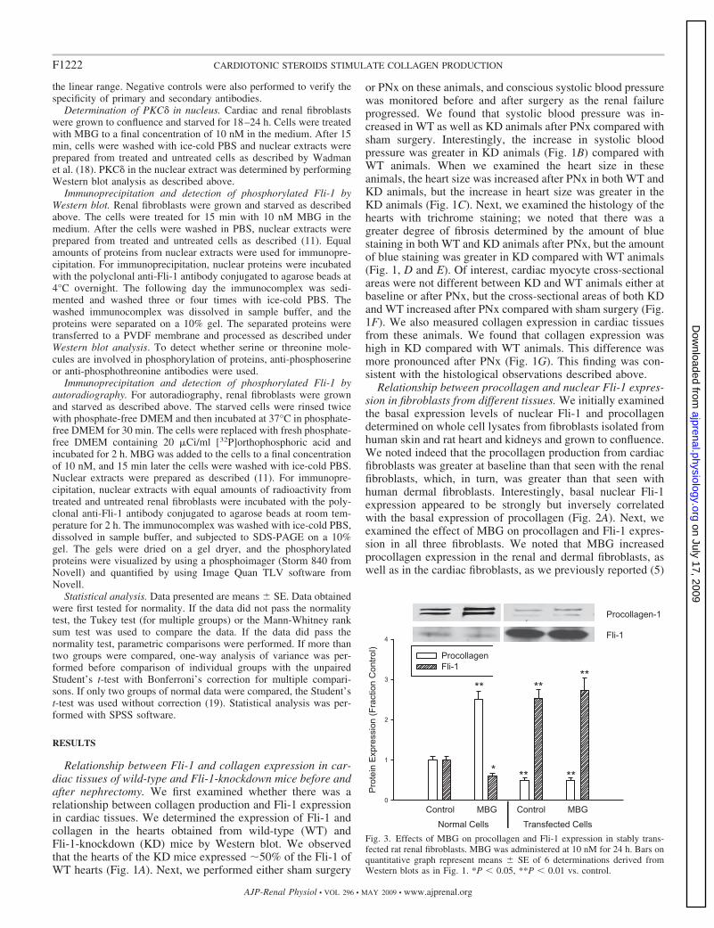
Relationship between procollagen and nuclear Fli-1 expres-sion in fibroblasts from different tissues. We initially examinedthe basal expression levels of nuclear Fli-1 and procollagendetermined on whole cell lysates from fibroblasts isolated fromhuman skin and rat heart and kidneys and grown to confluence.We noted indeed that the procollagen production from cardiacfibroblasts was greater at baseline than that seen with the renalfibroblasts, which, in turn, was greater than that seen withhuman dermal fibroblasts. Interestingly, basal nuclear Fli-1expression appeared to be strongly but inversely correlatedwith the basal expression of procollagen (Fig. 2A). Next, weexamined the effect of MBG on procollagen and Fli-1 expres-sion in all three fibroblasts. We noted that MBG increasedprocollagen expression in the renal and dermal fibroblasts, aswell as in the cardiac fibroblasts, as we previously reported (5)
Fig. 3. Effects of MBG on procollagen and Fli-1 expression in stably trans-fected rat renal fibroblasts. MBG was administered at 10 nM for 24 h. Bars onquantitative graph represent means SE of 6 determinations derived fromWestern blots as in Fig. 1. *P 0.05, **P 0.01 vs. control.
F1222 CARDIOTONIC STEROIDS STIMULATE COLLAGEN PRODUCTION
AJP-Renal Physiol • VOL 296 • MAY 2009 • www.ajprenal.org
on July 17, 2009 ajprenal.physiology.org
Dow
nloaded from
(Fig. 2B). The threshold for significant increases in procollagenexpression was at 0.1 nM in the renal and dermal fibroblasts,whereas it was 1.0 nM for the cardiac fibroblasts. Other cardiacglycosides (ouabain and digoxin) also induced increases inprocollagen expression in all three cell types (data not shown),as we had previously reported in cardiac fibroblasts (5). MBGadministered at a concentration of 10 nM resulted in markedand similar decreases in nuclear Fli-1 expression in all fibro-blasts studied (Fig. 2C).
Decreases in nuclear Fli-1 expression are necessary forMBG-induced increases in procollagen expression in a renalfibroblast line. To further examine the relationship between Fli-1expression and MBG-induced increases in procollagen produc-tion, we stably transfected the renal fibroblasts with a Fli-1expression vector coupled to an SV40 promoter. We found thatthe renal fibroblasts transfected with Fli-1 expression vectorshowed marked increase in nuclear Fli-1 expression, as expected.At the same time, the basal expression level of procollagen wassignificantly reduced. Furthermore, when these transfected cells
were exposed to MBG, MBG did not produce an increase inprocollagen expression. On the other hand, MBG produced asignificant increase in procollagen expression as well as a de-crease in nuclear Fli-1 expression in control renal fibroblasts(transfected with an empty vector, Fig. 3).
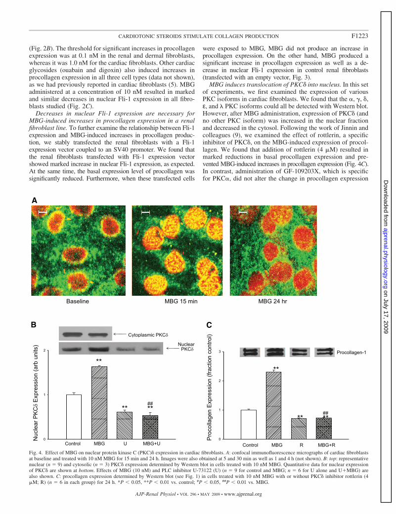
MBG induces translocation of PKC� into nucleus. In this setof experiments, we first examined the expression of variousPKC isoforms in cardiac fibroblasts. We found that the �, �, �,ε, and � PKC isoforms could all be detected with Western blot.However, after MBG administration, expression of PKC� (andno other PKC isoform) was increased in the nuclear fractionand decreased in the cytosol. Following the work of Jinnin andcolleagues (9), we examined the effect of rottlerin, a specificinhibitor of PKC�, on the MBG-induced expression of procol-lagen. We found that addition of rottlerin (4 �M) resulted inmarked reductions in basal procollagen expression and pre-vented MBG-induced increases in procollagen expression (Fig. 4C).In contrast, administration of GF-109203X, which is specificfor PKC�, did not alter the change in procollagen expression
Fig. 4. Effect of MBG on nuclear protein kinase C (PKC)� expression in cardiac fibroblasts. A: confocal immunofluorescence micrographs of cardiac fibroblastsat baseline and treated with 10 nM MBG for 15 min and 24 h. Images were also obtained at 5 and 30 min as well as 1 and 4 h (not shown). B: top: representativenuclear (n � 9) and cytosolic (n � 3) PKC� expression determined by Western blot in cells treated with 10 nM MBG. Quantitative data for nuclear expressionof PKC� are shown at bottom. Effects of MBG (10 nM) and PLC inhibitor U-73122 (U) (n � 9 for control and MBG; n � 6 for U alone and U MBG) arealso shown. C: procollagen expression determined by Western blot (see Fig. 1) in cells treated with 10 nM MBG with or without PKC� inhibitor rottlerin (4�M; R) (n � 6 in each group) for 24 h. *P 0.05, **P 0.01 vs. control; #P 0.05, ##P 0.01 vs. MBG.
F1223CARDIOTONIC STEROIDS STIMULATE COLLAGEN PRODUCTION
AJP-Renal Physiol • VOL 296 • MAY 2009 • www.ajprenal.org
on July 17, 2009 ajprenal.physiology.org
Dow
nloaded from

following MBG treatment for 24 h (relative expression tocontrol � 1.9 0.2; n � 3).
We next used confocal immunofluorescence microscopytechnique to follow the time course of the PKC� translocationto the nucleus. Confocal immunofluorescence microscopy ex-periments showed that that the nuclear PKC� expressionpeaked at �15 min and slowly decreased over the next 24 h ofexposure to MBG, and that this increase in nuclear expressionwas paralleled by a decrease in cytosolic expression (Fig. 4, Aand B). A virtually identical pattern was noted with renalfibroblasts (n � 5; data not shown). The finding of the confocalexperiment was supported by a Western blot experiment inwhich we found a significant increase in cardiac nuclear PKC�expression at 15 min accompanied by a decrease in cytosolicPKC� expression after exposure to 10 nM MBG. The increasein nuclear PKC� expression was blocked by coincubation withthe PLC inhibitor U-73122 (Fig. 4B).
Our next step was to examine whether the translocation ofPKC� into the nucleus causes phosphorylation of Fli-1. To testthis, cells were exposed to 10 nM MBG for 15 min and nuclearextracts were prepared. To enrich Fli-1, nuclear extract wasincubated with polyclonal anti-Fli-1 antibody. The resultingimmunocomplex was separated on a 10% gel and probed forphosphorylated proteins, particularly Fli-1, with an anti-phos-
phoserine antibody. We found a band with greater intensitycorresponding to the molecular mass of Fli-1 (Fig. 5A). Whenthe experiment was repeated in the presence of rottlerin orU-73122 (both n � 3), the intensity of the 50-kDa band was thesame or less than that seen with untreated cells (blot densitiesof 0.7 0.3 and 0.7 0.2 with rottlerin or U-73122 alone,respectively, and 0.6 0.3 and 0.7 0.2 when rottlerin orU-73122 was combined with MBG, respectively; data ex-pressed relative to control, both n � 3). When the immuno-complex was probed after separation of proteins with ananti-threonine antibody we did not see any increase in intensityof the band with 50-kDa mass after MBG treatment (blotdensity of 0.9 0.2 relative to control; n � 3).
To further demonstrate that MBG induced decreases in Fli-1expression through PKC�-mediated phosphorylation, we la-beled the cells with 32P and prepared nuclear extracts. Toenrich Fli-1, nuclear extracts were immunoprecipitated withpolyclonal anti-Fli-1 antibody and the immunocomplex wassubjected to gel electrophoresis. 32P-labeled proteins presenton the gel were quantified with a phosphoimager. We observedan increase in 32P-labeled protein at the level corresponding tomolecular mass of Fli-1 (Fig. 5B); this was attenuated to becomparable to that seen in control samples by coadministrationof rottlerin (blot density of 0.7 0.2 with rottlerin alone and0.6 0.3 with rottlerin MBG; data expressed relative tocontrol, n � 3).
DISCUSSION
Uremic cardiomyopathy is characterized by diastolic dys-function and left ventricular hypertrophy. Cardiac disease isresponsible for the high mortality seen in patients sufferingfrom kidney diseases (15). Our group and others have observedthat the cardiotonic steroid MBG, signaling through the Na-K-ATPase, is directly responsible for many features of experi-
Fig. 5. Effect of MBG on phosphorylated Fli-1 in renal fibroblasts.A: immunoblot against phosphoserine in nuclear extracts immunoprecipitatedwith polyclonal antibody against Fli-1; representative blot is shown at top andquantification of n � 4 determinations is shown at bottom. B: 32P autoradio-graph data obtained from cells treated with 32P-labeled phosphate for 2 h andMBG for 15 min. Top: representative autoradiograph. Bottom: quantification ofn � 5 experiments. *P 0.05 vs. control.
Fig. 6. Schematic describing how cardiotonic induced sodium pump signalingmay result in decreases in collagen production. In the presence of the car-diotonic steroid MBG, Na-K-ATPase is converted to a signal transducer thatcomplexes with Src and the EGF receptor (EGFR). A signal cascade is initiatedthat involves PLC, which results in the activation of PKC� and its translocationto the nucleus. In the nucleus, PKC� phosphorylates Fli-1. This, in turn, leadsto more rapid catabolism of Fli-1 and removal of Fli-1 inhibition on the Col1promoter and thus increases in collagen expression.
F1224 CARDIOTONIC STEROIDS STIMULATE COLLAGEN PRODUCTION
AJP-Renal Physiol • VOL 296 • MAY 2009 • www.ajprenal.org
on July 17, 2009 ajprenal.physiology.org
Dow
nloaded from

mental uremic cardiomyopathy. MBG directly induces cardiacfibroblasts to produce collagen, thus producing much of thecardiac fibrosis seen with experimental renal failure (5, 12).
Fli-1 is a member of the Ets oncogene family of proteins (1,4, 8, 16, 17) and normally competes with ETS-1 in a Sp-1-dependent manner to balance between stimulating and repress-ing the Col1a2 promoter. This transcription factor has beenclearly shown to play a role in dermal fibrosis, and a directinhibitory effect on collagen-1 synthesis has been demon-strated (3). In the present study, we extended this observationto an animal model and used Fli-1 KD mice, looking forcardiac fibrosis. First, we demonstrated that the viable het-erozygotes (Fli-1 /�) expressed �50% of cardiac Fli-1 com-pared with WT mice. These heterozygotes also had greaterdegrees of cardiac fibrosis and cardiac collagen expressioncompared with WT mice. The severity of cardiac fibrosis andthe amount of cardiac collagen expression were increased byPNx in both WT and heterozygote mice, thus supporting theconcept that the downregulation of nuclear Fli-1 expressioninduced by cardiotonic steroids plays a significant role in thecardiac fibrosis seen in experimental renal failure (5, 11). Thatsaid, we must point out that the systolic blood pressure wassignificantly higher in the Fli-1 heterozygote mice than in theWT mice after PNx, potentially contributing to the increasedfibrosis seen in these animals.
On the basis of these in vivo results, we examined therelevance to different types of fibroblasts, in particular cardiac,renal, and dermal fibroblasts. First, we demonstrated an inverserelationship between basal nuclear Fli-1 expression and colla-gen production in the three types of fibroblasts. We furtherdemonstrated that MBG induced corresponding decreases innuclear Fli-1 expression with increases in procollagen expres-sion in each of these fibroblasts, and we confirmed the impor-tance of Fli-1 with an overexpression transfection in a renalfibroblast cell line. These transfected cells showed higher basallevels of nuclear Fli-1 expression and lower basal levels ofprocollagen expression and did not show any significant in-crease in procollagen expression in response to MBG.
To further explore the signaling pathways leading to thisobservation, we first looked at the PKC family of proteins thatmediate the specific activation of a variety of transcriptionfactors. Specifically, Jinnin and colleagues (9) have shown thatPKC� stimulation can phosphorylate Fli-1 and increase colla-gen synthesis. Moreover, Zhang and Watson (22) suggestedthat a balance between protein kinase and protein phosphataseregulates the phosphorylation status and effectiveness of Fli-1in inhibiting collagen-1 synthesis. We observed that MBG-induced translocation of PKC� into the nucleus is a PLC-dependent process, a finding consistent with what Kometianiand coworkers (13) demonstrated for ouabain-induced activa-tion of the Na-K-ATPase signal cascade. U-73122 completelyprevented any increase in nuclear PKC�, whereas rottlerinprevented MBG-induced increases in procollagen expression,supporting this concept. We further determined that it is PKC�which phosphorylates Fli-1 as assessed by immunoprecipita-tion, immunoblotting, and 32P labeling. On the basis of thesedata as well as previous reports (3, 5, 12, 14), we wouldsuggest the schematic shown in Fig. 6.
Together with the existing literature, our findings stronglyimplicate the transcription factor Fli-1 in the progressive car-diac fibrosis seen with experimental uremia. If these data are
confirmed by further studies, we would suggest that the signalcascade that has been elucidated may serve as a therapeutictarget for clinical uremic cardiomyopathy.
ACKNOWLEDGMENTS
We thank Carol Woods for her excellent secretarial assistance.Portions of this work were presented at the 2006 and 2007 American
Society of Nephrology meetings in abstract form.
GRANTS
This work was supported by NIH Grants RO1-HL-67963 and P01-CA-78582 and supported in part by the intramural research program of the NationalInstitute on Aging.
REFERENCES
1. Blair DG, Athanasiou M. Ets and retroviruses—transduction and acti-vation of members of the Ets oncogene family in viral oncogenesis.Oncogene 19: 6472–6481, 2000.
2. Brilla CG, Zhou G, Matsubara L, Weber KT. Collagen metabolism incultured adult rat cardiac fibroblasts: response to angiotensin II andaldosterone. J Mol Cell Cardiol 26: 809–820, 1994.
3. Czuwara-Ladykowska J, Shirasaki F, Jackers P, Watson DK, TrojanowskaM. Fli-1 inhibits collagen type I production in dermal fibroblasts via an Sp1-dependent pathway. J Biol Chem 276: 20839–20848, 2001.
4. Dhulipal PD. Ets oncogene family. Indian J Exp Biol 35: 315–322, 1997.5. Elkareh J, Kennedy DJ, Yashaswi B, Vetteth S, Shidyak A, Kim EG,
Smaili S, Periyasamy SM, Hariri IM, Fedorova L, Liu J, Wu L,Kahaleh MB, Xie Z, Malhotra D, Fedorova OV, Kashkin VA, BagrovAY, Shapiro JI. Marinobufagenin stimulates fibroblast collagen produc-tion and causes fibrosis in experimental uremic cardiomyopathy. Hyper-tension 49: 215–224, 2007.
6. Fedorova OV, Lakatta EG, Bagrov AY. Endogenous Na,K pumpligands are differentially regulated during acute NaCl loading of Dahl rats.Circulation 102: 3009–3014, 2000.
7. He YY, Huang JL, Chignell CF. Delayed and sustained activation ofextracellular signal-regulated kinase in human keratinocytes by UVA:implications in carcinogenesis. J Biol Chem 279: 53867–53874, 2004.
8. Hromas R, Klemsz M. The ETS oncogene family in development,proliferation and neoplasia. Int J Hematol 59: 257–265, 1994.
9. Jinnin M, Ihn H, Yamane K, Mimura Y, Asano Y, Tamaki K.Alpha2(I) collagen gene regulation by protein kinase C signaling in humandermal fibroblasts. Nucleic Acids Res 33: 1337–1351, 2005.
10. Kennedy D, Omran E, Periyasamy SM, Nadoor J, Priyadarshi A,Willey JC, Malhotra D, Xie Z, Shapiro JI. Effect of chronic renal failureon cardiac contractile function, calcium cycling, and gene expression ofproteins important for calcium homeostasis in the rat. J Am Soc Nephrol14: 90–97, 2003.
11. Kennedy DJ, Elkareh J, Shidyak A, Shapiro AP, Smaili S, Mutgi K,Gupta S, Tian J, Morgan E, Khouri S, Cooper CJ, Periyasamy SM,Xie Z, Malhotra D, Fedorova OV, Bagrov AY, Shapiro JI. Partialnephrectomy as a model for uremic cardiomyopathy in the mouse. Am JPhysiol Renal Physiol 294: F450–F454, 2008.
12. Kennedy DJ, Vetteth S, Periyasamy SM, Kanj M, Fedorova L, KhouriS, Kahaleh MB, Xie Z, Malhotra D, Kolodkin NI, Lakatta EG,Fedorova OV, Bagrov AY, Shapiro JI. Central role for the cardiotonicsteroid marinobufagenin in the pathogenesis of experimental uremic car-diomyopathy. Hypertension 47: 488–495, 2006.
13. Kometiani P, Tian J, Li J, Nabih Z, Gick G, Xie Z. Regulation ofNa/K-ATPase beta1-subunit gene expression by ouabain and other hyper-trophic stimuli in neonatal rat cardiac myocytes. Mol Cell Biochem 215:65–72, 2000.
14. Kubo M, Czuwara-Ladykowska J, Moussa O, Markiewicz M, SmithE, Silver RM, Jablonska S, Blaszczyk M, Watson DK, TrojanowskaM. Persistent down-regulation of Fli1, a suppressor of collagen transcrip-tion, in fibrotic scleroderma skin. Am J Pathol 163: 571–581, 2003.
15. Sarnak MJ, Levey AS, Schoolwerth AC, Coresh J, Culleton B, HammLL, McCullough PA, Kasiske BL, Kelepouris E, Klag MJ, Parfrey P,Pfeffer M, Raij L, Spinosa DJ, Wilson PW. Kidney disease as a riskfactor for development of cardiovascular disease: a statement from theAmerican Heart Association Councils on Kidney in Cardiovascular Dis-ease, High Blood Pressure Research, Clinical Cardiology, and Epidemi-ology and Prevention. Hypertension 42: 1050–1065, 2003.
F1225CARDIOTONIC STEROIDS STIMULATE COLLAGEN PRODUCTION
AJP-Renal Physiol • VOL 296 • MAY 2009 • www.ajprenal.org
on July 17, 2009 ajprenal.physiology.org
Dow
nloaded from

16. Sato Y. Role of ETS family transcription factors in vascular developmentand angiogenesis. Cell Struct Funct 26: 19–24, 2001.
17. Truong AH, Ben-David Y. The role of Fli-1 in normal cell function andmalignant transformation. Oncogene 19: 6482–6489, 2000.
18. Wadman IA, Osada H, Grutz GG, Agulnick AD, Westphal H, Forster A,Rabbitts TH. The LIM-only protein Lmo2 is a bridging molecule assembling anerythroid, DNA-binding complex which includes the TAL1, E47, GATA-1 andLdb1/NLI proteins. EMBO J 16: 3145–3157, 1997.
19. Wallenstein S, Zucker CL, Fleiss JL. Some statistical methods useful incirculation research. Circ Res 47: 1–9, 1980.
20. Wang Y, Fan PS, Kahaleh B. Association between enhanced type Icollagen expression and epigenetic repression of the FLI1 gene in sclero-derma fibroblasts. Arthritis Rheum 54: 2271–2279, 2006.
21. Zhang XK, Gallant S, Molano I, Moussa OM, Ruiz P, SpyropoulosDD, Watson DK, Gilkeson G. Decreased expression of the Ets familytranscription factor Fli-1 markedly prolongs survival and significantlyreduces renal disease in MRL/lpr mice. J Immunol 173: 6481– 6489,2004.
22. Zhang XK, Watson DK. The FLI-1 transcription factor is a short-livedphosphoprotein in T cells. J Biochem 137: 297–302, 2005.
F1226 CARDIOTONIC STEROIDS STIMULATE COLLAGEN PRODUCTION
AJP-Renal Physiol • VOL 296 • MAY 2009 • www.ajprenal.org
on July 17, 2009 ajprenal.physiology.org
Dow
nloaded from