J Oral Maxillofac Surg 66:2050-2057, 2008 Conservative ... · PDF fileJ Oral Maxillofac Surg...
8
J Oral Maxillofac Surg 66:2050-2057, 2008 Conservative Treatment of Oral Ranula by Excision With Minimal Excision of the Sublingual Gland: Histological Support for a Traumatic Etiology Mark McGurk, MD, FRCS, FDSRCSEng,* Josiah Eyeson, PhD, FDSRCSEng,† Bethan Thomas, PhD, MFDSRCSEng,‡ and John D. Harrison, PhD, FDSRCSEng, FRCPath§ Purpose: This study investigates, clinically and histologically, a new conservative technique for the treatment of oral ranula based on the premise that a discrete unit of the sublingual gland feeds the ranula, which can therefore be treated by local removal with the attached part of the sublingual gland. Patients and Methods: The study group consisted of 8 patients with ranula treated by decompression of the ranula followed by local surgical removal together with the attached part of the sublingual gland. Detailed histologic examination of the entire specimen was undertaken in every case. Results: The treatment was successful in all the patients and there have been no recurrences after reviews of from 13 to 29 months (median, 26 months). Histologic examination of the entire specimen showed communication between the removed part of the sublingual gland and the ranula by way of a torn duct in every case. Conclusions: The premise that the ranula is fed by an attached, discrete unit of the sublingual gland has been vindicated and is the basis for the successful conservative treatment of ranula by decompression and local surgical removal together with the attached part of the sublingual gland. The finding of communication between the attached sublingual gland and ranula in every case indicates a traumatic etiology for these ranulas. © 2008 American Association of Oral and Maxillofacial Surgeons J Oral Maxillofac Surg 66:2050-2057, 2008 Most ranulas are large extravasation mucoceles that arise from the sublingual gland and are sufficiently ex- tensive to form a swelling that resembles the belly or vocal air sac of frog. They are cystic and are frequently blue owing to the Tyndall effect, whereby blue light is reflected more than red light at the interface of soft tissue and cyst. Most extravasation mucoceles occur in the lower lip 1 and are treated successfully by removal of the mucocele with the feeding minor salivary gland. Although the floor of the mouth is the second most common site for extravasation mucoceles, 1 the treat- ment of the ranula is varied and not always successful. 2-6 Treatment by incision, simple marsupialization, and ex- cision of the ranula alone have a high recurrence rate, whereas excision of the sublingual gland with or with- out the ranula is almost always successful. 7-9 Although the removal of the sublingual gland as the source of the extravasated mucus may be appealing, it is techni- cally demanding and associated with notable morbidity that can include damage to the lingual nerve, Wharton’s duct, submandibular gland, and blood vessels. 8,9 This has encouraged a search for a satisfactory conservative approach to treatment. Marsupialization with packing of the ranula is successful in about 90% of cases 2-4,6 and intracystic injection of the sclerosing-preparation OK- 432 has given variable results. 10,11 Received from King’s College London Dental Institute at Guy’s, King’s College and St Thomas’ Hospitals, London, England. *Professor and Consultant in Oral and Maxillofacial Surgery. †Lecturer in Oral and Maxillofacial Surgery. ‡Lecturer in Oral and Maxillofacial Surgery. §Reader and Consultant in Oral Pathology. Address correspondence and reprint requests to Dr Harrison: Department of Oral Pathology, Floor 28, Guy’s Tower, Guy’s Hos- pital, London, SE1 9RT, England; e-mail: [email protected] © 2008 American Association of Oral and Maxillofacial Surgeons 0278-2391/08/6610-0011$34.00/0 doi:10.1016/j.joms.2008.01.019 2050
Transcript of J Oral Maxillofac Surg 66:2050-2057, 2008 Conservative ... · PDF fileJ Oral Maxillofac Surg...

J6
MatvbrtttAcmT
R
K
Oral Maxillofac Surg6:2050-2057, 2008
Conservative Treatment of Oral Ranula byExcision With Minimal Excision of theSublingual Gland: Histological Support
for a Traumatic EtiologyMark McGurk, MD, FRCS, FDSRCSEng,*
Josiah Eyeson, PhD, FDSRCSEng,†
Bethan Thomas, PhD, MFDSRCSEng,‡ and
John D. Harrison, PhD, FDSRCSEng, FRCPath§
Purpose: This study investigates, clinically and histologically, a new conservative technique for thetreatment of oral ranula based on the premise that a discrete unit of the sublingual gland feeds the ranula,which can therefore be treated by local removal with the attached part of the sublingual gland.
Patients and Methods: The study group consisted of 8 patients with ranula treated by decompressionof the ranula followed by local surgical removal together with the attached part of the sublingual gland.Detailed histologic examination of the entire specimen was undertaken in every case.
Results: The treatment was successful in all the patients and there have been no recurrences afterreviews of from 13 to 29 months (median, 26 months). Histologic examination of the entire specimenshowed communication between the removed part of the sublingual gland and the ranula by way of atorn duct in every case.
Conclusions: The premise that the ranula is fed by an attached, discrete unit of the sublingual gland hasbeen vindicated and is the basis for the successful conservative treatment of ranula by decompressionand local surgical removal together with the attached part of the sublingual gland. The finding ofcommunication between the attached sublingual gland and ranula in every case indicates a traumaticetiology for these ranulas.© 2008 American Association of Oral and Maxillofacial Surgeons
J Oral Maxillofac Surg 66:2050-2057, 2008cwottctdhati4
D
p
©
0
ost ranulas are large extravasation mucoceles thatrise from the sublingual gland and are sufficiently ex-ensive to form a swelling that resembles the belly orocal air sac of frog. They are cystic and are frequentlylue owing to the Tyndall effect, whereby blue light iseflected more than red light at the interface of softissue and cyst. Most extravasation mucoceles occur inhe lower lip1 and are treated successfully by removal ofhe mucocele with the feeding minor salivary gland.lthough the floor of the mouth is the second mostommon site for extravasation mucoceles,1 the treat-ent of the ranula is varied and not always successful.2-6
reatment by incision, simple marsupialization, and ex-
eceived from King’s College London Dental Institute at Guy’s,
ing’s College and St Thomas’ Hospitals, London, England.
*Professor and Consultant in Oral and Maxillofacial Surgery.
†Lecturer in Oral and Maxillofacial Surgery.
‡Lecturer in Oral and Maxillofacial Surgery.
§Reader and Consultant in Oral Pathology. d
2050
ision of the ranula alone have a high recurrence rate,hereas excision of the sublingual gland with or with-ut the ranula is almost always successful.7-9 Althoughhe removal of the sublingual gland as the source ofhe extravasated mucus may be appealing, it is techni-ally demanding and associated with notable morbidityhat can include damage to the lingual nerve, Wharton’suct, submandibular gland, and blood vessels.8,9 Thisas encouraged a search for a satisfactory conservativepproach to treatment. Marsupialization with packing ofhe ranula is successful in about 90% of cases2-4,6 andntracystic injection of the sclerosing-preparation OK-32 has given variable results.10,11
Address correspondence and reprint requests to Dr Harrison:
epartment of Oral Pathology, Floor 28, Guy’s Tower, Guy’s Hos-
ital, London, SE1 9RT, England; e-mail: [email protected]
2008 American Association of Oral and Maxillofacial Surgeons
278-2391/08/6610-0011$34.00/0
oi:10.1016/j.joms.2008.01.019

rmalqw
P
polrcf
ewrg
ewta
btrtnpppcfi
wflcfcttassclg
wwwor
wusf
1scaw
LRCLRLVOT
Mf
M
McGURK ET AL 2051
This study describes a new method of treatinganulas based on principles applied to extravasationucoceles elsewhere in the mouth. The premise
dopted is that the ranula is fed by a discrete col-ection of sublingual tissue attached to it. Conse-uently it can be successfully treated by removalith only a portion of the gland.
atients and Methods
In the years from 2001 to 2006, 35 consecutiveatients were referred with extravasation mucocelesf minor salivary glands, most of which were in the
ower lip (Table 1). Records of these patients wereetrieved and reviewed retrospectively. This cohortomprised 21 males and 14 females and ranged in agerom 18 to 54 years (median, 31 years).
The treatment of these mucoceles was by surgicalxcision under local anesthesia. A standard approachas used whereby the mucocele was excised without
upture together with any attached minor salivarylands and the wound was closed directly.A similar approach was adopted for ranulas. How-
ver, to make the ranula amenable to local excision, itas decompressed by aspiration of some of its con-
ents before surgery, usually by 3 to 5 days, with theim of producing a visible, palpable cyst that had
Table 1. EXTRAVASATION MUCOCELES
Site of Mucocele Patients (n)
eft lower lip 15ight lower lip 11entre of lower lip 2eft buccal mucosa 3ight buccal mucosa 1eft floor of mouth 1entral surface of tongue 1rifice of right Wharton’s duct 1otal 35
cGurk et al. Treatment and Etiology of Ranula. J Oral Maxillo-ac Surg 2008.
Table 2. RANULAS
Gender Age (yr) Presenting Complaint
M 33 Recurrent swellingM 35 SwellingF 29 SwellingF 24 SwellingF 15 SwellingF 20 SwellingF 30 SwellingF 26 Swelling
cGurk et al. Treatment and Etiology of Ranula. J Oral Maxillofac Sur
een deflated by about a third and was small enougho be amenable to enucleation. A large cyst is prone toupture and it is important that the integrity is main-ained to identify the feeding sublingual tissue. It isot advisable to aspirate at operation because thisroduces a persistent leak of saliva from the punctureoint throughout the procedure unless the punctureoint is ligated. The cyst should not be evacuatedompletely, otherwise the capsule cannot be identi-ed.Surgery was under general anesthesia, the ranulaas approached through a longitudinal incision in theoor of mouth medial to the ranula. The cyst wasarefully freed from local tissues. This required care-ul and deliberate local dissection at the margin of theapsule, which was possible when the tension withinhe cyst had been released by the aspiration. Whenhe cyst was freed from local structures, it becamepparent that it was firmly attached to part of theublingual gland. At the point of attachment to theublingual gland, a portion of the gland was excised inontinuity with the ranula using cutting diathermy toiberate it from the main portion of the sublingualland.In 2005 and 2006, 8 consecutive patients referredith ranulas were treated in this manner and dataere collected prospectively (Table 2). The ranulasere typical oral ranulas lying in the paralingual spacef the floor of the mouth, not plunging, cervicalanulas.
All patients were reviewed at 1 week, and thoseho had ranulas were subsequently kept under reg-lar review. Patients with mucoceles of the minoralivary glands were surveyed by post or telephoneor evidence of recurrence.
The excised ranulas were fixed in formalin (Fig), sliced at about 4-mm intervals and labeled con-ecutively, embedded in wax, and sections wereut at intervals throughout the embedded materialnd stained with hematoxylin and eosin (H&E) orith Alcian blue at pH2.5 followed by periodic-
Site of RanulaDuration Before
Presentation (wks)
Right floor of mouth 6Right floor of mouth Patient unsureLeft floor of mouth 4Right floor of mouth 8Right floor of mouth 32Left floor of mouth 8Left floor of mouth 32Right floor of mouth 6
g 2008.

as
R
srfclsHq
e
wmeiwflo
evglttpt
Fo
M fac Sur
Fmego
M
2052 TREATMENT AND ETIOLOGY OF RANULA
cid-Schiff (ABPAS) for the demonstration of muco-ubstance.12
esults
Local surgical excision of mucoceles of the minoralivary glands was successful in all patients and noecurrence was reported at review, which rangedrom 11 to 65 months (median, 39 months). The onlyomplication of surgery was a local, minor sensoryoss in the mucosa of the lower lip at the site ofurgery that occurred in about one third of the cases.owever, it was so limited as to be of no conse-uence to the patient.The 8 patients with ranula were treated under gen-
ral anesthesia with 1 overnight stay. No recurrences
IGURE 1. Fixed specimen of ranula with attached part of sublinf gland (G) and ranula. Scale � mm.
cGurk et al. Treatment and Etiology of Ranula. J Oral Maxillo
IGURE 2. Section from transverse slice through ranula and attachucus in the acini of the gland (G) is stained royal blue. The grxtravasated mucus. An extensive region of extravasated mucus intland (G). A palely stained capsule of granulation tissue (arrows)f section stained with ABPAS. Magnification �13. Scale bar � 5
cGurk et al. Treatment and Etiology of Ranula. J Oral Maxillofac Sur
ere recorded at review, which ranged from 13 to 29onths (median, 26 months). No complications were
ncountered except for transient lingual paraesthesian 2 patients. In 1 case, the lingual nerve was bound
ith scar tissue as a result of previous surgery to theoor of the mouth and it had to be freed at the timef surgery.Histologic examination of the ranulas showed cystic
xtravasation mucoceles in all the cases (Fig 2). Theyaried in size from 25 to 50 mm. Lobules of sublingualland were associated with the mucoceles. In 1 case,obules of submandibular gland were found adjacent tohe sublingual lobules. The lumina of the cysts con-ained mucus in which there were conspicuous macro-hages. The capsules of the cysts consisted of granula-ion tissue in which there were macrophages. Mucus
and, which communicated with the ranula. Line indicates junction
g 2008.
of sublingual gland. Extravasated mucus is stained purple and theart of the ranula is a cyst (C), the lumen of which is filled withwith granulation tissue (*) is present between the cyst (C) and thee cyst and mucus intermixed with granulation tissue. Photograph
gual gl
ed parteater permixedlimits thmm.
g 2008.

if(
tOcctotnsTdrwicmnc
sTmd
iww
D
gdnr
celTtpsstgdcoo
Fbosg�
M fac Sur
McGURK ET AL 2053
ntermixed with granulation tissue was sometimesound adjacent to the cysts and in 1 case was extensiveFigs 2, 3).
Communication between the lumen of the cyst andhe sublingual gland was seen in all cases (Figs 3-6).ral mucosa was seen in 4 cases, in 3 of which aommunication was seen close to it (Fig 5). Twoommunications were seen in 3 cases. In one ofhese, 1 communication was superficial and close toral mucosa and the other was to the deep surface ofhe cyst (Figs 5, 6). In the other 2 cases, the commu-ications were in slices separated by an interveninglice, which indicates that they were over 4 mm apart.he communication was by an interlobular or mainuct that passed through the capsule of the cyst toeach the lumen (Figs 3-6) or into mucus intermixedith granulation tissue for 1 of the 2 communications
n 1 case. The epithelium that lined the duct at theommunication showed a characteristic squamousetaplasia and was sometimes seen to line the lumi-
al surface of the adjacent part of the capsule of theyst (Figs 4-6).There were variable inflammation, atrophy, fibro-
is, and ductal dilatation in the sublingual gland.here was a small amount of intralobular extravasateducus in 2 cases, which in 1 case was from acini and
IGURE 3. Section from transverse slice adjacent to that of Figurelue. The gland communicates with the ranula via a duct, the torn ef the cyst, which appears empty owing to a loss of mucus duringurrounds this extension. A palely stained capsule of granulation tissranulation tissue at the margin of the specimen. Photomicrographm.
cGurk et al. Treatment and Etiology of Ranula. J Oral Maxillo
ucts that consisted of flattened epithelium surround- p
ng a dilated lumen (Figs 7, 8), and in the other caseas from ducts with lymphoreticular metaplasia thatere surrounded by lymphoid follicles.
iscussion
The technique of limited removal of sublingualland is based on the premise that a ranula arises at aiscrete point along the sublingual gland. The combi-ation of histological findings and the lack of recur-ence confirms the validity of the premise.
The detailed anatomy of the sublingual gland is moreomplex than is generally appreciated. Leppi13 discov-red that the sublingual gland consists of a constant,esser sublingual gland and a greater sublingual gland.he latter is posterior to the lesser sublingual gland in
he paralingual space and was only found in 10 of 28eople and usually only unilaterally. Furthermore, theublingual gland is often continuous with the part of theubmandibular gland that is above the posterior part ofhe mylohyoid muscle. Bartholin’s duct runs from thereater sublingual gland to either join or open indepen-ently of Wharton’s duct. The lesser sublingual glandonsists of between 8 to 30 small glands, from every onef which a duct of Rivinus passes to open independentlyn the sublingual fold. Therefore, according to the
sublingual gland (G) contains mucus in acini that is stained royalhich (hollow arrow) communicates with the lumen of an extension
ssing. Purple stained mucus intermixed with granulation tissue (*)ows) encapsulates the main cyst (C) and the mucus intermixed withtion stained with ABPAS. Magnification �47. Scale bar � 1,000
g 2008.
2. Thend of wproceue (arrof sec
remise that the ranula arises from 1 small unit of gland,

otts
dup
Fwg
M Surg 2
FmS
M
2054 TREATMENT AND ETIOLOGY OF RANULA
nce the ranula is dissected free from local connectiveissue, only that portion of the sublingual gland attachedo the ranula needs to be excised and the majority of theublingual gland is preserved.
IGURE 4. Detail of section adjacent to that of Figure 3. The sublihich is seen (hollow arrow), shows squamous metaplasia and linranulation tissue between the lumen and the gland. Photomicrograp
cGurk et al. Treatment and Etiology of Ranula. J Oral Maxillofac
IGURE 5. Section from transverse slice through ranula. Oral mucoetaplasia indicative of a nearby tear and communication with the lcale bar � 500 �m.
cGurk et al. Treatment and Etiology of Ranula. J Oral Maxillofac Surg 2
However, the successful eradication of the ranulaepends on the successful identification of the secretorynit supplying it at operation. For this to be accom-lished, the circumference of the ranula has to be freed
land (G) communicates with the ranula via a duct, the torn end ofuminal surface of the capsule of the cyst. Mucus is intermixed withtion stained with H&E. Magnification �400. Scale bar � 200 �m.
008.
ers the cyst, close to which a duct (hollow arrow) shows squamoushotomicrograph of section stained with H&E. Magnification �118.
ngual ges the lh of sec
sa covumen. P
008.
facI
bTaa
FgP
M fac Sur
F(�
M
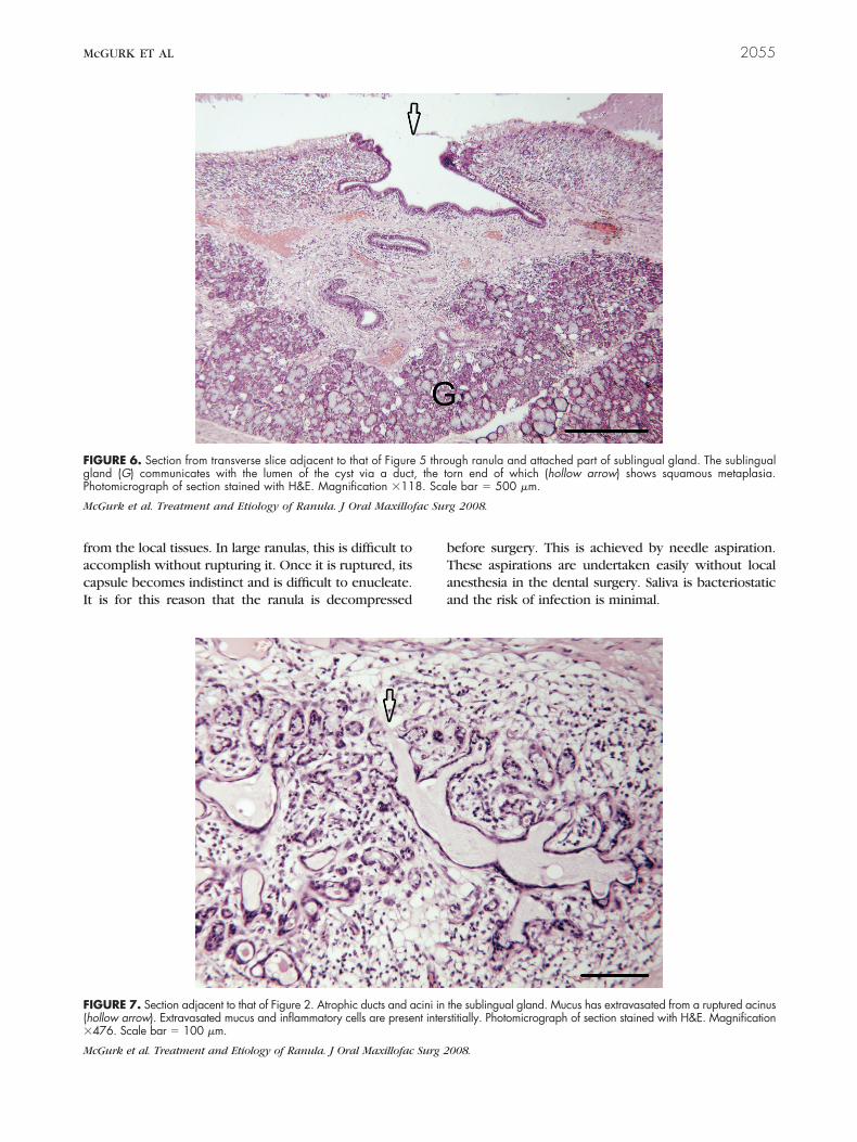
McGURK ET AL 2055
rom the local tissues. In large ranulas, this is difficult toccomplish without rupturing it. Once it is ruptured, itsapsule becomes indistinct and is difficult to enucleate.t is for this reason that the ranula is decompressed
IGURE 6. Section from transverse slice adjacent to that of Figureland (G) communicates with the lumen of the cyst via a ducthotomicrograph of section stained with H&E. Magnification �11
cGurk et al. Treatment and Etiology of Ranula. J Oral Maxillo
IGURE 7. Section adjacent to that of Figure 2. Atrophic ducts and ahollow arrow). Extravasated mucus and inflammatory cells are prese476. Scale bar � 100 �m.
cGurk et al. Treatment and Etiology of Ranula. J Oral Maxillofac Surg 2
efore surgery. This is achieved by needle aspiration.hese aspirations are undertaken easily without localnesthesia in the dental surgery. Saliva is bacteriostaticnd the risk of infection is minimal.
ugh ranula and attached part of sublingual gland. The sublingualrn end of which (hollow arrow) shows squamous metaplasia.
le bar � 500 �m.
g 2008.
the sublingual gland. Mucus has extravasated from a ruptured acinustitially. Photomicrograph of section stained with H&E. Magnification
5 thro, the to8. Sca
cini innt inters
008.

rmemmh2tgto
ssStaeFcirfitsctaw
aopditctrwalsoefommgcc
adtms
Fto
M fac Sur
2056 TREATMENT AND ETIOLOGY OF RANULA
The histological findings have confirmed that theanulas in the present investigation are extravasationucoceles that arise from the sublingual gland. The
tiology of extravasation mucoceles elsewhere in theouth and particularly in the lower lip is usuallyechanical trauma.1,9 However, Zhao et al7 found aistory of trauma to the floor of the mouth in only.7% of patients with ranulas. Furthermore, the ex-ravasation had often been found to occur deep in theland where it is unlikely to be mechanically trauma-ized.2,3,14 The etiology of the ranula was consideredbscure.3,15
One possible etiology is obstruction, for both theublingual and minor salivary glands are spontaneousecretors with a great resistance to obstruction.9,16
upportive evidence for this is that ductal ligation ofhe feline sublingual gland often results in an extrav-sation mucocele from an accumulation of mucus thatxtravasates from ruptured acini and not the ducts.16
urthermore, an increased incidence of ranulas asso-iated with untreated HIV infection in Zimbabwe17,18
s possibly a result of obstruction for the followingeasons: there is an increase of inflammation andbrosis in minor salivary glands of patients with un-reated HIV19 (this would also involve the biologicallyimilar sublingual glands), inflammation and fibrosisause obstruction of salivary glands,20-23 and obstruc-ion of the sublingual glands leads to extravasationnd possibly the development of ranulas in patients
IGURE 8. Detail of section of Figure 2. The mucus is stained fromhe lumina of the ducts and acini, extravasates through the rupturef section stained with ABPAS. Magnification �476. Scale bar �
cGurk et al. Treatment and Etiology of Ranula. J Oral Maxillo
ith untreated HIV infection. s
However, the findings in the present study supporttraumatic etiology for most ranulas. Thus, naturallyccurring ranulas have been found in the present andrevious investigations to be fed by a damageduct,14,15 and although extravasation from acini and
ntralobular ducts was found in 2 of the present cases,he amount of extravasated mucus was insignificantompared with the amount in the ranulas. The findinghat communication between sublingual gland andanula was close to the oral mucosa in 3 of 4 cases inhich oral mucosa was seen, and the finding of bothsuperficial and a deep communication between sub-
ingual gland and ranula in 1 case suggest that aublingual duct was originally severed close to theral mucosa and the 2 ends became separated by thexpansion of the ranula. This is the likely explanationor the previous finding that the extravasation oftenccurs deep in the gland where it is unlikely to beechanically traumatized,2,3,14 and for the finding ofore than 1 communication between sublingual
lands and ranulas in dogs.14 Thus it seems that me-hanical trauma to the floor of the mouth is moreommon than assumed and passes unnoticed.The investigation by Glen14 confirmed that ranulas
rise from the sublingual gland and not the subman-ibular, although he found a coexistent leakage fromhe anterior border of the submandibular gland in 1ucocele out of 58. The submandibular gland is not a
pontaneous secretor and is far less resistant to ob-
to royal blue, is present in reduced amount in atrophic acini, fillscinus (hollow arrow) and is present interstitially. Photomicrographm.
g 2008.
purpleof the a100 �
truction than the sublingual gland, and even when

tecwg
is
igtc
A
N
R
1
1
1
1
1
1
1
1
1
1
2
2
2
2
McGURK ET AL 2057
he submandibular gland is damaged and there isxtravasation of saliva, the ensuing granulation tissuean obstruct further extravasation and seal the leak,hich is an outcome less likely with the sublingual
land.9,15,16
This histological investigation indicates that traumas the usual etiology of the ranula, as it is of extrava-ation mucoceles elsewhere in the mouth.
The conservative, enucleation technique describedn this investigation preserves most of the sublingualland and has been successful in 8 consecutive pa-ients. This method merits further evaluation andomparison with other conservative techniques.
cknowledgment
We gratefully acknowledge the technical assistance of Mrs Deepaayar.
eferences1. Harrison JD: Salivary mucoceles. Oral Surg Oral Med Oral
Pathol 39:268, 19752. Baurmash HD: Marsupialization for treatment of oral ranula: A
second look at the procedure. J Oral Maxillofac Surg 50:1274,1992
3. Baurmash HD: Mucoceles and ranulas. J Oral Maxillofac Surg61:369, 2003
4. Baurmash HD: A case against sublingual gland removal as pri-mary treatment of ranulas. J Oral Maxillofac Surg 65:117, 2007
5. McGurk M: The surgical management of salivary gland diseaseof the sublingual gland and floor of mouth. Atlas Oral Maxillo-fac Surg Clin N Am 6:51, 1998
6. McGurk M: Management of the ranula. J Oral Maxillofac Surg65:115, 2007
7. Zhao Y-F, Jia Y, Chen X-M, et al: Clinical review of 580 ranulas.Oral Surg Oral Med Oral Pathol Oral Radiol Endod 98:281, 2004
8. Zhao Y-F, Jia J, Jia Y: Complications associated with surgical
management of ranulas. J Oral Maxillofac Surg 63:51, 20059. Harrison JD, Sowray JH, Smith NJD: Recurrent ranula. A casereport. Br Dent J 140:180, 1976
0. Fukase S, Ohta N, Inamura K, et al: Treatment of ranula withintracystic injection of the streptococcal preparation OK-432.Ann Otol Rhinol Laryngol 112:214, 2003
1. Rho J-L: Primary treatment of ranula with intracystic injectionof OK-432. Laryngoscope 116:169, 2006
2. Harrison JD, Auger DW, Paterson KL, et al: Mucin histochem-istry of submandibular and parotid salivary glands of man: Lightand electron microscopy. Histochem J 19:555, 1987
3. Leppi TJ: Gross anatomical relationships between primate sub-mandibular and sublingual salivary glands. J Dent Res 46:359,1967
4. Glen JB: Canine salivary mucoceles. Results of sialographicexamination and surgical treatment of fifty cases. J Small AnimPract 13:515, 1972
5. Harrison JD, Garrett JR: An ultrastructural and histochemicalstudy of a naturally occurring salivary mucocele in a cat.J Comp Pathol 85:411, 1975
6. Harrison JD, Garrett JR: Experimental salivary mucoceles in cat:A histochemical study. J Oral Pathol 4:297, 1975
7. Chidzonga MM, Rusakaniko S: Ranula: Another HIV/AIDS asso-ciated oral lesion in Zinbabwe? Oral Dis 10:229, 2004
8. Chidzonga MM, Mahomva L: Ranula: Experience with 83 casesin Zinbabwe. J Oral Maxillofac Surg 65:79, 2007
9. McArthur CP, Africa CWJ, Castellani WJ, et al: Salivary glanddisease in HIV/AIDS and primary Sjögren’s syndrome: Analysisof collagen I distribution and histopathology in American andAfrican patients. J Oral Pathol Med 32:544, 2003
0. Harrison JD, Epivatianos A, Bhatia SN: Role of microliths in theetiology of chronic submandibular sialadenitis: A clinicopatho-logical investigation of 154 cases. Histopathology 31:237, 1997
1. Antoniades D, Harrison JD, Epivatianos A, et al: Treatment ofchronic sialadenitis by intraductal penicillin or saline. J OralMaxillofac Surg 62:431, 2004
2. Harrison JD: Histology and pathology of sialolithiasis, in WittRL (ed): Salivary Gland Diseases. Surgical and Medical Manage-ment. New York, Stuttgart, Thieme, 2006, p 71
3. Harrison JD: Natural history of chronic sialadenitis and sialoli-thiasis, in Nahlieli O, Iro H, McGurk M, et al (eds): ModernManagement Preserving the Salivary Glands. Herzeliya, Isradon,
2007, p 93


![SHIFT mag [n°4] - Europe 2057](https://static.fdocuments.us/doc/165x107/568c4b761a28ab49169c4c25/shift-mag-n4-europe-2057-56f7710a974e5.jpg)