Isolation and Estimation of DNA Level in Coconut Leaf ...
4
Journal of Natural Sciences Research www.iiste.org ISSN 2224-3186 (Paper) ISSN 2225-0921 (Online) Vol.5, No.2, 2015 93 Isolation and Estimation of DNA Level in Coconut Leaf (Coccos Nucifera) AINA, V.O 1 IBRAHIM, M.B 2 ABDULSALAMI, M.S 3 ADEWUMI, A.A.J. 1 PETER M. WAZIRI 4 ISIYAKU MUHAMMED 4 1.Dept of Applied Science, College of Science and Technology, Kaduna Polytechnic, P.M.B 2021, Kaduna – Nigeria 2.Central Administration Kaduna Polytechnic, Kaduna 3.Dept of Biological Sciences, Nigerian Defence Academy, P.M.B. 2109, Kaduna – Nigeria 4.Dept of Biochemistry, Kaduna State University, P.M.B. 2339, Kaduna – Nigeria Corresponding Author Email: [email protected] Abstract The DNA Level of Coconut leaf was determined using agarose gel electrophoresis and UV-double beam spectrophotometer. 30µg leaf sample was weighed; chemical homogenization using mortar and pestle was done using lyses buffer and “Morning fresh” detergent. Whole sample was centrifuged at 10,000 rpm for 20 minutes; the supernatant was decamped into clean microcentruifuge tubes. Ethanol (500ml) was added and mixed thoroughly and incubated at room temperature for 30 minutes. DNA is insoluble in ethanol and so will appear as white precipitate at the bottom of the tube. Sample was centrifuged at 10,000 rpm for 5 minutes, thereafter the content was exposed to the atmosphere for 10 minutes to rid off remaining solvent (Ethanol). The pellets were dissolved in 50ml of TE (Tris-EDTA) buffer. The DNA (25ml) was taken and diluted in 1.75ml of TE Buffer and absorbance read at 260nm and 280nm with purity of DNA calculated followed by Electrophoresis. 25ml of the DNA sample was taken and ran on 0.8% agarose gel electrophoresis using a standard marker for 60 minutes. The analysis was done in triplicate with the ratio of absorbance at 260 and 280nm (1.79, 1.76 and 1.84) showing the purity of the DNA sample. Keywords: Coccos nucifera, Percentage Purity, DNA Sample, TE Buffer, Agarose Gel Electrophoresis. INTRODUCTION The coconut palm, Coccos nucifera is a member of the family Arecaceae (palm family). It is the only accepted species in the genus cocos (Hahn, 1997). The term coconut can refer to the entire coconut palm, the seed, or the fruit, which is not a botanical nut. Found across much of the tropic and subtropics area, the coconut is known for its great versatility as seen in many domestic commercial and industrial users of its different parts. Coconuts are part of the daily diet of many people. When young, the entire fruits are used as melons, when mature only the seeds are used as nuts. The oil and milk derived from it are commonly used in cooking and frying. Coconut oil is also widely used in soaps and cosmetics. The clean liquid coconut water within is a refreshing drink and can be processed to create alcohol. The husks and leaves can be used as material to make a variety of products for furnishing and decorating. It also has cultural and religious significance in many societies that use it (Grimwood et al, 1975). The determination of the concentration of DNA or RNA in solution is a fundamental task in molecular biology. The quantity of DNA is limiting in most experiments, therefore the knowledge of its concentration is critical. Determination of the DNA concentration can be estimated either by qualitatively comparing the fluorescence of DNA bands in an agarose gel to a standard or by spectrophotometric. Deoxyribonucleic Acid (DNA) is a nucleic acid containing the genetic instructions used in the development and functioning of all known living organisms (with the exception of RNA viruses). The DNA segments carrying this genetic information are called genes. Likewise, other DNA sequences have structural purposes, or are involved in regulating the use of this genetic information. Along with RNA and proteins, DNA is one of the three major macromolecules that are essential for all known form of life. DNA consists of two long polymers of simple units called nucleotides, with backbones made of sugars and phosphate groups joined by ester bonds. These two strands run in opposite directions to each other and are therefore anti-parallel. In living organisms, DNA does not usually exist as a single molecule, but instead as a pair of molecules that are held tightly together (Berg, et al, 2002). These two long strands entwine like vines, in the shape of a double helix. As a portable source of both food and water, the coconut played a critical role in the ability of humans to voyage, establish trade routes and colonize lands in regions throughout the Cold World Tropics (Harries, 1978). This species continues to have hundreds of uses as a source of food, drink, fiber construction matter, charcoal and coil. (Batugal, 2005). The history of dispersal and cultivation of this species is thus fundamentally intertwined with human history in the tropics.
Transcript of Isolation and Estimation of DNA Level in Coconut Leaf ...

Journal of Natural Sciences Research www.iiste.org
ISSN 2224-3186 (Paper) ISSN 2225-0921 (Online)
Vol.5, No.2, 2015
93
Isolation and Estimation of DNA Level in Coconut Leaf (Coccos
Nucifera)
AINA, V.O1 IBRAHIM, M.B
2 ABDULSALAMI, M.S
3 ADEWUMI, A.A.J.
1
PETER M. WAZIRI4
ISIYAKU MUHAMMED4
1.Dept of Applied Science, College of Science and Technology, Kaduna Polytechnic, P.M.B 2021, Kaduna –
Nigeria
2.Central Administration Kaduna Polytechnic, Kaduna
3.Dept of Biological Sciences, Nigerian Defence Academy, P.M.B. 2109, Kaduna – Nigeria
4.Dept of Biochemistry, Kaduna State University, P.M.B. 2339, Kaduna – Nigeria
Corresponding Author Email: [email protected]
Abstract
The DNA Level of Coconut leaf was determined using agarose gel electrophoresis and UV-double beam
spectrophotometer. 30µg leaf sample was weighed; chemical homogenization using mortar and pestle was done
using lyses buffer and “Morning fresh” detergent. Whole sample was centrifuged at 10,000 rpm for 20 minutes;
the supernatant was decamped into clean microcentruifuge tubes. Ethanol (500ml) was added and mixed
thoroughly and incubated at room temperature for 30 minutes. DNA is insoluble in ethanol and so will appear as
white precipitate at the bottom of the tube. Sample was centrifuged at 10,000 rpm for 5 minutes, thereafter the
content was exposed to the atmosphere for 10 minutes to rid off remaining solvent (Ethanol). The pellets were
dissolved in 50ml of TE (Tris-EDTA) buffer. The DNA (25ml) was taken and diluted in 1.75ml of TE Buffer
and absorbance read at 260nm and 280nm with purity of DNA calculated followed by Electrophoresis. 25ml of
the DNA sample was taken and ran on 0.8% agarose gel electrophoresis using a standard marker for 60 minutes.
The analysis was done in triplicate with the ratio of absorbance at 260 and 280nm (1.79, 1.76 and 1.84) showing
the purity of the DNA sample.
Keywords: Coccos nucifera, Percentage Purity, DNA Sample, TE Buffer, Agarose Gel Electrophoresis.
INTRODUCTION
The coconut palm, Coccos nucifera is a member of the family Arecaceae (palm family). It is the only accepted
species in the genus cocos (Hahn, 1997). The term coconut can refer to the entire coconut palm, the seed, or the
fruit, which is not a botanical nut. Found across much of the tropic and subtropics area, the coconut is known for
its great versatility as seen in many domestic commercial and industrial users of its different parts. Coconuts are
part of the daily diet of many people. When young, the entire fruits are used as melons, when mature only the
seeds are used as nuts. The oil and milk derived from it are commonly used in cooking and frying. Coconut oil is
also widely used in soaps and cosmetics. The clean liquid coconut water within is a refreshing drink and can be
processed to create alcohol. The husks and leaves can be used as material to make a variety of products for
furnishing and decorating. It also has cultural and religious significance in many societies that use it (Grimwood
et al, 1975).
The determination of the concentration of DNA or RNA in solution is a fundamental task in molecular
biology. The quantity of DNA is limiting in most experiments, therefore the knowledge of its concentration is
critical. Determination of the DNA concentration can be estimated either by qualitatively comparing the
fluorescence of DNA bands in an agarose gel to a standard or by spectrophotometric.
Deoxyribonucleic Acid (DNA) is a nucleic acid containing the genetic instructions used in the
development and functioning of all known living organisms (with the exception of RNA viruses). The DNA
segments carrying this genetic information are called genes. Likewise, other DNA sequences have structural
purposes, or are involved in regulating the use of this genetic information. Along with RNA and proteins, DNA
is one of the three major macromolecules that are essential for all known form of life.
DNA consists of two long polymers of simple units called nucleotides, with backbones made of sugars
and phosphate groups joined by ester bonds. These two strands run in opposite directions to each other and are
therefore anti-parallel. In living organisms, DNA does not usually exist as a single molecule, but instead as a pair
of molecules that are held tightly together (Berg, et al, 2002). These two long strands entwine like vines, in the
shape of a double helix.
As a portable source of both food and water, the coconut played a critical role in the ability of humans
to voyage, establish trade routes and colonize lands in regions throughout the Cold World Tropics (Harries,
1978). This species continues to have hundreds of uses as a source of food, drink, fiber construction matter,
charcoal and coil. (Batugal, 2005). The history of dispersal and cultivation of this species is thus fundamentally
intertwined with human history in the tropics.
Journal of Natural Sciences Research www.iiste.org
ISSN 2224-3186 (Paper) ISSN 2225-0921 (Online)
Vol.5, No.2, 2015
94
AIMS AND OBJECTIVES OF THE RESEARCH
These includes:
1. To isolate and estimate the DNA level of coconut leaves (Coccos nucifera)
2. To evaluate a basis for genetic analysis in the area of scientific, medical or forensic purposes such as
introduction of DNA into each of animals or plants or for diagnostic purposes.
MATERIALS AND METHODS
This research/bench work was carried out between May – November, 2012 in the Biochemistry Laboratories of
the Department of Biochemistry, Kaduna State University, Kaduna – Nigeria.
Principle of Extraction
The concentration of DNA sample can be checked by the use of UV spectrophotometry. DNA absorb UV light
very efficiently making it possible to detect and quantify at concentrations as low as 2.5µg/µl. The nitrogenous
bases has an absorption maximum at about 260nm, using a 1-cm light path, the extinction coefficient for
nucleotides of this wavelength is 20. Based on this, the absorbance at 260nm in a 1-cm quartz cuvette of a
50µg/ml solution of double stranded DNA or a 40µg/ml solution of single stranded RNA is equal to 1 or RNA in
the sample as follows:
DNA Concentration (µg/ml) =
(O.D260) x (dilution factor) x [50µg DNA/ml/(1 O.D260 unit)]
Coconut leaf (30mg) was weighed and homogenized using mortar and pestle. At pH8, 100µL of 0.1M
Tris HCl buffer was added, SDS (Sodium Dodecyl Sulphate) (50µl) was also added and mixed by inverting the
tube. Potassium acetate (mildly acidic) (100µl of 5M) was then added, mixed by inverting the tube and placed an
ice for 5 minutes. The whole tube was centrifuged at 10,000 rpm for 20 minutes supernatant was decanted into a
clean micro centrifuge tube.
Ethanol (500µl) was added, mixed thoroughly and incubated at room temperature for 30 minutes. DNA
is insoluble in ethanol and so will appear as white precipitate at the bottom of the tube. The sample was
centrifuged at 10,000 rpm for 5 minutes with after which the tube content was exposed to the atmosphere for 10
minutes in order to get rid of any remaining ethanol. The pellets were dissolved in 50µl of TE Buffer. The DNA
(25µl) was taken and diluted in 1.75ml of TE Buffer, with absorbance taken at 260nm and 280nm and purity
calculated, then followed by electrophoresis. (Lodhir M.A et al (1994).
TAE Buffer (400ml 1X) was prepared by adding 40ml 10X TAE Buffer to make a 1.0% agarose gel by
adding 1.0g of agarose powder to 50ml 1X TAE buffer in a 250ml Erlenmeyer flask, another 50ml 1X TAE
Buffer was added, swirled to suspend the agarose in the TAE buffer. This mixture was placed in a microwave
oven and heated for 2 minutes. After proper cooling on bench top, 10.0ul ethidium bromide (10.0µg/ml) was
added and swirled to mix. A Gel mold was assembled with the 1.0% agarose solution poured carefully into one
corner of the mold and allowing for even spread and allowing it to get firm. This was then placed into an
electrophoresis chamber with the remaining 500ml 1X TAE Buffer poured to cover the gel completely. In a
labeled centrifuge tube, 25µl sample DNA was mixed with 5µl sample loading dye. In another labeled centrifuge
tube, 15µl of DNA ladder and 5µl sample loading dye were mixed both sample DNA and DNA ladder were
centrifuged for 30 seconds at 5000 rpm to remove air bubbles.
Sample and Ladder DNA (25µl) each were placed into separate lanes avoiding overfilling the wells. A
lid was placed on the chamber and the appropriate terminals connected to a power supply. On a setting of 100V,
the DNA samples were ran through the gel until the loading dye was two-thirds of the way through the gel for 1
hour, with the DNA bands identified using an ultraviolet light box. (Lodhir M.A et al (1994).
RESULTS
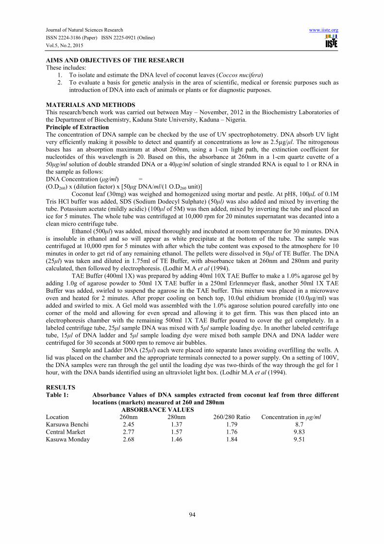
Table 1: Absorbance Values of DNA samples extracted from coconut leaf from three different
locations (markets) measured at 260 and 280nm
ABSORBANCE VALUES
Location 260nm 280nm 260/280 Ratio Concentration in µg/ml
Karsuwa Benchi 2.45 1.37 1.79 8.7
Central Market 2.77 1.57 1.76 9.83
Kasuwa Monday 2.68 1.46 1.84 9.51

Journal of Natural Sciences Research www.iiste.org
ISSN 2224-3186 (Paper) ISSN 2225-0921 (Online)
Vol.5, No.2, 2015
95
Agarose gel image of DNA isolated from coconut leaf from three different locations
Plate 1
DISCUSSION
From the results obtained, Table 1 shows the absorbance of the DNA samples at 260nm and 280nm. The
absorbance at 260nm indicated the presence of DNA whereas at 280nm indicated the presence of proteins. From
the values of 2.45, 2.77 and 2.68 obtained at 260nm, it showed that there was significant amount of DNA in the
samples from the three different locations and also the values of 1.37, 1.57 and 1.46 obtained at 280nm showed
the presence of protein in the DNA sample. From the 260/280 ratio obtained, the values of 1.79, 1.76 and 1.84
indicated the purity of the DNA samples since the range of purity was between. (1.65 – 1.85)
Table 1 also showed the DNA concentration in the three samples 8.7µg/ml from Kasuwa Berchi,
9.83µg/ml from the Central Market and 9.51µg/ml from Kasuwa Monday, being very significant
amounts/concentrations which could serve as a good source of DNA for Genetic engineering, Forensics and
possibly for cloning.
ACKNOWLEDGEMENT
The co-authors wish to thank and acknowledge with gratitude, the Head of Department, Department of
Biochemistry, Kaduna State University, Kaduna – Nigeria for allowing access to reagents and laboratory
facilities.
REFERENCES
Batugal (2005). Performance of Coconut Hybrids in some countries of Asia, Africa and Latin America in int.
plant Genetic Resour Newslett. 142, 47 – 54.
Berg, J., Tymoczko, J and Stryer, L. (2002): Biochemistry Freeman and Company 0 – 7167 -4955 – 6.
Edwards, KC., Johnson, C. and Thompson, C. (1991): A simple and rapid method for the preparation of plant
genomic DNA for PCR analysis. Nucl Acids res 19:23 – 49.
Grimwood, B.E., Ashman, F., Dendy, D.A.V., Jarman, C.G., Lttle, E.C.S and Timmins, W.H., (1975) Coconut
palm products – Their processing in developing countries. Rane FAO pp 3 – 4.
Hahn, W.J. (1997). Arecanae: The palms retrieved April 4, 2011 from the Tree of life project website.
Lodli, M.A, Ye, G.N., Weedan, N.F x Reish, B.I. (1994): A simple and efficient method of DNA extraction from
grapevine cultivas and vitis species, plant Mol. Biol Report (12) 6 – 13.

The IISTE is a pioneer in the Open-Access hosting service and academic event management.
The aim of the firm is Accelerating Global Knowledge Sharing.
More information about the firm can be found on the homepage:
http://www.iiste.org
CALL FOR JOURNAL PAPERS
There are more than 30 peer-reviewed academic journals hosted under the hosting platform.
Prospective authors of journals can find the submission instruction on the following
page: http://www.iiste.org/journals/ All the journals articles are available online to the
readers all over the world without financial, legal, or technical barriers other than those
inseparable from gaining access to the internet itself. Paper version of the journals is also
available upon request of readers and authors.
MORE RESOURCES
Book publication information: http://www.iiste.org/book/
Academic conference: http://www.iiste.org/conference/upcoming-conferences-call-for-paper/
IISTE Knowledge Sharing Partners
EBSCO, Index Copernicus, Ulrich's Periodicals Directory, JournalTOCS, PKP Open
Archives Harvester, Bielefeld Academic Search Engine, Elektronische Zeitschriftenbibliothek
EZB, Open J-Gate, OCLC WorldCat, Universe Digtial Library , NewJour, Google Scholar


![Corrosion Inhibition Performance of Coconut Leaf Extract ... › papers › vol15 › 150100001.pdf · corrosion current density is obtained by the following formula [20-23]: (%)](https://static.fdocuments.us/doc/165x107/5f20f2e14c644a7b175bf3e3/corrosion-inhibition-performance-of-coconut-leaf-extract-a-papers-a-vol15.jpg)


