Isolation and Analysis of Double-Stranded RNA from Virus-Infected Plant and Fungal Tissue€¦ ·...
5
Disease Detection and Losses Isolation and Analysis of Double-Stranded RNA from Virus-Infected Plant and Fungal Tissue T. J. Morris and J. A. Dodds Assistant Professor of Plant Pathology, Department of Plant Pathology, University of California, Berkeley, CA 94720; and assistant plant pathologist, Department of Plant Pathology and Botany, Connecticut Agricultural Experiment Station, New Haven, CT 06504. Accepted for publication 2 March 1979. ABSTRACT MORRIS, T. J., and J. A. DODDS. 1979. Isolation and analysis of double-stranded RNA from virus-infected plant and fungal tissue. Phytopathology 69: 854-858. A simple, rapid method for the isolation of double-stranded RNA identify dsRNA. The method permits rapid and efficient isolation and (dsRNA) from virus-infected plant and fungal tissues provides a new analysis of dsRNA from small amounts (I-10 g) of tissue and from multi- approach to virus detection and identification. Diseased tissue was phenol- ple samples using small amounts (0.1-2.5 g) of cellulose powder. Successful extracted to isolate cellular nucleic acids, and viral dsRNA was selectively isolation of dsRNA does not depend on the type of tissue processed and the purified from other nucleic acids by binding to cellulose powder in 15% method therefore is potentially useful for the study of RNA virus replica- ethanol either in small columns or by a batch procedure. The product was tion and for detection and diagnosis of virus infections directly from the analyzed first by gel electrophoresis and then by ribonuclease treatment to infected host tissues. Additional key word: disease detection. The isolation and properties of viral-specific double-stranded at 25 C for 48 hr in liquid complete medium (14). The dsRNA of RNA (dsRNA) from tissue infected with RNA viruses have been P1 strain has been characterized after preparation from tissue by a well documented (12). Analysis has most often inyolved lengthy more elaborate method that did not involve cellulose powder (9). isolation procedures that yield small quantities of dsRNA. The Both a virulent and a hypovirulent strain of Endothia parasitica quality of dsRNA has been uncertain for some viruses (2,13). The were grown as static cultures at 28 C for 7 days as described need for better recovery and quality of dsRNA has resulted in the elsewhere (3). U. maydis and E. parasitica tissue was washed in development here of a simple and rapid column or batch procedure 100 ml of extraction buffer and was not frozen before extraction. for the isolation of dsRNA. It has been used to isolate dsRNA from Nucleic acid extraction. Nucleic acids were extracted as several systems infected with either single-stranded RNA (ssRNA) described previously ( I I ). Fresh or frozen tissue ( 10 g or less) was or dsRNA viruses. Preliminary reports have been published (3,10). homogenized at low speed in a blender for 1 min or with a mortar The method is based on the affinity of cellulose powder for and pestle in the following mixture of buffer and solutions: lOml of nucleic acids (5) and, specifically, the adsorption of dsRNA at GPS buffer (0.2 M glycine, 0.1 M Na 2 HPO 4 , 0.6 M NaCl, pH 9.5), ethanol concentrations of 15%. The product is analyzed by gel I ml of 10% SDS, 0.1 ml of mercaptoethanol, 10 ml of water electrophoresis and dsRN A then is identified by ribonuclease treat- saturated phenol (containing 0.1% 8-hydroxyquinoline) and 10 ml ment. The method permits rapid, efficient isolation and analysis of of chloroform-pentanol (25:1). dsRNA from relatively small amounts of tissue from multiple sam- Mycelium of U. maydis and E. parasitica was homogenized with pies, and success appears to be independent of the type of tissue GPS buffer and 20 g of glass beads in a Braun homogenizer for 4 processed. Consequently it is a potentially useful tool for the study min. The other reagents were then added and the mixture was of RNA virus replication and for detection and diagnosis of virus occasionally shaken to form and maintain an emulsion for 30 min infections directly from host tissue. on ice. Homogenates were centrifuged at 8,000 g for 20 min at 4 C MATERIALS AND METHODS and the aqueous phase containing cellular nucleic acids was recovered. Virus culture. Bromegrass mosaic virus, the cowpea strain of Purification of dsRNA. The procedure of Franklin (5), used by southern bean mosaic virus and the cherry strain of tomato bushy Jackson et al (8), German and de Zoeten (6) and Dodds et al (4) for stunt virus were obtained from R. 1. Hamilton of Agriculture Can- isolating plant viral dsRNA was modified and shortened. Two pro- ada, Vancouver, and maintained in Hordeum vulgare, Vigna sinen- cedures were compared, both of which had the following features: sis and Nicotiana clevelandii, respectively. Tobacco necrosis virus Replicative intermediate and replicative form were not separated in with satellite virus, turnip yellow mosaic virus (TYMV), and the an attempt to recover as much dsRNA as possible. The interaction cowpea strain of tobacco mosaic virus were obtained from frozen of sample and cellulose was in the presence of 15% rather than isolates at Berkeley and propagated in Nicotiana tabacum L. higher ethanol concentrations. At this ethanol concentration, Xanthi, Brassica chinensis cv. Michili and V. sinensis, respectively. dsRNA is the only major class of nucleic acid bound to cellulose Isolations of alfalfa mosaic virus and cucumber mosaic virus were (5). The amount of cellulose powder used was reduced to a level made in California and the viruses were maintained in N. tabacum. sufficient to bind only the relatively small amount of dsRNA in the Inoculated plants were maintained in the greenhouse, harvested sample. 7 -14 days after inoculation, and stored frozen until use. A Method /. This was developed in Connecticut and used on U. mycovirus containing culture of Penicillium chrysogenum Thom maydis and E. parasitica. The cellular nucleic acids were collected (ATCC 9480) was obtained from B. Castanho, University of Cali- from the aqueous phase by centrifugation after ethanol precipita- fornia, Davis. The fungus was grown in potato dextrose broth for 2 tion, resuspended in and dialyzed against STE buffer (0. 1 M NaCI, wk, at which time the mycelium was collected by filtration and 0.05 M Tris, 0.001 M Na. EDTA, pH 7.0), adjusted to 15% ethanol stored frozen. Two strains of Ustilago maydis, the P 1 killer strain (STE/ 15% ethanol), and passed through a I 0-ml (2.5 g dry weight) that carries a viruslike particle containing dsRNA and the P2 column of chromatographic cellulose powder (Whatman CF-I I nonkiller strain that does not (9,16) were grown as shake cultures cellulose) equilibrated with STE/15% ethanol. The charged col- umn was washed with 80 ml of STE/15% ethanol and then the 00031-949X/79/000155$03.00/0 retained dsRNA was eluted with 15 ml of STE buffer without 01979 The American Phytopathological Society ethanol. 854 PHYTOPATHOLOGY
-
Upload
vuongduong -
Category
Documents
-
view
216 -
download
1
Transcript of Isolation and Analysis of Double-Stranded RNA from Virus-Infected Plant and Fungal Tissue€¦ ·...
Disease Detection and Losses
Isolation and Analysis of Double-Stranded RNA from Virus-Infected Plant and Fungal Tissue
T. J. Morris and J. A. Dodds
Assistant Professor of Plant Pathology, Department of Plant Pathology, University of California, Berkeley, CA 94720; and assistant plantpathologist, Department of Plant Pathology and Botany, Connecticut Agricultural Experiment Station, New Haven, CT 06504.
Accepted for publication 2 March 1979.
ABSTRACT
MORRIS, T. J., and J. A. DODDS. 1979. Isolation and analysis of double-stranded RNA from virus-infected plant and fungal tissue. Phytopathology 69:854-858.
A simple, rapid method for the isolation of double-stranded RNA identify dsRNA. The method permits rapid and efficient isolation and(dsRNA) from virus-infected plant and fungal tissues provides a new analysis of dsRNA from small amounts (I-10 g) of tissue and from multi-approach to virus detection and identification. Diseased tissue was phenol- ple samples using small amounts (0.1-2.5 g) of cellulose powder. Successfulextracted to isolate cellular nucleic acids, and viral dsRNA was selectively isolation of dsRNA does not depend on the type of tissue processed and thepurified from other nucleic acids by binding to cellulose powder in 15% method therefore is potentially useful for the study of RNA virus replica-ethanol either in small columns or by a batch procedure. The product was tion and for detection and diagnosis of virus infections directly from theanalyzed first by gel electrophoresis and then by ribonuclease treatment to infected host tissues.
Additional key word: disease detection.
The isolation and properties of viral-specific double-stranded at 25 C for 48 hr in liquid complete medium (14). The dsRNA ofRNA (dsRNA) from tissue infected with RNA viruses have been P1 strain has been characterized after preparation from tissue by awell documented (12). Analysis has most often inyolved lengthy more elaborate method that did not involve cellulose powder (9).isolation procedures that yield small quantities of dsRNA. The Both a virulent and a hypovirulent strain of Endothia parasiticaquality of dsRNA has been uncertain for some viruses (2,13). The were grown as static cultures at 28 C for 7 days as describedneed for better recovery and quality of dsRNA has resulted in the elsewhere (3). U. maydis and E. parasitica tissue was washed indevelopment here of a simple and rapid column or batch procedure 100 ml of extraction buffer and was not frozen before extraction.for the isolation of dsRNA. It has been used to isolate dsRNA from Nucleic acid extraction. Nucleic acids were extracted asseveral systems infected with either single-stranded RNA (ssRNA) described previously ( I I ). Fresh or frozen tissue ( 10 g or less) wasor dsRNA viruses. Preliminary reports have been published (3,10). homogenized at low speed in a blender for 1 min or with a mortar
The method is based on the affinity of cellulose powder for and pestle in the following mixture of buffer and solutions: lOml ofnucleic acids (5) and, specifically, the adsorption of dsRNA at GPS buffer (0.2 M glycine, 0.1 M Na 2HPO4, 0.6 M NaCl, pH 9.5),ethanol concentrations of 15%. The product is analyzed by gel I ml of 10% SDS, 0.1 ml of mercaptoethanol, 10 ml of waterelectrophoresis and dsRN A then is identified by ribonuclease treat- saturated phenol (containing 0.1% 8-hydroxyquinoline) and 10 mlment. The method permits rapid, efficient isolation and analysis of of chloroform-pentanol (25:1).dsRNA from relatively small amounts of tissue from multiple sam- Mycelium of U. maydis and E. parasitica was homogenized withpies, and success appears to be independent of the type of tissue GPS buffer and 20 g of glass beads in a Braun homogenizer for 4processed. Consequently it is a potentially useful tool for the study min. The other reagents were then added and the mixture wasof RNA virus replication and for detection and diagnosis of virus occasionally shaken to form and maintain an emulsion for 30 mininfections directly from host tissue. on ice. Homogenates were centrifuged at 8,000 g for 20 min at 4 C
MATERIALS AND METHODS and the aqueous phase containing cellular nucleic acids wasrecovered.
Virus culture. Bromegrass mosaic virus, the cowpea strain of Purification of dsRNA. The procedure of Franklin (5), used bysouthern bean mosaic virus and the cherry strain of tomato bushy Jackson et al (8), German and de Zoeten (6) and Dodds et al (4) forstunt virus were obtained from R. 1. Hamilton of Agriculture Can- isolating plant viral dsRNA was modified and shortened. Two pro-ada, Vancouver, and maintained in Hordeum vulgare, Vigna sinen- cedures were compared, both of which had the following features:sis and Nicotiana clevelandii, respectively. Tobacco necrosis virus Replicative intermediate and replicative form were not separated inwith satellite virus, turnip yellow mosaic virus (TYMV), and the an attempt to recover as much dsRNA as possible. The interactioncowpea strain of tobacco mosaic virus were obtained from frozen of sample and cellulose was in the presence of 15% rather thanisolates at Berkeley and propagated in Nicotiana tabacum L. higher ethanol concentrations. At this ethanol concentration,Xanthi, Brassica chinensis cv. Michili and V. sinensis, respectively. dsRNA is the only major class of nucleic acid bound to celluloseIsolations of alfalfa mosaic virus and cucumber mosaic virus were (5). The amount of cellulose powder used was reduced to a levelmade in California and the viruses were maintained in N. tabacum. sufficient to bind only the relatively small amount of dsRNA in the
Inoculated plants were maintained in the greenhouse, harvested sample.7 -14 days after inoculation, and stored frozen until use. A Method /. This was developed in Connecticut and used on U.mycovirus containing culture of Penicillium chrysogenum Thom maydis and E. parasitica. The cellular nucleic acids were collected(ATCC 9480) was obtained from B. Castanho, University of Cali- from the aqueous phase by centrifugation after ethanol precipita-fornia, Davis. The fungus was grown in potato dextrose broth for 2 tion, resuspended in and dialyzed against STE buffer (0. 1 M NaCI,wk, at which time the mycelium was collected by filtration and 0.05 M Tris, 0.001 M Na. EDTA, pH 7.0), adjusted to 15% ethanolstored frozen. Two strains of Ustilago maydis, the P 1 killer strain (STE/ 15% ethanol), and passed through a I 0-ml (2.5 g dry weight)that carries a viruslike particle containing dsRNA and the P2 column of chromatographic cellulose powder (Whatman CF-I Inonkiller strain that does not (9,16) were grown as shake cultures cellulose) equilibrated with STE/15% ethanol. The charged col-
umn was washed with 80 ml of STE/15% ethanol and then the00031-949X/79/000155$03.00/0 retained dsRNA was eluted with 15 ml of STE buffer without01979 The American Phytopathological Society ethanol.
854 PHYTOPATHOLOGY

Method 2. Developed in California, this even simpler method, in purified from 0.4 g of tissue. The pattern of dsRNA componentsaddition, avoids the concentration and dialysis of nucleic acids that from P1 is similar to that reported by Koltin and Day (9) whowas unnecessary and sometimes detrimental to maximum recovery purified dsRNA by the method described by Vodkin et al (15),of dsRNA. The aqueous phase was adjusted to 15% ethanol by which relies on the use of lithium chloride and enzyme digestion toaddition of absolute ethanol with stirring. Cellulose powder remove all other nucleic acids. In this study the single 2.6 X 10⁶(Whatman CF- 1 I cellulose or Biorad Cellex N-i) without pre- component of Koltin and Day (the second highest molecular weighttreatment, was then added at a rate of 0.25 g/20 ml of supernatant. band) was resolved as two distinct components. We detected anThe mixture was gently shaken, on ice, for 10 min and then cen- additional very minor component with a mobility between that oftrifuged at 6,000 g for 10 min. The supernatant was discarded and the 2.6 and 0.94 X 10⁶ components of Koltin and Day. This compo-the cellulose pellet was resuspended in 5 ml of STE/ 15% ethanol. nent is of some interest because it also was detected in the P2The cellulose was then placed in a small column and washed free of nonkiller strain used as the virus free control. No other componentsssRNA with 60 ml of STE/ 15% ethanol at room temperature. The were detected in P2.dsRNA was eluted from the cellulose by the addition of 5 ml of The results for E. parasitica using Method 1 also are shown inSTE buffer without ethanol. Traces of DNA were removed from Fig. I. The virulent strain contained no detectable dsRNA, but thethe dsRNA by adding MgCl, to a concentration of 0.03 M and hypovirulent strain contained three dsRNA components. VirulentDNAse (DNase I from Sigma) to a concentration of 10 Ag/ml and and hypovirulent strains of E. parasitica were analyzed in detailincubating the mixture for 20 min at 30 C. The dsRNA prepared and reported elsewhere (3).by either method was concentrated by ethanol precipitation and Comparison of dsRNA isolation procedures. The results for U.stored at -20 C. maydis dsRNA prepared by Method 1, compared with another
Gel electrophoresis. The dsRNAs were analyzed by electrophore- method (15), indicate that, similar results can be obtained usingsis in 2.4 or 5% polacrylamide gel in 0.04 M Tris, 0.02 M sodium CF-I I to isolate dsRNA. The CF- 1I cellulose method described byacetate, 0.001 M NaEDTA, pH 7.2, in the absence of SDS. Elec- Jackson et al (8) for isolating dsRNA from plant tissues involvestrophoresis was performed in cylindrical gels (0.6 X 10 cm) at 75 extensive purification of the cellular nucleic acids and two cycles ofV, 6mA per gel. Samples for electrophoresis were dissolved in 200pl of 1:5 concentration electrophoresis buffer with 10% sucrose and0.00 1% bromophenol blue. The gels were analyzed by scanning at280 nm with an ISCO Gel Scanner. Alternately, the nucleic acidwas fixed by soaking the gels in 0.2% hexadecyltrimethyl-ammo-nium bromide followed by thorough washing with distilled waterand staining in 0.1% toluidene blue 0 in water. Gels were destainedwith water and scanned at 620 nm. The concentration of thedsRNA was estimated by planimetry of the gel scans with refer-ence to a standard curve prepared from known concentrations ofTYMV dsRNA.
RESULTS 0.3-dsRNA from U. maydis and E. parastica. The results for U.
maydis P2 (nonkiller) and P1 (killer) using Method 1 are shown inFig. 1. The dsRNA sample applied to each gel was the amount a
E
0c..
0-,4
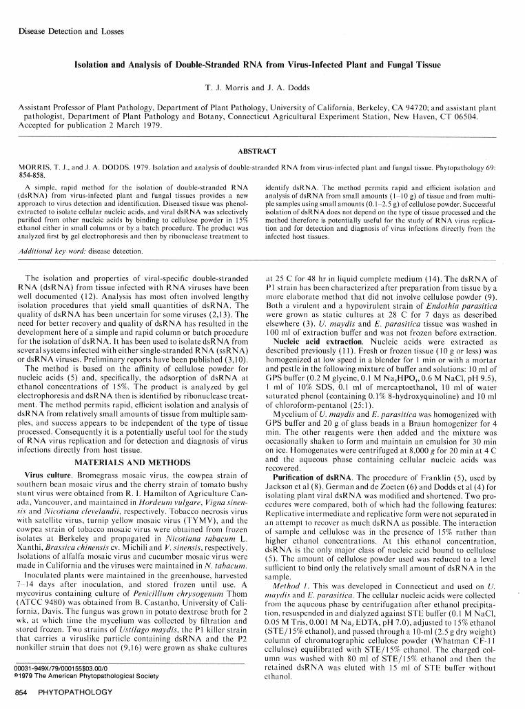
Fig. 1. Polyacrylamide gels showing double-stranded RNA prepared from Relative Mobility-..fungal mycelium by method 1. The RNA was electrophoresed into 5% gelsfor 6 hr at 6 mA per gel and the gels were stained with 0.1% toluidine blue Fig. 2. Scanning profile of 2.4% polyacrylamide gels showing double-0. The gels contain nucleic acid extracted from (left to right): Ustilago stranded RNA of turnip yellow mosaic virus prepared from 5 g of infectedmaydis P2 (nonkiller); U. maydis PI (killer); Endothia parasitica, virulent, leaf tissue. a, Isolation by the method of Jackson et al (9); b, Isolation byand E. parasitica, hypovirulent. Note the minor component (arrowed) com- method 2; c, Isolation from 10 g of healthy tissue by method 2. The RNAmon to both strains of U. maydis. was electrophoresed at 75 V, 6 mA per gel for 4 hr at 15 C.
Vol. 69, No. 8, 1979 855
cellulose chromatography. Their procedure was compared with the ssRNA was eliminated at 10% ethanol in GPS buffer, but theshorter batch method (Method 2) using several different types of recovery of dsRNA was also somewhat reduced. Several brands ofvirus-infected tissue. Equivalent amounts of TYMV-infected tissue chromatographic cellulose were compared and all were found towere processed for dsRNA by the two methods and electrophoresed bind dsRNA efficiently from solutions of 15% ethanol. Finer gradeson gels (Fig. 2). The shorter batch procedure consistently gave such as Biorad Cellex N-1 had greater binding capacity (20-2540 -80% greater recovery of TYMV dsRNA for all types of tissue g/l100 mg of cellulose) than did coarser grades such as Whatmantested, and the quality of the dsRNA was comparable as judged by CF- I I cellulose (5 gg/ 100 mg of cellulose), but the very fine gradesgel electrophoresis. Table I summarizes the recovery by the two had slow flow rates that increased the washing time during the 15%methods of dsRNA from plant and fungal tissues infected with elution step. Biorad Cellex N-I displayed the best combination ofthree different viruses. It is evident that the simpler procedure, capacity and flow rate and was used in all subsequent experiments.Method 2, consistently gave equal or better recoveries of dsRNA Temperature of incubation had little effect on the binding capac-than the Jackson method. ity of the cellulose for dsRNA (Table 2, h and i); therefore the
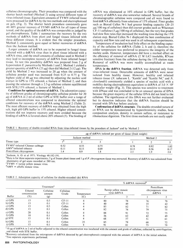
Larger amounts of dsRNA are to be expected in fungal tissue colder temperature was preferred to preserve the integrity of theinfected with a dsRNA virus than in plant tissue infected with a nucleic acids. However, temperature did have a marked effect onssRNA virus, and the small amounts of cellulose used in Method 2 the efficiency of removal of ssRNA (2 M LiCl insoluble, RNasemay lead to incomplete recovery of dsRNA from infected fungal sensitive fraction) from the cellulose during the 15% elution step.tissue. To test this possibility dsRNA was prepared from 2 g of Removal of ssRNA was more readily accomplished at roomhypovirulent E. parasitica by Methods 1 and 2. Method 1 yielded a temperature.greater quantity of dsRNA (25 gg) than did Method 2 (12 gg). The DNA in the dsRNA fraction. dsRNA was detected only fromyield by Method 2 was increased to 20 Ag when the amount of virus-infected tissue. Detectable amounts of dsRNA were nevercellulose powder used was increased from 0.25 to 0.75 g. The isolated from healthy tissue. However, healthy and infectedhighest yield of 40 pg was obtained by adjusting the nucleic acid tobacco tissue (N. tabacum L. 'Xanthi' and 'Xanthi NC' and N.extract in GPS to 15% ethanol, a feature of Method 2, and then clevelandii) consistently yielded a species of nucleic acid with apassing the solution through a 2.5-g cellulose column equilibrated mobility during electrophoresis equivalent to dsRNA of 3.0 X 10⁶with STE/ 15% ethanol, a feature of Method 1. molecular weight (Fig. 3). This species was sensitive to treatment
Conditions for optimal recovery of dsRNA. The adsorption capac- with DNase and was concluded to be an unusual species of DNAity of different grades of chromatographic cellulose powder for 20 because the great majority of the cellular DNA did not bind to themg of dsRNA in 2 ml of either GPS or STE buffer over a range of cellulose. The significance of this DNA was not further evaluatedethanol concentrations was compared to determine the optimal but its detection indicates that the dsRNA fraction should beconditions for recovery of the dsRNA using Method 2 (Table 2). treated with DNAse before analysis.The most efficient recovery of dsRNA was obtained from the high Confirmation of dsRNA structure. The double-stranded nature ofsalt, high pH GPS buffer in 15% ethanol. Higher ethanol concen- an RNA can be demonstrated by hyperchromicity studies, basetrations did not improve recovery and were avoided because the composition analysis, density in cesium sulfate, or resistance tobinding of ssRNA is favored above 20% ethanol (5). The binding of ribonuclease digestion. The first three methods are not easily appli-
TABLE 1. Recovery of double-stranded RNA from virus-infected tissues by the procedure of Jacksona and by Method 2
Ag of dsRNA isolated per gram of tissue (fresh weight)Jackson method Method 2
Tissue type I b 2 3 1 2 3TYMVc-infected Chinese cabbage 0.52 0.55 0.75 0.78 0.92 1.02AMVd-infected tobacco 0.11 0.15 0.09 0.15 0.26 0.17Penicillium chrysogenum 1.4 0.9 1.8 2.5 1.9 2.8a'Jackson, A. 0. et al 1971. Virology 48:182-191.bData is for three separate experiments; 5 g of frozen plant tissue and 1 g of P. chrysogenum tissue was extracted. Recovery of dsRNA was calculated fromplanimetry of gel scans recorded at 280 nm.CTYMV = turnip yellow mosaic virus.
dAMV = alfalfa mosaic virus.
TABLE 2. Adsorption capacity of cellulose for double-stranded (ds) RNA
% dsRNA recoveredb
Treatmenta PenicilliumEthanol Cellulose Cellulose Turnip yellow mosaic chrysogenum virus
concentration amount Grade Temperature virus dsRNA dsRNABuffer (%) (g) (C) Ic 2 1 2
a) GPS 15 0.5 CF-I 1 4 80 78 76 78b) GPS 15 0.1 CF-lI 4 40 49 35 42c) GPS 15 0.5 Cellex 4 85 82 71 82d) GPS 15 0.1 Cellex 4 82 76 79 75c) GPS 20 0.1 Cellex 4 88 91 72 75f) GPS 10 0.1 Cellex 4 56 62 42 49g) STE 15 0.1 Cellex 4 68 57 51 61h) GPS 15 0.1 Cellex 25 85 79 68 72i) GPS 15 0.1 Cellex 4 91 81 75 79
"10 pg of dsRNA in 2 ml of buffer adjusted to the ethanol concentration was incubated with the amount and grade of cellulose, collected by centrifugation,and eluted with STE buffer.
bRecovery calculated from the micrograms of dsRNA detected by gel electrophoresis compared with the amount of dsRNA in the initial solution.CTwo separate experiments performed.
856 PHYTOPATHOLOGY
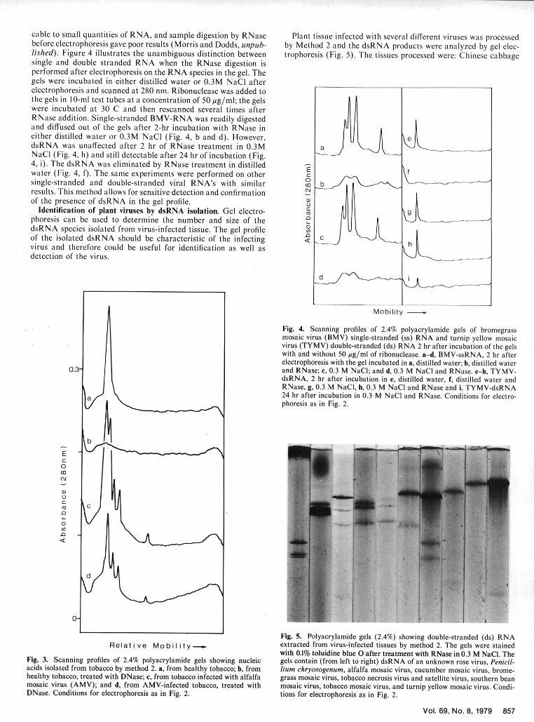
cable to small quantities of RNA, and sample digestion by RNase Plant tissue infected with several different viruses was processedbefore electrophoresis gave poor results (Morris and Dodds, unpub- by Method 2 and the dsRNA products were analyzed by gel elec-lished). Figure 4 illustrates the unambiguous distinction between trophoresis (Fig. 5). The tissues processed were: Chinese cabbagesingle and double stranded RNA when the RNase digestion isperformed after electrophoresis on the RNA species in the gel. Thegels were incubated in either distilled water or 0.3M NaCl afterelectrophoresis and scanned at 280 nm. Ribonuclease was added tothe gels in I 0-ml test tubes at a concentration of 50 Ag/ml; the gelswere incubated at 30 C and then rescanned several times afterRNase addition. Single-stranded BMV-RNA was readily digestedand diffused out of the gels after 2-hr incubation with RNase ineither distilled water or 0.3M NaCI (Fig. 4, b and d). However,dsRNA was unaffected after 2 hr of RNase treatment in 0.3MNaCI (Fig. 4, h) and still detectable after 24 hr of incubation (Fig.4, i). The dsRNA was eliminated by RNase treatment in distilled -Ewater (Fig. 4, f). The same experiments were performed on other C
0single-stranded and double-stranded viral RNA's with similar 0 bresults. This method allows for sensitive detection and confirmationof the presence of dsRNA in the gel profile.
Identification of plant viruses by dsRNA isolation. Gel electro- ,phoresis can be used to determine the number and size of the
0dsRNA species isolated from virus-infected tissue. The gel profileof the isolated dsRNA should be characteristic of the infecting <virus and therefore could be useful for identification as well asdetection of the virus.
Mobility
Fig. 4. Scanning profiles of 2.4% polyacrylamide gels of bromegrassmosaic virus (BMV) single-stranded (ss) RNA and turnip yellow mosaicvirus (TYMV) double-stranded (ds) RNA 2 hr after incubation of the gelswith and without 50 gg/ml of ribonuclease. a-d, BMV-ssRNA, 2 hr afterelectrophoresis with the gel incubated in a, distilled water; b, distilled water
0.3- and RNase; c, 0.3 M NaCI; and d, 0.3 M NaCI and RNase. e-h, TYMV-dsRNA, 2 hr after incubation in e, distilled water, f, distilled water andRNase, g, 0.3 M NaCI, h, 0.3 M NaCI and RNase and i, TYMV-dsRNA24 hr after incubation in 0.3 M NaCl and RNase. Conditions for electro-phoresis as in Fig. 2.
EC:0
CC.
0
.0
Fig. 5. Polyacrylamide gels (2.4%) showing double-stranded (ds) RNARel at iv e Mo b i It y t extracted from virus-infected tissues by method 2. The gels were stained
with 0.1% toluidine blue 0 after treatment with RNase in 0.3 M NaCI. TheFig. 3. Scanning profiles of 2.4% polyacrylamide gels showing nucleic gels contain (from left to right) dsRNA of an unknown rose virus, Penicil-acids isolated from tobacco by method 2. a, from healthy tobacco; b, from hum chryosogenum, alfalfa mosaic virus, cucumber mosaic virus, brome-healthy tobacco, treated with DNase; c, from tobacco infected with alfalfa grass mosaic virus, tobacco necrosis virus and satellite virus, southern beanmosaic virus (AMV); and d, from AMV-infected tobacco, treated with mosaic virus, tobacco mosaic virus, and turnip yellow mosaic virus. Condi-DNase. Conditions for electrophoresis as in Fig. 2. tions for electrophoresis as in Fig. 2.
Vol. 69, No. 8, 1979 857
infected with TYMV, cowpea infected with the cowpea strains of plants indicate that such an approach is possible, and preliminary
tobacco mosaic virus and of southern bean mosaic virus, tobacco identification of the casual virus can be made from molecular
tissue infected with both tobacco necrosis virus and satellite virus, weight estimates after gel electrophoresis. This approach is being
tomato bushy stunt virus, cucumber mosaic virus and alfalfa used to detect viruses in rose, grape and cherry tissues; the high
mosaic virus; barley infected with bromegrass mosaic virus; virus content of polysaccharides in these plants makes isolation of nucleic
infected P. chrysogenum, and rose tissue infected with an unknown acid difficult by previously applied procedures. In addition, the
virus. The dsRNA profile were quite characteristic for each of the method already has provided a useful approach for the detection
viruses tested (Fig. 5). These results confirm that the method is and identification of viruses with dsRNA genomes infecting fungi
useful for the detection and identification of RNA viruses. Presence from which conventional viruslike particles have not yet been puri-
of dsRNA in rose is an example of the detection of a possible virus fied (1,3).infection in tissue that has failed to yield infectious inocula bystandard virus isolation procedures (T. J. Morris, unpublished). LITERATURE CITED
I. CASTANHO, B., E. E. BUTLER, and R. J. SHEPHERD. 1978. The
DISCUSSION association of double-stranded RNA with Rhizoctonia decline.Phytopathology 68:1515-1519.
A method has been described for the rapid, efficient isolation of 2. DAWSON, W. 0., T. L. GERMAN, and D. E. SCHLEGEL. 1976.
dsRNA from plant leaves of various textures and fungal mycelium. Homogenization and susceptible components of tobacco mosaic virus
Successful isolation of dsRNA was independent of the type of replicative form RNA. J. Gen. Virol. 32:205-215.
tissue used and the method should give good results with other 3. DAY, P. R., J. A. DODDS, J. E. ELLISTON, R. A. JAYNES, and S.
tissue types. In addition, a sensitive in situ RNase digestion test has L. ANAGNOSTAK1S. 1977. Double-stranded RNA in Endothia
been developed for unambiguous evaluation of the double-stranded ~parasitica. Phytopathology 67:1393-1396.naturen ofee olaed norucleicmb ciguous. Thev o othod maydprovbe-ustr 4. DODDS, J. A., J. H. TREMAINE, and W. P. RONALD. 1977. Somenature of the isolated nucleic acids. The method may prove useful properties of carnation ringspot virus single-and double-strandedfor detection and identification of RNA virus infections. ribonucleic acid. Virology 83:322-328.
The batch method (Method 2) works well for all kinds of tissues 5. FRANKLIN, R. M. 1966. Purification and properties of replicative
including those that produce viscous extracts that would not pass intermediate of the RNA bacteriophage R17. Proc. Nati. Acad. Sci.
readily through a column. The amount of cellulose used, 0.25 g, is USA. 55:1504-1511.
sufficient to bind a quantity of dsRNA that can be detected by gel 6. GERMAN, T. L., and G. A. deZOETEN. 1975. Purification and
clectrophoresis, and this quantity can be recovered from as little as properties of replicative form and replicative intermediates of pea ena-
1.0 g of tissue. For tissues that contain high concentrations of tion mosaic virus. Virology 66:172-184.
dsRNA and do not produce viscous extracts, a small column proce- . HENRIQUES, M. I. C., T. J. MORRIS, and D. E. SCHLEGEL.d dure, which ntil usesprnly2.5godue vsceou s e e a ts, 1, wlol n pwel. 1977. An unexpected double-stranded RNA from tomato bushy stuntdure, which still uses only 2.5 g of cellulose (Method 1), works well. virus infected plant tissue. Proc. Am. Phytopathol. Soc. 4:160.
The methods have advantages over previously published procedures 8. JACKSON, A. 0., D. M. MITCHELL, and A. SIEGEL. 1971. Repli-(4,6,8,12) because they are shorter and therefore quicker, give cation of tobacco mosaic virus. 1. Isolation and characterization ofbetter recovery, and permit small multiple samples to be processed double-stranded forms of ribonucleic acid. Virology 45:182-191.
(3). These are important considerations if the presence of dsRNA 9. KOLTIN, Y., and P. DAY. 1976. Inheritance of killer phenotypes and
is to be used for diagnosis of virus diseases. The method was used to double-stranded RNA in Ustilago maydis. Proc. Nat, Acad. Sci.
detect the expected dsRNAs in a number of virus-infected plant U.S.A. 73:594-598.
tissues and also some unexpected low molecular weight components 10. MORRIS, T. J. 1977. Isolation of double-stranded RNA from virus
assumed to be dsRNA, such as in TYMV-infected tissue (Figs. 2 infected plants and fungi. Proc. Am. Phytopathol. Soc. 4:160.
and 5). We have been able to isolate such minor replicative forms in II. MORRIS. T. J., and E. M. SMITH. 1977. Potato spindle tuber dis-ease: Procedures for the detection of viroid RNA and certification of
other RNA plant virus infections (Fig. 5) using these methods, and disease-free potato tubers. Phytopathology 67:145-150.the possible significance of this observation has been discussed (7). 12. RALPH, R. K. 1969. Double-stranded viral RNA. Adv. Virus Res.These observations show that the method may also be useful in the 15:61-158.study of the replication of RNA viruses. 13. REZAIAN, M. A., and R. 1. B. FRANCKI. 1973. Replication of
Because dsRNA has been detected only in virus-infected plants, tobacco ringspot virus. 1. Detection of low molecular weight double-
the method provides a tool for the study of diseases suspected, but stranded RNA from infected plants. Virology 56:238-240.
not proved, to be of viral origin because of failure to purify virus 14. STEVENS, R. B. 1974. Page 691 in Mycology Guidebook. Univ.
particles. Successful isolation of viruses from tissues is dependent IWashington Press, Seattle, WA.onrthes. Sueoftissfue iatnd theoftabilityof viruses frm t s is ienglen 15. VODKIN, M., F. KATTERMAN, and G. R. FINK. 1975. Yeast killeron the type of tissue and the stability of the virus, and no single mutants with altered double-stranded ribonucleic acid. J. Bacteriol.
purification method can be used for all viruses. By contrast, a single 117:681-686.method for isolating dsRNA from various tissues infected with 16. WOOD, H. A., and R. F. BOZARTH. 1973. Heterokaryon transfer ofseveral different viruses was successful in this study. Preliminary virus-like particles associated with a cytoplasmically inherited determi-
results for several suspected virus diseases from diverse types of nant in Ustilago maydis. Phytopathology 63:1019-1021.
858 PHYTOPATHOLOGY