SUMMARY Double-stranded cDNA has been synthesized from RNA ...
11
volume 10 Number 24 1982 Nucleic Acids Research Molecular cloning and characterization of a complete DNA copy of potato spindle tuber viroid RNA Peter van Wezenbeek*, Pieter Vos, Jacques van Boom+ and Albert van Kammen§ Department of Molecular Biology, Agricultural University, De Dreijen 11, 6703 BC Wageningen, The Netherlands Received 28 October 1982; Accepted 16 November 1982 SUMMARY Double-stranded cDNA has been synthesized from RNA of a severe strain of potato spindle tuber viroid using a synthetic oligodeoxyribonucleotide as a primer. Upon cloning in bacteriophage M13mp9, two recombinant phages were se- lected to construct a pBR322-derived plasmid containing a complete viroid DNA copy. Elucidation of the nucleotide sequence revealed four differences with the previously established sequence of another PSTV strain, three of which were base exchanges and one a deletion. INTRODUCTION Viroids are small single-stranded circular RNA molecules causing several economically important diseases in cultivated plants (1). The complete molecu- lar structures of several viroids have been established including potato spin- dle tuber viroid (PSTV, ref 2), chrysanthemum stunt viroid (CSV, ref 3), citrus exocortis viroid (CEV, ref 4), avocado sunblotch viroid (ASBV, ref 5) and cadang-cadang viroid (6). These RNAs range in size from 246 to 371 residues and form extended rod-like structures with a high degree of intramolecular base-pairing. In its most severe form potato spindle tuber viroid (PSTV) causes general stunting of potato plant growth, deformity of the upper foliage and production of disfigured potatoes (7). As potatoes are vegetatively propagated, it is of major importance to exclude viroids from seed potatoes. Currently, detection of PSTV involves laborious and expensive assays including polyacrylamide gel electrophoresis of extracted nucleic acids (8, 9). Very recently, however, Owens and Diener (10) described an alternative method which is based on hybri- dization of radioactively labeled PSTV cDNA with RNA extracts from clarified plant sap bound to a solid support. This method is attractive since it is simple, sensitive, inexpensive and reproducible. On request of the Dutch Plant Protection Service (PD) and the Research Institute for Plant Protection (IPO), we constructed a plasmid containing a full-length DNA copy of PSTV which may © IRL Press Limited, Oxford, England. 7947 0305-1048/82/1024-79478 2.00/0 Downloaded from https://academic.oup.com/nar/article-abstract/10/24/7947/2379140 by guest on 17 March 2018
Transcript of SUMMARY Double-stranded cDNA has been synthesized from RNA ...
volume 10 Number 24 1982 Nucleic Ac ids Research
Molecular cloning and characterization of a complete DNA copy of potato spindle tuber viroidRNA
Peter van Wezenbeek*, Pieter Vos, Jacques van Boom+ and Albert van Kammen§
Department of Molecular Biology, Agricultural University, De Dreijen 11, 6703 BC Wageningen,The Netherlands
Received 28 October 1982; Accepted 16 November 1982
SUMMARYDouble-stranded cDNA has been synthesized from RNA of a severe strain of
potato spindle tuber viroid using a synthetic oligodeoxyribonucleotide as aprimer. Upon cloning in bacteriophage M13mp9, two recombinant phages were se-lected to construct a pBR322-derived plasmid containing a complete viroidDNA copy. Elucidation of the nucleotide sequence revealed four differenceswith the previously established sequence of another PSTV strain, three ofwhich were base exchanges and one a deletion.
INTRODUCTION
Viroids are small single-stranded circular RNA molecules causing several
economically important diseases in cultivated plants (1). The complete molecu-
lar structures of several viroids have been established including potato spin-
dle tuber viroid (PSTV, ref 2), chrysanthemum stunt viroid (CSV, ref 3), citrus
exocortis viroid (CEV, ref 4 ) , avocado sunblotch viroid (ASBV, ref 5) and
cadang-cadang viroid (6). These RNAs range in size from 246 to 371 residues
and form extended rod-like structures with a high degree of intramolecular
base-pairing.
In its most severe form potato spindle tuber viroid (PSTV) causes general
stunting of potato plant growth, deformity of the upper foliage and production
of disfigured potatoes (7). As potatoes are vegetatively propagated, it is of
major importance to exclude viroids from seed potatoes. Currently, detection
of PSTV involves laborious and expensive assays including polyacrylamide gel
electrophoresis of extracted nucleic acids (8, 9). Very recently, however,
Owens and Diener (10) described an alternative method which is based on hybri-
dization of radioactively labeled PSTV cDNA with RNA extracts from clarified
plant sap bound to a solid support. This method is attractive since it is
simple, sensitive, inexpensive and reproducible. On request of the Dutch Plant
Protection Service (PD) and the Research Institute for Plant Protection (IPO),
we constructed a plasmid containing a full-length DNA copy of PSTV which may
© IRL Press Limited, Oxford, England. 7947
0305-1048/82/1024-79478 2.00/0Downloaded from https://academic.oup.com/nar/article-abstract/10/24/7947/2379140by gueston 17 March 2018
Nucleic Acids Research
serve as a hybridization probe in such a nucleic acid spot hybridization
procedure.
Comparative sequence analyses of severe and mild PSTV strains have indicated
that their nucleotide sequences are very similar and that only few nucleotide
differences occur (11, 12). Generally, to be able to correlate nucleotide
changes in a defined region with a particular symptom pattern in the plant,
knowledge of structural details of a great number of isolates will be required.
In this respect, it was also of interest to establish the nucleotide sequence
of the PSTV strain used in this study which evokes the symptoms characteristic
for a severe strain. Moreover, construction of a full-length PSTV DNA copy
was of great importance since it might be successfully used to produce viroid
molecules upon transformation of potato cell protoplasts. If indeed such a
clone generates biologically active viroids analogous to what has been descri-
bed for full-length DNA copies of phage QB and poliovirus RNAs (13, 14), this
would enable a thorough study on the nature of PSTV pathogenicity by site-
directed mutagenesis on DNA level.
MATERIALS AND METHODS
Enzymes. E.ooli DNA polymerase I was obtained from Boehringer, Mannheim.
T4 DNA ligase was prepared from an induced lysogen of XVllig phage NM989 (15)
and further isolated with minor modifications according to the method of Panet
et al. (16). T4 polynucleotide kinase was purchased from P.L. Biochemicals and
the restriction endonucleases used in this study were from Boehringer or
Bethesda Research Laboratories Inc. AMV reverse transcriptase was obtained
from J.W. Beard, Institute of Life Sciences, St. Petersburg.
Preparation of nucleic acids. The severe strain of PSTV used in this study
(isolated from Solanum commersonii Dun. ref 17) was originally obtained from
P. Howell, Edinburgh. PSTV RNA, a kind gift of W. Mosch, Wageningen, was puri-
fied from infected tomato tissue (Ly coper sicum esoulentum Mill., cv Rutgers,
ref 18). Final purification involved two cycles of polyacrylamide gel electro-
phoresis, the second of which was performed under denaturing conditions to
obtain the circular form of PSTV (19). To remove gel impurities, purified RNA
was bound to DEAE-Sephacel (Pharmacia) in 50 mM Tris-HCl, pH 8.0, washed with
the same buffer and eluted with buffer containing 1.2 M NaCl.
Plasmid DNA was isolated essentially as described by Godson and Vapnek (20).
The plasmid DNA finally obtained was extracted twice with an equal volume of
phenol, three times with diethylether and precipitated with ethanol. Recombi-
nant M13 RF DNA was isolated from infected cells in a similar way.
7948
Downloaded from https://academic.oup.com/nar/article-abstract/10/24/7947/2379140by gueston 17 March 2018

Nucleic Acids Research
The 15-mer d(TTCTTTTTTCTTTTC) and the 18-mer d(GTAAAACGACGGCCAGTG) were
synthesized by the chemical phosphotriester method (21).
Synthesis of double-stranded cDNA. Single-stranded PSTV cDNA was synthesi-
zed in a reaction mixture (10 pi) containing 50 mM Tris-HCl (pH 8.3), 80 mM
KC1, 10 mM MgCl2, 1 mM DTT, 0.05% NP-40, 0.5 pg of circular PSTV, 1.5 pg of
primer, 0.5 mM each of dGTP, dCTP and dTTP, 0.1 mM labeled (a-32P)dATP (speci-
fic activity 10 Ci/mmol) and 20 units of reverse transcriptase. Prior to DNA
synthesis,tempi ate PSTV RNA was heated for 1 min at 100°C in the presence of
the chemically synthesized primer d(TTCTTTTTTCTTTTC) and immediately quenched
in ice-water. Enzymatic reactions were carried out at 37°C for 2 h. After
heat denaturation, PSTV template RNA was degraded by addition of RNase A to
the reaction mixture (final concentration 10 pg/ml) and incubation for 30 min
at 37°C. Single-stranded PSTV cDNA was purified on a Sephadex 6-50 column and
incubated with 5 units of DNA polymerase I in a volume of 50 pi containing
100 mM potassium phosphate (pH 7.0), 6.5 mM MgCl-,, 1 mM DTT and 250 mM each
of the four deoxyribonucleoside triphosphates. The reaction was carried out at
37°C for 2 h.
Construction of recombinant DNA molecules. Chemically synthesized DNA octa-
mers containing an EaoRl endonuclease cleavage site (Collaborative Research)
were phosphorylated at their 5'-end with ATP and T4 polynucleotide kinase by
incubation for 45 min at 37°C in a 10 pi mixture containing 25 mM Tris-HCl,
pH 7.6, 10 mM MgCl2, 10 mM DTT, 0.1 mM ATP, 20 pmoles of linker molecules and
5 units of enzyme. The reaction mixture was adjusted to 1 mM ATP and unfrac-
tionated cDNA and 1 pi of T4 DNA ligase (the optimal amount of enzyme needed
to ligate blunt-ended DNA fragments) were added. Blunt-end ligation was perfor-
med at 15°C by overnight incubation. Generation of cohesive ends was performed
by digestion with 10 units of endonuclease during 1 h at 37°C. Subsequent li-
gation to 50 ng of linearized M13mp9 RF DNA was carried out in a reaction
volume of 20 pi containing 25 mM Tris-HCl, pH 7.6, 10 mM MgClp, 10 mM DTT and
0.2 mM ATP. Ligation was performed by overnight incubation at 15°C using an
amount of T4 DNA ligase which was 10 times less than in the blunt-end ligation
reaction. Construction of the complete PSTV cDNA-containing plasmid pAV401
was performed under blunt-end ligation conditions by adding 0.1 pg of lineari-
zed pBR322 to a twenty-fold molar excess of fragments which were isolated from
M13mp9 recombinant phages.
Competent E.aoli HB101 or JM103 cells were used for transformation or trans-
fection, respectively. Transformants arisen by uptake of plasmids were selec-
ted by plating on nutrient agar containing 20 pg/ml ampicillin, whereas trans-
7949
Downloaded from https://academic.oup.com/nar/article-abstract/10/24/7947/2379140by gueston 17 March 2018
Nucleic Acids Research
fected cel ls were plated in soft agar in the presence of 0.16 mg/ml isopropyl-
thio-galactoside and 0.2 mg/ml 5-bromo-4-chloro-3-indolyl-g-D-galactoside to
select for recombinants causing insertional inactivation of the production of
B-galactosidase (22).
DNA sequencing. The PSTV sequence was determined by using the dideoxynucleo-
side triphosphate chain-termination method described by Sanger et at. (23).
Limited DNA synthesis was performed on M13mp9 recombinant phage DNA (22, 24)
annealed with the "universal" primer d(GTAAAACGACGGCCAGTG). Standard procedures
were used for preparing single-stranded phage DNA (25).
RESULTS AND DISCUSSION.
Synthesis of double-stranded PSTV cDNA. I t has been demonstrated previously
that the pentecaidecadeoxyribonucleotide d(TTCTTTTTTCTTTTC), which is comple-
mentary to the nucleotide numbers 49-63 of PSTV, can successfully be used as a
primer for the synthesis of the f i r s t strand (26). The sizes of cDNA synthesi-
zed using this primer are shown in Fig. lb. A d is t inct prominent band can be
seen representing fu l l - length cDNA. No single-stranded cDNA synthesis was de-
tected in the absence of the primer.
The short hairpin loops present at the 3'-termini of single-stranded DNA
were used to prime the synthesis of the second DNA strand by DNA polymerase I .
The products of this synthesis were analyzed by electrophoresis on a 5% poly-
acrylamide gel under denaturing conditions (Fig. l c ) . A number of discrete
cDNA classes are v is ib le , the slowest migrating band containing fragments of
approximately 710 bases as would be expected for fu l l - length unfolded double-
stranded PSTV cDNA.
A further characterization was obtained by testing the sensit iv i ty of the
unfractionated reaction products to cleavage by di f ferent endonucleases. The
largest fragments which should arise after cleavage with restr ic t ion enzymes
BamHl, Haelll or Hhal have lengths of approximately 330, 280 and 175 nucleo-
t ides, respectively, as deduced from the nucleotide sequence (ref. 2, Fig. 5).
Indeed, fragments of the predicted lengths were found upon digestion of the
double-stranded cDNA with these enzymes (Fig. 2). I t should be noted that the
unique BamHl s i te is located at a distance of only 24 nucleotides from the
5'-end of the primer sequence. The sl ight difference in electrophoretic mobil i-
ty between the most prominent bands in the patterns of BamHl-treated (lane a)
and untreated cDNA (lane d) again indicates that the products synthesized
were almost of fu l l - length size.
Molecular cloning of double-stranded PSTV cDNA. Since the BamHl recognition
7950
Downloaded from https://academic.oup.com/nar/article-abstract/10/24/7947/2379140by gueston 17 March 2018
Nucleic Acids Research
Fig. 1. Analysis of the reaction products^ ^ obtained after PSTV cDNA synthesis. Aliquots
8 4 9 - ^ ^ Qf ^e reaction mixtures of the first strand— synthesis (lane b) and the second strand
synthesis (lane c) were layered in 80%
f formamide, 8 mM EDTA on a denaturing 6%9f polyacrylamide gel containing 7 M urea. Prior
to loading, samples were heated for 2 min at°f o l
90°. Electrophoresis was performed at 250 Vduring 3 h using Tris-borate buffer (23).Lane a contains size markers which wereobtained by digesting M13 RF DNA withendonuclease Haelll (27).
158-
IB
site is located at only a short distance from the terminal loop formed after
the first strand synthesis we used this site for removing that loop by diges-
tion with endonuclease BamHl. This had the advantage over the conventionally
used nuclease SI treatment in that the linear double-stranded cDNA had one
cohesive end which can be directly ligated into a suitable vector molecule.
The other end of the cDNA was expected to be blunt.
Two approaches were followed for molecular cloning. Firstly, the BamHl
digested cDNA was ligated to M13mp9 RF cleaved with BamHl and Smal. Secondly,
in order to obtain cohesive ends at both termini of the cDNA, EaoRl linker
molecules were attached to the cDNA prior to BamHl digestion. Cohesive ends
were subsequently generated by cleavage with EooRl and BamHl. These molecules
were then inserted into M13mp9 RF linearized by the same two endonucleases.
Both ligation mixtures were used to transform competent E.ooli cells and white
plaques were selected on nutrient agar plates. Ultimately, three recombinants
were grown up for further investigation. They were called M13AV104 (isolated
from the first cloning method) and M13AV305 and M13AV312 (derived from the se-
cond cloning method).
The advantage of using bacteriophage M13mp9 as a vector is that single-
stranded recombinant phage DNA can easily be isolated and used as a template
in the rapid dideoxy sequencing method (23, 25). Determination of the nucleo-
7951
Downloaded from https://academic.oup.com/nar/article-abstract/10/24/7947/2379140by gueston 17 March 2018
Nucleic Acids Research
ongm- Fig. 2. Restriction enzyme analysis of double-stranded PSTV cDNA. Double-stranded cDNA (seeMaterials and Methods) was electrophorezed undernondenaturing conditions on a 6% polyacrylamidegel and visualized by autoradiography (lane d).Lanes a, b, c: double-stranded PSTV cDNAdigested with endonucleases BamHl, Haelll orHhal, respectively.
313-
158-
tide sequence allowed us to define precisely the lengths and origins of the
inserts. The insert of M13AV104 appeared to have a length of 290 nucleotides
and originated from the end of the BamHl site distal to the priming site
(Fig. 3). In M13AV305 a sequence of 334 nucleotides of PSTV cDNA was found
to be integrated which was the sequence extending from the 5'-end of the
primer towards the BamHl site in the 5' •+ 3' direction of the complementary
strand. It was expected that this recombinant phage would contain the maximum
length of PSTV sequence possible if BamHl is used to remove the terminal hair-
pin. Thus, the 24-nucleotide fragment located between the BamHl site and the
primer sequence could not be cloned.
However, M13AV312 contained 351 PSTV-characteristic nucleotides originating
from the primer-proximal end of the BamHl site and including therefore this
24-nucleotide fragment. The existence of this recombinant can only be explained
by assuming that occasionally the enzyme reverse transcriptase does not stop
after one round of synthesis but, instead, proceeds through the primer region
thereby displacing the original primer-extended first strand. Digestion of
this longer-than-unit-length double-stranded cDNA will then result in two
fragments, one of which extending from the primer sequence to the unique
BamHl site and identical to the insert of M13AV305. The second fragment ex-
7952
Downloaded from https://academic.oup.com/nar/article-abstract/10/24/7947/2379140by gueston 17 March 2018
Nucleic Acids Research
M13AV3O5
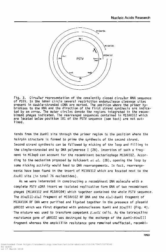
Fig. 3. Circular representation of the covalently closed circular RNA sequenceof PSTV. In the inner circle several restriction endonuclease cleavage sitespresent in double-stranded cDNA are marked. The position where the primer hy-bridizes to the RNA and the direction of the first strand synthesis are indica-ted by an arrow. The outer circles denote the regions integrated in the recom-binant phages indicated. The rearranged sequences contained in M13AV312 whichare located below position 181 of the PSTV sequence (see text) are not out-lined.
tends from the BcmHl site through the primer region to the position where the
hairpin structure is formed to prime the synthesis of the second strand.
Second strand synthesis can be followed by nicking of the loop and filling in
the single-stranded end by ONA polymerase I (28). Insertion of such a frag-
ment in M13mp9 can account for the recombinant bacteriophage M13AV312. Accor-
ding to the mechanism proposed by Volckaert et al. (28), opening the loop by
some nicking activity would lead to DNA rearrangements. In fact, rearrange-
ments have been found in the insert of M13AV312 which are located next to the
EcoRl site (in total 76 nucleotides).
As we were interested in constructing a recombinant DNA molecule with a
complete PSTV cDNA insert we isolated replicative form DNA of two recombinant
phages (M13AV312 and M13AV104) which together contained the whole PSTV sequence.
The Hindlll-Alul fragment of M13AV312 RF DNA and the Alul-BamWl fragment of
M13AV104 RF DNA were purified and ligated together in the presence of plasmid
pBR322 which was first digested with endonucleases BamWl and Hindlll (Fig. 4).
The mixture was used to transform competent E.aoli cells. As -the tetracycline
resistance gene of pBR322 was destroyed by the exchange of the BamWl-Hindlll
fragment whereas the ampicillin resistance gene remained unaffected, recombi-
7953
Downloaded from https://academic.oup.com/nar/article-abstract/10/24/7947/2379140by gueston 17 March 2018
Nucleic Acids Research
H A BEJ | ' j I M13AV10I.
=!=h
U W A U 9 - . PBR322 ( ^
*={ I pAVI.01
Fig. 4. Schematic diagram of the construction of plasmid pAV401 bearing thecomplete PSTV cDNA sequence. The two fragments of M13AV104 and M13AV312 whichwere used for the construction of pAV401 are underlined. Open boxes indicatePSTV cDNA sequences. Double-lines represent sequences derived from M13mp9.The single line refers to the pBR322 sequence. The rearranged PSTV cDNAsequences present in M13AV312 are indicated by a hatched region. It should bementioned that the orientation of the insert contained in M13AV104 is oppositeto the theoretically expected one. Several restriction enzyme cleavage sitesare denoted: H, Hindi 11; A, Alul; B, BamHl; E, EooRI. P refers to the posi-tion of the primer sequence.
nant plasmids would only confer ampicillin resistance to transformed E.ooli
cells. Transformants could therefore be selected on basis of their resistance
to ampicillin and sensitivity to tetracycline. One of the transformants con-
tained a plasmid referred to as pAV401. Characterization of this plasmid re-
vealed that the HindiII and BamHl cohesive ends and the blunt Alul ends used
in the ligation procedure were joined correctly and that these restriction
sites were restored. From alterations in the digestion patterns of Alul, Hhal
and Hpall as compared to pBR322 it could be unambigiously concluded that plas-
mid pAV104 contained the complete PSTV cDNA sequence (data not shown). The in-
sert could be excised precisely with endonuclease BamHl.
Nucleotide sequence. The nucleotide sequence of the PSTV insert in plasmid
pAV401 is presented in Fig. 5. It was deduced from the sequences of the M13
recombinant phages M13AV104, M13AV305 and M13AV312 which were determined by
making use of the dideoxynucleoside triphosphate sequencing method (23). The
inserts of these clones were completely sequenced and agreed with each other.
Thus, cloning errors due to the reverse transcriptase reaction can readily
be ruled out. Furthermore, with the exception of the 24 bases located between
the primer sequence and the BamHl site which were integrated only in M13AV312,
the sequence was independently determined in both strands.
Compared to the nucleotide sequence of the severe PSTV strain described by
Gross et al. (2), 4 nucleotide differences have been found in the S.commer-
7954
Downloaded from https://academic.oup.com/nar/article-abstract/10/24/7947/2379140by gueston 17 March 2018
Nucleic Acids Research
• P S T V - i n s e r t •PBR322 I
GCCGGCCACG ATGCGTCCGG CGIAGAGGAT CCCCGGGGM ACCTGGAGCG AACTGGCAATVAAGGACGGTG GGGAGTGCCC AGCGGCCGAC
Hpall BamHl H a e i l lHaelll _ j Smal '
151 AGGAGTAATT CCCGCCGAAA CAGGGTTTTC ACCCTTCCTT TCTTCGGGTG TCCTTCCTCG CGCCCGCAGG ACCACCCCTC GCCCCCTTTG
Hhal Avail
AC241 CGCTGTCGCT TCGGCTACTA CCCGGTGGAA ACAACTGAAG CTCCCGAGAA CCGCTTTTTC TCTATCTTCTATTGCTTCGGG GCGAGGGTGT
TCTHhal Hpall Alul
331 TTAGCCCTTG GAACCGCAGT TGGTTCCTCG GAACTAAACT CGTGGTTCCT GTGGTTCACA CCTGACCTCC TGAGCAGAAA AGAAAAAAGA
I M13mp9 I pBR322~
63 AGGCGGCTCG GAGGAGCGCT TCAGGGATCC GTCGACCTGC AGCCAAGCTT ATCGATGATA AGCTGTCAAA CATGAGAATT CTTGAAGACG
Fig. 5. Nucleotide sequence of the complete PSTV cDNA insert contained inplasmid pAV401 (located between the two BamHl sites). Nucleotide changes inthe severe strain determined by Gross et al. (2) are denoted on top of theline, differences in the mild strain sequence (12) are shown beneath eachline. The PSTV sequence indicated is homologous to the viroid RNA whereasthe pBR322 part is shown in an anti-clockwise direction (29). Due to thestrategy followed for the construction of pAV401 a small piece (20 bases) ofM13mp9 is conserved. The numbering of the nucleotides refer to the PSTV se-quence only. The asterisk denotes the reference point. The underlined sequencedenoted by P is complementary to the primer sequence. The sequences of severalendonuclease recognition sites are indicated by underlining.
sonii strain used in this study which is also considered to be a severe strain.
Three of these changes are substitutions whereas the fourth is a deletion.
For that reason, this PSTV strain is 1 nucleotide shorter and encompasses
only 358 nucleotides. It is interesting to note that the AA •+ U exchange at
position 120 is also found in the previously determined sequence of a mild
strain (12). This mild strain contains two other differences which are loca-
ted around position 309. One is an exchange (A •* U at position 309), the
other is an insertion of U at position 310-311. In the S.commersonii strain,
at positions 309-310 an AC •* CU exchange has been noted (see Fig. 5). Thus,
all three PSTV strains differ only within two regions which are located at
both sites of the highly conserved central loop of the secondary structure
model (2-6). In one region, the S.commersonii strain shares some nucleotide
differences with the mild strain whereas in the other region exchanges have
been found in all three strains. At least in that part of the sequence, the
numbers of nucleotides of the two severe strains agree with each other.
The question whether these nucleotides contribute in some way to the patho-
genic response in the host remains to be elucidated. More strains have to be
isolated to correlate nucleotide differences with the secondary structure of
the RNA and its pathogenicity. If the complete DNA copy of PSTV described
7955
Downloaded from https://academic.oup.com/nar/article-abstract/10/24/7947/2379140by gueston 17 March 2018
Nucleic Acids Research
here would produce biologically functional viroid RNA molecules in plant cells,
as has been found for cloned copies of phage Qf? and poliovirus RNAs in their
hosts (13, 14), it might be a powerful tool in generating PSTV mutants. Fur-
thermore, plasmid pAV401 can perfectly well be used as a hybridization probe
for the detection of potato spindle tuber viroid in potato tubers (10).
ACKNOWLEDGEMENTS
We thank Will em Mosch for the gift of PSTV RNA, Piet Madern for art work
and photography, Pirn Zabel and Rob Goldbach for critical reading the manus-
cript and Marie-Jose van Neerven for typing the manuscript. This work was sup-
ported by the Research Institute for Plant Protection (I.P.O.). Peter van
Wezenbeek is a postdoctoral fellow of the Netherlands Organisation for the
Advancement of Pure Research (Z.W.O.).
•Present address: Laboratory of Biochemistry, University of Nijmegen, 6525 EZ
Nijmegen, The Netherlands.+Gorlaeus Laboratories, State University of Leiden, P.O. Box 9502,
2300 RA Leiden, The Netherlands.
§To whom correspondence should be addressed.
REFERENCES1. Diener, T.O. (1979). Science 205, 859-866.2. Gross, H.J., Domdey, H., Lossow, C , Jank, P., Raba, M., Alberty, H.
and Sanger, H.L. (1978). Nature 273, 203-208.3. Haseloff, J. and Symons, R.H. (1981). Nucl. Acids Res. 9, 2741-2752.4. Visvader, J.E., Gould, A.R., Bruening, G.E. and Symons, R.H. (1982).
FEBS Lett. 137, 288-292.5. Symons, R.H. (1981). Nucl. Acids Res. 9, 6527-6537.6. Haseloff, J., Mohamed, N.A. and Symons, R.H. (1982). Nature 299, 316-321.7. Diener, T.O. (1979). Viroids and Viroid Diseases. (Wiley, New York).8. Morris, T.J. and Smith, E.M. (1977). Phytopathology 67, 145-150.9. Mosch, W.H.M., Huttinga, H., Hakkaart, F.A. and de Bokx, J.A. (1978).
Neth.J.Pl.Path. 84, 85-93.10. Owens, R.A. and Diener, T.O. (1981). Science 213, 670-672.11. Dickson, E., Robertson, H.D., Niblett, C.L., Horst, R.K. and Zaitlin, M.
(1979). Nature 277, 60-62.12. Gross, H.J., Liebl, U., Alberty, H., Krupp, G., Domdey, H., Ramm, K. and
Sanger, H.L. (1981). Biosci.Rep. 1, 235-241.13. Taniguchi, T., Palmieri, M. and Weissmann, C. (1978). Nature 274, 223-
228.14. Racaniello, V.R. and Baltimore, D. (1981). Science 214, 916-919.15. Wilson, G.G. and Murray, N.E. (1979). J.Mol.Biol. 132, 471-491.16. Panet, A., van de Sande, J.H., Loewen, P.C., Khorana, H.G., Raae, A.J.,
Lillehaug, J.R. and Kleppe, K. (1973). Biochemistry 12, 5045-5050.17. Gammack, R.H. and Harris, P.S. (1973). EPPO-Bull. 3, 117-118.18. Diener, T.O. , Hadidi, A. and Owens, R.A. (1977). In: Methods in Virolo-
gy, Maramorosch, K. and Koprowski, H., Eds. (Academic Press, New York)
7956
Downloaded from https://academic.oup.com/nar/article-abstract/10/24/7947/2379140by gueston 17 March 2018
Nucleic Acids Research
6, 185-217.19. Sanger, H.L., Ramm, K., Domdey, H., Gross, H.J., Henco, K. and Riesner,
D. (1979). FEBS Lett. 99, 117-122.20. Godson, G.N. and Vapnek, D. (1973). Biochim.Biophys.Acta 299, 516-520.21. Van der Marel, G.A., Wille, G., Marugg, J.E., De Vroom, E., Tromp, M.,
Van Boeckel, C.A.A. and Van Boom, J.H. (1982). Rec.Trav.Chim.Pays-Bas,in press.
22. Messing, J., Crea, R. and Seeburg, P.H. (1981). Nuci.Acids Res. 9,309-321.
23. Sanger, F., Nicklen, S. and Coulson, A.R. (1977). Proc.Natl.Acad.Sci.USA74, 5463-5467.
24. Messing, J. (1982). In: Genetic Engineering, Principles and Methods,Holiaender, A. and Setlow, J., Eds. (Plenum, New York) 4, in press.
25. Sanger, F., Coulson, A.R., Barrell, B.G., Smith, A.J.H. and Roe, B.A.(1980). J.Mol.Biol. 143, 161-178.
26. Rohde, W., Schnolzer, M., Rackwitz, H.-R., Haas, B., Seliger, H.and Sanger, H.L. (1981). Eur.J.Biochem. 118, 151-157.
27. Van Wezenbeek, P.M.G.F., Hulsebos, T.J.M. and Schoenmakers, J.G.G. (1980).Gene 11, 129-148.
28. Volckaert, G., Tavernier, J., Derynck, R., Devos, R. and Fiers, W. (1981).Gene 15, 215-223.
29. Sutcliffe, J.G. (1979). Cold Spring Harbor Symp. Quant. Biol. 43, 77-90.
7957
Downloaded from https://academic.oup.com/nar/article-abstract/10/24/7947/2379140by gueston 17 March 2018