The 32 nd International Hospital Federation Congress Hong Kong 15 – 18 May 2001 Eric Jackson

I N S I D E T H I S I S S U E
Editor’s Note . . . . . . . . . . . . . .2President’s Message . . . . . . . . . .22001 Congress Report . . . . . . . .3Your Committees at Work . . . . .42001-2003 Committee
Members Appointed . . . . . . . .5Knee Committee Hosts
Meeting in Florence . . . . . . . .6Symposia Summaries . . . . . . . .7Scenes From the 2001 Congress .12Current Concepts: Posterior
Cruciate Ligament Reconstruction . . . . . . . . . .19
Upcoming ISAKOS-Approved Courses . . . . . . . . . . . . . . . .20
Current Concepts: Modularity in Total Knee Arthroplasty . . .21
S U M M E R 2 0 0 1
Volume 5, Issue 1
E D I T O R
Stephen S. Burkhart, M.D., USA
A S S O C I A T E E D I T O R
Roland P. Jakob, M.D., Switzerland
E D I T O R I A L B O A R D
Moises Cohen, M.D., BrazilMark Ferguson, M.D., South Africa
Philippe Hardy, M.D., FranceNicola Maffulli, M.D., United Kingdom
Peter Myers, M.D., AustraliaMitsuo Ochi, M.D., Japan
Fernando Radice, M.D., ChileKurt Spindler, M.D., USATerry Whipple, M.D., USA
ISAKOSN E W S L E T T E R
Auckland, New Zealand, to Host 2003 Congress
Preparat ionsare under way
to bring theISAKOS Congressback to the Asia-Pacific Region. InMarch 2003,ISAKOS will trav-el to Auckland,New Zealand, therenowned host ofthe 2000 and 2003America’s Cup,the proclaimed"City of Sails."
New Zealand’s largest city is vibrant andclean, surrounded by subtropical islands,rainforests and shimmering harbors.Congress attendees are sure to enjoy therelaxed cafés, world-class wines, freshseafood, sand-filled beaches and art museums.Hike, kayak, bungy jump, sail, ride horseback,bicycle … the outdoor lifestyle of the resi-dents of Auckland combines perfectly with therelaxed sophistication of the urban advan-tages of this friendly South Pacific metropolis.ISAKOS attendees will find there is much todo and that a visit to this gorgeous city and
region is well worth a long airplane ride.Auckland alone claims 23 regional parks,
100 kilometers of coastline, 500 kilometers ofwalking and hiking paths and 48 volcaniccones. It has the largest concentration ofPolynesian people in the world. The "Kiwis"have much to be proud of, and they are eagerto welcome ISAKOS.
The ISAKOS Congress will be held at theAuckland Edge, the city’s center for conven-tions, cultural and entertainment events. Thecongress hotels are all nearby, within walkingdistance, and no place in Auckland is ever farfrom the waterfront. This unpretentious city iswell priced for the international traveler, andthe feel of the city is that of a cosmopolitansmall town. Guests will feel safe and welcomein Auckland.
Mark your calendars now – the FourthBiennial ISAKOS Congress will take placeMarch 10-14, 2003. (Note, this is two monthsearlier than usual. It does not conflict withthe AAOS Annual Meeting, which will be heldin February of that year.) The Call for Abstractswill be available this fall, and a PreliminaryProgram will be available in the fall of 2002.
We look forward to seeing you in Auckland.

F R O M O U R L E A D E R S H I P
ASSOCIATE MEMBERSSergio Aguirre, M.D. • Rodolfo Alonso, M.D. •Sergio Avondo, M.D. • Michel Bitar, M.D. • PaoloBulgheroni, M.D. • Antonio Dal Poggetto, M.D. •Masataka Deie, M.D., PhD • Sanjay Desai, M.D. •Aires Duarte Jr., M.D. • Rolaudo Fabian • PauloFaria, M.D. • Eisaku Fujimoto, M.D. • HugoGonzales, M.D. • Michiya Hara • Rikito Hokama,M.D. • Sadfumi Ichinohe, M.D. • Michael Iosifidis,M.D. • Takashi Kawada, M.D., Ph.D. • KenjiKobayashi, M.D. • Hideji Kura, M.D. •Cleberson Lima • Hin Fai Lung • LeopoldoMarco, M.D. • Mihkel Mardna, M.D. •Silvio Maschke, M.D. • Ferran Montserrat,M.D. • Hany Morsy, M.D. • HirotakaMutsuzaki, M.D. • Hironori Numazaki,M.D., Ph.D. • Afzal Osman, F.C.S.(SA)Orth.• Stergios Papastergiou, M.D. • DirkPetre, M.D. • Rubens Pitangueira, M.D. •Charles Ruotolo, M.D. • Esteban Santos,M.D. • Murilo Santos • Nilson Severino,M.D. • Masaki Sonoda, M.D., Ph.D. •Toshiaki Takahashi, M.D. • HideakiTakeda, M.D., Ph.D. • Enrique Troncoso,M.D. • Yuji Uchio, M.D., Ph.D. • BrunoWalser, M.D. • Andreas Weiler, M.D. •Ethan Wiesler, M.D. • Rogerio Winkler,M.D. • Haruyasu Yamamoto, M.D., Ph.D. •Shinichi Yoshiya, M.D. • AthanasiosZacharopoulos, M.D.
ACTIVE MEMBERSGustavo Acuna • Jose Alemparte • Abbas Al-Numairy • Alberto Araujo-Alvarez • GuillermoArce, M.D. • Pierre Bachelin, M.D. • Klaus Bak,M.D. • Fernando Barclay, M.D. • Gabriel Baron •Ariel Barrera Oro • Walter Besser, M.D. • CarlosBianchi • Seong-Il Bin, M.D. • Emanuele Bocchieri,M.D. • Anita Boecksteiner, F.R.A.C.S. (Ortho),M.B.B.S. • Julio Botello • Lecio Bourbon, M.D. •Edward Buess, M.D. • David Caborn, M.D. • SergioCanuto • Renato Castro, M.D. • Amit Chandratreya,FRCS • Chih-Hwa Chen, M.D. • Po-Yih Chen, M.D.• Pascal Christel, M.D. • Jaehoon Chung, M.D.,PhD • Daniel Comba, M.D. • Luis Costa, M.D. •Antonio Cruz, M.D. • Mario David • Joe De Beer,M.D. • Manuel Diaz-Samada • Oscar Donadio •Jose Duarte • Richard Feinstein, M.D. • HoracioGarcia • Mark Gittins, D.O. • Alberto Gobbi, M.D.• Alvarez Golano, • Joao Gomes, M.D. •Javier Gonzalez Garcia, M.D. • AdelHamed • Ezequiel Hidalgo, M.D. • KotaroIkeda, M.D. • Ernest Irha, M.D. • YasuyukiIshibashi, M.D. • Yasunobu Iwasaki, M.D.,PhD • Varughese Jacob • Anand Jadhav,F.R.C.S., M.Ch.Orth., D.Orth. • Jose Jones• Amos Kidron, M.D. • Kyung Kim, Ph.D.,• Roger Kwok, M.D. • Pablo Lacroze, M.D.• Philippe Landreau, M.D. • MiguelLapera • Gursel Leblebicioglu, M.D. •Dong Chul Lee, Associate Professor •Young-Soo Lee, M.D., Ph.D. • PedroLemos, M.D. • Wilson Li, M.D. • PauloLobo Jr., M.D. • Sergio Maass • PedroMachado, M.D. • Carlos Martin, M.D. •Tetsuya Matsuura, M.D. • John May •David McAllister, M.D. • Marco Merlo, M.D. •Ernst-Otto Muench, M.D. • Edward Nebel, M.D. •Roberto Negrin Vynreister, M.D. • Lei Ni, M.D. •Tahir Ogut, M.D. • Juraci Oliveira, M.D. • MargaretOlmedo, M.D. • Alejandro Orizola, M.D. • IlhanOzkan, M.D. • Vicente Paus • Ehud Rath, M.D. •Antonio Rosato • Nahum Rosenberg, M.D. •Ruben Ruiz • Christoph Saager, M.D. • Valdet Saciri,M.D. • Marc Safran, M.D. • Guillermo Sagasta,M.D. • Jose Serbino Jr., M.D. • Sahir Shaikh, M.D.• Carlos Silva, M.D. • Emir Soendoro, M.D. •Kevin Speer, M.D. • Klaus Steinbrueck, M.D. •Carlos Stierling • Yoshitsugu Takeda, M.D., Ph.D.• Omer Taser, M.D. • Christopher Tong • CarlosVandeputte, M.D. • Robert Verklin, M.D. • CarlosVottola • Nobuyuki Yoshino, M.D., Ph.D. •Giovanni Zaccherotti, M.D. • Oscar Zicaro
ISAKOS Welcomes New Members
Greetings to my friendsand colleagues fromAuckland, New Zealand,venue for our next con-gress. We have not longago returned home fromMontreux, Switzerland,where the 2001 ISAKOSCongress was an out-standing success. Morethan 1300 surgeons from66 countries attended our
largest meeting to date. The picturesque set-ting of lakeside Montreux was a perfectvenue. The scientific program was very strongand well-received. The President’s Dinner atthe Chillon Castle and the Farewell Banquetat the Nock Circus were unforgettable.
Our heartfelt thanks go to Roland andJeanette Jakob, our hosts in Switzerland.Roli’s leadership over the last two years hasbeen inspirational. His passionate commit-ment to ISAKOS has led to greater awarenessand recognition of our society in all cornersof the world. Membership has grown past
1500 and our future is secure. The strength of our society comes first
from our biennial congress and we must allpay tribute to Per Renström, M.D., Ph.D.,Program Chairman, and his team for the out-standing scientific and educational programin Montreux. I am delighted that JohnBartlett from Melbourne, Australia, will beProgram Chairman for the 2003 Congress inAuckland. We will make every effort to buildon the success of our last meeting.
I am committed to developing more ben-efits for members between each biennialcongress. Our Education Committee hasalready made progress with ISAKOS-approved teaching centers and ISAKOS-approved courses. Last year the KneeCommittee held a workshop focused on totalknee replacement in the relatively young andactive patient with osteoarthritis. This led toa symposium on the topic at the 2001ISAKOS Congress and the publication ofworkshop presentations in a booklet distrib-
President’s MessageBarry R. Tietjens, F.R.A.C.S., Auckland, New Zealand, 2001-2003 ISAKOS President
I have recently returnedfrom a truly remarkableorthopaedic meeting inone of the world’s mostbeautiful locations; avenue that far surpassedthe already high expecta-tions of the orthopaedicpilgrims who traveledfrom around the world toattend. It was a scientificassembly that achieved
new highs in education, entertainment andfellowship. For those of you who attended,you already know that I am referring to theISAKOS Congress in Montreux, Switzerland.For those who were unable to attend, I offermy condolences – you missed a good time.
From an educational perspective, the pro-gram was outstanding. The quality of thepapers, symposia and instructional courseswas very high. The program format of multi-ple lecture halls allowed for maximum flexi-bility of physicians to attend lectures of
interest, and the crisp digital presentationformat optimized the impact of each paper.
All of this was orchestrated in a seeming-ly effortless and seamless manner. Effortlessthough it seemed, I can assure the readerthat the smoothness of execution was theresult of continual hard work by a number ofkey people who deserve credit and recogni-tion: our President, Roland Jakob, MD, whoinspired us with his presidential address andwho, I am sure, never fully relaxed until theend; Program Chairman Per Renström, whoprepared a tremendous educational eventand who carefully watched over every detailthe entire time; our talented and tirelessExecutive Director Michele Johnson and herassociates Kathryn Grady and Gigi Agius,who put the ideas together and “made it allhappen”; and the digital audiovisual team ofBill Masheter, Mark Rosenthal and AlanDwan, who created ultimate digital serenityfrom the pandemonium of the speaker ready
Editor’s NoteStephen S. Burkhart, M.D., San Antonio, Texas, USA, ISAKOS Newsletter Editor
Continued on page 4
Continued on page 20

ISAKOS NEWSLETTER • SUMMER 2001 • 3
2 0 0 1 C O N G R E S S R E P O R T
The 2001 ISAKOS Congress was thelargest and most well-received to date,
as 1,386 orthopaedic surgeons convened inMontreux for the five-day scientific programchaired by Per Renström, M.D., Ph.D.,Sweden. An array of "optional events,” fromtours to banquets, kept attendees busy intothe evenings throughout the week.
More Countries Attend Than Ever BeforeSixty-six countries were represented in
Montreux, the greatest number to date.
Continued Growth From 1997 & 1999The 2001 ISAKOS Congress was a hall-
mark of growth as congress registrationscontinued to climb. More than 800 abstractswere submitted for consideration for the2001 Congress. This was a steep jump fromthe 1999 congress, where 400 abstracts hadbeen submitted. Surgeon attendance grewby 400, and poster submissions jumped,from 150 at the 1999 Congress to almost 500in Montreux.
ISAKOS Membership GrowsISAKOS membership continues to climb.More than 85 membership applications werecollected in Montreux, and more are expected.
Attendees Make Montreux a SuccessContinued Growth Expected: 98% of Evaluations Say Attendees “Would Recommend This Congress to Colleagues"
Surgeons: 1386
Exhibit Representatives: 305
Spouses: 150
Attendees in Montreux
0
10
20
30
40
50
60
70
80
66
54
39
1997 1999 2001
Total Number of Countries Represented
Argentina . . . . . . . . . . . .80Australia . . . . . . . . . . . . .35Austria . . . . . . . . . . . . . .13Belgium . . . . . . . . . . . . .35Brazil . . . . . . . . . . . . . . .65Canada . . . . . . . . . . . . .15Chile . . . . . . . . . . . . . . .31China . . . . . . . . . . . . . . . .7Croatia . . . . . . . . . . . . . . .2Czech Republic . . . . . . . .7Denmark . . . . . . . . . . . .10Egypt . . . . . . . . . . . . . . . .5England . . . . . . . . . . . . . .1Estonia . . . . . . . . . . . . . . .2Finland . . . . . . . . . . . . . .17France . . . . . . . . . . . . . .49Germany . . . . . . . . . . . .48Greece . . . . . . . . . . . . . .48Guatemala . . . . . . . . . . . .1Holland . . . . . . . . . . . . . .1Hong Kong . . . . . . . . . . . .3Hungary . . . . . . . . . . . . . .1
India . . . . . . . . . . . . . . . .6Indonesia . . . . . . . . . . . . .3Iran . . . . . . . . . . . . . . . . .4Israel . . . . . . . . . . . . . . .29Italy . . . . . . . . . . . . . . . .99Japan . . . . . . . . . . . . . .111Korea . . . . . . . . . . . . . . . .3Kuwait . . . . . . . . . . . . . . .2Latvia . . . . . . . . . . . . . . . .6Lebanon . . . . . . . . . . . . . .2Liechtenstein . . . . . . . . . .1Lithuania . . . . . . . . . . . . .1Luxembourg . . . . . . . . . . .1Malaysia . . . . . . . . . . . . .3Martinique . . . . . . . . . . . .1Mexico . . . . . . . . . . . . . . .4Netherlands . . . . . . . . . .20New Zealand . . . . . . . . . .7Norway . . . . . . . . . . . . . .3Peru . . . . . . . . . . . . . . . . .1Poland . . . . . . . . . . . . . .12Portugal . . . . . . . . . . . . .23
Puerto Rico . . . . . . . . . . .2Romania . . . . . . . . . . . . .3Russian Federation . . . . . .1Saudi Arabia . . . . . . . . . . .7Singapore . . . . . . . . . . . . .3Slovak Republic . . . . . . . .2Slovakia . . . . . . . . . . . . . .1Slovenia . . . . . . . . . . . . .10South Africa . . . . . . . . . .17South Korea . . . . . . . . . .28Spain . . . . . . . . . . . . . . .29Sweden . . . . . . . . . . . . .37Switzerland . . . . . . . . . .115Taiwan . . . . . . . . . . . . . . .5Thailand . . . . . . . . . . . . . .9Tunisia . . . . . . . . . . . . . . .1Turkey . . . . . . . . . . . . . .35United Kingdom . . . . . . .54Uruguay . . . . . . . . . . . . . .2USA . . . . . . . . . . . . . . .163Venezuela . . . . . . . . . . . .5Yugoslavia . . . . . . . . . . . .3
Attendance By Country
0 500 1000 1500 2000
Number of AbstractsSubmitted
Number of SurgeonsAttending
Number of Posters
Total Number ofRegistrants
1997 Buenos Aires
1999 Washington, DC
2001 Montreux
ISAKOS Congress Growth
0
200
400
600
800
1000
1200
1400
1600
1800
June2001
January2001
January2000
January1999
January1998
January1997
15251371
12821198
11201071
ISAKOS Membership by Year
2003 Call for Abstracts Available Online in OctoberThe Call for Abstracts for the 2003 ISAKOS Congress in Auckland, New Zealand, will be available
on the ISAKOS Web site in October 2001. Abstract submissions will only be accepted online.The deadline for receipt of abstracts is April 1, 2002.

4 • ISAKOS NEWSLETTER • SUMMER 2001
uted to everyone who attended the congress.Over the next two years all the clinical andscientific committees will be working onprojects to benefit all members of ISAKOS.
During the 2001 ISAKOS Congress, aninternational leaders’ meeting was heldunder the banner of the World Council ofOrthopaedic Sports Medicine, ably chairedby Rene Verdonk. It was agreed that in thefuture ISAKOS will host an InternationalPresidents’ Council, which will bring togeth-er leaders of the major continental societies
from North America (AOSSM and AANA),Europe (ESSKA), South America (SLARD)and the Asia-Pacific region (APOSSM,APOA). ISAKOS will maintain and publishon our Web site a meeting schedule to avoidclashes of major meetings. This council willcontinue to foster relationships betweenthese major organizations.
The Executive Board met with the Boardof Trustees of the journal Arthroscopy underthe chairmanship of Jerome Jennings. Ourgoal is to strengthen the relationshipbetween ISAKOS and our official journal forthe benefit of all members of ISAKOS.
The Executive Board also held preliminary
discussions with industry leaders to establishan education fund for ISAKOS. As soon as wecan improve our financial resources, we can,through our committee structure, developmore educational activities for ISAKOSmembers between each biennial congress.In November the Executive Board will bemeeting again with the journal’s Board ofTrustees and also with industry leaders.
It is a great honour for me to lead ISAKOSthrough to the next congress. I am proud torepresent my country and the Asia-PacificRegion. I shall do my very best to climb care-fully on the shoulders of the great leaderswho have gone before me.
The very successful meeting in Montreuxwas a sign of the current strength of
membership and the future of ISAKOS. Thisis an international organization that makesone proud. The number of papers and theexcellent symposia presented are a tributeto the Program Committee and to the facul-ty who presented. The international flavor ofthe venue was evident and made the meet-ing unique. This clearly is one of the meet-ings to attend in the future.
The Communication Committee is hop-ing to make it easier for you to reach yourcolleagues with a new initiative, the ISAKOSMailing List. This will be a list serve that anyISAKOS member with an e-mail address cansubscribe to. This service will only be avail-able to members, not to patients who wantto have their medical problems solvedonline. When any member has a clinical,
sports medicine, arthroscopy or knee prob-lem that they would like an opinion on fromthe members, they can e-mail a brief note toISAKOS, and that question will be sent to allmembers who have an e-mail address.Anyone who would like to reply can simplytype a reply and have his opinion sent toeveryone on the e-mail list. This stimulatesgreat discussion, is easy to do, and can be ashelpful as presenting a case at rounds. Youwill be receiving an e-mail when this serviceis available. Remember to keep the officeinformed of your new e-mail address.
The newsletter will continue to grow: thisissue features the summaries of the sym-posia, one of the main attractions of theMontreux meeting.
The ISAKOS Web site has a link to theArthroscopy journal, where the abstracts fromthe Montreux meeting have been posted.
Society Communications Continue to ExpandDon Johnson, M.D., Ottawa, Ontario, Canada, 2001-2003 Communications Committee Chairman
The Arthroscopy Committee will con-duct multi-center studies in the follow-ing areas:
• Endoscopic Apex Resections forInfrapatellar Tendinitis;
• Arthroscopic Treatment ofOsteochondromatosis of the Hip;and
• Revision Arthroscopy AfterMeniscectomy.
ISAKOS Members who are interestedin participating should contact AndreFrank, 2001-2003 ArthroscopyCommittee Chairman, [email protected].
President’s Message(Continued from page 2)
The Site Selection committee has chosena stunning new resort property in Fort
Lauderdale, Florida, USA, for the 2005ISAKOS Congress.
The 2005 Congress will be held from April9-13, 2005. The Saturday-Wednesday meet-ing pattern will allow surgeons to attendwithout missing many days of work.
The Diplomat Hotel, site of the 2005 con-gress, is set to open in January 2002. It is a
modern, lavish hotel and conference centerthat boasts 1,000 guest rooms and thou-sands of square feet of meeting space. Itsbeachfront location, comprehensive healthspa, private cabanas, two swimming pools,and adjoining country club will allow con-gress attendees to enjoy the advantages of aresort property when not attending confer-ence sessions.
Fort Lauderdale, Florida, Chosen as 2005 Meeting Site
Y O U R C O M M I T T E E S A T W O R K
Multi-Center Studiesto Commence

ISAKOS NEWSLETTER • SUMMER 2001 • 5
BOARD OF DIRECTORS
Executive BoardBarry R Tietjens, President, New ZealandPer A Renstrom, 1st Vice President, SwedenJohn A Bergfeld, 2nd Vice President, USADon H Johnson, Secretary, CanadaKai-Ming Chan, Treasurer, Hong KongFreddie H Fu, Vice Treasurer, USARoland P Jakob, Past President, Switzerland
Members-at-LargeF Alan Barber, USAJohn Bartlett, AustraliaGilberto Luis Camanho, BrazilRamon Cugat, SpainM Nedim Doral, TurkeyMark Ferguson, South AfricaAndre Frank, FranceAnastasios Georgoulis, GreeceMasahiro Kurosaka, JapanMitsuo Ochi, JapanAlberto Pienovi, ArgentinaW Jaap Willems, Netherlands
ARTHROSCOPYAndre Frank, Chairman, FranceRomain Seil, Deputy Chairman, GermanyJ.W. Thomas Byrd, USARodolfo Carpignano, ArgentinaEzequiel C. Hidalgo, VenezuelaBent Wulff Jakobsen, DenmarkHideshige Moriya, JapanHalit Pinar, TurkeyNiek van Dijk, NetherlandsEduardo Zamudio, Chile
BYLAWSPer A Renstrom, Chairman, SwedenJohn A Bergfeld, Deputy Chairman, USAJames Chiu-Yung Chow, USAMoises Cohen, Brazil
COMMUNICATIONSDon H Johnson, Chairman, CanadaF Alan Barber, Deputy Chairman, USAVladimir Bobic, United KingdomFernando Radice, ChileRonald M Selby, USA
EDUCATIONW Jaap Willems, Chairman, NetherlandsTorsten Wredmark, Deputy Chairman, SwedenRamon Cugat, SpainJames C Esch, USAVicente Gutierrez, ChileHideo Matsumoto, JapanDavid V Rajan, India
FINANCEKai-Ming Chan, Chairman, Hong KongFreddie H Fu, Deputy Chairman, USAJohn A Bergfeld, USARoland P Jakob, SwitzerlandDon H Johnson, CanadaPer A Renstrom, SwedenBarry R Tietjens, New Zealand
KNEEPaolo Aglietti, Chairman, ItalyJames Rand, Deputy Chairman, USADavid Stuart Barrett, United KingdomMichael A Kelly, USATomihisa Koshino, JapanUrs Munzinger, SwitzerlandPhilippe Neyret, FranceMitsuo Ochi, JapanPaulo Roberto Rockett, BrazilMichael Soudry, Israel
MEMBERSHIPMoises Cohen, Chairman, BrazilJose Mario Beca, PortugalWalton W Curl, USAMark Ferguson, South AfricaKyosuke Fujikawa, JapanRobert Wen-Wei Hsu, TaiwanDieter M Kohn, GermanyLuis A Vargas, USA
ORTHOPAEDIC SPORTS MEDICINEAnnunziato Amendola, Chairman, CanadaPeter T Myers, Deputy Chairman, AustraliaArnaldo Jose Hernandez, BrazilJose F Huylebroek, BelgiumHartmut E A Krahl, GermanyJoao Alves Grangeiro Neto, BrazilHans H Paessler, GermanyChrister Rolf, United KingdomKurt P Spindler, USA
PROGRAMJohn Bartlett, Chairman, AustraliaChristopher D Harner, Deputy Chairman, USARene Jorge Abdalla, BrazilPaolo Aglietti, ItalyAnnunziato Amendola, CanadaStephen S Burkhart, USAM Nedim Doral, TurkeyLars Engebretsen, NorwayAndre Frank, FranceAnastasios Georgoulis, GreeceStephen M. Howell, USAAlexandra Kirkley, CanadaMasahiro Kurosaka, JapanAnthony Miniaci, Canada
Alberto Pienovi, ArgentinaPer A Renstrom, SwedenKonsei Shino, JapanRussell JA Tregonning, New ZealandW Jaap Willems, NetherlandsSavio L-Y Woo, USA
SCIENTIFICAlexandra Kirkley, Chairman, CanadaNicola Maffulli, Deputy Chairman,
United KingdomLars Engebretsen, NorwayRobert J Johnson, USAJon Karlsson, SwedenJaime Mayer Wageck, BrazilSavio L-Y Woo, USAKazunori Yasuda, Japan
SITERoland P Jakob, Chairman, SwitzerlandJohn A Bergfeld, Deputy Chairman, USAPeter J Fowler, CanadaDon H Johnson, CanadaGary G. Poehling, USABarry R Tietjens, New Zealand
STRATEGIC PLANNINGGary G. Poehling, Chairman, USAWahid Al-Kharusi, Deputy Chairman, USAJohn A Bergfeld, USAGilberto Luis Camanho, BrazilBrian H Casey, AustraliaWalton W Curl, USADavid J Dandy, United KingdomKenneth E De Haven, USAFreddie H Fu, USAM Mike Malek, USAGideon Mann, IsraelJohn B McGinty, USAKonsei Shino, JapanRene E Verdonk, Belgium
UPPER EXTREMITYStephen S Burkhart, Chairman, USAPhilippe P Hardy, Deputy Chairman, FranceJames J Lam, ChinaMario Victor Larrain, ArgentinaDaniel Adolfo Slullitel, ArgentinaKevin P Speer, USAW Jaap Willems, Netherlands
2001-2003 Committee Members Appointed

6 • ISAKOS NEWSLETTER • SUMMER 2001
The Knee Committee of ISAKOS met inFlorence, Italy on January 11 to January
13, 2001 to discuss the issues of manage-ment of the young patient under age 55 withunicompartmental tibial femoral arthrosis.After extensive presentations and delibera-tion, a consensus has been reached.
OsteotomyPatient selection and surgical technique
are critical factors to obtain an optimalresult following tibial osteotomy. Patientsselected for osteotomy should be of physio-logical age of less than 60 to 70 although,occasionally, an older patient may be select-ed. Both men and women can be selected forosteotomy, although men are usually pre-ferred do to the concerns about cosmesis inwomen. Individuals who prefer heavy activi-ties, such as laborers or farmers, are betterselected for osteotomy than arthroplasty.Those individuals who wish to continue withhigh recreational activities, which includeimpact loading sports or rapid directionalchange, are better candidates for osteotomythan arthroplasty.
The patient should have pain localized tothe diseased compartment. Radiographsshould show Ahlback grade 1 to 3 arthrosis.Malalignment should not exceed 10 to 15degrees of varus or valgus. AP laxity of theknee should be less than 5 to 10 mm andmedial lateral laxity less than 10 degrees.Range of motion should be no more than a10-degree flexation contracture and flexionshould be at least 90 degrees.
Relative contraindications to osteotomywould be significant patellofemoral pain,advanced patellofemoral arthritis on radi-ographs, night or generalized pain.
Patient expectations following osteotomyshould include moderate to good pain relief,continued ability to participate in laboringactivities and impact loading sports. Cosmesisis fair. The durability of the osteotomy shouldbe at least 60 percent survival at ten years,but can be considerably longer, if ideal align-ment is achieved. Morbidity associated withosteotomy is moderate with a six monthrehabilitation time.
Options for osteotomy include closingwedge proximal tibial or distal femoralosteotomy, opening wedge, or occasionallyunicompartmental osteotomy to correct frac-ture malunion. The patient with severe axialmalalignment may require osteotomy in thedistal femur and proximal tibial as a com-bined procedure. The importance of overcorrecting limb alignment in the varus knee,such that the mechanical axial of the limbfalls in the lateral compartment was empha-sized. In the case of valgus deformity, correc-tion should be to 180 degrees, not overcor-rection as is done for valgus osteotomy formedial compartment disease. Some of thecommittee members felt that minimizingjoint line obliquity during osteotomy wasimportant. In the management of valgusdeformity, it is extremely important to avoidjoint line obliquity; therefore, distal femoralvarus osteotomy is preferred to proximaltibila varus osteotomy.
Unicompartmental Total Knee ArthroplastyThe patients selected for unicompart-
mental total knee arthroplasty are generallyover the age of 40. Females and males canboth be treated, but the female is more likelyto be selected for unicompartmental arthro-plasty than osteotomy to avoid cosmeticconcerns associated with the extent of cor-rection necessary for a good result afterosteotomy. The patients selected for uni-compartmental arthroplasty should besedentary of only involved in light labor.
Ideally, the patient with unicompartmen-tal arthroplasty has pain localized in the dis-eased compartment. Radiographs shouldshow Ahlback grade 1 to 3 arthrosis. Malalign-ment should not exceed 15 degrees. AP laxityshould be less than 5 mm and the anteriorcruciate ligament should be intact at thetime of surgery. Medial lateral laxity should beless than 10 degrees. Range of motion shouldbe no more than a 15 degree flexion contrac-ture and at least 90 degrees of flexion.
Relative contraindications to unicompart-mental total knee arthroplasty would be sig-nificant patellofemoral pain or advancedpatellofemoral arthritis on radiographs.
The patient can expect good pain reliefwith return to limited non-impact sports.Cosmesis following unicompartmentalarthroplasty is good, although the morbidityis moderate. The durability of unicompart-mental arthroplasty was debated. Somecommittee members felt that the unicom-partmental arthroplasty should be consid-ered as a pre-total knee arthroplasty andwould accept a duration of function of up to5 to 10 years. Other members felt that well-performed unicompartmental arthroplastycan provide very durable results with agreater than 90 percent success rate at 10years. The ability to perform a mini-invasivesurgical exposure for unicompartmentalarthroplasty in both flexion and extensionwas extremely important to obtain a satis-factory result.
Total Knee ArthroplastyPatients selected for total knee arthro-
plasty are generally older than 60 butyounger patients may be candidates if theyhave severe tricompartmental arthrosis.Either gender may be selected for kneearthroplasty. Patients selected for total kneearthroplasty should be sedentary in occupa-tion and only participate in light recreationalactivities. Impact loading sports should bediscouraged. Patients selected for total kneearthroplasty should have severe pain.Generalized or night pain is common in thisgroup of patients. The radiographic extent ofthe arthritis should be Achlback grade 3 to 4.Any degree of varus or valgus malalignmentcan be appropriately treated with TKA. Totalknee arthroplasty is able to manage laxity,flexion contracture, patients with limited flex-ion, and patellofemoral degenerative arthri-tis. In the young patient, the patella shouldnot be resurfaced if the cartilage damage ismild. The patella is congruent with thefemoral component and tracks correctly.
The patient’s expectations would includegood pain relief, limited non-impact sports,and fair to good cosmesis. The morbidityafter total knee arthroplasty is moderate, butthe results are durable with 90 percent suc-cess rate after 10 to 15 years.
Knee Committee Hosts Meeting in Florence:Total Knee Replacement in Relatively Young Patient With Osteoarthritis Generates Discussion and ConsensusJames Alan Rand, M.D., USA, 2001-2003 Knee Committee Deputy Chairman
ISAKOS NEWSLETTER • SUMMER 2001 • 7
S Y M P O S I A S U M M A R I E S
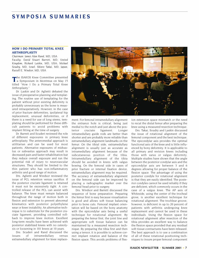
HOW I DO PRIMARY TOTAL KNEEARTHROPLASTYChairman: James Alan Rand, MD, USAFaculty: David Stuart Barrett, MD, UnitedKingdom; Richard Laskin, MD, USA; MichaelSoudry, MD, Israel; Shinro Takai, MD, Japan;Russell E. Windsor, MD, USA
The ISAKOS Knee Committee presenteda Symposium in Montreux on May 15
titled “How I Do a Primary Total KneeArthroplasty.”
Dr. Laskin and Dr. Aglietti debated theissue of preoperative planning and templat-ing. The routine use of templating for thepatient without prior existing deformity isprobably unnecessary as the bone is meas-ured intraoperatively. However, in the caseof prior fracture deformities, ipsilateral hipreplacement, unusual deformities, or ifthere is a need for use of long stems, tem-plating should be performed for these diffi-cult patients to avoid problems withimplant fitting at the time of surgery.
Dr. Barrett and Scuderi reviewed the roleof different exposures in primary kneearthroplasty. The anteromedial approach isutilitarian and can be used for mostpatients. Alternative exposures of midvas-tus or subvastus approach may result indecreased pain and faster rehabilitation butthey reduce overall exposure and run thepotential risk of injury to neurovascularstructures. They should be limited to thethin patient who has non-inflammatoryarthritis and good range of motion.
Drs. Aglietti and Windsor reviewed theissue of PCL retention versus sacrifice. Ifthe posterior cruciate ligament is retainedit must not be excessively tight. A con-trolled release of the PCL can assist withbalance. The knee must remain balancedthroughout the range of motion both inflexion and extension to prevent abnormalkinematics with posterior polyethylenewear or knee instability. An alternative tech-nique is to substitute for the posterior cru-ciate ligament, providing controlled roll-back to improve knee motion. Excellentlong-term results have been achieved withposterior stabilized knees with no osteoly-sis or loosening in 101 knees at 10 years.
Drs. Scuderi and Rand discussed theissues of intramedullary versusextramedullary alignment for knee replace-
ment. For femoral intramedullary alignmentthe entrance hole is critical, being justmedial to the notch and just above the pos-terior cruciate ligament. Longerintramedullary guide rods are better thanshorter and are probably more reliable thanextramedullary alignment landmarks on thefemur. On the tibial side, extramedullaryalignment is usually just as accurate asintramedullary alignment because of thesubcutaneous position of the tibia.Intramedullary alignment of the tibiashould be avoided in knees with valgusbowing. On the femoral side in cases ofprior fracture or internal fixation device,extramedullary alignment may be required.The accuracy of extramedullary alignmenton the femoral side can be improved byplacing a radiographic marker over thefemoral head prior to surgery.
Drs. Windsor and Barrett discussed thesequence of bone preparation. Preparingthe femur first is easy because visualizationis good and allows soft tissue balancingprior to bone cuts. Femoral implant orien-tation will be based on the bony anatomywith the epicondylar axis being a usefultechnique for rotational alignment. Bypreparing the femur first, the joint line andappropriate soft tissue balance can bemaintained by a measured resection tech-nique. By preparing the tibia first and thenusing a tensor, it is possible to achieve cor-rect implant rotation and balance of theflexion space. This avoids problems of flex-
ion extension space mismatch or the needto recut the distal femur after preparing theknee using a measured resection technique.
Drs. Takai, Soudry and Laskin discussedthe issue of rotational alignment of thefemoral component and the best technique.The epicondylar axis provides the optimalfunctional axis of the knee and is little influ-enced by bony deformity. It is applicable toall primary and revision knees includingthose with varus or valgus deformity.Multiple studies have shown that the anglebetween the posterior condylar axis and theepicondylar axis are between 3 and 6degrees allowing for proper balance of theflexion space. The advantage of using theposterior condyle for rotational alignmentis that they are easily identified. The poste-rior condyles cannot be used reliably if theyare deficient, which commonly occurs in thecase of a valgus knee. The AP axis ofWhiteside, using the center of the trochealgrove, is another excellent technique forrotational alignment. The trochlear groove,however, is deficient in up to 20 percent ofpatients with arthritis undergoing kneereplacement and cannot be used in theseindividuals. Using the flexion space forrotational alignment after resection of thetibia provides an excellent way to balancethe flexion space provided that any residualsoft tissue contractures have been released.The best approach is to use a combinationof all of these rotational positioning tech-niques to insure proper femoral component

8 • ISAKOS NEWSLETTER • SUMMER 2001
rotation.Drs. Rand and Windsor discussed the
issue of tibial component rotational align-ment. Rotational options include the tibialtubercle using the transmalleolar axis,aligning it congruent with the femoral com-ponent with the knee in extension, or torotate off the posterior aspect of the tibia.All these techniques can work reasonablywell for low conforming articulations. Inmore conforming articulations, a closerrotational alignment between the femoraland tibial component are required in orderto maximize contact areas and provide opti-mal mechanics. In these knees, careful rota-tion of the tibial components referable tofemur is essential.
Drs. Aglietti and Laskin discussed theassessment of soft tissue balance. Spacerblocks, trials, and tensors can all be utilizedto assess soft tissue balance. An equal flex-ion extension space must be the goalthroughout a range a motion not just at 90degrees and 0 degrees. It is extremelyimportant neither during the soft tissuebalancing process not to place the knee inexcessively tight, which will limit motion,nor to place it in lax, which can result insymptomatic instability.
Drs. Soudry and Windsor discussed softtissue balancing for the varus knee. The firststep in balancing the varus knee is removalof osteophytes from beneath the collateralligaments. In most knees release of thedeep portion of the medial collateral liga-ment from the tibia and, if necessary, theposterior medial corner of the knee willresult in soft tissue balance. Releasesshould be done in a staged manner withassessment of soft tissue balance betweeneach stage of the release. The pes anserinetendons are generally left intact to providesome intrinsic stability on the medial sideof the tibia. Medial epicondylar osteotomyis an advantage in the severe varus kneeand is done by release with an osteotomyleaving the periosteal and soft tissueattachments intact proximally. The epi-condylar osteotomy is repaired with suturesand has provided 83 percent good or excel-lent results.
Drs. Laskin and Aglietti discussed theissue of soft tissue balancing for the valgusknee. The technique of balancing the kneewith valgus deformity has evolved from
striping of the lateral femoral condyleattachments of the popliteus and fibularcollateral ligament to release of the capsulefrom the tibia with multiple pie crusting ofthe posterior lateral capsule. The pie crust-ing technique has the advantage of gradualrelease and reduced risk of flexion instabil-ity. Selective release of the valgus kneeshould be considered based on the extentof deformity. If the knee has fixed valgusonly in extension but the valgus is passive-ly correctable in flexion, only the IT bandand posterior lateral capsule need to bereleased. If the knee is fixed both in flexionand extension then the fibular collateral lig-ament and popliteus will need to bereleased from the lateral femoral epi-condyle. The IT band may need to bereleased in the case of severe contractureas a final step. The PCL can be retained inmany valgus knees and will provide someintrinsic stability. One must be careful to doall releases in the valgus knee sequentiallyto prevent an excessively lax knee.
An alternative in the elderly low demandpatient may be simply to do no releasesand use a more constrained prosthesis toprovide intrinsic stability in place of softtissue balancing.
Drs. Soudry, Scuderi and Rand discussedthe issue of patellar resurfacing. Dr. Soudryfelt that patellar resurfacing was not neces-sary if the patella had good articular carti-lage and tracked congruently with thefemoral component, especially in theyounger patient requiring knee arthroplas-ty. If patellar bone is insufficient, thenpatellar resurfacing should not be attempt-ed. In the patient with patellofemoralarthritis or inflammatory arthritis, patellarresurfacing should be utilized. Patellarresurfacing should be performed if there issevere damage to the articular cartilage ofthe patella, patellar maltracking or lack ofcongruence between the patellar geometryand the geometry of the trochlear aspect ofthe femoral component. During patellarresurfacing it is important not to thickenthe resurfaced patellar composite over itspreoperative value and to ensure absenceof patellar tilt or subluxation. The use of aninset patella should be considered in thosecases of patellofemoral arthritis with a con-cave patella and appears to provide less tiltand less subluxation that those knees in
which an onlay patellar design is utilized.The inset patellar has the disadvantage ofproviding less coverage of the patellar bonecompared to an onlay patellar implant.
In summary, there are multiple impor-tant steps in knee replacement includingcorrect alignment of the implants in thecoronal, sagittal and transverse planes,restoration of the joint line and soft tissuebalance that are critical to achieving opti-mum results. The selection of posterior cru-ciate retention, sacrifice or substitutionshould be dependent on the patient’sanatomy and the ability of the surgeon tocorrect the deformity.
PCL SURGERYChairman: John Bartlett, AustraliaFaculty: Christopher D. Harner, USA; Konsei Shino,MD, Japan
John Bartlett described the anatomy of thePCL and its broad attachment to the
medial femoral condyle, with the isometricpoint being mid-way along a line drawn par-allel to Blumensaat’s line from the widestpart of the medial femoral condyle posteri-orly. The antero lateral part of the PCL istight in flexion and three times strongerand stiffer (1120N, 120N/mm) then the pos-teromedial part (419N, 57N/mm). Themenisco femoral ligaments also contributeto the posterior stability (297N, 49N/mm).
Other structures resisting posteriortranslation and external rotation includethe lateral co-lateral ligament (750N) andthe popliteo fibular ligament (425M). Morethan 10 mm of posterior drawer, instabilityin extension, or excessive external rotationindicates a complex and combined injury tostructures other than the PCL. X-ray(including alignment, and stress views),MRI and arthroscopy all have a place indefining the pathology. The natural historyof “isolated” PCL is benign, but complexlesions will be disabling and may requirecorrective surgery.

ISAKOS NEWSLETTER • SUMMER 2001 • 9
EVALUATION AND MANAGEMENT OF EARLY OSTEOARTHRITIS IN THE ACTIVE POPULATIONSymposium Chairman: Joseph A Buckwalter, MD,USASymposium Faculty: Annunziato Amendola, MD,Canada; K.M. Chan, MD, Hong Kong; HaraldRoos, MD, PhD, Sweden; Kurt P. Spindler, MD,USA
This symposium focused on methods ofaiding patients with mild to moderate
osteoarthritis with maintaining a high levelof activity. This is an important goal, ashelping these individuals allows them toenjoy life, improve their overall generalhealth, and possibly increase their longevity.
Speakers identified a number of strate-gies. One strategy included avoiding activi-ties that expose joints to a level of torsionand impact loading, possibly causing oraccelerating the progression of osteoarthri-tis. Other strategies included weight loss,appropriate shoe wear, use of heel wedgesand knee braces and the importance ofmuscle strengthening for treatment of mildto moderate osteoarthritis. The role of glu-cosamine and hyaluronan was discussed atlength, as well as the role of anti-inflamma-tory medications and analgesics.
Presentations were given concerning therole of osteotomy in the treatment of mildto moderate osteoarthritis in patients whowant to maintain a high level of activity, aswell as the roles of joint debridement andabrasion. The panel agreed that the strate-gies for maintaining high levels of activityfor patients with mild to moderateosteoarthritis should become a central partof sports medicine practice.
TREATMENT OF ROTATOR CUFF DISEASEChairman: Stephen S. Burkhart, MD, USAFaculty: James Esch, MD, USA; PeterHabermeyer, MD, Germany; Philippe P. Hardy,MD, France; Kevin Speer, MD, USA
The goal of this symposium was to coverthe spectrum of pathology (subacromial
impingement, complete and partial rotatorcuff tears, subscapularis tears, biceps
pathology) and the broad range of treat-ment options (open, mini-open, and arthro-scopic).
Dr. Hardy gave the opening presentationon current arthroscopic techniques of sub-acromial decompression. He discussed anumber of technical hints to enable the sur-geon to perform this procedure more easily,and demonstrated the advantages ofarthroscopic subacromial decompressionover open techniques.
Dr. Habermeyer then discussed mini-open rotator cuff repair techniques andresults, pointing out the decreased deltoidmorbidity with this technique compared tostandard open techniques.
Dr. Esch presented his philosophy andtechnique of arthroscopic rotator cuffrepair, pointing out that even large andmassive tears are amenable to arthroscopicrepair. Following Dr. Esch, Dr. Kevin Speerdiscussed pathologic features of the longhead of the biceps. He showed arthroscop-ic examples of failure of the medial “sling”or “pulley” of the biceps (composed of thesuperior glenohumeral ligament and cora-cohumeral ligament), which allowed thebiceps to sublux over the superolateral bor-der of the subscapularis, eventually causingsubscapularis disruption.
Finally, Dr. Burkhart presented his tech-nique and preliminary results of arthro-scopic subscapularis repair, a techniquewhich is the first of its kind for the sub-scapularis.
A panel discussion focused on specificareas of interest from the audience. It wassuggested that good results can beobtained by open, mini-open, and arthro-scopic techniques, but that the trend istoward greater use of arthroscopic repair.
OSTEOCHONDRITIS DISSECANS OF THE ANKLEChairman: K.M. Chan, MD, Hong KongFaculty: Andre Frank, MD, France; Mitsuo Ochi,MD, Japan; Christer Rolf, MD, PhD, UnitedKingdom
We began with an overview of the histo-ry of the first recognition of the condi-
tion, and proposed etiologies mentioned inthe literature. The clinical features, classifi-cation systems used based on plain radi-
ographic and MRI images, and controver-sies regarding the natural history and clini-cal significance of such lesions were high-lighted. For our treatment strategy, weraised the question of which parameters(clinical or radiological) should be the mainconsideration in decision-making.
Mitsuo Ochi demonstrated how he treat-ed one case successfully by autologouschondrocyte transplantation into the lesionin the talus. It cannot be concluded at thisstage if this should be the standard treat-ment since the sample size was too smalland there was too short a follow-up time.
Andre Frank gave a brief introductorysummary of the condition. He regardedanterolateral talar lesions as fractures andseparated this group from the posteromedi-al group. His clinical study included a largenumber of osteochondral fractures result-ing from Pott’s fractures and he useddrilling, curettage for a number of his cases.It was therefore not clear if his interpreta-tion of ankle OCD was the same as theother speakers’ or not.
Christer Rolf pointed out that osteo-chondral fractures resulting from Pott`sfractures are different. He also queried theclinical significance of OCD found inciden-tally on diagnostic arthroscopy for otherconditions. In fact, he believed that many ofthe symptoms of pain might be attributableto unrecognized syndesmosis injuries.
The treatment of OCD by mosaicplastywas not discussed in great detail althoughone member of the audience from Koreaapparently had a reasonable amount ofexperience.
THE KNEE AFTER MENISCECTOMYChairman: K.E. DeHaven MD, USAFaculty: C.D. Harner MD, USA; J.R. AndrewsMD, USA
This symposium addressed three optionsfor patients who continue to have sig-
nificant disability and functional limita-tions following meniscectomy, typically dueto the development of degenerativechanges in that compartment.
Dr. DeHaven discussed the CollagenMeniscus Implant (currently in FDA trial inthe US) as an example of tissue engineeringto replace lost meniscus tissue. It is made
10 • ISAKOS NEWSLETTER • SUMMER 2001
of reprocessed bovine collagen, inducescellular ingrowth and tissue regeneration,and is biodegradable. The indications, tech-nique, aftercare and current update of theUS FDA trial were presented. Based on theclinical and second-look arthroscopic datato date, this approach appears to havepromise as a means to decrease post-meniscectomy problems in the future.
Dr. Harner discussed the role of allograftmeniscus transplantation using fresh-frozen, non-irradiated allografts from young(age 15-35) donors. The important selectionfactors of age, localized pain after menis-cectomy, state of articular cartilage (nomore than moderate loss), and neutral limbalignment were stressed. Donor/recipientmatching, surgical technique, aftercare andoutcomes were presented. He concludedthat meniscus transplantation remains aviable option in this select group of patients.
Dr. Andrews discussed the more severeend of the arthritis spectrum and comparedthe advantages and disadvantages ofosteotomy, total knee replacement and uni-compartmental replacement. He specifical-ly addressed the option of uni-compart-mental replacement and how more recentrefinements of component design,improved patient selection, stricter indica-tions and improved techniques haveimproved outcomes. Current indications,contraindications and personal experiencesince 1997 were presented. He concludedthat in properly selected patients uni-com-partmental replacement is a practical alter-native for the treatment of uni-compart-mental arthritis of the knee.
KNEE BRACESCo-Chairmen: Per Renström, MD, PhD, Swedenand Braden C. Fleming, PhD, USA
Functional knee braces (FKBs) are com-monly used to protect the anterior cruci-
ate ligament (ACL), to protect an ACL graftparticularly during healing, and to restorefunction to the ACL-deficient knee. Kneebracing remains controversial. This symposiumwas organized to look at knee bracing frombiomechanical and clinical perspectives.
Biomechanical evaluations of kneebraces should be performed under weightbearing, muscle activated conditions since
athletes depend on them this way. The bio-mechanical questions addressed were: 1)Does a FKB protect an ACL-intact or ACL-reconstructed knee during weight bearing?and 2) Does a FKB restore normal kinemat-ics to the ACL-deficient knee during weightbearing?
The first question was addressed bymeasuring the strain in the ACL-intact kneeassuming that these data would be similarto a properly positioned ACL graft. Subjectswere tested both with and without a bracewhile the knee was weight bearing (WB) ornon-weight bearing (NWB) and in combina-tion with externally applied loads (Fleminget al, Am J Sports Med 28:815, 2000). It wasdetermined that the knee brace significantlyreduced but did not eliminate ACL strainsas the knee made the transition from non-weight bearing to weight bearing. The bracealso reduced ACL strain values when ante-rior shear loads were applied to both theNWB and WB knee.
The second question involved ACL-deficient patients. Anterior-posterior (A-P)laxity values were measured when the kneewas WB and NWB. The braces restored theA-P laxity of the ACL-deficient knee to nor-mal. However, the braces did not eliminatethe anterior shift of the tibia relative to thefemur that occurs as the knee makes thetransition from NWB to WB.
Biomechanically, there are some ques-tions that still need to be answered. Can FKBsprotect an ACL, or ACL graft, during loadsthat are sufficient to produce injury? Canthey reduce the incidence of knee re-injuryand arthritis in the ACL-deficient knee?
ARTHROSCOPY AND ANTERIOR INSTABILITY OF THE SHOULDERSymposium Presented by the FrenchArthroscopy Association (SFA)Symposium Chairman: Andre Frank, MDSymposium Faculty: M. Allard, P. Beaufils, P. Boileau, O. Chaix, H. Coudane, P. Desmoineaux,P. Devallet, P.H. Flurin, G.Gartsman, D. Gazielly,P. Gleyze, Ph. Hardy, A. Imhoff, A. Iserin, F. Kelbérine, F. Kempf, L. Lafosse, C.Levigne, S. Messens, J.C. Meynet, D. Molé, H. Thomazeau,B. Toussaint, G. Walch
The French Arthroscopy Association pre-sented the results of several different
studies in its afternoon symposium atISAKOS:
CORRELATION BETWEEN CLINICALAND ARTHROSCOPIC FINDINGS INSHOULDER ANTERIOR CHRONIC INSTABILITYA Prospective Multicenter Study of 224 Cases
The purpose of this multicenter prospec-tive study was to compare anatomical find-ings to clinical data in 224 patients whounderwent an arthroscopic stabilization ofthe shoulder. Each procedure was recordedon videotape, showing the lesions and thefinal aspect after treatment.
Ten centers took part in this continuousstudy from October 1999 to October 2000.243 shoulders were operated on; the twofirst authors controlled the videotapes. 224cases were included into this study. Meanage was 27.5 (16 to 56); 74% of the patientswere males; 84% were involved in sportsactivities and 70% were involved in over-head sports. Indications for surgery includ-ed: recurrent dislocation (56%), subluxation(26%), and painful shoulder (18%). A bonylesion was found in 28% of the cases on theglenoid rim and, in 58% of the cases, on thehumeral head. The average delay betweenthe first symptoms and the index procedurewas three to five years.
To assess the location of the periglenoidlesion we divided the glenoid rim into 6sections (A,B,C,D,E,F) of 2 hours. The firstsection (A), located from 11 to 1 o’clock,was correlated to a SLAP lesion. Significantcorrelations were found between preopera-tive clinical symptoms and anatomical find-ings:
• Anterior lesions “BC” (32%) were 4/5 cor-related to painful shoulders or subluxa-tions, and only 1/5 were correlated todislocations;
• Anterior lesions with SLAP extension“ABC” (30%) represented 80% of thepainful shoulders;
• Anterior lesions with inferior extension“BCD” (22%) were correlated to disloca-tions or subluxations (95%), and only 5%were correlated to painful shoulders;
• Anterior-inferior lesions with posteriorextension “BCDE” (9%) were also corre-

ISAKOS NEWSLETTER • SUMMER 2001 • 11
lated to dislocations (62%);• In 88% of the cases, global detachment
“ABCDEF” (4%) was correlated to dislo-cations or subluxation;
• 3% of the cases had no detachment.
Anterior ligaments (axillary poach, ante-rior band of LGHI, MGHI) were considerednormal in 50%, torn in 39%, or stretched in11% of the cases. The humeral attachmentof the anterior glenohumeral ligament wastorn in 22% of the cases.
A classification of the arthroscopic find-ings has been established in order toimprove our indications:
• Type A – (50%) Periglenoid lesion only,with no ligament injury, nor humeraldetachment. This type seems to be thebest indication for an arthroscopic stabi-lization.
• Type B – (28%) Periglenoid lesion +ligament lesion. The low quality of thesoft tisues may be responsible for recur-rent instability after an arthroscopic pro-cedure.
• Type C – (22%) Ligament detachmenton the humeral side. Isolated arthro-scopic periglenoid ligament reattach-ment is not indicated; open surgery isnecessary.
In conclusion, correlation betweenarthroscopic anatomical findings and clini-cal symptoms has been found. A classifica-tion according the arthroscopic lesions hasbeen proposed to allow better indications.
The arthroscopic visualization of boththe lesions and the final aspect after repairrepresents an important database that willallow a better assessment of the clinicalresults after a minimum two-year follow-up.
ARTHROSCOPIC SHOULDER STABILIZATION IN CHRONIC ANTERIOR SHOULDER INSTABILITY
209 patients with a chronic anteriorshoulder instability entered into a multi-center retrospective study. Four techniquesusing an arthroscopic procedure were per-formed in four different centers: Fastak (35cases), Mitek G II (75 cases), Suretac (58cases) and Shrinkage (42 cases). The aver-age age of the patients was 28 years old (14
to 59) and 78% were involved in sportsactivities.
Surgery was indicated for: recurrent dis-location (63%), subluxation (21%), andpainful shoulder (16%). In 80% of the casesbony lesions were present on the glenoidrim, and in 86% of the cases they were pres-ent on the humeral head (Hill Sachslesion). Delay between first symptoms andsurgery was three to five years.Immobilization varied from three to sixweeks. Recurrent dislocation occurred in13% of the cases (Suretac 12%, Fastak 23%,Mitek 14%, Shrinkage 9%).
Comparisons between the different tech-niques used in this series are difficultbecause the indications were different. Inthe Mitek and Fastak groups there weremore dislocations than in the Shrinkageand Suretak groups. Patients were gradedaccording to the Duplay score. The averagescore was 82/100 (Suretac 85.4, Fastak 71.9,Mitek 89.3, Shrinkage 85.8). 71% returned tothe same sport at the same level, 75% hadno pain and the average range of motionwas 176° in elevation with a mean loss of 8°in external rotation. The results accordingthe Duplay score were excellent in 49%,good in 31%, fair in 6% and unsatisfactory in15% of the cases. 82% were very satisfied orsatisfied. Results were badly influenced bydifferent factors: age inferior to 25, strenu-ous sports, throwing athletes, high rate ofdislocation and hyperlaxity.
In conclusion, arthroscopic stabilizationis efficient in more than 80% of the case.Improvement in techniques and indicationsshould improve our results.
ACUTE FIRST ANTERIOR GLENOHUMERAL DISLOCATION: ARTHROSCOPIC ANATOMICAL FINDINGS. A PROSPECTIVE MULTICENTER STUDY OF 30 CASES.
Thirty patients underwent an arthro-scopic evaluation without stabilization afteran acute primary anterior glenohumeraldislocation. The purpose of this study wasto assess the initial anatomical lesions andto correlate them to the clinical outcomeafter a conservative treatment.
Material: All patients were male under30 years old (average age: 22 years). 81%were involved in sports activities.
The post-arthroscopic conservativetreatment consisted of sling immobilizationfor 4 weeks. A CT scan was performed sixweeks later.
Arthroscopic findings:• Anteroinferior Labrum detachment +
IGHL: 89%• IGHL detachment on the humeral side:
23%• Anterosuperior Labrum detachment +
MGHL: 66%• SLAP: 33%• Hill Sachs: 100% (Minor 81%, Major 19%)• Glenoid rim avulsion: 7%
Results: 21 patients were reviewed after24 months mean follow-up. A recurrent dis-location occurred in two cases (15 and 27months). The Apprehensive Test was posi-tive in four other cases. Fifteen patients(71%) had a perfect stable shoulder.
Prognosis factors: Glenoid rim avulsion,major Hill Sachs lesion, extended lesion tothe anterosuperior part of the labrum, andSLAP lesion did badly influence the finalresults.
Conclusions of this study: In acute ante-rior glenohumeral dislocation, arthroscopyshows a great variety of lesions.
The rate of recurrent dislocation in thisstudy is low: 9.5%. Why? Arthroscopiclavage? Level of activity? Four weeks ofpost-operative immobilization?
Such studies remain necessary in orderto understand the recurrence factors beforeachieving initial stabilization.
LESIONS OF THE GLENOID LABRUM (STABLE SHOULDER).ARTHROSCOPIC TREATMENT
In 1998, the Society of FrenchArthroscopy organized a symposium devot-ed to isolated lesions of the glenoid labrumin the stable shoulder.
This was a multicenter study with arthro-scopic shoulder surgeons from six centersparticipating. (Fourteen were solicited.)Approximately 3000 shoulder arthroscopiesperformed over a three-year period werereviewed, ultimately yielding 151 cases (5%)of isolated labral lesions.
Continued on page 14

12 • ISAKOS NEWSLETTER • SUMMER 2001
S C E N E S F R O M T H E 2 0 0 1 C O N G R E S S
Drs. Paessler and Thal perform a Live Surgical Demonstration
President’s Reception
Past President Roland P. Jakob introduces current President Barry R. Tietjens
Fun Run
Past President Roland P. JakobPoster Exhibits
Farewell Banquet at the Nock Circus
Welcome Reception at the Petit Palais
Honorary Members (left to right) LambertoPerugia, James Andrews for Jack Hughston,
John B. McGinty, and Robert Jackson
Attendees in the Exhibit Hall

ISAKOS NEWSLETTER • SUMMER 2001 • 13
S C E N E S F R O M T H E 2 0 0 1 C O N G R E S S
500 posters were on display
President’s Reception at the Chillon Castle Mini Hands-on Workshops Multimedia Center
Poster & New Member Reception
Stravinsky Auditorium
Technical Exhibits
Knot-Tying Station

14 • ISAKOS NEWSLETTER • SUMMER 2001
Frequency of isolated pathogenic labrallesions:
Posterior superior lesions 115 cases 70%(posterosuperior impingement)
SLAP lesions 16 cases 10%Anterior superior lesions 22 cases 15%Posterior inferior lesions 8 cases 5%
Posterior Superior LesionsPosterior superior lesions of the glenoidlabrum were defined as “pathologicallesions of the segment of glenoid labrum,from the insertion of the long head of thebiceps superiorly, extending posteriorly tothe equator of the glenoid”. One hundredfifteen cases constitute this group and rep-resent our 1998 experience with theselesions and their treatment.
This study demonstrated that posteriorsuperior lesions of the glenoid labrumoccur mainly in the overhead athlete andare the result of posterosuperior glenoidimpingement as described by Walch.Arthroscopy is not necessary to establishthis diagnosis if the clinical, radiographic,and computed tomographic arthrographydata are consistent with posterosuperiorglenoid impingement.
When the diagnosis is established, atrial of functional rehabilitation is alwaysindicated. Failing nonoperative treatment,arthroscopic debridement of the labral androtator cuff lesions gives inconsistentresults, but remains an acceptable opera-tive alternative. Surgical treatment usinghumeral derotational osteotomy asdescribed by Walch can only be recom-mended in selected cases in which thepatient has already failed less invasive ther-apeutic alternatives.
Lesions of the Superior Part of theGlenoid Labrum
Snyder originally described theselesions, located at the insertion of the longhead of the biceps, in 1990. Since that time,controversy has existed regarding thislesion’s etiology, frequency, and clinical andpathological relevance.
In this series, the authors found sixteenSLAP lesions as described by Snyder. Thesepatients were reviewed extensively, includ-ing preoperative, intraoperative, and post-operative findings, insuring that these were
isolated SLAP tears and that these patientsdid not have any associated pathologicallesions, i.e. instability, rotator cuff pathology.
From the evaluation of these patients, itis apparent that the preoperative findingsin patients with this lesion are nonspecific.Despite this lack of specificity, an individualbetween the ages of twenty and forty yearsold sustaining a traumatic injury, usually asa result of athletics involving the upperextremity, with mechanical shoulder paincaused by the cocking maneuver, palpationof the bicipital groove, or pain with resistedforward flexion of the shoulder with asupinated extremity, must arouse suspicionof a SLAP lesion.
Computed tomographic arthrographyand MRI demonstrate a SLAP lesion inapproximately 50% of the cases. Ultimately,it is arthroscopy that establishes the diag-nosis. We discovered four Type I, nine TypeII, one Type III, and two Type IV SLAPlesions in our series. In all cases the lesionappeared indisputably pathologic, in con-trast to a normal anatomic variant. Becauseof this necessary distinction, preciseanatomical criteria must be considered.
Analysis of the results of this seriesaffirmed that debridement of Type IIIlesions and repair of Type II and Type IVlesions are appropriate treatment.
When we evaluated the postoperativeresults according to the Constant score, weobtained twelve “excellent” and “good”results, two “fair” results, and two “poor”results. Overall, the results were satisfacto-ry in 75% of the cases, with resumption ofprevious employment in 90% of the casesand resumption of sports activity at the pre-injury level in 75% of the cases.
In conclusion, lesions of the superiorglenoid labrum exist but are rare. Whenthey are present, SLAP lesions can be effec-tively treated by arthroscopy.
Anterior Superior LesionsTwenty-two cases of lesions in the ante-
rior superior segment of the glenoid labrumwere found. These consisted of a completedisruption in eleven cases, a flap tear inseven cases, fibrillation of the labrum intwo cases, and a labral fissure in one case.
These isolated lesions of the anteriorsuperior labrum without known instabilityare rare in France, especially if lesionswhich extend superiorly or inferiorly areexcluded from this category. The greater fre-quency of these lesions in the English liter-ature is probably related to several factors:throwing sports are less commonly prac-ticed in France, a less rigorous selection cri-teria for lesions located in this zone, andunclear distinctions between fibrillation,flap tears, and complete disruption of thelabrum may exist.
The complete tear seems to be a distinctentity with, perhaps, a degenerative mecha-nism. Treatment with simple resectionyields good results. Attention must begiven to the status of the superior and mid-dle glenohumeral ligaments, which may beinvolved in this lesion.
Lesions of the Posterior Inferior LabrumPosterior inferior labral lesions are
defined as all lesions within zones VI and Vof Snyder. Often these labral lesions areassociated with chondral pathology in theadjacent glenoid and/or a posterior capsu-lar abnormality, usually in the form of cap-sular distension.
Since the arthroscopic identification ofposterior capsular distension is extremelydifficult, we were not able to establishwhether these lesions were associated withposterior instability. We have thereforeincluded all of the lesions within thisanatomic location in this study.
After thoroughly reviewing the 3000
Keep Your Member Information Current!ISAKOS Members: Help us keep your information current! Please visit the ISAKOS Website at www.isakos.com and check your listing in the online Membership Directory to ver-ify that we have your correct address, telephone numbers and e-mail address. Pleasenotify the ISAKOS office if any of your information is missing or incorrect.

ISAKOS NEWSLETTER • SUMMER 2001 • 15
cases collected from the six centers ofarthroscopic surgery, we discovered onlyeight isolated posterior inferior labrallesions. Epidemiologically, it was a veryhomogeneous population. The diagnosiswas suspected in all cases on computedtomographic arthrography and confirmedarthroscopically.
• Six cases were isolated lesions withoutany apparent posterior capsular pathol-ogy. Arthroscopic resection of the lesionwas successful in all cases.
• Two cases were associated with a subjec-tively-appearing distended posteriorcapsule, although no findings on preop-erative clinical examination were sug-gestive of posterior instability.Treatment was successful only if the pos-terior capsular was plicated.
Lesions of the posterior inferior glenoidlabrum exist, however, they rarely necessi-tate surgical treatment (0.3% of the shoul-der arthroscopies we reviewed). Overall, ourresults confirmed that of the available liter-ature, which indicates arthroscopic treat-ment of these lesions yields good resultsprovided that occult posterior instability isaddressed.
ARTHROSCOPY OF THE SHOULDER AND ROTATOR CUFF TEARS
The tremendous development of endo-scopic procedures represents a recent andmajor evolution in the treatment of therotator cuff pathology. To investigate itsability to substitute for open procedures,the French Arthroscopy Society conducted amulticenter symposium to retrospectivelyreview 487 patients with three major ques-tions:
• Is arthroscopy reliable enough to assessthe anatomical extent of a rotator cufftear (i.e., will it be able to become the“Gold Standard” for scientific prospec-tive investigations)?
• Is Arthroscopic Subacromial Decom-pression (ASD) without repair a long-term efficient procedure for the treat-ment of rotator cuff tears?
• Are arthroscopic repair proceduresanatomically and functionally efficient?
The following articles offer their find-ings.
ARTHROSCOPIC ASSESSMENT OF FULL-THICKNESS ROTATOR CUFFTEARS. (117 cases)
Evaluation of open or endoscopic proce-dures requires a precise, reliable and repro-ducible intraoperative assessment of therotator cuff lesions. It is now well estab-lished that the anatomical extent of a full-thickness rotator cuff tear represents themajor predictive factor of the anatomicaland functional results following treatment.Description of this tear can no longer belimited to a simple classification usinggrades or centimeters, but must specify theanatomical state of each structure of therotator cuff, including the rotator interval.
To evaluate the reliability of the arthro-scopic assessment of full-thickness rotatorcuff tears, 117 cases were prospectivelyinvestigated with imaging arthroscopy,immediately followed by open repair. Theconfidence of the operator, his accuracy, andthe operator-dependent character of arthro-scopic assessment were evaluated for thedescription of the main anatomical param-eters.
Confidence in the method of investiga-tion was judged on the percentage of non-communicated responses (nc) for each ofthe 3 types of investigation (imaging, endo-scopic, open surgery). Comparing endo-scopic data to open surgery data taken as areference test then assessed accuracy. Re-analyzing the “confidence” and “accuracy”criteria of the operators who were dividedin 4 different groups assessed the operator-dependent character of the endoscopicevaluation.
The operators felt confident and wereaccurate in diagnosing a full-thickness tearof the supraspinatus, but they clearlyunderestimated its coronal and sagittalextent, and its reducibility to the greattuberosity. Conversely this techniqueappeared very accurate for the descriptionof the rotator interval. Endoscopic assess-ment was particularly operator-dependentfor the antero-posterior analysis of the tear.
This study demonstrated the limits ofthe endoscopic assessment of rotator cufffull-thickness tears. The main limitations
are the sagittal analysis of the tear of SSN,especially when the lesion extends to theISN and to a lesser degree to the SSC. Thisdifficulty, already significant in this studydespite experienced and motivated opera-tors, is probably worse for less experiencedoperators. The influence of sagittal extentof the lesion on the final prognosis of arotator cuff repair emphasizes the impor-tance of a perfect and precise preoperativeimaging. It also requires an adequatearthroscopic technique with a good knowl-edge of the limits of endoscopic assess-ment, and of the normal and pathologicalanatomy of the rotator cuff. These condi-tions are essential to precisely appreciatethe lesion and its reparability before dis-cussing the indication and the type ofrepair if chosen.
ARTHROSCOPIC SUBACROMIAL DECOMPRESSION WITHOUT REPAIR OFFULL-THICKNESS ROTATOR CUFFTEARS (283 cases)
The purpose of this study was to assessresults and clinical evolution of full-thick-ness rotator cuff tears treated byArthroscopic Subacromial Decompression(ASD), an/or biceps tenotomy without ten-don repair, with a minimal 3 years follow-up, and to discuss our surgical indicationsaccording to the patient and the anatomicallesions.
283 patients entered this study, with amean age of 63.1 years (range 32 to 82). Acomplete tear of the supra-spinatus tendonin the sagittal plane was noticed in 93.2% ofthe cases. The lesions extended to the infra-spinatus tendon in 57.3% of the cases, andto the sub-scapularis tendon in 29%. Thelong head of the biceps was stable andintact in only 27% of the cases.
All patients were evaluated pre andpost-operatively using the Constant scoreand a radiological A-P view for sub-acromi-al space measurement. 218 ASD and 116biceps tenotomies were performed. The twoprocedures were combined in nearly 25% ofthe cases. At the final revision (mean fol-low-up of 4.77 years), 98 patients (34.6%)were very satisfied, 122 (43.1%) satisfied, 43(15.2%) moderately satisfied and 20 (7.1%)were disappointed. The average Constantscore was 67.6 points/100 with a final gain

16 • ISAKOS NEWSLETTER • SUMMER 2001
of +27.3 points. The radiological studyshowed a 1 mm narrowing of the sub-acro-mial space (7.6mm to 6.6mm).
Patients were divided in 4 groupsaccording to their age: Group I (19 casesless than 50 years), Group II (88 cases from51 to 60 years), Group III (58 cases from 61to 65 years) and Group IV (118 cases over 65years). The anatomical extent of the tearincreased with age and the final objectiveresult depended more on the extension ofthe lesions than on age (except for Group I).
This series did not demonstrate anymajor secondary clinical or radiologicaldeterioration after the arthroscopic treat-ment without repair. Subacromial decom-pression and/or biceps tenotomy indica-tions depend on age and extension of thetear. ASD can be considered as a reliablebut also a “limited goal” procedure for thetreatment off full-thickness rotator cufftears (final Constant score: 67.6 points).Repair must be discussed for youngerpatients .
ARTHROSCOPIC ROTATOR CUFFREPAIR: WHEN AND WHY ?
In the presence of a symptomatic tear of therotator cuff, should the cuff be repaired andthe humeral head re-covered? In theory, theanswer can only be affirmative, for is it notour role to restore function and, if possible,anatomy?
This is not always technically easy, how-ever. Is it always necessary ?
An MRI study of 96 totally painlessshoulders revealed that after the age of 60years 28% of the individuals have a fullthickness rotator cuff tear involving thesupraspinatus tendon.
Another well-known study clearlydemonstrated that there are 50% of recur-rences after the repair of tears extendingbeyond the supraspinatus.
Various other studies showed the bene-fits of arthroscopic “debridement” withoutrepair of rotator cuff tears, especially painrelief.
Recent studies also showed excellentresults after anatomical repair (open, mini-open, arthroscopic)
Therefore, the fundamental questionsare: Repair: when and why? If repair, thenwhen and why arthroscopic?
Repair: When and Why ?Depends on the lesion and the patient:
• The lesion: Size, Tissue Trophicity(Imaging +++)
• The patient (Age, Activity, FunctionalDemand…)
What can we expect after treatment?Arthroscopic subacromial decompression(ASD): palliative treatment for pain ++.Good and durable results.Repair: Better functional result ++If repairable and non degenerative tissue =excellent result
Arthroscopic Repair: When and Why
• Arthroscopy + Miniopen. Simple.Excellent exploration. Permit any kind ofsuture
• All arthroscopic: Recent studies areencouraging with promising results.
• Necessity of anatomical control +++.Rerupture = 7 to 50 %
• Ideal indication = Small non retractedsupra-spinatus lesion
CONCLUSIONSThe operative indication must be evaluatedconsidering all pertinent parameters: age,pain, physical examination data (strength,mobility, etc.), tear extension (arthrography,arthroCT-scan, MRI), social and profession-al background, etc.
An operative indication is therefore easyto establish for younger manual workerswho have a post-traumatic tear with a cuffsusceptible to easy reinsertion (by open orarthroscopic technique). It is likewise easyto establish for the elderly patient whosefunctional demands are more modest butwho complains about pain and for whom anarthroscopic debridement should provide apositive analgesic effect.
Between these two extreme situations,the definition of the operative indicationbecomes more complex, particularlybecause one must take into considerationthe intratendineous degenerative disease(MRI and computerized arthrotomography)so that the repair is lasting. The future willcertainly enable us to specify the respectiveoperative indications, restricting the debateto “paliative” or “curative” treatment, andnot to “arthroscopic” or “open surgery”.
GOTS SYMPOSIUMSymposium Chairman: Ernst-Otto Muench, MD,GermanyFaculty: Manfred Dingerkus, MD, Germany;Hubert Hoerterer, MD, Germany; Karl-HeinzKristen, MD, Austria
About GOTS:
GOTS stands for the Gesellschaft fürOrthopädisch-Traumatologische-
Sportmedizin, meaning the “Society ofOrthopedic Sports Medicine andTraumatology”. Physicians of sports medi-cine from Germany, Austria and Switzerlandfounded the German-speaking society in1986. With more than 600 members workingin private hospitals, sports federations,clubs, and at universities for the improve-ment of diagnosis, therapy and rehabilita-tion in amateur and professional sports,GOTS is behind the American OrthopaedicSociety for Sports Medicine (AOSSM) as theworld’s largest and most important societyfor orthopaedics and trauma in sports.
GOTS understands sports medicine as afar-reaching and dialogue-oriented medi-cine. The society strives to unite the syner-gies of orthopaedists, surgeons, physio-therapists, psychologists and sports sci-ence researchers.
The self-image and function of GOTS ischaracterized by a combination of research,sports medicine, special events, work withinternational institutes, procurement ofcurrent research results for sports medicineapplications, integration of industry, anddialogue with athletes, trainers, federationsand the public.
The GOTS Symposium at ISAKOS: Chairman of the GOTS Symposium at
the ISAKOS Congress was former Vice-President E. Otto Muench. HubertHoerterer, Manfred Dingerkus and Karl-Heinz Kristen presented epidemiology,technique, injury patterns and preventionof injuries for sports skiing, inline skatingand carving skiing.
Dr. Hoerterer demonstrated the differ-ence between carving skies and traditionalskies. Carving skies are characterized bybroader ski-tips and tails and are usuallyshorter than traditional skies. It is easier forbeginners to start skiing with this kind of ski

ISAKOS NEWSLETTER • SUMMER 2001 • 17
and more fun for the advanced skier. Dr.Hoerterer stated that he couldn’t see nega-tive aspects in the new development, butthere is not enough data at present to pointout a difference in injury patterns in carvingskies compared to traditional skies. Kneeinjuries and especially ruptures of the ACLare still the most common injuries, occur-ring about 40% of the time.
In snowboarding, injuries are more fre-quently located at the upper extremity. Thisis also the case in inline skating. The mostcommon and typical severe injury is thefracture of the distal radius.
The estimated number of active inlineskaters, alpine skiers and snowboarders isabout 50 million worldwide. All of thesekinds of sports have a high potential forinjury. A lot can be done for prevention byusing protective equipment and devices,learning and using a proper technique, andtraining for physical fitness all year round.
Sports medicine physicians should pos-sess a good knowledge of differing sportstechniques, the mechanisms of injuries andthe possibilities of injury prevention. Onlythen is adequate treatment likely.
CRITERIA FOR RETURN TO PLAYChairman: W. Ben Kibler, MD, USAFaculty: John Bergfeld, MD, USA; Barry Tietjens,FRACS, New Zealand
This symposium presented informationregarding the planning and implemen-
tation of strategies to safely and effectivelyreturn athletes to normal athletic function.It defined “function” as the capability towithstand the anatomic, physiological andbiomechanical demands inherent in thesport or activity. The general criteria wereoptimization of the anatomy, by surgery orother treatment, and normalization of thephysiological alterations, such as musclestrength, flexibility, or endurance, and biome-chanical alterations, such as reduced jointmotion or adaptations in jumping, running,or throwing.
W. Ben Kibler, MD, USA, presented infor-mation about return for throwing activities.He discussed the need to develop normalkinetic chain activations from the lowerextremity, because this is the source of over1/2 of the power development. Other issues
are accurate repair of all of the injuriesaround the shoulder and stabilization ofthe scapula before trying to optimize rota-tor cuff strength and balance. Return tothrowing progressions are well establishedand should be gradually accomplishedbefore full play.
John A. Bergfeld, MD, USA, then present-ed information about return to play for aspecific sport: basketball. He emphasizedthe endurance and lower body and upperbody strength requirements of basketballand the importance of healing of the tissuesbefore return to play. He illustrated a pro-gram of progressions from work in the reha-bilitation facility (strength, flexibility, power)to an on-court session emphasizing activi-ties of running, pivoting, and shooting.
Barry Tietjens, FRACS, New Zealand,presented information about return to playfor a specific injury: ACL injury. A successfuloutcome requires a high strength graft withstable fixation, followed by a physiologicrehabilitation program emphasizing fullweight bearing and early restoration of fullmotion. Patients can expect to return toplay between 5-10 months, depending oncompetition and physical status.
The discussion session concentrated onthe gray areas of when to allow play whiletissues were still healing, and when bracesshould be used. It ended with a discussionof how vigorous the rehabilitation proto-cols should be, and what should be the con-tent of the protocols, thus re-emphasizingthe importance of rehabilitation in return-ing athletes to optimal function.
ARTHROSCOPIC TREATMENT OFTRAUMATIC ANTERIOR SHOULDERINSTABILITYChairman: Mario V. Larrain MD, ArgentinaFaculty: Philippe P. Hardy MD, France; Seung-HoKim MD, Korea; Anthony A Romeo MD, USA.
Topic and discussion highlights at thissymposium included the following:
Traumatic Anterior Shoulder Instability:has high incidence of labrum detachment
Bone Defects: Rx, CT scan andArthroscopic evaluationHumeral: engaging Hill Sachs lesion over
25% / Increasing risk of redislocation
Glenoid Deficiency: > 25% / considerOPEN SURGERY
Anatomical Restoration: labrum GHL com-plex attachment, adequate capsule and lig-aments tension
Biomechanical Restoration: dish effect,effective glenoid arc, concavity-compres-sion, and intraarticular pressure.
Acute Repair After First Dislocation:Good and excellent results with differentarthroscopic techniques (suture anchor, trans-glenoid repair) because the tissues are inthe best condition (major healing potential,minor capsular plastic deformity, less bonealterations).
Suture Anchor Technique:Correct Arthroscopic Diagnosis(type and grade of instability, evaluation ofbone effects)
Proper Tissue Release
Accurate Placement and SufficientNumber of Anchors(at least 3, just at the edge of the cartilagesurface on the osteochondral junction)
Good Knot-Tying Technique, Non-Slidingand Sliding Knots (must be able to tie both types of knots)
Adequate Reduction and Tension of theCapsulolabral Complex(not to lateral - reduce the articular surface.Not to medial create and ALPSA lesion)
Capsulorrhaphy: Special instrumentrequired. Plication 0.5 to 1.5 cm.
Thermal Assisted Capsulorrhaphy: Usefuladjunct in some surgeon’s hands. Be awareof potential complications (axillary neuropa-thy).
Results with suture plications in thehands of an experienced shoulder arthro-scopist will be better than thermal capsu-lorrhaphy in anyone’s hands, and may equalresults of open surgery.

18 • ISAKOS NEWSLETTER • SUMMER 2001
With correct patient selection, identifi-cation of the type and characteristic of thelesions, and an accurate reconstruction sur-gical technique, we can obtain a 90% ratingof good and excellent results in anteriortraumatic instability, even in collisionsports athletes.
Revision surgery: Identify the cause offailure of the primary Bankart repair (Pre-Operative, Intra-Operative, and/or Post-Operative)
If it is possible to correct and treat thecause of failure with arthroscopic technique(suture anchor), then we can perform anarthroscopic revision surgery when takinginto account the type of patient population.
OSTEOTOMIES ABOUT THE KNEEFOR INSTABILITY AND ARTHROSISChairmen: Peter Fowler, FRCS, Canada andGiancarlo Puddu, MD, Italy
The symposium, “Osteotomies About theKnee for Instability and Arthrosis” was
divided into three parts.“Osteotomy Planning” was presented by
Giancarlo Puddu and was dedicated to theradiographic assessment of lower limbalignment through full standing radi-ographs. Puddu recommended doublestance, full length anteroposterior radi-ographs showing both lower extremitiesfrom femoral heads to the ankle joints. Theuse of a single stance radiograph was notrecommended because of the increased lat-eral joint opening that occurs with deficientlateral ligament restraints.
To provide satisfactory clinical resultsHTO must create a valgus alignment to thelower extremity. The target of the correctionhas to be the movement of the mechanicalaxis from the medial compartment to 62%of the lateral tibial plateau. To the contrary,if the knee needs an antivalgus osteotomy,the mechanical axis has to be translated tothe center of the tibial plateaux or within afew degrees of varus. A valgus knee shouldnever be transformed in a varus knee.
“Upper Tibial Valgus ProducingOsteotomies,” presented by Peter Fowler,was dedicated to the indications: malalign-ment and arthrosis, malalignment andinstability, and malalignment and arthrosisand instability.
The medial tibial opening wedge tech-nique was suggested because fibularosteotomy can be avoided, it is easier to dofor small corrections, and there is only onecut. Dealing with arthrosis and instability,Fowler dedicated an important part of hispresentation to the tibial slope and its rela-tionship with the anterior tibial translationin the ACL deficient knee.
He suggested that surgeons keep inmind the following: increasing posterior tib-ial slope increases tendency for anterior tib-ial translation; and increasing posterior tib-ial slope worsens ACL deficit and helps PCLdeficit.
Fowler preferred the opening wedge sur-gical technique for HTO, while Puddu pre-ferred plate fixation, allograft or bone sub-stitute graft.
The third part of the symposium waspresented by Puddu and was dedicated to“Tibial and Femoral AntivalgusOsteotomies.” Puddu suggested using aclosing wedge medial tibial osteotomy incases of a minor valgus knee, as it is effi-cient in both extension and in flexion. If theosteotomy has to produce more than 10° ofcorrection, it creates an oblique joint linethat can be dangerous for the future of theknee.
To the contrary, a opening wedge lateralosteotomy is efficient only in extension butdoes not create an oblique joint line. Puddusuggested this type of osteotomy using thefemoral Arthrex opening wedge plates, as itis an easy and more precise operation inrespect of the medial femoral closing wedge.
HTO and antivalgus tibial and femoralosteotomies have very precise and usefulroles in the modern surgery of the kneedealing with initial arthrosis. With respectto the past, the age class of patients isyounger and the HTO especially has to bemade in the unstable knee in associationwith ACL reconstruction, with special atten-tion to the tibial slope.
After a lateral meniscectomy the carti-lage damage of the lateral compartmentcan be very frequent and it happens veryquickly: in these cases an antivalgusosteotomy can be very useful and has to bedone as soon as possible.
ISAKOS NewsletterTranslated to Spanish
ISAKOS thanks Fernando Radice,M.D., Chile, for translating the upcom-ing issues of the newsletter toSpanish.
The Spanish version of the ISAKOSNewsletter will be available on theISAKOS Web site in September.
New MembershipDirectory AvailableEarly September
The 2001-2003 ISAKOS MembershipDirectory will be printed and mailed toall ISAKOS members by earlySeptember.
In addition to a complete memberlisting, the directory will include thesociety bylaws and committee mem-ber listings.
Get a Free, ReliableE-mail AddressISAKOS Encourages All Members toGet Online for Better Communication
As ISAKOS continues to grow, it willcontinue to communicate via e-mail.Sign up for a free and reliable e-mailaddress today.
1. Visit http://mail.yahoo.com/ orhttp://www.hotmail.com.
2. Follow the steps to "Sign Up Now".It’s anonymous and free of charge.
You can then access your e-mailaccount from any computer, anywherein the world, if that computer hasInternet access.

ISAKOS NEWSLETTER • SUMMER 2001 • 19
C U R R E N T C O N C E P T S
Posterior Cruciate Ligament Reconstruction:Single vs. Double Bundle TechniquePeter J. Fowler, F.R.C.S., London, Ontario, Canada, and David A. Parker, M.D., Canada
Historically,m a n y
observers havec o n s i d e r e dinjuries to theposterior cruci-ate ligament(PCL) to be rela-tively benign.However, inves-tigators such asHarner and his
colleagues have shown more recently that inthe longer term, instability and arthrosis canresult. Most surgeons would agree that PCLreconstruction or repair is indicated in themultiple ligament-injured knee. It is thetreatment of isolated tears that remains con-troversial. This is especially so since resultsof PCL reconstructions have not been as sat-isfactory as those of ACL reconstruction inrestoring and maintaining stability. Despiteprocedures that have been technically wellcarried out, residual laxity or over-constraintof the joint have been significant problems.Studies of the functional anatomy of the PCLhave prompted a rethinking about the ade-quacy of single bundle reconstruction tech-niques to restore its function.
Covey et al. and others have shown incadaver studies that the attachments of thePCL at both femur and tibia are quite exten-sive and that it is not possible to achievetrue single isometric tunnel sites for recon-struction. The PCL consists of an anterolat-eral and a posteromedial bundle which arestructurally and functionally distinct. Eachoccupies approximately one-half of the liga-ment insertion site. The anterolateral bundleis twice the size of the posteromedial at mid-substance, has stronger mechanical proper-ties and is tight in flexion. It is the aim ofmost single bundle techniques to recon-struct this component. The posteromedialbundle is tight in extension, has a more pos-terior and distal femoral attachment and amore medial tibial attachment. The femoralattachment of the PCL spans approximately32mm, is 8-10mm posterior to the articularmargin and large enough to accommodate atwo bundle reconstruction. The tibial attach-ment is approximately 15mm distal to the
tibial plateau, 12mm across and oval-shaped.
The results of cadaver studies comparingsingle to double bundle reconstructionstrongly support the latter. Harner et al.found that this technique best reproducednormal knee kinematics throughout fullrange of motion and restored normal trans-lation from 0-12° of flexion. They also con-cluded that the posteromedial bundle isimportant in resisting posterior tibial loadsin flexion and hypothesized that load-shar-ing between the two bundles may offer pro-tection from excessive stress during grafthealing. In a similar study Race et al. com-pared “isometric” grafts to single and doublebundle grafts. They found that the single“isometric “ grafts over constrained the kneein extension, became lax in flexion and pro-duced near normal constraint only from 0 to60°. On the other hand, there was near nor-mal constraint across full range of motionwith the double bundle grafts. They conclud-ed, as did Harner et al., that an isometricreconstruction was not possible and con-firmed the role of the posteromedial bundlein resisting posterior translation at higherflexion angles. Although not yet supportedclinically, the data from these in vitro studiesclearly suggest that PCL reconstruction tech-niques, which address both bundles, maylead to the satisfactory results that have notas yet been realized.
The many technical aspects of such areconstruction require careful preoperativeplanning and intra-operative attention todetail. Graft sources, which include autograftor allograft hamstrings, patellar or quadri-ceps tendon, or allograft Achilles tendon, areall suitable options with varied risks andbenefits. Selection should be based on theindividual patient. Tunnel placement is obvi-ously critical and should, as closely as possi-ble, mimic normal anatomy. The doublebundle technique can utilize two femoraland one tibial tunnel. The work of Covey etal., which demonstrated that the femoralattachment is the primary determinant offibre length, supports this approach. Ideally,the anterolateral tunnel should be placed 8-10mm behind the articular margin near thejunction of the roof and side wall of the
femoral notch, with the posteromedial tun-nel distal and posterior to this. The bonybridge between the tunnels should be atleast 3-4mm. Making the tibial tunnel paral-lel to the tibiofibular joint and aiming for thelateral and distal margin of the footprintfrom a start point near the midline on thetibia will decrease somewhat the so-called“killer curve.” Miller et al. have described thetibial inlay technique using a bone blockwith patellar tendon to avoid this curve. Thisdoes require a separate posterior approach.With respect to fixation there are numeroussuitable choices. It is critical however, to ten-sion each bundle individually. Harner rec-ommends tensioning the anterolateral bun-dle at 90° with an anterior drawer and theposteromedial at 30°.
Techniques of PCL reconstruction contin-ue to evolve. Current procedures have notbeen successful in consistently restoring sta-bility and range of motion – a fact that tendsto influence the indications for reconstruc-tion. Cadaver study data have provided anexplanation for the unsatisfactory results ofsingle bundle reconstruction and certainlysupport the transition to a two bundle pro-cedure. As with all innovative surgical tech-niques, clinical experience in multiple cen-ters with sufficient patient numbers andlong-term follow-up is necessary to accu-rately determine its feasibility and success.
References:1. Harner CD, Janaushek MA, Kanamori A,
Yagi M, Vogrin TM, Woo S-Y: Biomechanicalanalysis of a double-bundle posteriorcruciate ligament reconstruction. Am JSports Med 2001; 28: 144-151.
2. Covey DC, Sapega AA, Sherman GM:Testing for isometry during reconstruc-tion of the posterior cruciate ligament.Anatomic and biomechanical considera-tions. Am J Sports Med 1996; 24: 740-746.
3. St.Pierre P, Miller M: Posterior cruciateligament injuries. Clinics in SportsMedicine 1999; 18: 199-221.
4. Race A, Amis A: PCL reconstruction. Invitro biomechanical comparison of ‘iso-metric’ versus single and double-bundled‘anatomic’ grafts. JBJS (B) 1998; 80: 173-179.

20 • ISAKOS NEWSLETTER • SUMMER 2001
room.The entertainment was magnificently pre-
sented by our host, ISAKOS PresidentRoland Jakob. Throughout the elegantPresident’s Reception, the President’sDinner at Chillon Castle, and the FarewellDinner at the Nock Circus, Dr. Jakob was theepitome of European grace, charm and hos-pitality. And in the end, at the Nock Circus,he proved to be quite a showman as heincorporated his official presidential dutiesinto the circus acts. But the entertainment-was not limited to official ISAKOS functions.Many of the physicians and their familieswere able to venture out into the countrysideto enjoy the magnificent sightseeing oppor-tunities offered by the Swiss Alps and LakeGeneva region.
Finally, there was the fellowship. A majorfactor in the appeal of ISAKOS is the oppor-tunity for professional interchange and inter-action with colleagues from around theworld. This has brought a spirit of cama-raderie to ISAKOS that is unmatched inother organizations. Nowhere was this
camaraderie and fellowship more evidentthan at the Farewell Banquet at the NockCircus. Throughout the circus tent, physi-cians and their families enjoyed visiting withtheir counterparts from far-flung regions ofthe globe. Language and geography were notbarriers.
An unexpected opportunity presenteditself when an orthopaedic surgeon fromRussia sat down across from me at our table.We began a discussion about the status ofarthroscopy in Russia and compared theRussian system of health care delivery withthe American system. Such a conversationbetween American and Russian orthopaedicsurgeons could not have taken place a fewshort years ago. How the world has changed.
Modern travel and instantaneous com-munication have brought us all much closertogether. We truly are becoming a worldwideorthopaedic community, a global communi-ty of peers and of friends.
The spirit of Montreux stirred an oldmemory from my past. When I was in collegeat Rice University, an engraved plaque out-side the history lecture hall displayed aquote by Ralph Waldo Emerson: “The edu-cated man is at home in the world; his
hearth the earth, his roof the azure dome.” Itwas good to be in Montreux with friends, athome in the world, under such a peacefuland beautiful azure dome.
To further communication betweensocieties of related specialties,
ISAKOS has listed international soci-eties, their presidents, and theirupcoming meetings on the ISAKOSWeb site.
Members are encouraged to visitthe Web site at www.isakos.com.Societies that are not listed may sub-mit a request to the ISAKOS Office.Online forms will be available inNovember 2001.
InternationalSocieties, Presidentsand Meetings Online
Editor’s Note(Continued from page 2)
Knee Cadaveric Training CourseOctober 20-21, 2001Singapore General HospitalSINGAPORE
Registration Fee: SING $950.00. For fur-ther information, please contact:
C.M. Wang1 Jalan Kilang Timor#08-03 Pacific Tech CentreSingapore 159303SINGAPORETel: 65 270 0552Fax: 65 272 6698E-mail: [email protected]
18th International JerusalemSymposium on Sports Medicine March 20-21, 2002Jerusalem, ISRAEL
For further information, please visitwww.sportsmedicine.co.il or contact:
Dr. Gideon MannUnit of Knee Surgery & Arthroscopy,Meir Hospital, Kfar Saba, IsraelRegin Medical CenterP.O. Box 34400, Givat ShaulJerusalem 95483, ISRAELFax: +972-2-6528231E-mail: [email protected]
Argentina 2002 Arthroscopy Congress May 22-25, 2002Sheraton Hotel & Convention CenterBuenos Aires, ARGENTINA
For further information, please contact:
ASOCIACION ARGENTINA DE ARTROSCOPIAMontevideo 1546 1st floor1018 - Capital FederalBuenos Aires - ArgentinaTel: +54 11 4811-2089Tel: +54 11 4816-8191Fax: +54 11 4811-2389E-mail: [email protected]
Upcoming ISAKOS-Approved Courses

ISAKOS NEWSLETTER • SUMMER 2001 • 21
C U R R E N T C O N C E P T S
Total kneearthoplasty
with modern eradesigns beganwith the totalcondylar pros-thesis with a onepiece all poly-ethylene tibialcomponent. Thelong-term resultswith the original
total condylar prosthesis have been excel-lent with implant survivorship to revision of89-96% at fifteen to twenty years (1,2). Metalbacking was added to the tibial componentbased on laboratory studies that showeddecreased stresses in the cancellous boneand the cement of the proximal tibia (3,4).The addition of the metal backing to the tib-ial component was anticipated to decreasethe prevalence of mechanical loosening. Twoseries of total condylar prostheses compar-ing an all polyethylene to a metal backed tib-ial component have found no significant dif-ferences in survival between the two designs(5,6).
Metal backing of the tibial componenthas some disadvantages. For any giventhickness of the component, polyethlyenethickness will be diminished by the thick-ness of the metal backing. Failure from wearof the polyethylene through to the metalbacking has been identified with several dif-ferent implant designs (7, 8,9,10). The cost ofmanufacture of the metal backed tibial com-ponent is increased compared to an all poly-ethylene implant. Initially, the polyethylenewas fixed to the metal backed tibial compo-nent at the factory. The results of fixed metalbacked tibial components have been goodwith survivorship to revision of 88 to 96 % at10 to 15 years with several designs(11,12,13). The use of metal backed tibialcomponents compared to all polyethylenecomponents of newer implant designs hasnot have been prospectively studied formany designs. In one randomized, prospec-tive study of 213 knees at three years, therewere no significant differences in survivor-ship or radiolucency between the metal
backed and all polyethylene tibial compo-nents (14). The Anatomic GraduatedComponent (AGC) design has a compressionmolded polyethylene metal-backed tibialcomponent. The results of the AGC metal-backed component have been excellent withsurvivorship of 98% at ten years (15).However, when the AGC prosthesis with anall polyethylene component of the samedesign was utilized in a series of 536 knees,failure occurred in 95 knees at four years(16). Therefore, the total condylar experiencecannot be generalized to all other implantdesigns.
The addition of a metal backing to thetibial component provided the option formodularity. Modularity was first applied tothe polyethylene for the tibial component.There are several advantages to a modularpolyethylene. A modular polyethyleneallows the ability to adjust soft tissue ten-sion once the implants are fixed to bone.Acrylic bone cement can be removed fromthe back of the joint prior to placement ofthe polyethylene to prevent third body wearfrom entrapped cement particles. The con-formity of the polyethylene can be varied toprovide differing constraint options for liga-ment laxity or deficiency. The disadvantageof a modular polyethylene relates to thelocking mechanism of the polyethylene tothe metal tibial tray. The locking mechanismmust allow ease of insertion of the polyeth-ylene at the time of implantation. However,the locking mechanism for the polyethylenemust be secure enough to prevent dislodge-ment of the polyethylene in vivo. A variety oflocking mechanisms have been used includ-ing snap-fit, pins, clips, and combinations ofthese mechanisms. Dislodgement of poly-ethylene components from the tibial trayhave occurred in vivo. Motion between theundersurface of the polyethylene articulatingagainst the metal tray has been recognizedas a generic problem in total knee designand as a source of wear debris (17,18). In arecent study, micromotion between the poly-ethylene and the tibial tray in a newlyassembled implant was 70mm (19). Motionwas increased upon retrieval at revision to413mm or at autopsy to 420mm (19). The
micromotion between the polyethylene andthe metal tray is a source of wear debriswhich has been associated with osteolysisand failure of some implant designs. In arecent study of a posterior stabilized pros-thesis design, the failure rate at five years ofthe metal reinforced tibial component was2.9% of 557 knees at 56 months for looseningand osteolysis (20). Motion and wear on theundersurface of the polyethylene was con-sidered as a factor contributing to these fail-ures (20). In a comparison of metal backedand all polyethylene posterior stabilizedcomponents of one design in 243 knees at 5years, survivorship was 96% for the all poly-ethylene and 75% for the metal-backed com-ponents (21). Therefore, the use of a modu-lar polyethylene has had some unanticipat-ed adverse consequences in some designs.
Should modular polyethylene tibial com-ponents be abandoned in favor of either allpolyethylene or non-modular metal backedtibial components? The answer is difficult todetermine as some designs with a modularpolyethylene tibial component have hadexcellent results. For one posterior cruciatepreserving design with a semi-conformingtibial geometry, the survivorship at ten yearsin 1000 knees was 97% (22). Therefore, pros-thetic geometry is an important variableinfluencing the potential for wear betweenthe polyethylene and the metal tray. Othervariables that must be considered are thequality of finish of the tibial tray, the finish ofthe undersurface of the polyethylene, andthe adequacy of the locking mechanism forthe polyethylene to the metal backing. It isessential that as new designs are introducedinto the market place that both laboratorytesting and clinical trials be performed todocument the performance of these designsboth for all polyethylene and metal-backedtibial trays.
Another advantage of a modular designhas been in the management of complexknee deformities and revisions. The additionof modular augments for bone deficiency toassist in bone defect management and jointline restoration has facilitated revision totalknee arthroplasty. Metaphyseal non-unionsand osteotomies can be managed by fixation
Modularity in Total Knee Arthroplasty:An Essential Advance or a Source of More Problems?James Alan Rand, M.D., Scottsdale, Arizona, USA

22 • ISAKOS NEWSLETTER • SUMMER 2001
with modular stems at the time of total kneearthroplasty. Curved stems can be used togain fixation in the presence of offsetsbetween the metaphysis and diaphysis thatoccur after prior fracture. However the lock-ing mechanism of these modular implantsmust be adequate to prevent fretting withthe creation of wear debris or dissociation invivo. Recent reports have identified problemswith dissociation of locking screws and frac-ture through the coupling mechanism ofmodular stems utilized in revision (23,24).Therefore, appropriate laboratory testingand correct clinical use of modularity in revi-sion and complex total knee arthroplasty isessential to prevent these failures.
In summary, modularity is widely acceptedin total knee arthroplasty design at the pres-ent time. The surgeon must carefully evalu-ate the design characteristics of eachimplant and not generalize from one designto another. An all polyethylene componentof a moderately conforming design similar tothe total condylar is a reasonable choice formany primary knee arthroplasty patients.The use of a modular metal backed tibialcomponent for the patient with osteoporoticbone or need for modular augments orstems is an appropriate choice. Eachimplant must be carefully evaluated for itsdesign characteristics and clinical results.
References:1. Ranawat, C.S.; Flynn,W.F.; and Deshmukh,
R.G. Impact of Modern Technique onLong-Term Results of Total CondylarKnee Arthroplasty. Clin. Orthop. 309:131-135, 1994.
2. Gill, G. and Joshi, A. Performance of theCruciate Retaining Knee Arthroplasty:15Years and Beyond. Presented at theAnnual Meeting of the AmericanAcademy of Orthopedic Surgeons 2000.
3. Walker, P.S.; Greene, D.; Reilly, D.;Thatcher, J.; Ben-Dov, M.; and Ewald, F.C.Fixation of the Tibial Components ofKnee Prostheses. J. Bone and Joint Surg.63A:258-267, 1981.
4. Bartel, D.L.; Burstein, A.H.; Santavicca,E.A.; and Insall, J.N. Performance of theTibial Component in Total KneeReplacement. J. Bone and Joint Surg.64A:1026-1033, 1982.
5. Appel. D. M.; Tozzi, J.M.; and Dorr, L.D.Clinical Comparison of All-Polyethylene
and Metal-Backed Tibial Components inTotal Knee Arthroplasty. Clin. Orthop.273:243-252, 1991.
6. Rand, J.A. Comparison of Metal-backedand All-polyethylene Tibial Components inCruciate Condylar Total Knee Arthroplasty.J. Arthroplasty :307-313, 1993.
7. Kilgus, D.J.; Moreland, J.R.; Finerman,G.A.; Funahashi, T.T.; and Tipton, J.S.Catastrophic Wear of Tibial PolyethyleneInserts. Clin. Orthop.273:223-231,1991.
8. Peters, P.C.; Engh, G.A.; Dwyer, K.A.; andVingh, T.N. Osteolysis after Total KneeArthroplasty without Cement. J. Boneand Joint Surg. 74A:864-876, 1992
9. Tsao, A.; Mintz, L.; McRae, C.R.; Stulberg,D.S.; and Wright, T. J Bone and Joint Surg.75A:19-26, 1993.
10. Cadambi, A.; Engh, G.A.; Dwyer, K.A.; andVinh, T.N. Osteolysis of the Distal FemurAfter Total Knee Arthroplasty. J.Arthroplasty 9:579-594, 1994.
11. Malkani, A.; Rand, J.A.; Bryan, R.S.; andWallrichs, S.L. Total Knee Arthroplastywith the Kinematic Condylar Prosthesis.J. Bone and Joint Surg. 77A:423-431,1995.
12. Colizza, W.A.; Insall, J.N.; Scuderi, G. R.The Posterior Stabilized Total KneeProsthesis J.Bone and Joint Surg.77A:1713-1720, 1995.
13. Thadani, P.J.; Vince, K.G.; Ortaaslan, S.G.;Blackburn, D.C.; and Cudiamat, C.V. Ten-to 12-Year Followup of the Insall-Burstein I Total Knee Prosthesis Clin.Orthop. 380:17-29, 2000.
14. Gioe, T.J. and Bowman, K.R. ARandomized Comparison of All-Polyethylene and Metal-Backed TibialComponents. Clin. Orthop. 380:108-115,2000.
15. Ritter, M.A.; Worland, R.; Saliski, J.;Helphenstine, J.V.; Edmondson, K.L.;Keating, E.M.; Faris, P.M.; and Meding,J.B. Flat on Flat, NonconstrainedCompression Molded Polyethylene TotalKnee Replacement. Clin. Orthop. 321:70-85, 1995.
16. Ritter, M.A. The Cemented All-poly Tibia.Orthopedics 17:841, 1994.
17. Wasielewski, R.C.; Parks, N.; Williams, I.;Surprenant, H.; Collier, J.P.; and Engh, G.Tibial Insert Undersurface as aContributing Source of Polyethylene WearDebris. Clin. Orthop. 345:53-59, 1997.
18. Parks, N.L.; Engh, G.A.; Topoleski, L.D.T.;and Emperdo, J. Modular Tibial InsertMicromotion. Clin. Orthop. 356:10-15,1998.
19. Engh, G.A.; Lounici, S. and Rao, A. InVivo Deterioration of ContemporaryTibial Baseplate Locking Mechanismswith Modular Total Knee Components.Presented at the Knee Society, SanFrancisco 2001.
20. Mikulak, S.A.; Mahoney, O.M.; Dela Rosa,M.A.; and Schmalzried, T.P. Looseningand Osteolysis with the Press-FitCondylar Posterior-Cruciate-SubstitutingTotal Knee Replacement. J. Bone andJoint Surg. 83A:398-403, 2001.
21. Rodriquez, J.A.; Baez, N.; Rasquinha, V.;and Ranawat, C.S. Modular Metal-Backed and All-Polyethylene TibialComponents in the PFC Knee. Presentedat the Annual Meeting of the AmericanAcademy of Orthopedic Surgeons, SanFrancisco 2001.
22. Berry, D. and Whaley, A. Ten Year Resultsof the Press Fit Condylar Total Knee.Presented at the Annual Meeting of theAmerican Association of Hip and KneeSurgeons, Dallas 2000.
23. Westrich, G.H.; Kidaka, C.; and Windsor,R.E. Disengagement of a Locking Screwfrom a Modular Stem in Revision TotalKnee Arthroplasty. J. Bone and JointSurg. 79A:254, 1997.
24. Lim, L.A.; Trousdale, R.T.; Berry, D.J.; andHanssen, A.D. Failure of the Stem-Condyle Junction of a Modular FemoralStem in Revision Total Knee Arthroplasty.J. Arthroplasty 16:128-132, 2001.
Search the ISAKOSMembershipDirectory OnlineISAKOS encourages members who arelooking for e-mail addresses or othercontact information for colleagues toutilize the online ISAKOS membershipdirectory. Visit www.isakos.com and clickon the "Directory" icon.

www.endoscopy1.com
Smith & Nephew, Inc., Endoscopy Division is a world leader in the development and commercialization of endoscopic techniques.
As the recognized world leader in Arthroscopy, Smith & Nephew offers a complete line of anatomically targeted products.
*No. 1 in Arthroscopy, U.S. and European markets, Millenium Research Group, USOVE, March, 2000; EUEOO, February, 2000.
The First Choicein Arthroscopy*

Inte
rnat
ional
Soc
iety
of
Art
hro
scop
y,
Knee
Surg
ery
and
Ort
hop
aedi
c S
port
s M
edic
ine
145
Tow
n &
Cou
ntry
Drive
, Su
ite
106
Dan
ville
, CA
9452
6-39
63USA
Tele
phon
e: +
1 (9
25)
314-
7920
Fax:
+1
(925
) 31
4-79
22E-
mai
l: is
akos
@is
akos
.com
Web
: w
ww.is
akos
.com
SUM
MER
200
1 IS
AKO
S NEW
SLET
TER
20
03
IS
AK
OS
CO
NG
RE
SS
MARC
H 1
0-14
, 20
03AU
CKLA
ND, NEW
ZEA
LAND
CALL
FOR A
BST
RAC
TS
AVAIL
ABLE
OCT
OBER
200
1