Irradiation damage and swelling of carbon-doped ...
14
METALS & CORROSION Irradiation damage and swelling of carbon-doped Fe 38 Mn 40 Ni 11 Al 4 Cr 7 high-entropy alloys under heavy ion irradiation at elevated temperature Shangkun Shen 1,3 , Feida Chen 1,3, *, Xiaobin Tang 1,2, *, Guojia Ge 1 , Jing Gao 1 , and Zhangjie Sun 1 1 Department of Nuclear Science and Technology, Nanjing University of Aeronautics and Astronautics, Nanjing 211106, China 2 Key Laboratory of Nuclear Technology Application and Radiation Protection in Astronautics (Nanjing University of Aeronautics and Astronautics), Ministry of Industry and Information Technology, Nanjing 211106, China 3 Jiangsu Engineering Laboratory of Nuclear Energy Equipment Materials, Nanjing 211106, China Received: 22 June 2020 Accepted: 30 July 2020 Published online: 16 September 2020 Ó Springer Science+Business Media, LLC, part of Springer Nature 2020 ABSTRACT Interstitial strengthening is one of the main approaches to improving the mechanical properties of high-entropy alloys (HEAs), but its effects on the irradiation resistance of HEAs need further study. Here, we investigated the irradiation-induced defects and swelling of Fe 38 Mn 40 Ni 11 Al 4 Cr 7 HEAs with different carbon contents under 5 MeV Xe 23? irradiation at 300 °C and 500 °C. Results show that the irradiation-induced swelling was significantly suppressed as the carbon content increased. Under the observation of TEM, the size of irradiation-induced dislocation loops also decreases with increasing carbon content. By comparing the effects of carbon content at different temperatures on the evolution of defects, the pinning effect of interstitial carbon on irradiation- induced defects of HEAs was proposed and analyzed. Carbon atoms, which are stabilized in the octahedron clearance of HEAs with FCC structure, not only promote the recombination of point defects by enhancing the sluggish diffusion effect of HEAs, but also pin the common 1/3 \ 111 [ faulted loops caused by irradiation. This pinning effect is the main mechanism of interstitial carbon for improving the irradiation resistance of HEAs below 300 °C. In summary, this study provides an essential experimental basis for the irradiation effects of carbon-doped HEAs and strives to reveal the effect of interstitial carbon on irradiation-induced defects at different temperatures. Handling Editor: Sophie Primig. Address correspondence to E-mail: [email protected]; [email protected] https://doi.org/10.1007/s10853-020-05229-7 J Mater Sci (2020) 55:17218–17231 Metals & corrosion
Transcript of Irradiation damage and swelling of carbon-doped ...
METALS & CORROSION
Irradiation damage and swelling of carbon-doped
Fe38Mn40Ni11Al4Cr7 high-entropy alloys under heavy ion
irradiation at elevated temperature
Shangkun Shen1,3, Feida Chen1,3,*, Xiaobin Tang1,2,*, Guojia Ge1, Jing Gao1, andZhangjie Sun1
1Department of Nuclear Science and Technology, Nanjing University of Aeronautics and Astronautics, Nanjing 211106, China2Key Laboratory of Nuclear Technology Application and Radiation Protection in Astronautics (Nanjing University of Aeronautics
and Astronautics), Ministry of Industry and Information Technology, Nanjing 211106, China3Jiangsu Engineering Laboratory of Nuclear Energy Equipment Materials, Nanjing 211106, China
Received: 22 June 2020
Accepted: 30 July 2020
Published online:
16 September 2020
� Springer Science+Business
Media, LLC, part of Springer
Nature 2020
ABSTRACT
Interstitial strengthening is one of the main approaches to improving the
mechanical properties of high-entropy alloys (HEAs), but its effects on the
irradiation resistance of HEAs need further study. Here, we investigated the
irradiation-induced defects and swelling of Fe38Mn40Ni11Al4Cr7 HEAs with
different carbon contents under 5 MeV Xe23? irradiation at 300 �C and 500 �C.Results show that the irradiation-induced swelling was significantly suppressed
as the carbon content increased. Under the observation of TEM, the size of
irradiation-induced dislocation loops also decreases with increasing carbon
content. By comparing the effects of carbon content at different temperatures on
the evolution of defects, the pinning effect of interstitial carbon on irradiation-
induced defects of HEAs was proposed and analyzed. Carbon atoms, which are
stabilized in the octahedron clearance of HEAs with FCC structure, not only
promote the recombination of point defects by enhancing the sluggish diffusion
effect of HEAs, but also pin the common 1/3\111[ faulted loops caused by
irradiation. This pinning effect is the main mechanism of interstitial carbon for
improving the irradiation resistance of HEAs below 300 �C. In summary, this
study provides an essential experimental basis for the irradiation effects of
carbon-doped HEAs and strives to reveal the effect of interstitial carbon on
irradiation-induced defects at different temperatures.
Handling Editor: Sophie Primig.
Address correspondence to E-mail: [email protected]; [email protected]
https://doi.org/10.1007/s10853-020-05229-7
J Mater Sci (2020) 55:17218–17231
Metals & corrosion
GRAPHICAL ABSTRACT
Introduction
Structural materials for nuclear reactor applications
are exposed to prolonged irradiation at elevated
temperatures, which could cause the swelling [1, 2],
hardening [1, 3], and irradiation assisted stress cor-
rosion cracking [4] of materials, thereby endangering
the safe and long-term operation of nuclear reactors.
According to previous research, high-entropy alloys
(HEAs) are considered one of the candidate struc-
tural materials for next-generation nuclear reactors
due to their superior mechanical properties [5, 6] and
good irradiation resistance [7, 8] compared with
those of conventional metal materials. The current
research on the irradiation resistance of HEAs mainly
focuses on the effects of irradiation temperature and
dose [9, 10], alloying elements [11, 12], and so on;
however, studies about the effects of interstitials on
the irradiation resistance of HEAs are very limited.
Recently, carbon-doped HEAs have attracted
widespread attention for various applications due to
the combination of their excellent mechanical prop-
erties and chemical stability at normal and elevated
temperatures [13–15]. In addition, Lu et al. [16]
recently reported that the interstitial carbon atoms in
FeMnNiCoCr HEA enhance the recombination of
point defects and inhibit the accumulation of vacan-
cies under proton irradiation, which were observed
using positron annihilation spectroscopy. Similarly,
in our previous experiment [17], we found that car-
bon-doped HEAs exhibit improved irradiation resis-
tance in comparison with undoped HEAs, especially
inhibiting the growth of irradiation defects and irra-
diation hardening under heavy ion irradiation at
room temperature. The specific data are shown in
Figs. S3 and S4 in Supplementary material.
To further evaluate the feasibility of using carbon-
doped HEAs as structural materials in nuclear
applications, their irradiation effects at elevated
temperature, such as irradiation-induced swelling,
segregation, and hardening, need to be studied.
Based on previous reports, it is demonstrated that the
unique chemical disorder and lattice distortion of
HEAs reduce the mobility of interstitial atoms [18]
that increase the defect formation barrier and pro-
mote the annihilation of irradiation-induced defects.
Furthermore, as a widely used approach to
strengthening alloys, minor interstitial carbon addi-
tion could increase the chemical disorder and lattice
distortion, which may further enhance the inherent
sluggish diffusion effect of HEAs and improve their
J Mater Sci (2020) 55:17218–17231 17219

irradiation resistance. Moreover, interstitial carbon
atoms in NiFe-based alloy have been proved to
interact with irradiation-induced vacancies and form
C-vacancy complexes [19], which could increase the
barrier of vacancy migration and suppress the
growth of irradiation-induced voids [20, 21]. In
addition, we believe that pinning is the main effect of
interstitial carbon on large-sized irradiation defects,
such as dislocation loops and net dislocation. This
work seeks to confirm this view by changing the
irradiation temperature.
In our present work, the evolution of irradiation-
induced defects and swelling in FeMnNiAlCr HEAs
with and without carbon doping irradiated at ele-
vated temperatures is characterized by using trans-
mission electron microscopy (TEM) and atomic force
microscopy (AFM). As for choosing Fe38Mn40Ni11-Al4Cr7 as the HEA matrix, there are two main rea-
sons: on the one hand, the carbon-doped HEAs are
expected to be used as structural material in nuclear
system; thus, the superiority of their mechanical
properties is a more significant concern in engineer-
ing field. Based on the reports of Wang et al. [22, 23],
the carbon-doped Fe40.4Ni11.3Mn34.8Al7.5Cr6 HEAs
have excellent mechanical properties that may be
able to meet the requirements of structural materials.
On the other hand, Co and Ni elements are unfa-
vorable for nuclear applications due to their high
neutron activation. Therefore, we chose the HEAs
without Co element and a small amount of Ni ele-
ment, which are similar to the HEAs with excellent
mechanical properties in Wang’s work. In addition,
the effects of the varying carbon contents in HEAs on
defects evolution at different irradiation tempera-
tures are investigated.
Methods
Three types of Fe38Mn40Ni11Al4Cr7 HEAs with
0 at.%, 0.5 at.%, and 1.0 at.% carbon doping contents
were used in the present study. The ingots were
prepared by the vacuum levitation melting process
and then re-melted at least three times to ensure
homogeneity before drop cast into a copper mold.
Subsequently, all the ingots were processed into
1 cm 9 1 cm 9 0.1 cm sheets by wire electrical dis-
charge machining. The specific chemical composi-
tions of the alloys are found in Tab. S1 in
Supplementary material. In the following, C0, C0.5, and
C1.0 represent the samples with 0 at.%, 0.5 at.%, and
1.0 at.% carbon doping contents, respectively. The
alloys were irradiated with 5 MeV Xe23? ion beam to
fluences of 1.4 9 1015 cm-2 at 300 �C and 500 �C on
the 320-kV platform at the Institute of Modern Phy-
sics, Chinese Academy of Sciences (CAS). The cor-
responding irradiation-induced damage profile and
the Xe concentration were calculated using SRIM-
2013 at the Quick Kinchin-Pease mode. Given the
negligible effect of trace carbon on the calculation
results of SRIM, only the result of C0.5 is shown in
Fig. 1d. During irradiation, the particle flux was set
to * 1.38 9 1011 ions/cm2 s (3.75 9 10–4 dpa/s). The
irradiation temperatures are approximately equal to
0.25Tm and 0.40Tm for all the alloys (Tm is the melting
point of the material). To ensure the same irradiation
conditions, the three samples were pieced together in
the middle of the irradiation target with the ion beam
at spot size of 1.8 9 1.8 cm2. The heating rate of the
sample target was controlled to 1 �C per 5 s with
vacuum in the target chamber maintained at
approximately 10–6 Pa.
Cross-sectional TEM foils were prepared through
the focused ion beam (FIB) lift-out technique on a FEI
Helios workstation at Shanghai Institute of Optics
and Fine Mechanics, CAS. During the FIB process, 30
and 5 keV Ga ions were used to thin the foils and
remove the amorphous layer caused by the FIB. In
the final milling, 2-keV Ga ions were used to reduce
the effect of FIB damage on the TEM foils. TEM (FEI
Talos F200X of Suzhou Institute of Nano-Tech and
Nano-Bionics, CAS) with a two-beam diffracting
condition in the bright field (BF) was adopted to
characterize the microstructures of the irradiation-
induced defects in irradiated samples. Considering
that the dislocation loops in f.c.c alloys caused by
irradiation are mainly faulted loops with b = 1/3
\111[ and perfect loops with b = 1/2 \110[, the
diffraction vector g = 200 is not extinct with the
above dislocation loops, so the both types of dislo-
cation loops can be observed in the field view of
TEM. The size and density of the dislocation loops
were estimated in the Nano-measurement software,
and the data for each sample were taken from at least
10 TEM images to prevent accidents. In addition,
AFM (Bruker Dimension Icon) was adopted to mea-
sure the step height of grain boundaries (GBs) in
irradiation regions. Among them, the average step
heights of GBs were measured from at least 10 dif-
ferent positions of the irradiated region for each
17220 J Mater Sci (2020) 55:17218–17231

sample to avoid accidents. The changes in height at
different measurement positions are presented as
standard deviations.
Results and discussion
Irradiation-induced defects
Figure 1a–c, e–g shows the cross-sectional BF TEM
images of the irradiation-induced damage bands in
the C0, C0.5, and C1.0 samples irradiated at 300 �Cand 500 �C, respectively. In the case of irradiation at
300 �C, the damage bands of the samples were
mainly concentrated in the depth region of approxi-
mately 400–1000 nm, while under the 500 �C irradi-
ation condition, the irradiation-induced defects were
scattered in a deeper region than the case of 300 �Cirradiation, approximately 400–1200 nm. As shown
in Fig. 1d, the defect distributions in both cases are
within the theoretical damage depth range calculated
by SRIM. However, the distribution of defects
expanded as the temperature of irradiation increased,
which may be attributed to the increased mobility of
defects with increasing temperature. Clearly, whe-
ther the samples were irradiated at 300 �C or 500 �C,typical dislocation loops, such as 1/3\111[ faulted
loops, are the main visible defects, while no void was
observed in our work. Previous reports [24, 25]
indicate that in conventional metal materials,
vacancies can gather to form clusters or voids when
the irradiation temperature T = 0.23–0.35 Tm (Tm is
the melting point of the material) and continue to
grow until saturation when T[ 0.35 Tm. In our work,
300 �C and 500 �C, respectively, correspond to
approximately 0.25 Tm and 0.4 Tm, but no voids were
observed. This may be attributed to the unique lattice
distortion and sluggish diffusion effect of HEAs [26],
which further hinder the migration of vacancies,
thereby suppressing the formation and growth of
voids at elevated temperatures. Furthermore, irradi-
ation-induced dislocation loops tend to accumulate
and grow into large-sized defects. The comparison of
the defect distribution of the samples with different
carbon contents at the same irradiation condition
indicates that the damage bands of the samples were
consistent in the case of 300 �C irradiation, but when
the temperature increased to 500 �C, the C0.5 and
C1.0 samples exhibited more dispersed defect distri-
butions than that of C0 sample. This phenomenon
suggests that interstitial carbon atoms may inhibit the
accumulation of irradiation-induced defects, result-
ing in the dispersed defects of carbon-doped samples,
and the effect of interstitial carbon on distribution of
defects was more significant at higher temperature in
our work. To reveal the specific effects of interstitial
carbon on irradiation-induced defects, we investi-
gated the microstructure and evolution of defects in
Figure 1 Cross-sectional BF TEM images of the C0, C0.5, and
C1.0 samples irradiated with 5 MeV Xe23? to a fluence of
1.4 9 1015 cm-2 at 300 �C and 500 �C. a–c are the case of
300 �C irradiation condition. e–g are the case of 500 �C
irradiation condition. d is the calculation result of SRIM for the
C0.5 sample corresponding to the above irradiation condition. The
defect distributions of the samples are consistent with the SRIM
calculation result.
J Mater Sci (2020) 55:17218–17231 17221

the samples with different carbon contents at 300 �Cand 500 �C irradiation conditions.
Figure 2a–c shows the enlarged TEM images of the
irradiation-induced damage bands of the C0, C0.5,
and C1.0 samples irradiated at 300 �C. All these TEM
images were taken near the [011] zone axis with
g = 200 (as shown in Fig. 2e). In this case, the damage
bands were mainly composed of high-density dislo-
cation loops, and the number density of the disloca-
tion loops increased visually with the carbon content.
According to a previous report [27], these loops are
considered to be interstitial-type dislocation loops,
which are formed by the accumulation of interstitial
atoms. We counted the dislocation loops in the
damage bands of the C0, C0.5, and C1.0 samples and
measured their size, as shown in Fig. 2d. The average
sizes of dislocation loops in the three samples were
estimated to be 12.06, 11.20, and 10.79 nm, respec-
tively. Notably, the distributions of the dislocation
loop sizes of C0, C0.5, and C1.0 samples were all
mainly concentrated in the 10–14 nm interval,
accounting for 42.1%, 36.1%, and 37.4%, respectively.
However, Compared with C0 sample, the propor-
tions of small-sized (\ 10 nm) dislocation loops in the
C0.5 and C1.0 samples were higher. The proportions
of the large-sized ([ 14 nm) dislocation loops in the
C0, C0.5, and C1.0 samples were estimated as 30.9%,
25.8%, and 22.4%, respectively. These results indicate
that the carbon-doped HEAs exhibited smaller defect
sizes under irradiation at 300 �C than the undoped
sample. Since the effect of trace carbon addition on
the formation of irradiation-induced point defects is
negligible, we assumed that the number of initial
point defects (self-interstitials and vacancies) in C0,
C0.5, and C1.0 samples is the same under the same
irradiation condition. In this case, interstitial carbon
atoms provided nucleation sites for the dislocation
loops and inhibited their movement, resulting in
higher number densities and smaller sizes of dislo-
cation loops in the doped samples than those in the
undoped sample. In addition, the mobility of large-
sized dislocation loops is originally weaker than that
of small-sized ones, and a large-sized dislocation
loop may be pinned by multiple interstitial carbons.
However, the small-sized dislocation loops are highly
mobile at elevated temperatures. Considering the
evolution of dislocation loops, the movement and
aggregation of small-sized dislocation loops are the
Figure 2 Enlarged BF TEM images for the damage peak regions
of a C0, b C0.5, and c C1.0 samples irradiated with 5 MeV Xe23?
to a fluence of 1.4 9 1015 cm-2 at 300 �C. d is statistical result
for the size distribution of dislocation loops in C0, C0.5, and C1.0
samples based on at least 10 TEM images. e are SAED patterns,
showing the two-beam diffracting condition of TEM images: all
the images were taken near the [011] zone axis with g = 200.
17222 J Mater Sci (2020) 55:17218–17231
main reason for the growth of dislocation loops with
large sizes. Comparing the sizes of dislocation loops
in C0.5 and C1.0 samples, we found that as the car-
bon content increased, the proportion of large-sized
dislocation loops decreased. This phenomenon sug-
gested that as the carbon content increases, the pin-
ning strength of interstitial carbons on small-sized
dislocation loops was enhanced, which hindered the
movement and aggregation of these dislocation
loops.
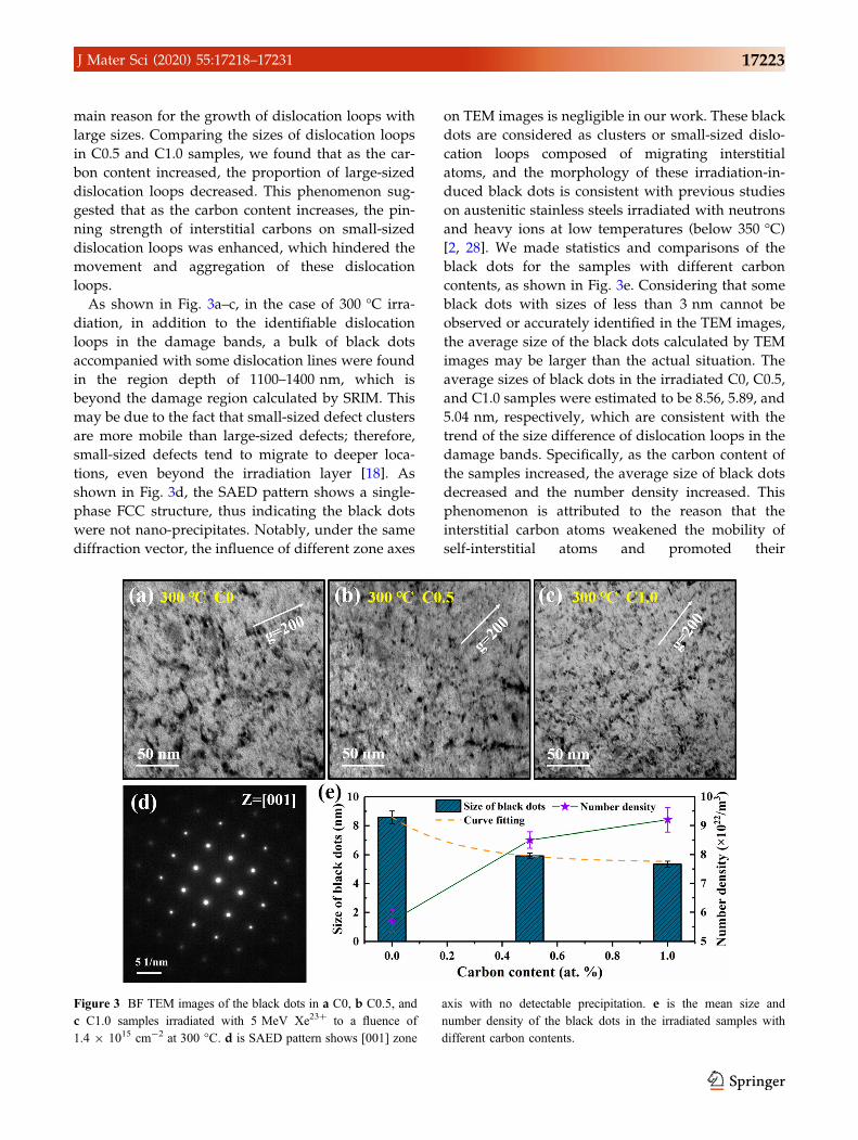
As shown in Fig. 3a–c, in the case of 300 �C irra-
diation, in addition to the identifiable dislocation
loops in the damage bands, a bulk of black dots
accompanied with some dislocation lines were found
in the region depth of 1100–1400 nm, which is
beyond the damage region calculated by SRIM. This
may be due to the fact that small-sized defect clusters
are more mobile than large-sized defects; therefore,
small-sized defects tend to migrate to deeper loca-
tions, even beyond the irradiation layer [18]. As
shown in Fig. 3d, the SAED pattern shows a single-
phase FCC structure, thus indicating the black dots
were not nano-precipitates. Notably, under the same
diffraction vector, the influence of different zone axes
on TEM images is negligible in our work. These black
dots are considered as clusters or small-sized dislo-
cation loops composed of migrating interstitial
atoms, and the morphology of these irradiation-in-
duced black dots is consistent with previous studies
on austenitic stainless steels irradiated with neutrons
and heavy ions at low temperatures (below 350 �C)[2, 28]. We made statistics and comparisons of the
black dots for the samples with different carbon
contents, as shown in Fig. 3e. Considering that some
black dots with sizes of less than 3 nm cannot be
observed or accurately identified in the TEM images,
the average size of the black dots calculated by TEM
images may be larger than the actual situation. The
average sizes of black dots in the irradiated C0, C0.5,
and C1.0 samples were estimated to be 8.56, 5.89, and
5.04 nm, respectively, which are consistent with the
trend of the size difference of dislocation loops in the
damage bands. Specifically, as the carbon content of
the samples increased, the average size of black dots
decreased and the number density increased. This
phenomenon is attributed to the reason that the
interstitial carbon atoms weakened the mobility of
self-interstitial atoms and promoted their
Figure 3 BF TEM images of the black dots in a C0, b C0.5, and
c C1.0 samples irradiated with 5 MeV Xe23? to a fluence of
1.4 9 1015 cm-2 at 300 �C. d is SAED pattern shows [001] zone
axis with no detectable precipitation. e is the mean size and
number density of the black dots in the irradiated samples with
different carbon contents.
J Mater Sci (2020) 55:17218–17231 17223

annihilation during the 3D migration path, reducing
the number of migratable defects. However, specific
numerical values of the migration energy of self-in-
terstitial atoms in the samples with different carbon
contents are difficult to provide, thus requiring fur-
ther verification by simulation methods.
As shown in Fig. 4a–c, typical large-sized 1/3
\111[ faulted loops are distributed in the damage
bands of the samples irradiated at 500 �C. The aver-
age sizes of the dislocation loops in C0, C0.5, and C1.0
samples were estimated to be 17.5, 15.8, and 15.2 nm,
respectively, and the trend of this result is similar to
that in the case of 300 �C irradiation. However, in the
case of 500 �C irradiation, the difference in average
defect sizes of the C0.5 and C1.0 samples was very
small because the pinning effects of interstitial carbon
on irradiation-induced dislocation loops were affec-
ted by temperature. Specifically, when the irradiation
temperature is high enough, interstitial carbon atoms
begin to migrate [29], causing the invalidation of the
pinning effect of interstitial carbon on dislocation
loops. Moreover, the elevated temperature intensified
the thermal motion of molecules and reduced the
migration energy of irradiation defects, which may
result in the unpinning of many dislocation loops. In
this case, the content of interstitial carbon would no
longer influence the pinning strength of the disloca-
tion loops, greatly weakening the response of the
dislocation loop size to the carbon content.
Stacking fault tetrahedron (SFT) is a kind of
vacancy-type and stable defect in FCC crystal, which
is formed by the migration of vacancy clusters or the
evolution of Frank loops. As shown in Figs. 5a, b,
SFTs were observed in the C0 samples irradiated at
300 �C and 500 �C, while no SFT was observed in the
C0.5 and C1.0 samples at both 300 �C and 500 �Cirradiation temperatures. This may be attributed to
the reason that interstitial carbon atoms had inhibi-
tory effects on the above two formation mechanisms
of SFTs. Firstly, interstitial carbon atoms could com-
bine with the vacancies to form C-vacancy com-
plexes, thereby inhibiting the accumulation and
movement of vacancies. Reportedly, the vacancies in
the austenitic steel can stably bind two carbon atoms
Figure 4 Enlarged BF TEM images for the damage peak regions
of a C0, b C0.5, and c C1.0 samples irradiated with 5 MeV Xe23?
to a fluence of 1.4 9 1015 cm-2 at 500 �C. d is SAED patterns,
showing the two-beam diffracting condition of TEM images: all
the images were taken near the [011] zone axis with g = 200. e is
statistical result for the size distribution of dislocation loops in C0,
C0.5, and C1.0 samples based on at least 10 TEM images.
17224 J Mater Sci (2020) 55:17218–17231
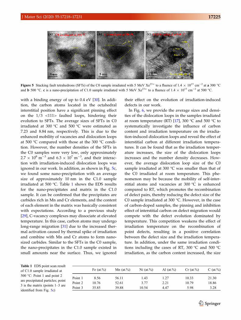
with a binding energy of up to 0.4 eV [30]. In addi-
tion, the carbon atoms located in the octahedral
interstitial position have a significant pinning effect
on the 1/3 \111[ faulted loops, hindering their
evolution to SFTs. The average sizes of SFTs in C0
irradiated at 300 �C and 500 �C were estimated as
7.23 and 8.84 nm, respectively. This is due to the
enhanced mobility of vacancies and dislocation loops
at 500 �C compared with those at the 300 �C condi-
tion. However, the number densities of the SFTs in
the C0 samples were very low, only approximately
2.7 9 108 m-3 and 6.3 9 107 m-3, and their interac-
tion with irradiation-induced dislocation loops was
ignored in our work. In addition, as shown in Fig. 5c,
we found some nano-precipitation with an average
size of approximately 10 nm in the C1.0 sample
irradiated at 500 �C. Table 1 shows the EDS results
for the nano-precipitates and matrix in the C1.0
sample. It can be confirmed that the precipitates are
carbides rich in Mn and Cr elements, and the content
of each element in the matrix was basically consistent
with expectations. According to a previous study
[29], C-vacancy complexes may dissociate at elevated
temperature. In this case, carbon atoms may undergo
long-range migration [31] due to the increased ther-
mal activation caused by thermal spike of irradiation
and combine with Mn and Cr atoms to form nano-
sized carbides. Similar to the SFTs in the C0 sample,
the nano-precipitates in the C1.0 sample existed in
small amounts near the surface. Thus, we ignored
their effect on the evolution of irradiation-induced
defects in our work.
In Fig. 6, we provide the average sizes and densi-
ties of the dislocation loops in the samples irradiated
at room temperature (RT) [17], 300 �C and 500 �C to
systematically investigate the influence of carbon
content and irradiation temperature on the irradia-
tion-induced dislocation loops and reveal the effect of
interstitial carbon at different irradiation tempera-
tures. It can be found that as the irradiation temper-
ature increases, the size of the dislocation loops
increases and the number density decreases. How-
ever, the average dislocation loop size of the C0
sample irradiated at 300 �C was smaller than that of
the C0 irradiated at room temperature. This phe-
nomenon may be because the mobility of self-inter-
stitial atoms and vacancies at 300 �C is enhanced
compared to RT, which promotes the recombination
of defect pairs, thereby reducing the defect size of the
C0 sample irradiated at 300 �C. However, in the case
of carbon-doped samples, the pinning and inhibition
effect of interstitial carbon on defect migration would
compete with the defect evolution dominated by
temperature. This competition weakens the effect of
irradiation temperature on the recombination of
point defects, resulting in a positive correlation
between the defect size and the irradiation tempera-
ture. In addition, under the same irradiation condi-
tions including the cases of RT, 300 �C and 500 �Cirradiation, as the carbon content increased, the size
Figure 5 Stacking fault tetrahedrons (SFTs) of the C0 sample irradiated with 5 MeV Xe23? to a fluence of 1.4 9 1015 cm-2 at a 300 �Cand b 500 �C. c is a nano-precipitation of C1.0 sample irradiated with 5 MeV Xe23? to a fluence of 1.4 9 1015 cm-2 at 500 �C.
Table 1 EDS point scan result
of C1.0 sample irradiated at
500 �C: Point 1 and point 2
are precipitated particles; point
3 is the matrix (points 1–3 are
identified from Fig. 5c)
Fe (at.%) Mn (at.%) Ni (at.%) Al (at.%) Cr (at.%) C (at.%)
Point 1 8.56 56.11 1.43 1.27 10.33 21.30
Point 2 10.76 52.61 3.77 2.21 10.79 18.86
Point 3 35.85 39.88 10.54 4.47 5.98 3.28
J Mater Sci (2020) 55:17218–17231 17225

of the dislocation loops decreased and the number
density increased, proving that interstitial carbon
atoms inhibit the growth of irradiation-induced
defects and improve the irradiation resistance of
carbon-doped HEAs.
Irradiation-induced swelling
Irradiation-induced swelling of material is remark-
able at elevated temperature (0.3–0.6 Tm, where Tm is
the melting point of the material), negatively affect-
ing the performance of structural materials in nuclear
applications. According to previous reports [32, 33],
there are two causes of irradiation swelling: void-in-
duced swelling and bubble-induced swelling. The
degree of swelling strongly depends on the density
and size of the voids or bubbles. Considering that the
Xe irradiation fluence of all the samples is
1.4 9 1015 cm-2 and no Xe bubbles were observed in
the TEM images, the swelling of the irradiated sam-
ples in our work is considered to be caused by the
voids and the migration of interstitial atoms. In
addition, Terasawa et al. [32] reported that GBs
exhibit a different step structure from matrix under
N? irradiation at 773 K, which could reflect the irra-
diation swelling of the sample. This is because the
GBs absorb a large number of interstitials and
vacancies and prompt them to annihilate there,
resulting in no evolution of irradiation-induced
defects at GBs [34]. On the basis of the above research
results, we explored the swelling at the GBs of the
samples with different carbon contents after Xe23?
irradiation at elevated temperature, so as to explain
the effect of carbon doping on irradiation-induced
swelling. However, we did not find any
detectable swelling of the samples irradiated at
300 �C (* 0.25 Tm). Thus, we only provide the
swelling of the samples irradiated at 500 �C.Figure 7a–c shows 3D AFM images for the GBs of
the C0, C0.5, and C1.0 samples irradiated at 500 �C.Among them, the illustrations are corresponding
cross-sectional data of the scanning line in the AFM
images. Clearly, the characteristic grooves at the GBs
were observed in all the samples, while different
volume swelling appeared on both sides of the GBs in
each sample. For ease of expression, we defined the
average height above the both sides of grooves as the
GBs’ step height (HGBs), as shown in Fig. 7d. To avoid
accidental experimental data, we used the above
method to test 10 different GBs of each sample and
calculated the average swelling height. The average
swelling heights of the C0, C0.5, and C1.0 sample
were estimated to be 40.2 nm, 32.5 nm, and 13.3 nm,
respectively. Furthermore, on the basis of the calcu-
lation result of SRIM and TEM observation, the depth
of irradiated region (hirr) was determined to be
approximately 1250 nm. Thus, the degrees of swel-
ling at GBs (Dh = HGBs/hirr 9 100%) were calculated
to be 3.22%, 2.60%, and 1.06% for the C0, C0.5, and
C1.0 samples, respectively. Notably, the swelling
degree of C1.0 sample dropped to 33% of the C0
sample, suggesting that the volume swelling of the
sample was suppressed under Xe23? irradiation at
500 �C with increasing the carbon concentration. We
Figure 6 a Mean size and
b number density of
irradiation-induced dislocation
loops of the samples with
different carbon contents
under room temperature (RT)
[17], 300 �C and 500 �Cirradiation conditions. The
irradiation fluence of all the
above samples is
1.4 9 1015 cm-2, which
corresponds to the peak
damage dose of 3.8 dpa. The
images and data of the
irradiated samples at RT are
found in Supplementary
material.
17226 J Mater Sci (2020) 55:17218–17231

estimated the Dh/dpa (%/dpa) values of the irradi-
ated C0, C0.5, and C1.0 samples as 0.85, 0.68, and
0.28, respectively.
According to previous studies, there are two pos-
sible factors affecting the void-induced swelling.
Mansur et al. [35] proposed a key parameter
expression affecting the growth of voids, as shown in
Eq. (1), which considers the sink strength of defects.
dR=dt¼XDiDV
2RKVi
1þ 4KVi K0
DiDVSiSV
� �1=2
�1
" #ZVSV�ZiSið Þ
ð1Þ
where R is the radius of voids, X is the atomic vol-
ume, and Si and SV are sink strengths of interstitials
and vacancies.Di and DV are the diffusion coefficients
of interstitials and vacancies. K0 and ðZV�ZiÞ are the
temperature-independent formation rate of defects
and the dislocation bias factor, respectively. KVi is the
recombination or annihilation rate of point defects. In
this case, according to the sign of ZVSV�ZiSið Þ in
Eq. (1), the voids may be growing or shrinking. Fur-
thermore, the greater the sink strength (Si and SV) is,
the slower the swelling rate is, because point defects
are absorbed by the sinks rather than being voids.
Interstitial carbon can be considered as absorption
sinks for self-interstitials and vacancies. On the one
hand, it is because carbon atoms provide the sites
where self-interstitials or clusters nucleate as dislo-
cation loops, thereby absorbing lots of self-intersti-
tials. On the other hand, interstitial carbon could
combine with vacancies to form C-vacancy com-
plexes and hinder the movement of vacancies. The
experimental results and theoretical analysis above
indicate that interstitial carbon could increase the
sink strength of HEAs; that is, it absorbs self-inter-
stitials atoms and vacancies and promotes their
recombination and annihilation. However, a bulk of
black dots were observed in TEM images, which are
considered as interstitial clusters or small-sized dis-
location loops. Therefore, if we consider the influence
of interstitial clusters and their mobility on Eq. (1),
Figure 7 3D AFM images for the GBs of the a C0, b C0.5, and
c C1.0 samples irradiated at 500 �C. The insets in a–c are the data
result corresponding to the scanning line. d shows the swelling
height and degree of swelling of the samples with different carbon
contents irradiated at 500 �C.
J Mater Sci (2020) 55:17218–17231 17227

then the equation can be transformed into (2) and (3)
[36, 37].
dS=dt ¼ DvCvzvv
� �k2v �DgCgxgkgpR
2vqv ð2Þ
dS=dt ¼ Keff0
�egi
�ZVv k
2v
ZVv k
2v þ Zd
vqd� pR2
VqVkg
�
þ ð1� egi ÞPl
ZVv k
2vZ
dvqd
ZVi k
2v þ Zd
i qd� �
ZVv k
2v þ Zd
vqd
�� �
ð3Þ
where Dg and Cg are the diffusion coefficient and
concentration of interstitial clusters. xg is the size of
the cluster. The perturbation of cluster recombination
is ignored, and Eq. (2) is expanded to obtain Eq. (3).
There are three key parameters in Eq. (3), which are
the efficiency of Frenkel pairs Keff0 , the proportion of
the 1D migration of interstitial clusters egi , and the
proportion of clusters that migrate in 3D pathð1� egi Þ.Considering the same irradiation condition and
similar alloy components, the efficiency of defect
pairs remains unchanged in our work. Under this
premise, the factor affecting swelling rate is the
migration (1D or 3D path) of interstitial clusters. In
HEAs, most interstitial clusters exhibit 3D migra-
tions, which can also be confirmed in the TEM results
in our work. Thus, the value of egi is negligible. The
cluster size observed in TEM decreases with
increasing carbon content, indicating that the
absorption intensity of voids to point defects (ZVi k
2v) is
improved. Moreover, no voids were found in all the
irradiated samples, which means that the void bias of
all the samples can be considered as the same value.
Therefore, the value of the last term on the right side
of Eq. (3) decreases as the carbon content increases,
indicating the decrease in swelling rate. In this way,
we explain why the sample swelling rate decreases
with the increasing carbon content from a theoretical
perspective.
Carbon doping would increase the migration
energy of point defects, which is conducive to the
nucleation but not to the growth of dislocation loops.
Studies have shown that the reduced mobility of
dislocation loops would suppress irradiation swel-
ling [38, 39]. Considering the inherent sluggish
diffusion effect in HEAs, carbon doping may further
aggravate lattice distortion and significantly reduce
the migration rate of interstitial atoms. Therefore, a
large number of uncaptured interstitial atoms would
provide numerous recombination positions for
vacancies, promoting the annihilation of interstitial
atoms and vacancies. Meanwhile, octahedral inter-
stitial carbon atoms have a pinning effect on irradi-
ation-induced interstitial atom clusters and
dislocation loops, which hinders the accumulation
and growth of irradiation-induced defects. Further-
more, interstitial carbon atoms could combine with
vacancies and form C-vacancy complexes, which
would inhibit the evolution of vacancies and hinder
the growth of voids even at elevated temperatures.
As shown in Fig. 8, it is believed that the recombi-
nation of the defects significantly reduces the number
of interstitial atoms migrating to the surface, and the
pinning effect of interstitial carbon on clusters inhi-
bits the growth of irradiation-induced defects,
thereby reducing the swelling of the carbon-doped
HEAs irradiated at 500 �C in our work.
Figure 8 Schematic of the evolution of irradiation-induced
defects in HEAs with and without carbon doping. The case of
undoped HEA is shown on the left, and the carbon-doped HEAs
are on the right. The orange balls on the right represent interstitial
carbon atoms.
17228 J Mater Sci (2020) 55:17218–17231

Conclusion
In this study, the irradiation-induced defects and
swelling in Fe38Mn40Ni11Al4Cr7 HEAs with different
carbon contents were compared at 300 �C and 500 �Cwith 5 MeV Xe23? ions irradiated to a fluence of
1.4 9 1015 cm-2. TEM and AFM were used to pro-
vide essential evidence. On this basis, we revealed
the effects of interstitial carbon on the irradiation
resistance of HEAs at elevated temperature and dis-
cussed the evolution of irradiation-induced defects
and swelling of carbon-doped HEAs at different
temperatures. This study shows the positive effects of
carbon doping on irradiation resistance and provides
theoretical and experimental bases for the potential of
carbon-doped HEAs as reactor structural materials.
The specific research conclusions are as follows:
1. Both in the case of 300 �C and 500 �C irradiation,
carbon-doped HEAs exhibited smaller and den-
ser irradiation-induced dislocation loops than the
undoped HEAs. This phenomenon may be
attributed to the pinning effect of interstitial
carbon on dislocation loops, which hinders the
accumulation and growth of irradiation defects.
2. In the case of HEAs with the same carbon
content, elevated temperature would promote
the growth of irradiation-induced dislocation
loops, resulting in the larger-sized and lower-
density dislocation loops in HEAs irradiated at
500 �C than those under the 300 �C irradiation
condition.
3. At a low irradiation temperature (300 �C), the
inhibitory effects of interstitial carbon on the
dislocation loops are mainly manifested in pro-
moting the recombination of defects and the
pinning effect on dislocation loops. However, in
the case of 500 �C irradiation, the pinning effect
of interstitial carbon is weakened due to the
increased mobility of carbon atoms at elevated
temperature, resulting in the nearly constant size
of dislocation loops with the changing carbon
content.
4. Under the 500 �C irradiation condition, swelling
was observed on both sides of the grain bound-
aries in the irradiated samples, while no
detectable swelling was found at 300 �C irradia-
tion. The swelling at 500 �C decreased with the
increase in carbon content. This is attributed to
the reason that interstitial carbon atoms hindered
the long-distance migration of self-interstitials,
promoted the recombination of Frenkel pairs, and
reduced the number of self-interstitials that can
move to the surface and cause swelling.
Acknowledgements
This work is supported from the National Natural
Science Foundation of China (Grant No. 11705087),
the Natural Science Foundation of Jiangsu Province
(Grant No. BK20170776), and the project supported
by the Foundation of Graduate Innovation Center in
NUAA (Grant No. kfjj20190604). In addition, the
authors would like to thank Senior engineer Niu of
Suzhou Institute of Nano-Tech and Nano-Bionics
(CAS) for his help in TEM characterization. We are
grateful to the Institute of Modern Physics (CAS) for
providing irradiation experiments.
Electronic supplementary material: The online
version of this article (https://doi.org/10.1007/s108
53-020-05229-7) contains supplementary material,
which is available to authorized users.
References
[1] Jin K, Lu C, Wang L, Qu J, Weber WJ, Zhang Y, Bei H
(2016) Effects of compositional complexity on the ion-irra-
diation induced swelling and hardening in Ni-containing
equiatomic alloys. Scripta Mater 119:65–70
[2] Lin JW, Chen FD, Tang XB, Liu J, Shen SK, Ge GJ (2020)
Radiation-induced swelling and hardening of 316L stainless
steel fabricated by selected laser melting. Vacuum
174:109183
[3] Holmes JJ, Robbins RE, Brimhall JL, Mastel B (1968)
Elevated temperature irradiation hardening in austenitic
stainless steel. Acta Mater 16(7):955–967
[4] Was GS, Ashida Y, Andresen PL (2011) Irradiation-assisted
stress corrosion cracking. Corros Rev 29:7–49
[5] Tsai MH, Yeh JW (2014) High-entropy alloys: a critical
review. Mater Res Lett 2(3):107–123
[6] Zhang WR, Liaw PK, Zhang Y (2018) Science and tech-
nology in high-entropy alloys. Sci China Mater 61(1):2–22
[7] Yang L, Ge H, Zhang J, Xiong T, Jin QQ, Zhou Y, Shao XH,
Zhang B, Zhu Z, Zheng SJ, Ma XL (2019) High He-ion
irradiation resistance of CrMnFeCoNi high-entropy alloy
J Mater Sci (2020) 55:17218–17231 17229

revealed by comparison study with Ni and 304SS. J Mater
Sci Technol 35(3):300–305
[8] Egami T, Guo W, Rack PD, Nagase T (2014) Irradiation
Resistance of Multicomponent Alloys. Metall Mater Trans A
45(1):180–183
[9] Yang T, Xia S, Guo W, Hu R, Poplawsky JD, Sha G, Fang Y,
Yan Z, Wang C, Li C et al (2018) Effects of temperature on
the irradiation responses of Al 0.1 CoCrFeNi high entropy
alloy. Scripta Mater 144:31–35
[10] Yang TN, Lu CY, Velisa G, Jin K, Xiu PY, Zhang YW, Bei
HB, Wang LM (2019) Influence of irradiation temperature
on void swelling in NiCoFeCrMn and NiCoFeCrPd. Scripta
Mater 158:57–61
[11] Yang TF, Xia SQ, Liu S, Wang CX, Liu SS, Fang Y, Zhang
Y, Xue JM, Yan S, Wang YG (2016) Precipitation behavior
of AlxCoCrFeNi high entropy alloys under ion irradiation.
Sci Rep 6:32146
[12] Guo Z, Zhang A, Han J, Meng J (2019) Effect of Si additions
on microstructure and mechanical properties of refractory
NbTaWMo high-entropy alloys. J Mater Sci
54(7):5844–5851. https://doi.org/10.1007/s10853-018-0328
0-z
[13] Stepanov ND, Shaysultanov DG, Chernichenko RS, Yurch-
enko NY, Zherebtsov SV, Tikhonovsky MA, Salishchev GA
(2017) Effect of thermomechanical processing on
microstructure and mechanical properties of the carbon-
containing CoCrFeNiMn high entropy alloy. J Alloys
Compd 693:394–405
[14] Wang Z, Bei H, Baker I (2018) Microband induced plasticity
and the temperature dependence of the mechanical properties
of a carbon-doped FeNiMnAlCr high entropy alloy. Mater
Charact 139:373–381
[15] Li JB, Gao B, Tang S, Liu B, Liu Y, Wang Y, Wang JW
(2018) High temperature deformation behavior of carbon-
containing FeCoCrNiMn high entropy alloy. J Alloys
Compd 747:571–579
[16] Lu E, Makkonen I, Mizohata K, Li Z, Raisanen J, Tuomisto
F (2020) Effect of interstitial carbon on the evolution of
early-stage irradiation damage in equi-atomic FeMnNiCoCr
high-entropy alloys. J Appl Phys 127:025103
[17] Shen S, Chen F, Tang X, Lin J, Ge G, Liu J (2020) Effects of
carbon doping on irradiation resistance of Fe38Mn40-
Ni11Al4Cr7 high entropy alloys. J Nucl Mater 540:152380
[18] Lu CY, Niu LL, Chen NJ, Jin K, Yang TN, Xiu PY, Zhang
YW, Gao F, Bei HB, Shi S, He MR, Robertson IM, Weber
WJ, Wang LM (2016) Enhancing radiation tolerance by
controlling defect mobility and migration pathways in mul-
ticomponent single-phase alloys. Nat Commun 7:13564
[19] Wolff J, Franz M, Kluin JE, Schmid D (1997) Vacancy
formation in nickel and a-nickel-carbon alloy. Acta Mater
45(11):4759–4764
[20] Cermak J, Kral L (2014) Effect of carbon-supersaturation
upon the carbon diffusion in 9Cr–1Mo ferrite steel. Mater
Lett 116:402–404
[21] Kresse T, Borchers C, Kirchheim R (2013) Vacancy–carbon
complexes in bcc iron: correlation between carbon content,
vacancy concentration and diffusion coefficient. Scripta
Mater 69(9):690–693
[22] Wang Z, Baker I, Cai Z, Chen S, Poplawsky JD, Guo W
(2016) The effect of interstitial carbon on the mechanical
properties and dislocation substructure evolution in Fe40.4-Ni11.3Mn34.8Al7.5Cr6 high entropy alloys. Acta Mater
120:228–239
[23] Wang Z, Baker I (2016) Interstitial strengthening of a f.c.c.
FeNiMnAlCr high entropy alloy. Mater Lett 180:153–156
[24] Ehrhart P, Averback RS (1989) Diffuse X-ray scattering
studies of neutron- and electron-irradiated Ni, Cu and dilute
alloys. Philos Mag A 60(3):283–306
[25] Theis U, Wollenberger H (1980) Mobile interstitials pro-
duced by neutron irradiation in copper and aluminium.
J Nucl Mater 88(1):121–130
[26] Tsai KY, Tsai MH, Yeh JW (2013) Sluggish diffusion in Co–
Cr–Fe–Mn–Ni high-entropy alloys. Acta Mater
61(13):4887–4897
[27] Bae DS, Nahm SH, Lee HM, Kinoshita H, Shibayama T,
Takahashi H (2004) Effect of electron-beam irradiation
temperature on irradiation damage of high Mn–Cr steel.
J Nucl Mater 329–333:1038–1042
[28] Zinkle SJ, Maziasz PJ, Stoller RE (1993) Dose dependence
of the microstructural evolution in neutron-irradiated auste-
nitic stainless steel. J Nucl Mater 206(2–3):266–286
[29] Terentyev D, Martin-Bragado I (2015) Evolution of dislo-
cation loops in iron under irradiation: the impact of carbon.
Scripta Mater 97:5–8
[30] Hepburn DJ, Ferguson D, Gardner S, Ackland GJ (2013)
First-principles study of helium, carbon, and nitrogen in
austenite, dilute austenitic iron alloys, and nickel. Phys Rev
B 88(2):024115
[31] Arakawa K, Hatanaka M, Mori H, Ono K (2004) Effects of
chromium on the one-dimensional motion of interstitial-type
dislocation loops in iron. J Nucl Mater 329–333:1194–1198
[32] Terasawa M, Mitamura T, Liu L, Tsubakino H, Niibe M
(2002) Metal surface swelling by heavy charged particle
irradiation. Nucl Instrum Methods B 193:329–335
[33] Little EA, Stow DA (1979) Void-swelling in irons and fer-
ritic steels: II. An experimental survey of materials irradiated
in a fast reactor. J Nucl Mater 87(1):25–39
17230 J Mater Sci (2020) 55:17218–17231
[34] Shimada M, Nakahigashi S, Terasawa M (1975) On the
surface denudation of voids induced by 200 keV C? ion
irradiation. Radiat Eff Defects Solids 25(4):283–285
[35] Mansur LK (1994) Theory and experimental background on
dimensional changes in irradiated alloys. J Nucl Mater
216:97–123
[36] Singh BN, Golubov SI, Trinkaus H, Serra A, Osetsky YN,
Barashev AV (1997) Aspects of microstructure evolution
under cascade damage conditions. J Nucl Mater
251:107–122
[37] Golubov SI, Singh BN, Trinkaus H (2000) Defect accumu-
lation in fcc and bcc metals and alloys under cascade damage
conditions: Towards a generalisation of the production bias
model. J Nucl Mater 276(1):78–89
[38] Terentyev D, Olsson P, Malerba L, Barashev AV (2007)
Characterization of dislocation loops and chromium-rich
precipitates in ferritic iron-chromium alloys as means of void
swelling suppression. J Nucl Mater 362(2):167–173
[39] Garner FA, Toloczko MB, Sencer BH (2000) Comparison of
swelling and irradiation creep behavior of fcc-austenitic and
bcc-ferritic/martensitic alloys at high neutron exposure.
J Nucl Mater 276(1):123–142
Publisher’s Note Springer Nature remains neutral with
regard to jurisdictional claims in published maps and
institutional affiliations.
J Mater Sci (2020) 55:17218–17231 17231