Iodine- 131-Labeled MAb F(ab ג€˜)2Fragments Are More Efficient ...
11
therapy trials of hepatocarcinoma, melanoma, colon carcinoma, and lymphoma (1—9). We and others have previously shown that iodine labeled F(ab')2 fragments of MAbs injected in tumor bearing nude mice can give higher tumor-to-normal tissue ratios than their intact antibody counterparts (10—13).F(ab')2 fragments may therefore offer an ad vantage over intact antibodies for radioimmunotherapy (14—1 7). Another theoretical advantage of F(ab')2 frag ments is the absence of the Fc portion, which might lead to decreased radioactivity accumulation in the reticuloendothelial system. We have evaluated the therapeutic value ofa pool of 131I-labeledMAt, F(ab')2 in nude mice bearing large human colon carcinoma and compared its efficiency with that of pooled, corresponding ‘31I-labeled intact MAbs. METHODS Duringoneweek,beginning18 daysaftertransplantation, nude mice bearing human colon carcinoma ranging from 115to943mm3(mean335mm3)weretreatedbyrepeated intravenousinjections of either iodine-131-(131l) labeled intactantibodiesor1311-labeled correspondingF(ab')@ frag ments of a pool of four monoclonalantibodies(MAbs) directedagainstdistinctepitopesof carcinoembryonic an tigen (CEA). Complete tumor remission was observed in 8 of 10 miceaftertherapywith F(ab')@ and 6 of the animals survived 10 mo in good health. In contrast, after treatment with intact MAbs, tumors relapsed in 7 of 8 mice after remission periods of 1 to 3.5 mo despite the fact that body weightlossanddepressionof peripheral whitebloodcells, symptomsofradiationtoxicity,andthecalculatedradiation dosesfor liver,spleen,bone,andbloodwereincreasedor equal in these animals as compared to mice treated with F(ab')@. J NucIMed 1990;31:1035—1044 dioimmunotherapy using antibodies labeled with isotopes such as iodine-l3 1 (‘@‘I) or yttrium-90 (@°Y), which emit medium- to high-energy beta-particles, of fers the theoretical advantage that these electron parti des can penetrate and damage several cell layers within a solid tumor. Thus, due to the crossfire of the beta radiation, cells expressing low amounts of antigen or cells which are inaccessible to antibodies can be lethally hit by electrons emitted from distant monoclonal anti bodies (MAbs). Polyclonal antibodies and MAbs car rying such isotopes have been used in clinical tumor ReceivedJun. 2, 1989; revision accepted Jan. 16, 1990. Forreprintscontact:F.Buchegger, MD,InstituteofBiochemistry, CH 1066 Epalinges,Switzerland. Monoclonal Antibodies Intact antibodies and F(ab')2 fragments offour MAbS(MAb 35, CE25-B7, B17, and B93) (18, 19) directed against four independent epitopes ofcarcinoembryonic antigen (CEA) (ep itopes Gold 1—4, (20)) have been selected according to criteria required for therapeutic injection of patients (21—23). These criteria include: high binding rate to purified CEA and no binding to: 1. Cross-reacting antigens NCA-55 and NCA-95 (24). 2. Biliary glycoprotein (25). 3. Fresh human granulocytes (26). The ‘31I-labeled individualantibodieslocalizeequallywell within human colon carcinoma xenografts yielding high tu mor-to-whole body ratios ranging from 25 to 40 and from 35 to 50 for intact MAbS and F(ab')2, respectively (27). Radioimmunotherapy with 131l-MAb F(ab')@ • Bucheggeretal 1035 Iodine- 131-Labeled MAb F(ab ‘)2 Fragments Are More Efficient and Less Toxic Than Intact Anti-CEA Antibodies in Radioimmunotherapy of Large Human Colon Carcinoma Grafted in Nude Mice Franz Buchegger, AndréPèlegrin,Bernard Delaloye, Angeika Bischof-Delaloye, and Jean-Pierre Mach Institute ofBiochemistry, University ofLausanne; Swiss Institutefor Experimental Cancer Research, Epalinges, Switzerland; and Division ofNuclear Medicine, Centre Hospitalier Universitaire Vaudois, Lausanne, Switzerland by on February 20, 2018. For personal use only. jnm.snmjournals.org Downloaded from
Transcript of Iodine- 131-Labeled MAb F(ab ג€˜)2Fragments Are More Efficient ...

therapy trials of hepatocarcinoma, melanoma, coloncarcinoma, and lymphoma (1—9).
We and others have previously shown that iodinelabeled F(ab')2 fragments of MAbs injected in tumorbearing nude mice can give higher tumor-to-normaltissue ratios than their intact antibody counterparts(10—13).F(ab')2 fragments may therefore offer an advantage over intact antibodies for radioimmunotherapy(14—17). Another theoretical advantage of F(ab')2 fragments is the absence of the Fc portion, which mightlead to decreased radioactivity accumulation in thereticuloendothelial system.
We have evaluated the therapeutic value ofa pool of131I-labeledMAt, F(ab')2 in nude mice bearing largehuman colon carcinoma and compared its efficiencywith that of pooled, corresponding ‘31I-labeledintactMAbs.
METHODS
Duringoneweek,beginning18 daysaftertransplantation,nude mice bearing human colon carcinoma ranging from115 to 943mm3(mean335mm3)weretreatedbyrepeatedintravenousinjectionsof either iodine-131-(131l)labeledintactantibodiesor 1311-labeledcorrespondingF(ab')@fragments of a pool of four monoclonalantibodies(MAbs)directedagainstdistinctepitopesof carcinoembryonicantigen (CEA). Complete tumor remission was observed in 8of 10 miceafter therapywith F(ab')@and6 of the animalssurvived 10 mo in good health. In contrast, after treatmentwith intact MAbs, tumors relapsed in 7 of 8 mice afterremission periods of 1 to 3.5 mo despite the fact that bodyweightlossanddepressionof peripheralwhitebloodcells,symptomsof radiationtoxicity,andthecalculatedradiationdosesfor liver,spleen,bone,andbloodwereincreasedorequal in these animals as compared to mice treated withF(ab')@.
J NucIMed 1990;31:1035—1044
dioimmunotherapy using antibodies labeled withisotopes such as iodine-l3 1 (‘@‘I)or yttrium-90 (@°Y),which emit medium- to high-energy beta-particles, offers the theoretical advantage that these electron partides can penetrate and damage several cell layers withina solid tumor. Thus, due to the crossfire of the betaradiation, cells expressing low amounts of antigen orcells which are inaccessible to antibodies can be lethallyhit by electrons emitted from distant monoclonal antibodies (MAbs). Polyclonal antibodies and MAbs carrying such isotopes have been used in clinical tumor
ReceivedJun. 2, 1989; revision accepted Jan. 16, 1990.Forreprintscontact:F.Buchegger,MD,Instituteof Biochemistry,CH
1066 Epalinges,Switzerland.
Monoclonal AntibodiesIntact antibodies and F(ab')2 fragments offour MAbS(MAb
35, CE25-B7, B17, and B93) (18, 19) directed against fourindependent epitopes ofcarcinoembryonic antigen (CEA) (epitopes Gold 1—4,(20)) have been selected according to criteriarequired for therapeutic injection of patients (21—23).Thesecriteria include: high binding rate to purified CEA and nobinding to:
1. Cross-reacting antigens NCA-55 and NCA-95 (24).2. Biliary glycoprotein (25).3. Fresh human granulocytes (26).
The ‘31I-labeledindividualantibodieslocalizeequallywellwithin human colon carcinoma xenografts yielding high tumor-to-whole body ratios ranging from 25 to 40 and from 35to 50 for intact MAbS and F(ab')2, respectively (27).
Radioimmunotherapywith 131l-MAbF(ab')@•Bucheggeret al 1035
Iodine- 131-Labeled MAb F(ab ‘)2FragmentsAre More Efficient and Less Toxic Than IntactAnti-CEA Antibodies in Radioimmunotherapyof Large Human Colon Carcinoma Grafted inNude MiceFranz Buchegger, AndréPèlegrin,Bernard Delaloye, Angeika Bischof-Delaloye, and Jean-Pierre Mach
Institute ofBiochemistry, University ofLausanne; Swiss Institutefor Experimental Cancer Research,Epalinges, Switzerland; and Division ofNuclear Medicine, Centre Hospitalier Universitaire Vaudois,Lausanne, Switzerland
by on February 20, 2018. For personal use only. jnm.snmjournals.org Downloaded from

imal percentageof tumor localizationwas reached (Ref. 14and resultsshownhere),the micewerekilledand radiolabeledantibody distribution was measured in tumor and normaltissues and expressed in percentage of injected dose per gram(%ID/g).Tumor localizationdata wereplottedagainsttumorsize and then compared with different equations on a curvefiuing program.
Therapeutic Injection of 1311-MAbs,F(ab')@andControlProteins
Eighteen days after tumor transplantation (tumor T380,passage52),30animalsbearingexponentiallygrowingtumors(transplanted the same day with the same inoculum) wererandomly distributed in five groups except for the six micewith largesttumors whichweredistributedinto the two therapy arms for being injected with either ‘3I-intactMAbs or‘31I-MAbF(ab')2. The injected baseline ‘@‘Iradioactivity wasselected in order to deliver similar radiation doses to eachtumor. Our previous experiences (14, 27) and further prelimmary results had shown that we needed four to five timesmore 1311F(ab')2than intact antibodies.In addition, sincethepercentage of ID/g of tumor decreases with increasing tumorsize (see Fig. 1), we had to adapt injected radioactivity foreach mouse followingthe formulashownbelow.
For ‘31I-intactMAt, injections, a baseline 500-@iCidose wassupplemented with 0.25 MCi/mg tumor weight (TW) according to:
Total activityintact MAb(in XCi)=
500+ 0.25x TW (in mg).
Two-thirds ofthis dose was injected on Day 18 and onethird on Day 25 after tumor transplantation.
For 131I-MAbF(ab')2injections,the formula wassimilarlychosen according to:
Total activity F(ab')2 (in @iCi)= 2,200 + 1.1 x TW (in mg).
This amount was split in three identical doses injected onDays 18, 20, and 25.
For both injections of intact MAbs and F(ab')2 by usingthese formulas, the injected dose was increasedby 50% formice bearing l-g tumors as compared to mice with very smalltumors. All the fractionated doses were injected within 1 wkbecause in patients development of human anti-mouse IgGantibodies (HAMA), often limits this time period.
The activities of ‘‘iwere calculated for mice of 30-g bodyweight. For smaller and heavier animals, the amount wascorrected according to weight difference from 30 g.
Four control mice were injected with ‘31I-n.IgGF(ab')2together with 300 @gunlabeled anti-CEA MAb F(ab')2. Theamount of microcuries@ ‘Iand of microgram proteins wascalculated as it was for ‘31I-MAbF(ab')2, and was injected inthree doses at the same times.
Three control mice were injected with ‘31I-intactn.IgGtogether with 75 @tgunlabeled intact anti-CEA MAb, as in‘311-intactMAb injections. It also was given in two doses. Thiscontrol group exhibited the strongest bone marrow depression(very low peripheral white blood cells (pWBCs) and hemoglobin) and two micedied shortlyafter BMCtransplantation.Inthe third mouse, the tumor relapsed rapidly after a shortremission period (data not shown).
Preparationof IntactMAbsand F(ab')2FragmentsThe intact four MAbs (all of the IgG1 subclass) or normal
IgG3(n.IgG) were prepared from ascites of the four hybridomas or of the IgG1-secretingmouse myeloma P3x63 byprecipitation with ammonium sulfate. Redissolved IgG waspurified by DE 52 (Whatman, Balston, UK) ion-exchangechromatography. F(ab')2 fragments were obtained from ascitesby ammonium sulfate precipitation, digestion of the redissolved sediment with pepsin (Sigma, St. Louis, MO) andpurification by chromatography on Sephadex G-150 (Pharmacia, Uppsala, Sweden) and DE 52 ion-exchange column(27). Purified antibodies (intact and F(ab')2) gave a homogeneous band on SDS-polyacrylamide gel electrophoresis (polyacrylamide 10%,(28)) including>95% of the proteins. Fortherapy and for time-course studies of tissue distribution, thefour MAbs were used as a pool, mixed together in equalamounts, either in intact form or as F(ab')2.
Labelingof MAbsand F(ab'h FragmentsBatches of 1 mg protein ofthe four pooled anti-CEA MAbS
or fragments or of control n.IgG were labeled by the chloramineT methodusing10mCiof ‘@‘I,yieldinga specificactivityof 8—9@tCi/zg.The immunoreactivity of radiolabeled intactMAbs and F(ab')2 was determined as described earlier (27):In the direct binding assay of radiolabeledMAbSto CEAinsolubilized on CNBr-Sepharose (Pharmacia) intact MAbSbound to 71.8% ±6.8% and F(ab')2 fragments to 73.6% ±6.0%. Nonspecific binding of n.IgG and F(ab')2 was <2%.Analytical chromatography of ‘311-labeledproteins on Sephadex G-200gavea singlepeak without detectableamounts ofaggregates.
Nude MouseTumorModelThe human colon carcinoma T380 was serially trans
planted subcutaneously into the right flank of 7—9-wk-oldmale “Swiss―homozygous nu/nu mice (Iffa Credo, L'Arbresle,France).Tumor T380 contains almost no necrotic areas upto large tumor sizes, is moderately differentiated and containsnumerouspseudoluminawhichare rich in CEA.From 1g oftumor -@40 @LgCEA can be extracted (29).
Mice were kept in aseptic conditions using filter papertopped cages and fed with standard, vitamin-supplemented,irradiated food. In addition, drinking water was supplementedwith a polyvitamin preparation (Protovit iv, Roche, Basel,Switzerland, 0.3 ml/300 ml during 4 days of each secondweek). Lugol iodine solution (5%) was added into drinkingwater (0.2 ml/300 ml) 3 days before and up to 6 wk afterinjection of ‘311-labeledproteins.
Eight to 10 days after inoculation of minced T380 tumor(50 mm3) ‘@-90%of the transplants started to grow exponentially and were selected for therapy.
Tumor Size Influence on Antibody LocalizationIt has been shown that the percentageof injecteddose of
antibody localization per gram of tumor is inversely relatedto the size of tumor when ‘‘‘In-labeledantibodies are used(30). We have used ‘@‘Ilabeled anti-CEA MAt, F(ab')2 toverify if this rule is also true in our model.
One hundred microgramsof anti-CEA MAb F(ab')2 together with 1.5 @Ci‘311-labeledMAb F(ab')2 were injectedintravenously into mice bearing colon carcinoma T-380 tumors of different size. Eight hours after injection, when max
1036 TheJournalof NuclearMedicine•Vol.31 •No.6 •June1990
by on February 20, 2018. For personal use only. jnm.snmjournals.org Downloaded from

The last control group consisted of five tumor-bearinganimals which were not injected.
Tumor-SizeMeasurementsThree diameters of the tumors (which can be easily moved
around subcutaneously), were measured twice a week for 50days and then once a week. Tumor volume (V) was calculatedby:
V=4/3@rXr1 xr2xr3,
where r is the tumor radius.The accuracy ofthese measurements was ±l0%—15%when
performed by different observers and by comparing resultsobtained by external tumor measurements and direct weighingof 20 dissected tumors.
ToxicityEvaluationin AntibodyTreated Mice andControls
Whole-body counting was performed immediately afterinjecting ‘3@I-intactantibodies and F(ab')2 and every 1 to 2days thereafter using a dose calibrator (Isotope Assayer 1,RADX, Houston, TX).
On the day of injection and every 2 to 3 days thereafter,mice were weighed until they recovered from initial weightloss.
On Days 7, 14, 19, 22, 27, and 43 after beginning 131!injections, blood was taken from a tail vein and peripheralpWBCswerecounted.
BoneMarrowTransplantationNude micewith pWBCfallingbelow 1000cells/mm3were
transplanted with bone marrow cells (BMC) to prevent microbial infections. Donor nude mice of the same “Swiss―geneticbackground were killed and BMC washed out from long bones(femur, tibia, and humerus) using a small volume of serumfree culture medium. About 12 x 106BMC were recoveredfrom a donor mouse and 4 x 10⁶ BMC were injected intravenously per recipient mouse. None of the mice treated with‘31I-anti-CEAMAb F(ab')2 and only one in the group treatedwith ‘311-anti-CEAintact MAbs needed BMC transplantation.In contrast, several control mice injected with ‘3I-n.IgGinintact form or as F(ab')2 had to be transplanted.
Dosimetry for Tumor and Normal OrgansCalculation ofradiation doses for tumor and normal tissues
were based on time-course studies of ‘@Itissue distribution inmice injected with trace amounts of ‘31I-MAbtogether withunlabeled antibodies either in intact form or as F(ab')2.
Twenty-seven and 18 mice were injected with 10 @iCi1311..intact anti-CEA MAb or anti-CEA MAb F(ab')2 together with50@ unlabeledintactMAbor 100 @gunlabeledMAbF(ab')2,respectively. At different times postinjection (1, 4, 8, 12, 24,48, 80, 120, and 168 hr) groups of three mice were killed,dissected, and tissue distribution of radioactivity was measured. From the amount of radioactivity measured directly atthese timepoints, an integral activity in @iCix h was calculatedper gram of tumor and normal tissues. Tissue-absorbed betaradiation for tumor, normal organs, and whole body was thencalculated according to (31):
D@= 2.13 x ,@Ci/gX h x E@rad[E@of ‘@‘I= 0. 19 g/(@Ci x h)].
An additional gamma-radiation was assumed to be equallydistributed in the whole animal. The gamma-radiation represents-@@l0%ofthe betawholebodyradiationfora 30-gmouse(27).
A further, comparable time-course study after injecting 18mice with ‘311-MAbF(ab')2 mixed with 270 @igunlabeledMAb F (ab')2 has been published by us (14).
StatisticalAnalysisResults were analyzed quantitatively and qualitatively using
the Student t-test and the x2 test, respectively. Weight dataand pWBCs measured on different days were also comparatively analyzed by a two-factors-analysis of variance.
RESULTS
Influenceof TumorSize on AntibodyLocalizationFifteen mice bearing tumors of different size were
injected with 100 sg unlabeled MAb F (ab')2 togetherwith trace amounts of ‘@‘I-(1.5 MCi)anti-CEA MAbF(ab')2. Eight hours after antibody injection, the micewere killed and tumor as well as normal tissue radioactivity distribution was measured and expressed in% ID/g. Tumor radioactivity was high in mice bearingsmall tumors (25%—38%ID/g) and exponentiallydecreased in mice with larger tumors, reaching 5.4%ID/g in a mouse bearing a 7.9-g tumor (Fig. 1). Bestcurve fitting was obtained to a line following the equation:
AY = (B + X)'
4 eTUMOR WEIGHT IN GRAMS
FIGURE 1Influenceof tumor size on antibody localization.Percent injected dose of ‘31l-anti-CEAMAb F(ab')@per gram of tumorwas determined in 15 nude mice bearing colon carcinomaT380transplantsofdifferentsizevaryingbetween0.2to7.9g. One hundred micrograms unlabeledMAb F(ab')@,mixedwith 1.5 zCi(2kg)1311-MAbF(ab')@were injectedintravenously,andtissuedistributionin the animalswas measuredat 8 hrafter injection.The percentageof ID/g of tumor (•)is plottedagainsttumor size.The regressioncurve is determinedby theequation:
53.2% ID/gtumor = .
1.44 + tumor weight (mg)
0
I-
4
Uia.UiU)000UiI-0Ui-,z
40•
30
• .•
20@a
•
• a • •10 S
S
0• , , , ,0 2 e
1037Radioimmunotherapy with 1311-MAbF(ab')@•Buchegger et al
by on February 20, 2018. For personal use only. jnm.snmjournals.org Downloaded from
where Y is the % ID/g tumor, X being tumor weightin grams, and A and B are constants with values of53.2 1 and 1.44, respectively. The coefficient of determination for this regression was very high with r =0.95. The expected localizations calculated from thisequation for infinitely small tumors, tumors of 0.3 gand of 1 g would be 36.9%, 30.6%, and 21.8% ID/g,respectively.
Tumor Size Evolution in Mice Treated with131l-Iabeled Anti-CEA MAb F(ab')@Fragments
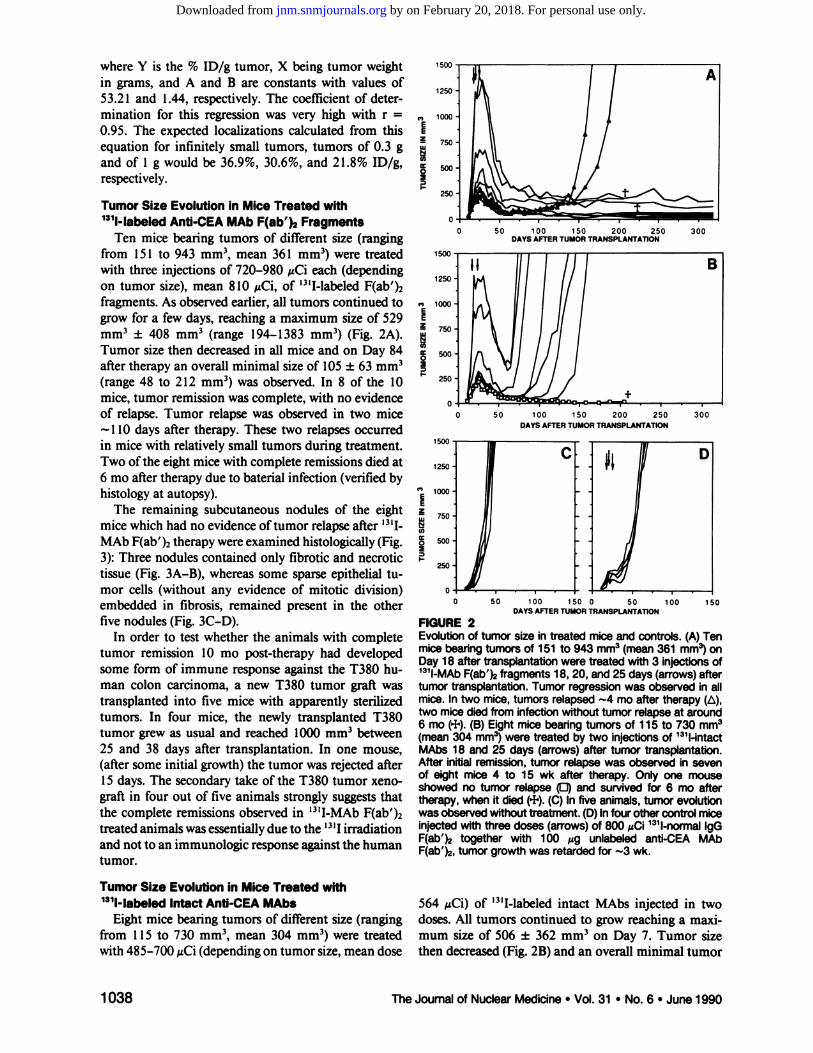
Ten mice bearing tumors of different size (rangingfrom 151 to 943 mm3, mean 361 mm3) were treatedwith three injections of 720—980 @Cieach (dependingon tumor size), mean 810 @zCi,of ‘31I-labeledF(ab')2fragments. As observed earlier, all tumors continued togrow for a few days, reaching a maximum size of 529mm3 ±408 mm3 (range 194—1383mm3) (Fig. 2A).Tumor size then decreased in all mice and on Day 84after therapy an overall minimal size of 105 ±63 mm3(range 48 to 212 mm3) was observed. In 8 of the 10mice, tumor remission was complete, with no evidenceof relapse. Tumor relapse was observed in two mice
@ 110 days after therapy. These two relapses occurredin mice with relatively small tumors during treatment.Two ofthe eight mice with complete remissions died at6moaftertherapyduetobaterialinfection(verifiedbyhistology at autopsy).
The remaining subcutaneous nodules of the eightmice which had no evidence oftumor relapse after ‘@‘IMAt, F(ab')2 therapy were examined histologically (Fig.3): Three nodulescontained only fibrotic and necrotictissue (Fig. 3A—B),whereas some sparse epitheial tumor cells (without any evidence of mitotic division)embedded in fibrosis, remained present in the otherfive nodules (Fig. 3C—D).
In order to test whether the animals with completetumor remission 10 mo post-therapy had developedsome form of immune response against the T380 human colon carcinoma, a new T380 tumor graft wastransplanted into five mice with apparently sterilizedtumors. In four mice, the newly transplanted T380tumor grew as usual and reached 1000 mm3 between25 and 38 days after transplantation. In one mouse,
(after some initial growth) the tumor was rejected after15 days. The secondary take of the T380 tumor xeno
graft in four out of five animals strongly suggests thatthe complete remissions observed in ‘311-MAbF(ab')2treated animals was essentially due to the ‘@‘Iirradiationand not to an immunologic response against the humantumor.
TumorSize Evolutionin Mice Treated with1311-IabeledIntact Anti-CEA MAbs 564 MCi) of ‘311-labeledintact MAbs injected in two
Eight mice bearing tumors of different size (ranging doses. All tumors continued to grow reaching a maxifrom 115 to 730 mm3, mean 304 mm3) were treated mum size of 506 ±362 mm3 on Day 7. Tumor sizewith 485-700 @Ci(depending on tumor size, mean dose then decreased (Fig. 2B) and an overall minimal tumor
50 100 150 200 250 300DAYS AFTER TUMOR TRANSPLANTATiON
0 50 100 150 200 250 300DAYS AFTER TUMOR TRANSPLANTATION
50 100 150 0 50DAYS AFTER TUMOR TRANSPLANTATION
C,
EE
I
I
Cl)
0
I-
0 100 150
FIGURE2Evolution of tumor size in treated mice and controls. (A) Tenmicebearingtumorsof 151to 943mm3(mean361mm@)onDay I 8 after transplantationwere treated with 3 injectionsof131I-MAbF(ab')@fragments 18, 20, and 25 days (arrows)aftertumor transplantation.Tumor regressionwas observed in allmice. In two mice, tumors relapsed @-4mo after therapy (is),two micedied from infectionwithout tumor relapseat around6 mo (+). (B) Eight mice bearingtumors of I 15 to 730 mm3(mean304 mm3)were treated by two injectionsof 131l-intactMAbs18 and25 days(arrows)aftertumortransplantation.Afterinitialremission,tumorrelapsewasobservedin sevenof eight mice4 to 15 wk after therapy.Onlyone mouseshowed no tumor relapse (0) and survived for 6 mo aftertherapy, when it died (+). (C) In five animals, tumor evolutionwasobservedwfthouttreatment.(D)lnfourothercontrolmiceinjectedwith three doses (arrows)of 800 @iCi131l-normallgGF(ab')@together with 100 @gunlabeledanti-CEAMAbF(ab')@,tumorgrowthwasretardedfor @3wk.
1038 The Journal of Nuclear Medicine •Vol. 31 •No. 6 •June 1990
by on February 20, 2018. For personal use only. jnm.snmjournals.org Downloaded from

size of 15 1 ±141 mm3 was measured 42 days afterstarting therapy. Remission lasted from 1 to 3.5 mo,
.@ then seven ofeight tumors relapsed. The time of relapse
, . depended on the initial tumor size during therapy,larger tumors relapsing earlier than smaller ones (Fig.2B). Only onemouseremainedin completeremissionand in apparent good health for 6 mo. It then died,probably from an infectious disease (infectious agentnot determined).
The difference in the number of mice without tumorrelapse 6 mo after therapy with ‘311-MAbF(ab')2 (8 of10 mice), and with ‘311-intactMAbs (1 of 8 mice) washighly significant (p < 0.005).
Tumor Size Evolution in Control MiceFive mice transplanted with colon tumor T380 on
the same day as treated animals were observed withoutany treatment (Fig. 2C). The 5 tumors grew rapidly andreached 2 g between Days 45 and 57 (mean 50 days).
Four other control mice bearing tumors of 113—202mm3 were injected with three equal doses of ‘@@800@tCi‘31I-n.IgGF(ab')2, together with 100 @gofthe unlabeledanti-CEA MAb F(ab')2 (Fig. 2D). In these animals,tumor growth was retarded by -@-3wk. All four tumorsreached 2 g between Days 64 and 81 (mean 71) aftertransplantation.
Three additional tumor-bearing mice were injectedon Days 18 and 25 after transplantation, with a total of#%@53Ø@Ci‘31I-intactn.IgG and 75 @Lgofthe pooled fourunlabeled anti-CEA MAbS. Toxic side effects in thesethree animals were very important and two animalsdied after bone marrow transplantation. Tumor growthin the remaining animal was retarded by --30 days (datanot shown).
Side Effectsof Treatmentswith 131l-intactMAbsand 131l-F(ab')2Fragments
Side effects of treatment with ‘311-labeledintact antibodies as well as with ‘31I-F(ab')2fragments includedweight loss and bone marrow depression, the latterbeing detected through decrease of pWBC counts.
Weight loss after treatment with intact antibodieswas most pronounced four days after the second injection when it was 8% (range from 3% to 11%) (Fig. 4A).
@. After treatment with F(ab')2 fragments, weight loss was
@..,4%(range 0%—7%)four daysafter the last injection.
S
t
‘4
B-@ -4-v @,.‘@ @,1@' (
‘ ‘--a...@
@ ...1' I@
..@, .@ a@@ iL.-@
@.—-#-@ . - b @: , /V‘:-@‘@@ @@:>r@―@ @j:@@@ ‘
;-,4 , t @;@ ‘@ p@
-- 9 . 10
@1,@@@ ..@@
.@ .-_;,,--@ :-@‘@‘1p*@
!@‘V@'@ ‘.@ S.,@ @‘C―@@'@ . 4 1,,.
@ ( /S #@ * i:
@ f@@ @‘:@
, (‘4 t :. @‘- @,\@@ ‘
‘.,,-@,‘ A@4@ ‘I@ .4\@.@ .@@@@ .@ @-,.@‘\; @.‘ :@
@.@@
-S,,,@@@@@ .-. . . @f .@
@*@@ j,@,.
@:;T)@‘4@'• ., @., /@ ‘.4e@' @.,.
@ . C; • ,@ •fr4@@@@ ‘@-, ,@ .@@
‘ @. ?@•@ ,.,,@@ .@
@‘;:‘(@ @.@ @,),@- :@@ ,:@ @:@-@ .@‘T;@
ti@ ( ,@ ,. 4 . :@
D.' ‘@‘@ fr. .@‘,. :.r@.
@ .@ .; .@@@
@%@@@ . .. . .-@ . ‘ -q
@@ . 1 .A.@. . , .
@@@ , t, ‘.
@@ ,,@$ , @.@ S.,-@@ ‘\@‘@ .@
;@ @@:4tt\ ,.@: :@.@.;
FIGURE 3Histology of tumor nodules remaining after treatment withF(ab')@fragments. Histologicsections of 3 @mfrom nodulesof eightmicewithouttumorrelapse10 moaftertreatmentwith 131I-MAbF(ab')@are shown.Absenceof tumor cells wasobservedin three nodules,where only fibrosis, necrosis,andsomemusclecells were seen, illustratedby imagesA and B.In C and D, a singlegroup of epitheliallike cellsembedded‘inlarge fibrosis is shown, representativeof the other five nodules. Such epithelial cells were rare in the large areas offibrosisand they showed no evidenceof cell division.
Radioimmunotherapywith 131I-MAbF(ab')2 •Buchegger et al 1039
by on February 20, 2018. For personal use only. jnm.snmjournals.org Downloaded from

28 ‘-;15 42 Hemoglobin also was decreased in treated animals as
compared to untreated mice. This decrease was muchless marked than that of pWBC. Hemoglobin was decreased to 78% ±11% after treatment with intactantibodies and to 88% ±4% after treatment withF(ab')2 fragments.
0 7 14 21
ment (total of 180 weight measurements), indicated ahighly significant difference (p <0.0002).
Peripheral WBC reached its lowest values at Days
14, 19, and 22 after beginning oftherapy in both groups(Fig. 4B). The values observed on these days should becompared with the increasing values in tumor transplanted mice which were not treated. From all treatedmice, only one animal in the group injected with intactantibodies required bone marrow transplantation because pWBC fell below 1000 cells/mm3. The lowest
) value measured in mice treated with fragments was
2500 cells/mm3.As shown for weight loss, it appears that the decrease
in pWBC counts was also more pronounced after treatment with ‘31I-intactMAbs as compared to fragments.A two-factors-analysis of variance comparing all datashown in Figure 4B (total of 108 pWBC measurements)was highly significant (p < 0.0005). However, thepWBC counts at the start of therapy are not known(they should, in principle, be similar in the two randomized groups). We, thus, conclude that the higher dosesof ‘311-F(ab')2were, at least, not more toxic than intactMAbs.
Side Effects in Control Mice Injected with 131l-n.IgGTogether with Unlabeled MAb and Comparison toUninjected Mice
In uninjected, tumor-bearing mice, some variationin the weightwasobserved(Fig. 4A). The pWBC ofthese animals increased continuously from 10.1 x l0@cells/mm3 (range 8.4 to 12.9 x l0@) on Day 25 aftertransplantation, to 18.3 x l0@cells/mm3 (range 15.2—22 x 10@)on Day44 (Fig.4B).Micekeptin the samefacility without transplanted tumors had 8.3 x i0@pWBC/mm3 (range 3.3—15.6x i0@,n = 8).
In mice injected with ‘311-n.IgGF(ab')2 together withunlabeled anti-CEA MAb F(ab')2, weight loss andpWBC decrease were slightly more pronounced as cornpared to mice treated with ‘311-anti-CEAMAb F(ab')2.The higher toxicity of ‘311-n.IgGF(ab')2 correlates witha longer whole-body half-life of radioactivity as cornpared to mice bearing tumors of similar size treatedwith ‘311-anti-CEAMAb F(ab')2 (data not shown).
In mice injected with ‘311-intactn.IgG and unlabeledanti-CEA MAb, toxicity was very high as ifiustrated byweight loss (@4@7%),and particularly pWBC counts thatfell below 1000 cells/mm3 in all three mice. Low pWBCwere paralleled by very low hemoglobin values (38%-58% when compared to untreated animals). Two ofthese mice died shortly after BMC transplantation,
6
DAYS AFTER TREATMENT
.70
EE
LIIa.Cl)
@1-IUI00
FIGURE 4Comparisonof weight loss and peripheralWBC counts in thetherapy groups and in untreated animals. All mice wereweighedimmediatelybeforeand every2 to 3 days aftertherapy(A).Weightlosswas morepronouncedinmicetreatedwith intact antibodies(meanloss 8%), ( J@, •)as comparedtomicetreatedwith F(ab')2(meanloss4%),(@,x). Untreatedanimalsalso showed some weight variations, but less pronounced (A). At 7, 14, 19, 22, 27, and 43 days after thebeginningof therapy,pWBCwerecounted(B).Miceinjectedwith131l-intactMAbs(J@) areindicatedby(•);thoseinjectedwith 131I-F(ab')@fragments (@)by (x). Decreaseof pWBC inmicetreatedwithintactantibodieswasmorepronouncedascomparedto mice treated with F(ab')@.In untreated, tumorbearingcontrolmice,pWBCswere increasingduringtheobservationtime (A). Decreaseof pWBCin miceinjectedwith131I-intactn.IgG(0) and 131l-n.IgGF(ab')@(0) was more pronouncedascomparedto anti-CEAantibodytreatedanimals.Twomiceinjectedwith intactn.IgGwerelost(+) afterbonemarrow transplantation,which had to be given to severalanimalsfromthe controlgroupsbecauseof a very low pWBC.The varianceof pWBCmeasurementsin the smallcontrolgroupswas relativelyhigh(-@.@30%of the mean).For the largerantibodytherapygroups,verticalbarsshowthes.e.m.
1040 TheJournalof NuclearMedicine•Vol.31 •No.6@ June1990
A T
@@ T@@i
“I TT@T T/@'@
TT T4
‘:@\,,Ii\@J'/' I—@@@'.L@X
k@-―*if/II8S_I@ 1/1
I
I
I@ I I I0 10 20 30 40 50
DAYS AFTER TREATMENT
280
@ 27
26'
Weight loss in mice treated with ‘31I-labeledintactMAbs and with F(ab')2 fragments was statistically compared. Mean body weight was identical in the twogroups at the beginning of therapy. Weight loss wasmore marked in mice treated with intact antibodiesalready after the first injection. The difference becamesignificant after 7 days (2p < 0.02) and remained significant thereafter for all the measurements shown inFigure 4A. The two-factors-analysis ofvariance, considering all weight data from 7 up to 57 days after treat
by on February 20, 2018. For personal use only. jnm.snmjournals.org Downloaded from
concomitant with the lowest measured hemoglobin values of38% and 41%.
DosimetryTime-course studies of the biodistribution of ‘@‘I
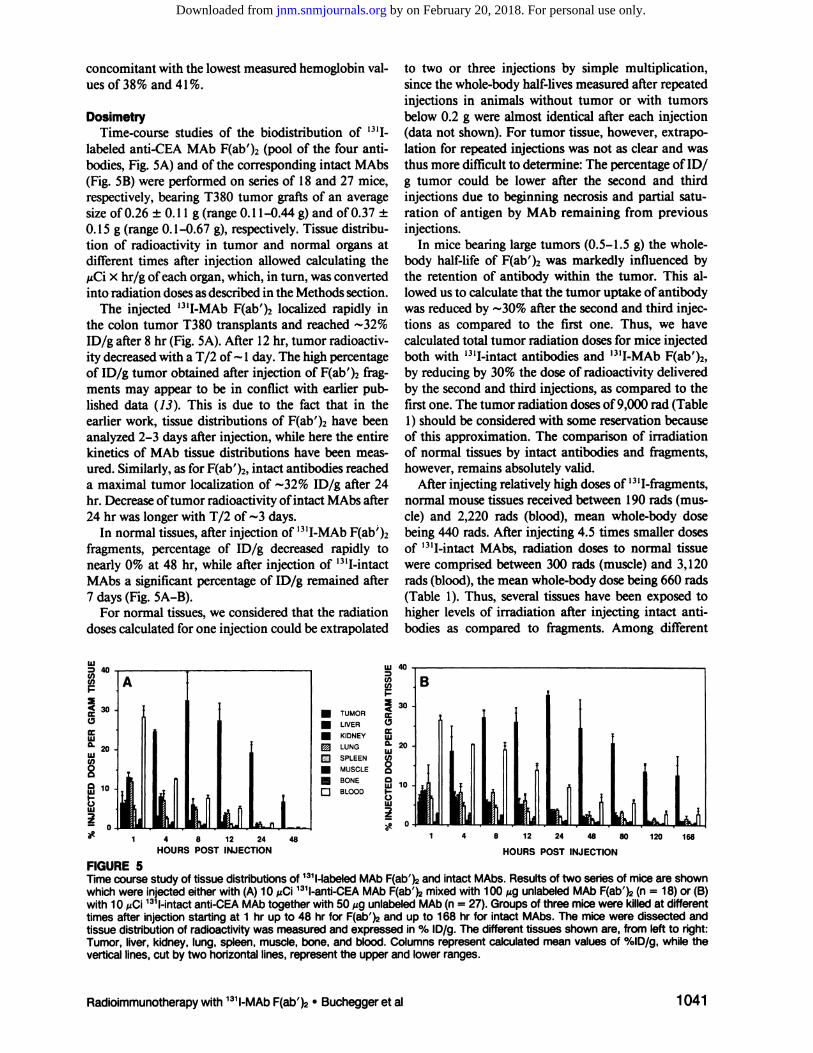
labeled anti-CEA MM, F(ab')2 (pool of the four antibodies, Fig. 5A) and of the corresponding intact MAbs(Fig. SB) were performed on series of 18 and 27 mice,respectively, bearing T380 tumor grafts of an averagesize ofO.26 ±0.1 1 g (range 0.1 1—0.44g) and ofO.37 ±0. 15 g (range 0. 1—0.67g), respectively. Tissue distribution of radioactivity in tumor and normal organs atdifferent times after injection allowed calculating the@zCiX hr/g ofeach organ, which, in turn, was convertedinto radiation doses as described in the Methods section.
The injected 1311-MAbF(ab')2 localized rapidly inthe colon tumor T380 transplants and reached -@32%ID/g after 8 hr (Fig. 5A). After 12 hr, tumor radioactivity decreased with a T/2 of'@-l day. The high percentageof ID/g tumor obtained after injection of F(ab')2 fragments may appear to be in conflict with earlier published data (13). This is due to the fact that in theearlier work, tissue distributions of F(ab')2 have beenanalyzed 2—3days after injection, while here the entirekinetics of MAb tissue distributions have been measured. Similarly, as for F(ab')2, intact antibodies reacheda maximal tumor localization of @@.-32%ID/g after 24hr. Decrease oftumor radioactivity ofintact MAbs after24 hr was longer with T/2 of @@-3days.
In normal tissues, after injection of ‘31I-MAbF(ab')2fragments, percentage of ID/g decreased rapidly tonearly 0% at 48 hr, while after injection of ‘311-intactMAbs a significant percentage of ID/g remained after7 days(Fig.SA—B).
For normal tissues, we considered that the radiationdoses calculated for one injection could be extrapolated
to two or three injections by simple multiplication,since the whole-body half-lives measured after repeatedinjections in animals without tumor or with tumorsbelow 0.2 g were almost identical after each injection(data not shown). For tumor tissue, however, extrapolation for repeated injections was not as clear and wasthus more difficult to determine: The percentage of ID/g tumor could be lower after the second and thirdinjections due to beginning necrosis and partial saturation of antigen by MAb remaining from previousinjections.
In mice bearing large tumors (0.5—1.5g) the wholebody half-life of F(ab')2 was markedly influenced bythe retention of antibody within the tumor. This allowed us to calculate that the tumor uptake of antibodywas reduced by -..-30%after the second and third injections as compared to the first one. Thus, we havecalculated total tumor radiation doses for mice injectedboth with ‘31I-intactantibodies and ‘311-MAbF(ab')2,by reducing by 30% the dose of radioactivity deliveredby the second and third injections, as compared to thefirst one. The tumor radiation doses of9,000 rad (Table1) should be considered with some reservation becauseof this approximation. The comparison of irradiationof normal tissues by intact antibodies and fragments,however, remains absolutely valid.
After injecting relatively high doses of ‘311-fragrnents,normal mouse tissues received between 190 rads (muscle) and 2,220 rads (blood), mean whole-body dosebeing 440 rads. After injecting 4.5 times smaller dosesof ‘31I-intactMAbS, radiation doses to normal tissuewere comprised between 300 rads (muscle) and 3,120rads (blood), the mean whole-body dose being 660 rads(Table 1). Thus, several tissues have been exposed tohigher levels of irradiation after injecting intact antibodies as compared to fragments. Among different
LU
Cl)Cl)
4
LUa.LUU)
20LU
LU
1 4 8 12 24 48 80 120 168
HOURSPOSTINJECTION
LU40
Cl)U)I-
@ 30
0LU
LUU)000LU 10I-0LU
. TUMOR
. LIVER
. KIDNEYD LUNG@ SPLEEN. MUSCLE
@ BONE
0 BLOOD
FIGURE5Timecoursestudyof tissuedistributionsof 131I-labeledMAbF(ab')@andintactMAbs.Resultsof twoseriesof miceareshownwhich were injectedeitherwith (A) 10 @Ci131l-anti-CEAMAb F(ab')@mixedwith 100 @gunlabeledMAb F(ab')@(n = 18) or (B)with 10 @iCi131I-intactanti-CEAMAbtogetherwith 50 @igunlabeledMAb(n = 27). Groupsof three micewere killedat differenttimes after injectionstarting at 1 hr up to 48 hr for F(ab')@and up to 168 hr for intact MAbS.The mice were dissected andtissue distributionof radioactivitywas measuredand expressedin % ID/g. The differenttissues shown are, from left to right:Tumor,liver,kidney,lung,spleen,muscle,bone,andblood.Columnsrepresentcalculatedmeanvaluesof %ID/g,whiletheverticallines,cut by two horizontallines,representthe upperand lower ranges.
1 4 8 12 24
HOURSPOSTINJECTION48
Radioimmunotherapywith 131I-MAbF(ab')2 •Buchegger et al 1041
by on February 20, 2018. For personal use only. jnm.snmjournals.org Downloaded from

TABLEITumorandNormalTissueAbsorbedRadiationDoses(in
rads) Estimated from BiodistributionStudies131l-F(ab')@
131I-intactMAbs
low toxicity of high doses of ‘31I-MAbF(ab')2 wasconfirmed by the fact that in these animals pWBC neverfell below 2,500 cells/mm3 and none needed bonemarrow transplantation. Furthermore, weight lossduring treatment was minimal and except for twomice which died from infection at 6 mo post-therapy,no other side effects developed during 10 mo of observation.
Several reports have described delayed tumor growthand, in some cases, complete remissions of solid tumortransplants by treatments using various radiolabeledMAbs (15, 16, 32—37).Complete remissions, however,have often been obtained by initiating antibody therapywithin 24 hr after tumor transplantation, or, in othercases, by injecting very large amounts of radiolabeledantibodies, which caused severe radiation toxicity anddeath ofa high number ofanimals (34).
Recent efforts are therefore directed toward reducingbone marrow toxicity from radiolabeled MAbs. Aninteresting approach is the injection of Interleukin-lbefore and/or during radioimmunotherapy (38). Injection of biotinylated, radiolabeled antibodies followedby a secondary injection of avidin has been proposedas a mean to provoke a rapid clearance of circulatingantibodies (39). Along similar lines, a secondary injection of anti-mouse-IgG antibodies had been given toproduce a more rapid blood clearance of radiolabeled
first antibodies (5, 40). It has also been proposed toinject unlabeled antibodies prior to radiolabeled MAbin order to reduce liver uptake ofradiolabeled antibody(41).
A more direct manner of decreasing marrow andliver uptake ofradiolabeled antibody is to use fragmentsofMAbs. F(ab')2 and Fab fragments give higher tumorto-normal tissue ratios in experimental tumor models(10-13) and are widely used for immunoscintigraphyin patients (2, 22, 23). Very few experimental results,however, have been published on their use for therapy.Our recent results (14) and the present study demonstrate experimentally the superiority of F(ab')2 overintact MAbS in radioimmunotherapy. These results arein agreement with those obtained in a model of colontumor transplants in hamsters (16), with a dosimetricstudy performed in a glioma xenograft model in nudemice brains (15), and with a rat model system (17).These three studies too compared MAb F(ab')2 tissuedistributions with the corresponding intact antibodies.
Larson et al. made a logical choice when they usedradiolabeled fragments (Fab) for radioimmunotherapyof melanoma patients (2). In our hands, however, thehalf-life of Fab fragments from anti-CEA MAbs is tooshort to deliver sufficient amount ofradioactivity to thetumor.
Here, in matched groups of tumor-bearing nudemice, we essentially show that i.v. injections of largedoses of ‘311-MAbF(ab')2 give a much better therapeutic
Tumor91709420Blood2220(4.l)t3120(3.0)@+41%@Kidney1
450 (6.3)1 150(8.2)—21%Lung1400 (6.6)1 580(6.0)+13%Stomach1040 (8.8)480 (19.6)—54%Liver580
(15.8)1 120(8.4)+93%Spleen510 (18.0)790 (11.9)+55%Bone310
(29.6)500(18.8)+61%Smallintestine340 (26.9)400(23.6)—18%Largeintestine220 (41.7)250(37.7)—14%Muscle1
90 (48.3)300 (31.4)+58%Wholemouse440 (20.8)660 (14.3)+50%
. Tumor and normal tissue irradiations are given in rads.
t Tumor-to-normal tissue irradiation ratios are indicated in pa
rentheses.4 Percent increase/decrease of irradiation for normal tissues
after injectionof 131I-intactantibodiesas comparedto irradiationafterinjectionof 131l-F(ab')@fragments.
tissues the increase for bone was 6 1%, for blood 41%,for spleen 55%, and forliver 93% (Table 1).Conversely,radiation exposure after injecting F(ab')2 was 116%higher for stomach and 26% higher for kidneys ascompared to intact MAbs.
Radioactivity localized in bone marrow was notmeasured directly. However, irradiation doses calculated for bones, blood, and spleen were definitivelylower after injecting fragments as compared to intactMAbs. This strongly suggests that bone marrow received less irradiation when fragments were used.
DISCUSSION
We have previously shown that nude mice bearingsmall human colon carcinoma transplants could becured by i.v. injections of ‘31I-labeledanti-CEA MAbF(ab')2 fragments (14). These results were encouragingbut they had been obtained by starting treatment onDays 9 to 10 after transplantation when the coloncarcinoma T380 xenografts, albeit in exponentialgrowth, were still relatively small (mean maximal size:102 ±50 mm3).
In the present experiment, therapy was begun 18days after transplantation, when the tumors were muchlarger, allowing us to confirm the therapeutic effectiveness of 131I-anti-CEA MAb F(ab')2 fragments on micebearing tumors which reached a mean maximal size of530 mm3 (range 190 to 1400 mm3). Complete remissions without tumor relapse were observed in 8 of 10mice and this does not appear to be due to an immunologic rejection, since the majority of successfullytreated mice readily accepted a subsequent challengewith the same human colon carcinoma xenograft. The
1042 TheJournalof NuclearMedicine•Vol.31 •No.6 •June1990
by on February 20, 2018. For personal use only. jnm.snmjournals.org Downloaded from
efficiency together with similar or reduced general toxicity, as compared to ‘311-intactMAbs. The lower toxicity of F(ab')2 fragments is in agreement with thereduced radiation doses calculated for vital organs suchas liver, spleen, bone, and blood when compared totreatment with intact antibodies. Due to the difficultyof calculating tumor radiation doses after repeated injections, the value of -@@9,000rad, both for treatmentwith intact MAbs and F(ab')2 fragments, represents anapproximation. The superior anti-tumor effect ofF(ab')2 (as compared to intact MAbs) could be duetherefore to at least three factors:
1. The real tumor dose obtained after injection offragments could be slightly higher than that obtamed with intact MAbS.
2. A deeper penetration of F(ab')2 is likely to haveproduced a more homogenous distribution of radiation within tumor tissue, as shown earlier byautoradiography (13). The fact that larger tumorstreated with intact antibodies relapsed more rapidly (Fig. 2B), while this was not the case withF(ab')2, supports this hypothesis.
3. Due to their very short biologic half-life, the radiation dose was delivered by the high amount of1311 F(ab')2 within a shorter period of time and
with higher energy flux (peak localizations in tumor reached 200—280@Ci/gafter the three injections) than after injection ofthe lower amounts of131I-intact antibodies (peak localizations were-@420 @iCi/gtumor). This may have allowed lessDNA repair to occur within the tumor after injection of F(ab')2.
Our experimental results with 1311-labeledMAbF(ab')2 demonstrate the feasibility of successful radioimmunotherapy of large colon carcinoma xenografts.These results, however, cannot be directly extrapolatedto clinical therapy because: (a) human bone marrow isknown to be 2—3times more sensitive to ionizingradiation as compared to murine marrow, and (b) thepresence ofsome CEA in normal human colon mucosa(42), results in a slightly increased localization of antiCEA antibodiesin this tissue as comparedto others(22, 23) (the clinical relevance of this accumulationremains unclear).
Our clinical experience in treating liver metastases ofcolon carcinoma by injecting large doses of ‘31I-antiCEAMAbin intactformand as F(ab')2fragmentsisindeed limited, and we have not yet obtained significanttumor remission (7). Theoretically, the absoluteamount of injected antibody per gram of tumor inpatients is expected to be @@-2,0®times lower than thatin mice as a consequence of the different body weights(43). In contrast, tumor-to-normal tissue radioactivityratios could be similar in mice and men. Indeed, ourresults in patients indicate, that this is the case: This is
illustrated by measurements of the % ID/g in early (at24 hr after injection) resected tumors of patients reaching 0.02 percent (23), and in some favorable cases0.03% and 0.06% (personal observations), while thecalculated mean whole-body retention at 24 hr [email protected]% ID/g (for a 70.000-g patient with a wholebody ‘@‘Iretention of 70%). Bone marrow toxicity
remains thus the major dose-limiting side effect inpatients. This toxicity might be overcome by autologousbone marrow transplantation.
ACKNOWLEDGMENTS
The authors thank Mrs. K. Fournier, F. Prevel, and M.Pagani for excellenttechnical assistance.We are grateful toDrs. J.F. Valley and S. Raimondi, Institute of Applied Radiophysics,EPFL,Lausanne,for helpfulsuggestionsconcerningdosimetry.
REFERENCES
1. Order SE, Klein JL, Leichner PK, Ettinger DS. The treatmentof hepatocellular cancec a clinical model for radiolabelledantibody therapy. In: Proceedings ofihe International Symposium on. Labelled and Unlabelled Antibodies in CancerDiagnosisand Therapy.Nail Cancerlnst Monogr 1987;3:37—41.
2. LarsonSM,CarrasquilloJA,KrohnKA, etal. Localizationof ‘311-labeledP97-specificFab fragments in human melanoma as a basisfor radiotherapy.JClin Invest 1983;72:2101—2114.
3. EpenetosAA, HammersmithOncologyGroup. Antibodyguidedirradiation ofmalignant lesions:three casesillustratinga new method oftreatment. Lancet 1984;i:1441—1443.
4. Lashford L, Jones D, Pritchard J, Gordon I, Breatnach F,KemsheadJT.Therpaeuticapplicationofradiolabeledmonoclonal antibody UJ13A in children with disseminated neuroblastoma. Nail Cancer Inst Monogr 1987;3:53—57.
5. BegentRHJ, BagshaweKD, PedleyRB, et al. Use of secondantibody in radioimmunotherapy. Nail Cancer Inst Monogr1987; 3:59—61.
6. De NardoSi, De NardoGL, O'GradyLF, etal. Treatmentof a patient with B cell lymphoma by 1-131LYM-l monoclonal antibodies. mi J BiolMarkers 1987;2:49—53.
7. Mach J-P, Buchegger F, Bischof-Delaloye A, et al. Progressin diagnostic immunoscintigraphy and first approach to radioimmunotherapy of colon carcinoma. In: Srivastava S, ed.Radiolabeledmonoclonalantibodiesfor imaging and therapy.New York: Plenum Publishing Corp; 1988:95—109.
8. MUller-GartnerH-W,MontzR, KlapdorR, et al. Radioimmunbehandlung solitärerLebermetastasen mittels intratumoraler Instillation ‘31J-markiertermonokionaler Antikörper—ErsteErgebnisse einer klinischen Studie. Nuc! Med1988; 27:258—265.
9. Kalofonos HP, PawlikowskaTR, Hemingway A, et al. Antibody-guideddiagnosisand therapyof brain gliomasusingradiolabeled monoclonal antibodies against epidermal growthfactor receptor and placental alkaline phosphatase. J NuclMed 1989;30:1636—1645.
10. Colcher D, Zalutsky M, Kaplan W, Kufe D, Austin F, SchlomJ. Radiolocalizationof human mammary tumors in athymicmice by a monoclonal antibody. Cancer Res 1983;43:736—742.
11. Wahl RL, Parker CW, Philpott GW. Improved radioimaging
Radioimmunotherapywith 131I-MAbF(ab')@•Bucheggeret al 1043
by on February 20, 2018. For personal use only. jnm.snmjournals.org Downloaded from
and tumor localization with monoclonal F(ab')2. J Nuci Med1983;24:316—325.
12. Herlyn D, Powe J, Alavi A, et al. Radioimmunodetection ofhuman tumor xenografts by monoclonal antibodies. CancerRes 1983; 43:2731—2735.
13. Buchegger F, Haskell CM, Schreyer M, et al. Radiolabeledfragments of monoclonal antibodies against carcinoembryonic antigens for localization of human colon carcinomagraftedintonudemice.JExp Med 1983;158:413—427.
14. Buchegger F, Pfister C, Foumier K, et al. Ablation of humancolon carcinoma in nude mice by ‘311-labeledmonoclonalanti-carcino-embryonicantigen antibody F(ab')2 fragments.J C/inInvest1989;83:1449—1456.
15. Colapinto EV, Humphrey PA, Zalutsky MR, et al. Comparative localization of murine monoclonal antibody Mel-14F(ab')2 fragment and whole IgG2a in human glioma xenografts.CancerRes 1988;48:5701—5707.
16. Blumenthal RD, Sharkey RM, Kashi R, Goldenberg DM.Comparison of therapeutic efficacy and host toxicity of twodifferent ‘311-labeledantibodies and their fragments in theGW-39 colonic cancer xenograft model. mt j Cancer 1989;44:292—300.
17. Walker KZ, Seymour-Munn K, Towson JE, et al. Radioimmunolocalization and selective delivery of radiation in a ratmodel system: comparison ofintact and fragmented antibody.Nuc/earMedComm 1988; 9:517—526.
18. Haskell CM, Buchegger F, Schreyer M, Carrel S, Mach J-P.Monoclonal antibodies to carcinoembryonic antigen: ionicstrength as a factor in the selection of antibodies for immunoscintigraphy. Cancer Res 1983; 43:3857—3864.
19. Buchsbaum D, Lloyd R, Juni J, et al. Localization andimaging of radiolabeled monoclonal antibodies against cobrectal carcinoma in tumor bearing nude mice. Cancer Res1988; 48:4324—4333.
20. Hammarstrom S, Shively JE, Paxton Ri, et al. Antigenic sitesin carcinoembryonic antigen. Cancer Res 1989; 49:4852—4858.
21. Mach J-P, Buchegger F, Forni M, et al. Use of radiolabeledmonoclonal anti-CEA antibodies for the detection of humancolon carcinomas by external photoscanning and tomoscintigraphy.Immuno/Today1981;2:239—249.
22. DelaloyeB,Bischof-DelaloyeA, BucheggerF, et al. Detectionof colorectal carcinoma by emission computerized tomography after injection of ‘231-labeledFab or F(ab')2 fragmentsfrom monoclonal anti-carcino-embryonic antigen antibodies.J C/inInvest1986;77:301—311.
23. Bischof.Delaloye A, Delaloye B, Buchegger F, et al. Clinicalvalue of immunoscintigraphy in colorectal carcinoma patients: a prospective study. J Nuc/ Med 1989; 30:1646—1656.
24. BucheggerF, Schreyer M, Carrel S, Mach J-P. Monoclonalantibodies identify a CEA crossreacting antigen of 95 kD(NCA-95) distinct in antigenicity and tissue distribution fromthe previously described NCA of 55 kD. liii J Cancer 1984;33:643—649.
25. Svenberg T. Carcinoembryonic antigen-like substances ofbile: isolation and partial characterization. mt J Cancer 1976;17:588—596.
26. Buchegger F, Fournier K, Schreyer M, Carrel S, Mach J-P.Swine monocbonal antibodies of high affinity and specificityto carcinoembryonic antigen. J Nail Cancer inst 1987; 79:337—342.
27. Buchegger F, Vacca A, Carrel 5, Schreyer M, Mach J-P.Radioimmunotherapy of human colon carcinoma by ‘@Ilabeled monoclonal anti.CEA antibodies in a nude mouse
model.mt j Cancer1988;41:127—134.28. Laemmli UK. Cleavage of structural proteins during the
assembly of the head of bacteriophage T4. Nature 1970;227:680—685.
29. Martin KW, Halpern SE. Carcinoembryonic antigen production, secretion and kinetics in BALB/c mice and a nudemouse-human tumor model. Cancer Res 1984; 44:5475—5481.
30. Hagan PL, Halpern SE, Dillmann RO, et al. Tumor size:effect on monoclonal antibody uptake in tumor models. JNuciMed 1986; 27:422—427.
31. Johns HE, Cunningham JR. The physics of radiology. In:Friedmann M, ed. Monograph in the Bannerstane division ofamerican lectures in radiation therapy, 3rd edition. Springfield, IL: Charles C Thomas Publ.; 1978:564—574.
32. Goldenberg DM, Gaffar SA, Bennett SJ, Beach JL. Experimental radioimmunotherapy ofa xenografted human colonictumor (GW-39) producing carcinoembryonic antigen. CancerRes 1981;41:4354—4360.
33. Jones DH, Goldman A, Gordon I, Pritchard J, Gregory BJ,Kemshead JT. Therapeutic application of a radiolabeledmonoclonal antibody in nude mice xenografted with humanneuroblastoma: tumoricidal effects and distribution studies.IntfCancer1985;35:715—720.
34.CheungN-K,LandmeierB,NeelyJ,etal.Completetumorablation with iodine 131-radiolabeled disiaboganglioside GD2-specific monoclonal antibody against human neuroblastomaxenograftedin nude mice. J Nail Cancer Inst 1986;77:739—745.
35. Esteban JM, SchbomJ, Mornex F, Coicher D. Radioimmunotherapy ofathymic mice bearing human colon carcinomaswith monocbonal antibody B72.3: histological and autoradiographic study of effectson tumors and normal organs. EurJ CancerC/in Oncol 1987;6:643—655.
36. Ceriani RL, Blank EW. Experimental therapy of humanbreast tumors with ‘311-labeledmonocbonal antibodies prepared against the human milk fat globule. Cancer Res 1988;48:4664—4672.
37. Senekowitsch R, Reidel G, Möllenstädt5, Kriegel H, PabstH-W. Curative radioimmunotherapy of human mammarycarcinoma xenografts with iodine-l3l-labeled monocbonalantibodies. JNuclMed 1989;30:531—537.
38. Blumenthal RD, Sharkey RM, Snyder DJ, Hansen HJ,Goldenberg DM. Reduction of radioantibody-induced myebotoxicityin hamsters by recombinant Interleukin-l . CancerRes 1988;43:5403—5406.
39. Sinitsyn VV, Mamontova AG, Checkneva YY, Shnyra AA,Domogatsky SP. Rapid blood clearance of biotinylated IgOafter infusion ofavidin. J NuclMed 1989; 30:66—69.
40. Blumenthal RD, Sharkey RM, Snyder D, Goldenberg DM.Reduction by anti-antibody administration of the radiotoxicity associated with ‘311-labeledantibody to carcinoembryonic antigen in cancer radioimmunotherapy. J Nat! Cancerlnsi1989;81:194—199.
41. Beatty BG, Beatty JD, Williams LE, Paxton Ri, Shively JE,O'Connor-TresselM. Effectofspecific antibody pretreatmenton liver uptake ‘‘In-labeledanticarcinoembryonic antigenmonocbonal antibody in nude mice bearing human coloncarcinoma xenografts.CancerRes 1989;49:1587—1594.
42. FritschéR, Mach J-P. Isolation and characterization of carcinoembryonic antigen (CEA)extracted from normal humancolon mucosa. Immunochemistry 1977; 14:119—127.
43. Fischman AJ, Khaw BA, Strauss HW. Quo vadis radioimmune imaging?JNuclMed1989;30:1911—1915.
1044 The Journal of Nuclear Medicine•Vol.31 •No. 6 •June1990
by on February 20, 2018. For personal use only. jnm.snmjournals.org Downloaded from

1990;31:1035-1044.J Nucl Med. Franz Buchegger, André Pèlegrin, Bernard Delaloye, Angelika Bischof-Delaloye and Jean-Pierre Mach Grafted in Nude MiceIntact Anti-CEA Antibodies in Radioimmunotherapy of Large Human Colon Carcinoma
Fragments Are More Efficient and Less Toxic Than2)′Iodine-131-Labeled MAb F(ab
http://jnm.snmjournals.org/content/31/6/1035This article and updated information are available at:
http://jnm.snmjournals.org/site/subscriptions/online.xhtml
Information about subscriptions to JNM can be found at:
http://jnm.snmjournals.org/site/misc/permission.xhtmlInformation about reproducing figures, tables, or other portions of this article can be found online at:
(Print ISSN: 0161-5505, Online ISSN: 2159-662X)1850 Samuel Morse Drive, Reston, VA 20190.SNMMI | Society of Nuclear Medicine and Molecular Imaging
is published monthly.The Journal of Nuclear Medicine
© Copyright 1990 SNMMI; all rights reserved.
by on February 20, 2018. For personal use only. jnm.snmjournals.org Downloaded from

![[1-ג€˜ג€˜C] DLג€”VaIIne, A Potential Pa ncreasג€”lmagin g Agent](https://static.fdocuments.us/doc/165x107/58a178ff1a28abb24d8c056c/1-c-dlvaiine-a-potential-pa-ncreaslmagin-g-agent.jpg)