Investigations of caspr2, an autoantigen of encephalitis and neuromyotonia
9
ORIGINAL ARTICLE Investigations of Caspr2, an Autoantigen of Encephalitis and Neuromyotonia Eric Lancaster, MD, PhD, 1 Maartje G. M. Huijbers, BS, 1 Vered Bar, PhD, 2 Anna Boronat, BS, 1 Andrew Wong, BS, 1 Eugenia Martinez-Hernandez, MD, 1 Christina Wilson, MD, PhD, 1 Dina Jacobs, MD, 1 Meizan Lai, MD, 1 Russell W. Walker, MD, 3 Francesc Graus, MD, 4 Luis Bataller, MD, 5 Isabel Illa, MD, 6 Sander Markx, MD, 7 Kevin A. Strauss, MD, 8,9,10 Elior Peles, PhD, 2 Steven S. Scherer, MD, PhD, 1 and Josep Dalmau, MD, PhD 1 Objective: To report clinical and immunological investigations of contactin-associated protein-like 2 (Caspr2), an autoantigen of encephalitis and peripheral nerve hyperexcitability (PNH) previously attributed to voltage-gated potassium channels (VGKC). Methods: Clinical analysis was performed on patients with encephalitis, PNH, or both. Immunoprecipitation and mass spectrometry were used to identify the antigen and to develop an assay with Caspr2-expressing cells. Immunoabsorption with Caspr2 and comparative immunostaining of brain and peripheral nerve of wild-type and Caspr2-null mice were used to assess antibody specificity. Results: Using Caspr2-expressing cells, antibodies were identified in 8 patients but not in 140 patients with several types of autoimmune or viral encephalitis, PNH, or mutations of the Caspr2-encoding gene. Patients’ antibodies reacted with brain and peripheral nerve in a pattern that colocalized with Caspr2. This reactivity was abrogated after immunoabsorption with Caspr2 and was absent in tissues from Caspr2-null mice. Of the 8 patients with Caspr2 antibodies, 7 had encephalopathy or seizures, 5 neuropathy or PNH, and 1 isolated PNH. Three patients also had myasthenia gravis, bulbar weakness, or symptoms that initially suggested motor neuron disease. None of the patients had active cancer; 7 responded to immunotherapy and were healthy or only mildly disabled at last follow-up (median, 8 months; range, 6–84 months). Interpretation: Caspr2 is an autoantigen of encephalitis and PNH previously attributed to VGKC antibodies. The occurrence of other autoantibodies may result in a complex syndrome that at presentation could be mistaken for a motor neuron disorder. Recognition of this disorder is important, because it responds to immunotherapy. ANN NEUROL 2011;69:303–311 W e recently reported that leucine-rich glioma inacti- vated 1 (Lgi1) is the main autoantigen of limbic encephalitis previously attributed to voltage-gated potas- sium channels (VGKC). 1 In that study, we also demon- strated that neither Lgi1 nor VGKC are the target anti- gens in patients with disorders other than limbic encephalitis who also have antibodies previously attrib- uted to VGKC. A direct immunoprecipitation technique showed that 1 of these antigens is contactin-associated protein-like 2 (Caspr2), but the clinical and immunologi- cal associations of these antibodies were not further examined. Here we describe the index patient whose se- rum was used to isolate Caspr2, and examine the clinical and immunological associations in 7 additional patients View this article online at wileyonlinelibrary.com. DOI: 10.1002/ana.22297 Received Aug 5, 2010, and in revised form Sep 17, 2010. Accepted for publication Oct 1, 2010. Address correspondence to Dr Dalmau, Department of Neurology, 3 W. Gates (Division of Neuro-oncology), University of Pennsylvania, 3400 Spruce Street, Philadelphia, PA 19104. E-mail: [email protected] From the 1 Department of Neurology, University of Pennsylvania School of Medicine, Philadelphia, PA; 2 Department of Molecular Cellular Biology, Weizmann Institute of Science, Rehovot, Israel; 3 Barrow Neurological Institute, St. Joseph’s Hospital and Medical Center, Phoenix, AZ; 4 Service of Neurology, Hospital Clinic, University of Barcelona and Institut d’Investigacions Biome ` diques August Pi i Sunyer (IDIBAPS), Barcelona ¨ , Spain; 5 Department of Neurology, University Hospital La Fe, Valencia, Spain; 6 Department of Neurology, Hospital de Sant Pau, Autonoma University of Barcelona, Barcelona, Spain; 7 Department of Psychiatry, Columbia University, College of Physicians and Surgeons, New York, NY; 8 Clinic for Special Children, Strasburg, PA; 9 Department of Biology, Franklin and Marshall College, Lancaster, PA; and 10 Lancaster General Hospital,Lancaster, PA. Additional supporting information can be found in the online version of this article. V C 2011 American Neurological Association 303
-
Upload
eric-lancaster -
Category
Documents
-
view
217 -
download
5
Transcript of Investigations of caspr2, an autoantigen of encephalitis and neuromyotonia

ORIGINAL ARTICLE
Investigations of Caspr2, an Autoantigenof Encephalitis and Neuromyotonia
Eric Lancaster, MD, PhD,1 Maartje G. M. Huijbers, BS,1 Vered Bar, PhD,2 Anna Boronat, BS,1
Andrew Wong, BS,1 Eugenia Martinez-Hernandez, MD,1 Christina Wilson, MD, PhD,1
Dina Jacobs, MD,1 Meizan Lai, MD,1 Russell W. Walker, MD,3 Francesc Graus, MD,4
Luis Bataller, MD,5 Isabel Illa, MD,6 Sander Markx, MD,7 Kevin A. Strauss, MD,8,9,10
Elior Peles, PhD,2 Steven S. Scherer, MD, PhD,1 and Josep Dalmau, MD, PhD1
Objective: To report clinical and immunological investigations of contactin-associated protein-like 2 (Caspr2), anautoantigen of encephalitis and peripheral nerve hyperexcitability (PNH) previously attributed to voltage-gatedpotassium channels (VGKC).Methods: Clinical analysis was performed on patients with encephalitis, PNH, or both. Immunoprecipitation andmass spectrometry were used to identify the antigen and to develop an assay with Caspr2-expressing cells.Immunoabsorption with Caspr2 and comparative immunostaining of brain and peripheral nerve of wild-type andCaspr2-null mice were used to assess antibody specificity.Results: Using Caspr2-expressing cells, antibodies were identified in 8 patients but not in 140 patients with severaltypes of autoimmune or viral encephalitis, PNH, or mutations of the Caspr2-encoding gene. Patients’ antibodiesreacted with brain and peripheral nerve in a pattern that colocalized with Caspr2. This reactivity was abrogated afterimmunoabsorption with Caspr2 and was absent in tissues from Caspr2-null mice. Of the 8 patients with Caspr2antibodies, 7 had encephalopathy or seizures, 5 neuropathy or PNH, and 1 isolated PNH. Three patients also hadmyasthenia gravis, bulbar weakness, or symptoms that initially suggested motor neuron disease. None of thepatients had active cancer; 7 responded to immunotherapy and were healthy or only mildly disabled at last follow-up(median, 8 months; range, 6–84 months).Interpretation: Caspr2 is an autoantigen of encephalitis and PNH previously attributed to VGKC antibodies. Theoccurrence of other autoantibodies may result in a complex syndrome that at presentation could be mistaken for amotor neuron disorder. Recognition of this disorder is important, because it responds to immunotherapy.
ANN NEUROL 2011;69:303–311
We recently reported that leucine-rich glioma inacti-
vated 1 (Lgi1) is the main autoantigen of limbic
encephalitis previously attributed to voltage-gated potas-
sium channels (VGKC).1 In that study, we also demon-
strated that neither Lgi1 nor VGKC are the target anti-
gens in patients with disorders other than limbic
encephalitis who also have antibodies previously attrib-
uted to VGKC. A direct immunoprecipitation technique
showed that 1 of these antigens is contactin-associated
protein-like 2 (Caspr2), but the clinical and immunologi-
cal associations of these antibodies were not further
examined. Here we describe the index patient whose se-
rum was used to isolate Caspr2, and examine the clinical
and immunological associations in 7 additional patients
View this article online at wileyonlinelibrary.com. DOI: 10.1002/ana.22297
Received Aug 5, 2010, and in revised form Sep 17, 2010. Accepted for publication Oct 1, 2010.
Address correspondence to Dr Dalmau, Department of Neurology, 3 W. Gates (Division of Neuro-oncology), University of Pennsylvania,
3400 Spruce Street, Philadelphia, PA 19104. E-mail: [email protected]
From the 1Department of Neurology, University of Pennsylvania School of Medicine, Philadelphia, PA; 2Department of Molecular Cellular Biology,
Weizmann Institute of Science, Rehovot, Israel; 3Barrow Neurological Institute, St. Joseph’s Hospital and Medical Center, Phoenix, AZ; 4Service of
Neurology, Hospital Clinic, University of Barcelona and Institut d’Investigacions Biomediques August Pi i Sunyer (IDIBAPS), Barcelona, Spain; 5Department
of Neurology, University Hospital La Fe, Valencia, Spain; 6Department of Neurology, Hospital de Sant Pau, Autonoma University of Barcelona, Barcelona,
Spain; 7Department of Psychiatry, Columbia University, College of Physicians and Surgeons, New York, NY; 8Clinic for Special Children, Strasburg, PA;9Department of Biology, Franklin and Marshall College, Lancaster, PA; and 10Lancaster General Hospital,Lancaster, PA.
Additional supporting information can be found in the online version of this article.
VC 2011 American Neurological Association 303

with this autoimmunity. We also demonstrate that anti-
bodies to Caspr2 specifically account for the reactivity of
these patients’ cerebrospinal fluid (CSF) or sera against
brain and peripheral nerve.
Patients and Methods
The term antibodies attributed to VGKC is used in this study to
define antibodies identified by a radioimmunoassay based on the
immunoprecipitation of brain protein complexes containing
VGKC labeled with 125I-a-dendrotoxin (125I-a-dendrotoxin RIA)
or by immunohistochemical methods.2–4 Because specific assays
show that in patients with limbic encephalitis and antibodies
attributed to VGKC the main target antigen is Lgi1, and because
Caspr2 was precipitated with serum of a patient with encephalitis
and peripheral neuropathy,1 a goal of the current study was to
determine whether patients with encephalitis or peripheral nerve
dysfunction suspected to be related to VGKC antibodies in fact
had Caspr2 antibodies. The term peripheral nerve hyperexcitability
(PNH) was used to include acquired neuromyotonia (Isaacs syn-
drome) or partial manifestations of this disorder such as cramps
and fasciculations without clear evidence of neuromyotonia. The
groups of patients examined to identify the 7 additional cases
included 49 patients with limbic encephalitis previously attributed
to VGKC antibodies, 18 with acquired PNH, 15 with encephali-
tis and atypical antibodies against the neuropil of hippocampus,
and 6 with Morvan syndrome. As control samples, we used se-
rum or CSF from 19 patients with other autoimmune encephali-
tis associated with N-methyl-d-aspartate (n ¼ 7), a-amino-3-
hydroxy-5-methylisoxazole-4-propionic acid (n ¼ 6), or gamma-
aminobutyric acid B (n ¼ 6) receptor antibodies, 13 patients
with Rasmussen encephalitis, and 6 patients with viral encephali-
tis. Additionally, because some disorders with genetic mutations
(eg, hereditary ataxias) are associated with a tendency to autoim-
munity,5 and patients with mutations of CNTNAP2 (the gene
that codes for Caspr2) develop symptoms resembling those of
patients with Caspr2 antibodies, including seizures, hyperactivity,
encephalopathy,6 and sometimes neuromyotonia (discussed later),
we examined 21 patients with polymorphisms or mutations of
CNTNAP2 for antibodies to Caspr2. Studies were approved by
the University of Pennsylvania Institutional Review Board.
Immunocytochemistry onCaspr2-Expressing CellsTo generate a diagnostic cell-based assay, human embryonic kidney
293 cells (HEK293) were transiently transfected using Lipofectamine
2000 (Invitrogen, Carlsbad, CA) and a plasmid containing human
Caspr2.1 Patients’ CSF (diluted 1:5) or sera (1:200) and a commer-
cial rabbit antibody to Caspr2 (1:10,000; Abcam, Cambridge, MA;
ab93228) were applied to cells, followed by fluorescein isothiocya-
nate (FITC)-conjugated antihuman immunoglobulin (Ig)G
(1:1,000; Molecular Probes, Eugene, OR) and tetramethylrhod-
amine isothiocyanate (TRITC)-conjugated antirabbit secondary anti-
bodies (1:1,000; Jackson Laboratories, Bar Harbor, ME). Images of
transfected cells were captured with an epifluorescence microscope
using Zeiss Axiovision software (Zeiss, Thornwood, NY).
Immunohistochemistry on Brain andPeripheral NerveAdult rat brains were prepared for immunohistochemical screen-
ing.4 Female Wistar rats were sacrificed without perfusion, the
brains removed and immersed in 4% paraformaldehyde at 4�C
for 1 hour, cryoprotected in 40% sucrose for 24 hours, snap fro-
zen in chilled isopentane, and sectioned. Caspr2-null mice and
their wild-type littermates were generated and genotyped as
reported,7 and the brains were prepared and sectioned as above.
For studies labeling peripheral nerve with patients’ antibodies,
unfixed teased rat or mouse sciatic nerve fibers were prepared as
previously described for paraformaldehyde fixed nerves.8
Immunohistochemistry was done using a standard avidin-
biotin peroxidase method, using serum (1:200) or CSF (1:5)
on 7lm sagittal brain sections followed by the appropriate sec-
ondary antibodies.4
Teased nerve fibers were permeabilized with acetone
(�20�C for 10 minutes), washed with phosphate buffered saline
(5 minutes � 3), blocked with 5% goat serum (1 hour, room
temperature), then incubated with serum (1:200 to 1:4,000) or
CSF (1:5) and a rabbit antibody to Caspr2 (Abcam; ab93228;
diluted 1:300) overnight at 4�C, followed by FITC-conjugated
antihuman IgG (1:200; Molecular Probes) and TRITC-conju-
gated antirabbit secondary antibody (1:300; Jackson Laboratories)
for 1 hour at room temperature. An antihuman IgM antibody
(1:200; Molecular Probes) was used for some experiments to con-
firm that patients’ antibodies were of the IgG type. In some
FIGURE 1: Patients’ antibodies react with cells expressingCaspr2. HEK cells were transiently transfected to expresshuman Caspr2, and labeled with the index patient’scerebrospinal fluid (CSF) (A; green) or serum from a differentpatient (B; green) and a rabbit antibody to Caspr2 (A, B; red),and counterstained with DAPI. Merged images (A, B; yellow)demonstrate the overlap of patients’ antibody staining withCaspr2 expression. (C) CSF from a control patient did notreact with cells expressing Caspr2. Scale bar 5 20lm.
ANNALS of Neurology
304 Volume 69, No. 2

experiments, paranodes were labeled with a monoclonal anti-
body to Caspr (1:50) described previously,9 followed by a
Cy5-conjugated goat antimouse secondary antibody (Jackson
Laboratories; 1:200). Images of peripheral nerve were captured
with an epifluorescence microscope using Openlab 3.1.7 soft-
ware (Perkin Elmer, Waltham, MA). Immunoabsorption was
carried out with serial incubations of patient’s CSF in 6 wells
containing fixed, permeabilized HEK293 cells expressing
Caspr2 or a control plasmid without an insert. After sequen-
tial passes of 1 hour each, the CSF was applied to brain or pe-
ripheral nerve as described above.
Results
Index Patient (Case #1)A 68-year-old man was evaluated for seizures and mem-
ory difficulties. Over the previous 2 years he had had 3
seizures, consisting of staring and unresponsiveness fol-
lowed by generalized tonic-clonic convulsions. Approxi-
mately 1 year before evaluation, he developed progressive
memory difficulties. He often repeated himself, and had
difficulty learning new information and recalling names
of close acquaintances. He occasionally became lost while
TABLE 1: Demographic and Clinical Features
Patient Age, yr Sex Symptoms/Signs Other Clinical andImmunological Features
1 68 M 3 years of thermal allodynia, 2 years ofintermittent complex partial seizures, and1 year of progressive memory difficulties.
VGKC antibodies.a CSF: 2 WBC/ll,protein 44mg/dl, glucose 65mg/dl.Oligoclonal bands negative.
2 46 F 2 years of progressive fatigue, personalitychanges, increased sweating, dysphagia withsubstantial weight loss, dysarthria, andintermittent ptosis and diplopia. Progressedto prolonged respiratory failure, visualhallucinations, and delusions of persecution.
Diagnosed with myasthenia gravis.MuSK antibodies 89nmol/l(normal <0.5). AChR antibodies247nmol/l (normal <0.5).VGKC antibodies.a
3 66 M 1 year of progressive lower extremity cramps,worse with exercise, and sensory-predominantpolyneuropathy.
VGKC antibodies not detected.a
4 52 M 5 years of progressive encephalopathy,myokymia, and weakness. Progressed tocritical illness with seizures, severe bulbarweakness, and encephalitis.
VGKC antibodies.a GAD65 antibodies,AChR antibodies. SIADH, peripheralneuropathy (type 1 diabetes).
5 77 M 1 year of 70 pounds weight loss, diffusesweating, twitching of hands and feet.Progressed to severe jerking at night, evenduring sleep, difficulty walking, falls,difficulty concentrating, confusion,and psychomotor slowing.
VGKC antibodies.a Serum sodium129 mmol. CSF: no cells, protein34mg/dl, glucose 58mg/dl.
6 60 M Agitation, amnestic periods, seizures.No peripheral nervous system symptoms.
VGKC antibodies not tested. CSF:4 WBC/ll, protein 35mg/dl, glucose75mg/dl. Elevated PSA-negative prostatebiopsy, negative PET scan and CT ofabdomen and pelvis.
7 61 M Myokymia, fasciculations, cramps,involuntary lingual movements. Developedseizures and encephalitis after 1 year.Progressive dysphagia, dysarthria,myoclonus, and ataxia after 6 years.
VGKC antibodies.a CSF: 15 WBC/ll,normal protein and glucose.
8 59 M Generalized seizures and severeamnestic syndrome.
VGKC antibodies.a
aVGKC antibodies detected by the 125I-a-dendrotoxin radioimmunoassay; normal values differed in different laboratories.VGKC ¼ voltage-gated potassium channels; CSF ¼ cerebrospinal fluid; WBC ¼ white blood cells; MuSK ¼ muscle-specifickinase; AChR ¼ acetylcholine receptor; SIADH ¼ syndrome of inappropriate antidiuretic hormone secretion; PSA ¼ prostate-spe-cific antigen; PET ¼ positron emission tomography; CT ¼ computed tomography.
Lancaster et al: Caspr2 Encephalitis
February 2011 305
driving in familiar surroundings. Starting 3 years earlier,
he had painful hypersensitivity to warm temperatures
affecting his hands and feet that persisted for 2 years and
spontaneously resolved. Twelve months after the onset of
seizures, he had low-grade, noninvasive bladder cancer
that was treated with local scraping and cauterization;
this has not recurred. He smoked (40–50 packs/year)
and drank approximately 8 alcoholic drinks per week.
His Mini Mental State Examination was 29/30
(missed recall of 1 item at 5 minutes). He was able to
name 12 of 15 objects on the Boston Naming test. Neuro-
psychological testing revealed a mild memory deficit, with
difficulties in visuospatial and nonverbal memory. Cranial
nerve function, strength, and muscle tone were normal.
Sensation of vibration was decreased on the toes, but sensa-
tion of cold, proprioception, and pinprick was intact.
Reflexes were 2/5 and symmetrical. His gait was slightly
wide-based, and he had difficulty performing tandem gait.
Brain magnetic resonance imaging (MRI) demon-
strated bilateral mesial temporal lobe abnormalities, par-
ticularly in the amygdala, with extension posteriorly
through the hippocampus, as well as mild cortical atro-
phy. An electroencephalogram (EEG) demonstrated left
frontal polar and bitemporal epileptiform discharges but
no electrographic seizures. A lumbar puncture revealed
an opening pressure of 156mm H20, 2 white blood cells/
ll, 0 red blood cells/ll, protein 44mg/dl, and glucose
65mg/dl. CSF cytology and flow cytometry were nega-
tive, and there were no oligoclonal bands. 125I-a-dendro-toxin RIA for VGKC antibodies in serum was positive
(0.19nmol/l, upper limit of normal, 0.02nmol/l). CSF
polymerase chain reaction was negative for West Nile vi-
rus, Tropheryma whipplei, and arboviruses. Serum vitamin
B12 level was normal, and the following antibodies were
negative: ANA, SSB, ds-DNA, RNP, Scl 70, and Jo-1.
Elevated SSA/Ro antibodies (3U; upper limit of normal,
0.9U) were noted in serum; complement C3/C4 was
within normal limits. Tumor markers CEA, CA19-9,
CA125, a-fetoprotein, b-hCG, and paraneoplastic anti-
bodies (Hu, Ma2, CRMP5, amphiphysin, Yo, Ri) were
all negative. Computed tomography and fluorodeoxyglu-
cose positron emission tomography did not reveal a neo-
plasm. Nerve conduction velocities showed no evidence
of polyneuropathy, and needle electromyogram (EMG)
showed no evidence of PNH.
One month following this evaluation, he pre-
sented with a cluster of 3 generalized tonic-clonic seiz-
ures, including 1 lasting 20 minutes, followed by non-
convulsive status epilepticus. His seizures were
controlled with lorazepam, levetiracetam, valproic acid,
and phenytoin. Repeat CSF and brain MRI studies
were unchanged. A 5-day course of methylprednisolone
(1g daily) was initiated, with marked improvement in
his cognitive function. He was slowly tapered from
prednisone 60mg to 40mg, which led to increased
confusion. Prednisone 60mg was reinitiated, with some
improvement in mental function. In an effort to
decrease his steroid dependence, he was initiated on
weekly rituximab for a 4-week cycle. At the last visit
(3 months after rituximab), he was seizure free for >6
months and was able to fluently converse, balance a
check book, and independently manage his daily
affairs. His chief concern was continuing difficulty
recalling events from several years ago.
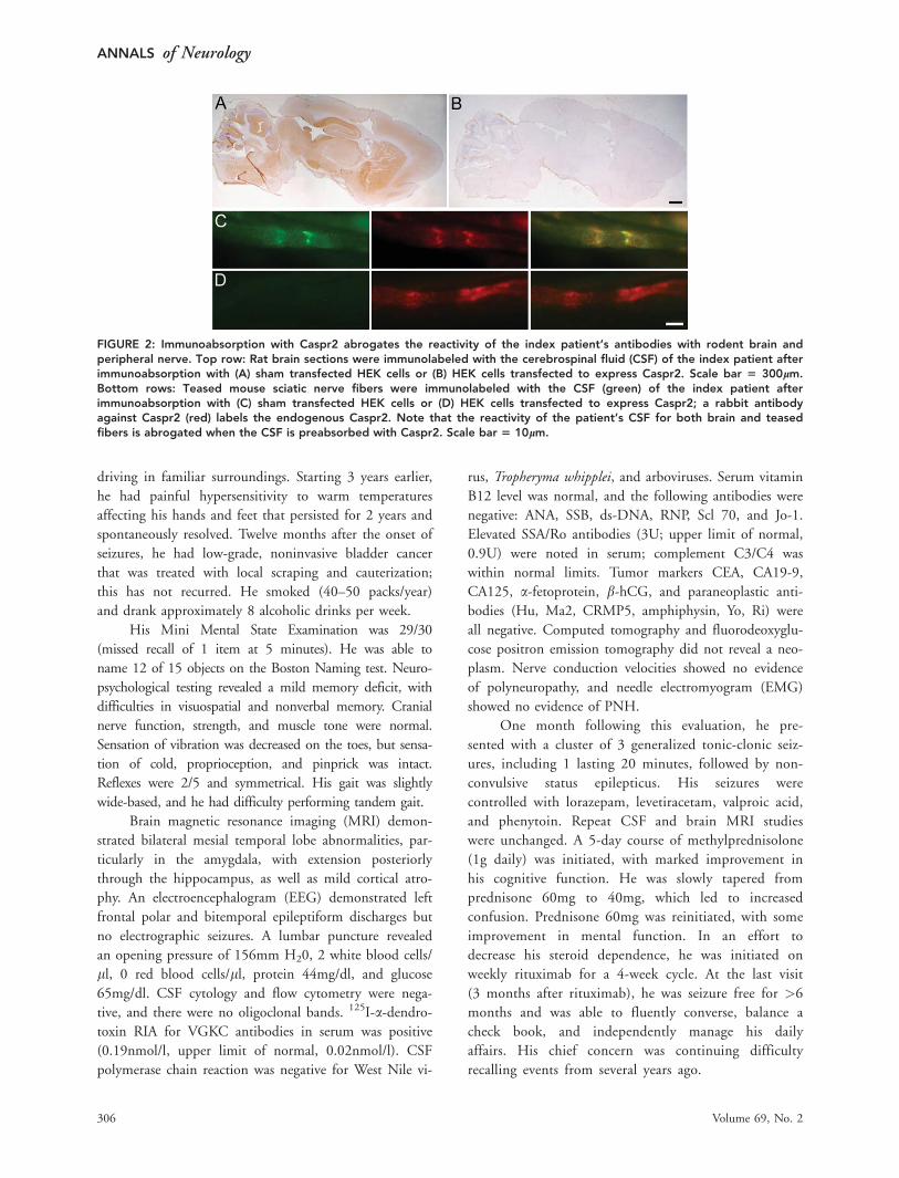
FIGURE 2: Immunoabsorption with Caspr2 abrogates the reactivity of the index patient’s antibodies with rodent brain andperipheral nerve. Top row: Rat brain sections were immunolabeled with the cerebrospinal fluid (CSF) of the index patient afterimmunoabsorption with (A) sham transfected HEK cells or (B) HEK cells transfected to express Caspr2. Scale bar 5 300lm.Bottom rows: Teased mouse sciatic nerve fibers were immunolabeled with the CSF (green) of the index patient afterimmunoabsorption with (C) sham transfected HEK cells or (D) HEK cells transfected to express Caspr2; a rabbit antibodyagainst Caspr2 (red) labels the endogenous Caspr2. Note that the reactivity of the patient’s CSF for both brain and teasedfibers is abrogated when the CSF is preabsorbed with Caspr2. Scale bar 5 10lm.
ANNALS of Neurology
306 Volume 69, No. 2

Identification of Caspr2 as a TargetAutoantigenThe serum and the CSF from the index case reacted with
a neuronal surface antigen expressed on live, nonpermeabi-
lized rat hippocampal neurons (data not shown) that was
precipitated and characterized by mass spectrometry as
Caspr2.1 Subsequently, HEK293 cells transfected to express
human Caspr2 were used as a diagnostic assay that confirmed
the presence of Caspr2 antibodies in the index patient (Fig 1),
and served to identify 7 additional cases among the indicated
cohort of patients (Table 1). These 7 patients’ sera did not
react with nontransfected cells, cells transfected with the volt-
age-gated potassium channel subunits Kv1.1 and Kv1.4, or
cells transfected with Lgi1 (data not shown).
Patients’ Antibodies React with PeripheralNerve and Colocalize with theJuxtaparanodal Expression of Caspr2Because Caspr2 is concentrated at the juxtaparanodal
region of myelinated axons7 and the sera of some
patients with antibodies previously attributed to VGKC
labeled myelinated axons,10 we examined whether
patients’ antibodies colocalized with Caspr2 at the juxta-
paranodal region of mouse sciatic nerve fibers. The
patients’ serum and CSF, but not those from controls,
robustly stained the juxtaparanodal regions of myelinated
fibers in a pattern that overlapped with that of Caspr2
(Supplementary Fig). Patients’ antibodies bound to
Caspr2 at the juxtaparanodal region were labeled by a
secondary antihuman IgG antibody but not a secondary
antihuman IgM antibody, indicating that they are of the
IgG type (data not shown).
Caspr2 Is the Target Autoantigenin Brain and NerveWe next used 2 different approaches to demonstrate that
the reactivity of patients’ antibodies with brain and nerve
is directed against Caspr2. First, we immunoabsorbed a
patient’s CSF with Caspr2, and found that this specifi-
cally abrogated the reactivity of patient’s antibodies with
brain and peripheral nerve (Fig 2). As a further test, we
immunostained brains and teased nerve fibers from
Caspr2-null mice and their wild-type littermates. Patients
with coexisting antibodies to other antigens (acetylcholine
receptor [AChR], muscle-specific kinase [MuSK], glu-
tamic acid decarboxylase) were excluded from this analy-
sis (#2 and #4). These studies showed that sera from the
remaining 5 patients immunostained the neuropil of
brain in a pattern expected for Caspr2 (Fig 3). Similar
experiments with peripheral nerve showed that patients’
sera also labeled the juxtaparanodes of myelinated periph-
eral nerve fibers of wild-type mice in a pattern expected
for Caspr2 reactivity (Fig 4). In contrast, these patterns
of reactivity were not seen in Caspr2-null mice, indicat-
ing that the immunostaining was due to specific Caspr2
antibodies (see Figs 3 and 4). Peripheral nerve fibers of
wild-type and Caspr2-null mice were costained with a
monoclonal antibody to the paranodal protein Caspr,
which is related to Caspr2.9 Caspr staining did not
colocalize with patients’ antibody staining and was
unchanged in Caspr2-null mice, further confirming the
specificity of patients’ antibodies for Caspr2.
FIGURE 3: Patients’ antibodies react with wild-type but notCaspr2-null mouse brains. The (A) cerebrospinal fluid (CSF)and (B) serum from 2 different patients with Caspr2antibodies immunostain the hippocampus of a wild-typemouse in a pattern identical to that obtained with (C) arabbit polyclonal antibody against Caspr2. This staining isnot seen in sections from a Caspr2-null mouse (A–C,columns on the right). (D) The pattern of staining with a CSFfrom a patient with limbic encephalitis associated with Lgi1antibodies is subtly different from Caspr2, and it is notaffected using wild-type or Caspr2-null mice. (E) The CSF ofa control individual without Caspr2 or Lgi1 antibodies doesnot label either sample. Scale bar 5 100lm.
Lancaster et al: Caspr2 Encephalitis
February 2011 307

Characteristics of Patients withCaspr2 AntibodiesThe clinical characteristics of the patients with Caspr2
antibodies are described in Tables 1 and 2. Five patients
had symptoms involving both the central nervous system
(CNS) (encephalitis, seizures) and peripheral nervous sys-
tem (PNS) (hyperexcitability, neuropathy, thermal allody-
nia). Two additional patients had a purely CNS syn-
drome. Only 1 patient (#3) had acquired neuromyotonia
without CNS symptoms. Two patients (#2 and #4) had bul-
bar weakness consistent with myasthenia gravis, and other au-
toantibodies against the skeletal muscle AchR and/or MuSK.
One of these patients (#2) was described in a case report prior
to the identification of Caspr2 antibodies.11 Case #7 had
severe progressive bulbar weakness but was not evaluated for
myasthenia. A diagnosis of motor neuron disease was initially
considered in these 3 patients (#2, #4, and #7).
Of the 5 patients with CNS symptoms who had
brain MRI, 2 had T2 hyperintensities affecting the medial
temporal lobes, 1 developed generalized atrophy, and 2
were normal. EEG studies of these 5 patients showed focal
epileptiform activity in 3, diffuse slowing in 1, and no ab-
normality in 1. Of the 5 patients with PNS symptoms
who had EMG/nerve conduction studies, 4 had electro-
physiological evidence of PNH, and 1 (who only had tran-
sient thermal allodynia) had a normal study.
Six of 7 patients tested for VGKC antibodies using
the 125I-a-dendrotoxin RIA had positive results, although
none of them recognized HEK293 cells transfected with
VGKC subunits (data not shown). None of the patients
had a tumor except for past history of low-grade bladder
cancer in the index case. Seven patients improved with
immunotherapy and were either healthy or only mildly
disabled at the last follow-up (median, 8 months; range,
6–84 months). The patient with poor outcome (#7) had
progressive bulbar weakness, never received immunother-
apy, and was lost to follow-up after 7 years.
Other Groups of PatientsAmong the groups of patients studied, Caspr2 antibodies
were not identified in 2 of 6 patients with Morvan syn-
drome, 17 of 18 patients with acquired PNH, and 13 of
15 patients with encephalitis associated with atypical
antibodies against the neuropil of hippocampus. None of
the 49 patients with limbic encephalitis and antibodies
attributed to VGKC had Caspr2 antibodies; however,
they all had Lgi1 antibodies. Two patients with PNH
and 1 patient with Morvan syndrome without Caspr2 or
Lgi1 antibodies had a positive 125I-a-dendrotoxin RIA,
suggesting they had antibodies against other components
of the VGKC-protein complex.
Discussion
This study demonstrates that Caspr2 is a brain and pe-
ripheral nerve autoantigen in a subgroup of disorders
previously attributed to VGKC antibodies, including en-
cephalitis, peripheral nerve dysfunction, or a combination
of both (Morvan syndrome). Symptoms may include
cognitive impairment, memory loss, hallucinations, delu-
sions, seizures, PNH, and axonal sensorimotor neuropa-
thy. The presence of other autoantibodies may result in a
complex syndrome with clinical and electrophysiological
FIGURE 4: Patients’ antibodies label teased fibers from wild-type but not Caspr2-null nerves. (A, B) The sera (green) of 2patients with Caspr2 antibodies immunolabel the juxtaparanodes of teased sciatic nerve fibers from wild-type mice in apattern similar to that of a rabbit antibody against Caspr2 (red), but different from the paranodal staining seen with a mousemonoclonal against Caspr (blue). In contrast, neither these patients’ sera nor the rabbit antibody against Caspr2 label teasedsciatic nerve fibers from Caspr2-null mice (columns on the right). (C, D) Sera from 2 controls do not label teased nerve fibers.Scale bar 5 5lm.
ANNALS of Neurology
308 Volume 69, No. 2
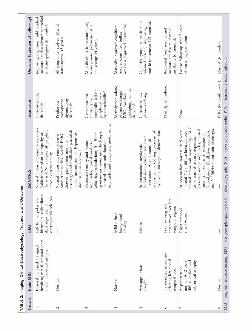
TABLE2:Im
aging,ClinicalElectrophysiology,Treatm
ent,
andOutcome
Patient
Brain
MRI
EEG
EMG/N
CS
Treatments
Outcome(durationoffollow-up)
1BilateralincreasedT2signal
involvingmesialtemporallobes,
andmildcorticalatrophy.
Leftfron
talpolar
and
bitemporalepileptiform
dischargesbu
tno
electrographic
seizures.
Normalmotor
andsensory
respon
ses
(asidefrom
medianneuropathyat
thewrist).Noevidence
ofperipheral
nerve
hyperexcitability.
Corticosteroids,
rituximab.
Improvingcognition;mildresidual
mem
orydeficits;seizurescontrolled
withantiepileptics
(6mon
ths).
2Normal.
—Normalmotor
andsensory
nerve
conductionstudies.NeedleEMG
show
edspon
taneousmotor
unit
dischargesandfibrillation
potentials,
butnoneuromyotonia.Repetitive
stim
ulation
was
normal.
Prednison
e,cyclosporine,
thym
ectomy,
rituximab.
Allsymptomsresolved.Mental
statusnormal(5
years).
3—
—Decreased
sensory
andmotor
amplitudes,normalconduction
velocities,fasciculation
s,5–
150H
zspon
taneousmotor
unitdischarges,
increasedmotor
unitduration
,am
plitude,andpolyphasicmotor
units.
Carbamazepine,
amitriptyline,
pregabalin(allfor
peripheral
nerve
hyperexcitability).
Milddisabilityfrom
continuing
sensory-m
otor
polyneuropathy
andcram
ps(4
years).
4Normal.
Milddiffuse
background
slow
ing.
Methylprednisolon
e,plasm
aexchange,
IVIG
,low-dose
cyclophospham
ide,
rituximab.
Markedly
improvedcognition,
seizurescontrolled,bu
lbar
weaknessim
proved(6
mon
ths).
5Age-appropriate
atrophy.
Normal.
Atpresentation
:myokymia,
fasciculation
s,chronicandacute
denervation
.After
1mon
thof
treatm
ent:markedim
provementof
myokymia,nosignsof
denervation
.
Corticosteroids,
plasm
aexchange.
Cognitivesymptomspersist(not
orientedto
time),im
proving
musclemovem
ents(12mon
ths).
6T2increasedintensities
affectingleftmedial
temporallobe.
Focalslow
ingand
sharpwaves
over
left
temporalregion
.
Methylprednisolon
e.Recovered
from
seizuresand
mem
orydeficits;mildmood
instability(6
mon
ths).
7Atpresentation
:normal.At2years:
diffuse
corticaland
subcorticalatrophy.
Righttemporal
sharpwaves.
Atpresentation
:normal.At5years:
normalNCS,
diffuse
fasciculation
s,normalmotor
unitmorphology.At7
years:normalsensory
respon
ses,
decreased
motor
amplitudes,normal
conductionvelocities,widespread
fasciculation
s,fibrillation
potentials,
and5–
150H
zmotor
unitdischarges.
Non
e.Lostto
follow-upafter7years
ofworseningsymptoms.
8Normal.
——
IVIG
(6-m
onth
cycles).
Normal(6
mon
ths).
MRI¼
magneticresonance
imaging;
EEG
¼electroencephalography;
EMG
¼electrom
yography;
NCS¼
nerve
conductionstudies;IV
IG¼
intravenousim
munoglobu
lin.

features suggesting a motor neuron disorder. Three dif-
ferent sets of experiments establish Caspr2 as an autoan-
tigen of these disorders, (1) specific immunostaining of
HEK293 cells expressing Caspr2 with serum or CSF of
patients; (2) specific abrogation of patients’ serum or
CSF antibody reactivity after immunoabsorption with
Caspr2-expressing cells; and (3) comparative brain and
nerve immunostaining of wild-type and Caspr2-nullmice, demonstrating lack of reactivity of patients’ serum
and CSF with Caspr2-null mice.
Caspr2 has a critical role in concentrating VGKC
and other proteins in the juxtaparanodal region of my-
elinated axons in both the PNS and the CNS.9,12 One
patient with homozygous deletion of CNTNAP2(OMIM 604569), the human gene that encodes Caspr2,
had a history of seizures and developed over 6 months
progressive painful peripheral neuropathy and neuromyo-
tonia to the point that it interfered with the patient’s gait
(K. Strauss, unpublished data). However, knockout mice
with disruption of the murine homolog encoding
Caspr27 had normal peripheral nerve conduction, sug-
gesting that in patients with genetic or autoimmune dis-
ruption of Caspr2 the occurrence of peripheral neuropa-
thy may be due to the involvement of other proteins that
interact with Caspr2. Caspr2 is also expressed in the hip-
pocampus,13 and mutations and polymorphisms of
CNTNAP2 have been linked to schizophrenia, psychosis,
intractable focal seizures, autism, mental retardation, and
cortical dysplasia.6,14–16 In this respect, the CNS symp-
toms of patients with antibodies to Caspr2 recapitulate
some of the clinical features of genetic disruption of the
gene. In 1 patient with mutated CNTNAP2, the resem-
blance of symptoms with ‘‘VGKC-antibody associated
encephalitis’’ led his neurologist to suspect this disorder
(S. Markx, unpublished observation). Subsequent testing
for VGKC antibodies using 125I-a-dendrotoxin RIA as
well as for Caspr2 antibodies produced negative results.
We examined 20 additional patients with CNTNAP2mutations or polymorphisms, and all were negative for
Caspr2 antibodies.
Juxtaparanodal immunostaining of peripheral nerve
by sera of patients with PNH has been previously attrib-
uted to antibodies against VGKC.10 Our data show that
an actual antigenic target is Caspr2. Other investigators
recently confirmed that Lgi1 is a major CNS target of
VGKC complex-binding autoantibodies, and independ-
ently reported the paranodal protein Caspr2 as a second
antigenic target.17 In clinical practice, the diagnosis of
patients with Caspr2 antibodies can be more complicated
than that of patients with classical limbic encephalitis
and Lgi1 antibodies. This is due to the frequent occur-
rence of Caspr2 with other autoantibodies, resulting in a
complex disorder that can manifest with motor weakness,
atrophy, fasciculations, and bulbar symptoms, leading
one to suspect an irreversible motor neuron disorder (see
pictures and detailed information of case #2 in Diaz-
Manera et al11). However, this patient and most patients
of the current study had dramatic responses to immuno-
therapy. It has been suggested that Caspr2 antibodies of-
ten occur in association with tumors (mostly thymoma)
but our study shows otherwise.17 Extensive tumor screen-
ing, sometimes with a long follow-up, did not reveal a
tumor in most patients, an observation that is empha-
sized by our referral pattern that likely favors patients
with paraneoplastic disorders.
Our findings help to clarify the perplexing diversity
of symptoms in patients previously diagnosed with
VGKC antibodies. We suspect that other components of
the Lgi1 or Caspr2 protein complex may be target anti-
gens in other subgroups of patients pending characteriza-
tion. For example, Lgi1 is a neuronally secreted protein
that interacts with pre- and postsynaptic proteins, organiz-
ing a trans-synaptic protein complex with multiple com-
ponents.18 There are cases of epilepsy or rapidly progres-
sive dementia, different from limbic encephalitis, reported
in association with antibodies to VGKC.19 It is unclear
whether in these cases the target antigen is Lgi1, Caspr2,
or another cell surface protein. Because only 1 of 18
patients with acquired PNH had Caspr2 antibodies and
another 2 had positive 125I-a-dendrotoxin RIA, other
autoantigens may account for the majority of these cases.
Additional work is required to clarify the patho-
genic mechanisms of autoantibodies to Caspr2, and
determine how they may cause central and peripheral
nerve dysfunction. Based upon the response of most
patients with Caspr2 antibodies to immunotherapy and
what is known about the direct effects of other cell sur-
face antibodies on the target antigens,20–22 functional dis-
ruption of Caspr2, as opposed to neuronal destruction, is
a likely mechanism.
Acknowledgments
This work was supported by RO1CA89054-06A2 and
1RC1NS068204 to J.D.; National Institutes of Health
NS43174 to S.S.S.; National Institute of Neurological
Disorders and Stroke NS50220 and a grant from the
Israel Academy of Sciences to E.P.; and a Dana Founda-
tion Neuro-immunology Award to E.L.
We thank the physicians who provided clinical in-
formation, and the patients and their families.
Authorship
E.L. and M.G.M.H. contributed equally.
ANNALS of Neurology
310 Volume 69, No. 2

Potential Conflicts of Interest
E.L. has received grant support from Talecris, a company that
sells human immunoglobulin. A patent application for the use
of Lgi1 antibody detection in patients’ CSF and sera has been
filed by J.D., whose laboratory has received grant support from
Euroimmun. No other authors have conflicts of interest.
References1. Lai M, Huijbers MG, Lancaster E, et al. Investigation of Lgi1 as the
antigen of limbic encephalitis previously attributed to potassiumchannels. Lancet Neurol 2010;9:776–785.
2. Buckley C, Oger J, Clover L, et al. Potassium channel antibodiesin two patients with reversible limbic encephalitis. Ann Neurol2001;50:73–78.
3. Tan KM, Lennon VA, Klein CJ, et al. Clinical spectrum of voltage-gated potassium channel autoimmunity. Neurology 2008;70:1883–1890.
4. Ances BM, Vitaliani R, Taylor RA, et al. Treatment-responsive lim-bic encephalitis identified by neuropil antibodies: MRI and PETcorrelates. Brain 2005;128:1764–1777.
5. Bushara KO, Goebel SU, Shill H, et al. Gluten sensitivity in sporadicand hereditary cerebellar ataxia. Ann Neurol 2001;49:540–543.
6. Strauss KA, Puffenberger EG, Huentelman MJ, et al. Recessivesymptomatic focal epilepsy and mutant contactin-associated pro-tein-like 2. N Engl J Med 2006;354:1370–1377.
7. Poliak S, Salomon D, Elhanany H, et al. Juxtaparanodal clusteringof Shaker-like Kþ channels in myelinated axons depends onCaspr2 and TAG-1. J Cell Biol 2003;162:1149–1160.
8. Brown AA, Xu T, Arroyo EJ, et al. Molecular organization of thenodal region is not altered in spontaneously diabetic BB-Wistarrats. J Neurosci Res 2001;65:139–149.
9. Poliak S, Gollan L, Martinez R, et al. Caspr2, a new member ofthe neurexin superfamily, is localized at the juxtaparanodes of my-elinated axons and associates with Kþ channels. Neuron 1999;24:1037–1047.
10. Kleopa KA, Elman LB, Lang B, et al. Neuromyotonia and limbicencephalitis sera target mature Shaker-type Kþ channels: subunit
specificity correlates with clinical manifestations. Brain 2006;129:1570–1584.
11. Diaz-Manera J, Rojas-Garcia R, Gallardo E, et al. Antibodies toAChR, MuSK and VGKC in a patient with myasthenia gravis andMorvan’s syndrome. Nat Clin Pract Neurol 2007;3:405–410.
12. Scherer SS, Arroyo EJ. Recent progress on the molecular organi-zation of myelinated axons. J Peripher Nerv Syst 2002;7:1–12.
13. Bel C, Oguievetskaia K, Pitaval C, et al. Axonal targeting ofCaspr2 in hippocampal neurons via selective somatodendriticendocytosis. J Cell Sci 2009;122:3403–3413.
14. Alarcon M, Abrahams BS, Stone JL, et al. Linkage, association,and gene-expression analyses identify CNTNAP2 as an autism-susceptibility gene. Am J Hum Genet 2008;82:150–159.
15. Friedman JI, Vrijenhoek T, Markx S, et al. CNTNAP2 gene dosagevariation is associated with schizophrenia and epilepsy. Mol Psy-chiatry 2008;13:261–266.
16. Zweier C, de Jong EK, Zweier M, et al. CNTNAP2 and NRXN1 aremutated in autosomal-recessive Pitt-Hopkins-like mental retarda-tion and determine the level of a common synaptic protein inDrosophila. Am J Hum Genet 2009;85:655–666.
17. Irani SR, Alexander S, Waters P, et al. Antibodies to Kv1 potas-sium channel-complex proteins leucine-rich, glioma inactivated 1protein and contactin-associated protein-2 in limbic encephalitis,Morvan’s syndrome and acquired neuromyotonia. Brain 2010;133:2734–2748.
18. Fukata Y, Lovero KL, Iwanaga T, et al. Disruption of LGI1-linkedsynaptic complex causes abnormal synaptic transmission and epi-lepsy. Proc Natl Acad Sci U S A 2010;107:3799–3804.
19. Geschwind MD, Tan KM, Lennon VA, et al. Voltage-gated potas-sium channel autoimmunity mimicking Creutzfeldt-Jakob disease.Arch Neurol 2008;65:1341–1346.
20. Dalmau J, Gleichman AJ, Hughes EG, et al. Anti-NMDA-receptorencephalitis: case series and analysis of the effects of antibodies.Lancet Neurol 2008;7:1091–1098.
21. Lai M, Hughes EG, Peng X, et al. AMPA receptor antibodies inlimbic encephalitis alter synaptic receptor location. Ann Neurol2009;65:424–434.
22. Lancaster E, Lai M, Peng X, et al. Antibodies to the GABA(B) re-ceptor in limbic encephalitis with seizures: case series and charac-terisation of the antigen. Lancet Neurol 2009;9:67–76.
Lancaster et al: Caspr2 Encephalitis
February 2011 311
![NIH Public Access autoantigen provides clues to etiology and ......Natural autoantibodies reactive with GBM were found and purified from normal human sera [34,35•], although at much](https://static.fdocuments.us/doc/165x107/60b3d428b5e95914983bdbc5/nih-public-access-autoantigen-provides-clues-to-etiology-and-natural-autoantibodies.jpg)