Contribution of Myelin Autoantigen Citrullination to T Cell
9
of November 22, 2018. This information is current as Central Nervous System the Citrullination to T Cell Autoaggression in Contribution of Myelin Autoantigen Anderton Antonio Carrillo-Vico, Melanie D. Leech and Stephen M. http://www.jimmunol.org/content/184/6/2839 doi: 10.4049/jimmunol.0903639 February 2010; 2010; 184:2839-2846; Prepublished online 17 J Immunol References http://www.jimmunol.org/content/184/6/2839.full#ref-list-1 , 20 of which you can access for free at: cites 58 articles This article average * 4 weeks from acceptance to publication Fast Publication! • Every submission reviewed by practicing scientists No Triage! • from submission to initial decision Rapid Reviews! 30 days* • Submit online. ? The JI Why Subscription http://jimmunol.org/subscription is online at: The Journal of Immunology Information about subscribing to Permissions http://www.aai.org/About/Publications/JI/copyright.html Submit copyright permission requests at: Email Alerts http://jimmunol.org/alerts Receive free email-alerts when new articles cite this article. Sign up at: Print ISSN: 0022-1767 Online ISSN: 1550-6606. Immunologists, Inc. All rights reserved. Copyright © 2010 by The American Association of 1451 Rockville Pike, Suite 650, Rockville, MD 20852 The American Association of Immunologists, Inc., is published twice each month by The Journal of Immunology by guest on November 22, 2018 http://www.jimmunol.org/ Downloaded from by guest on November 22, 2018 http://www.jimmunol.org/ Downloaded from
Transcript of Contribution of Myelin Autoantigen Citrullination to T Cell
of November 22, 2018.This information is current as
Central Nervous SystemtheCitrullination to T Cell Autoaggression in
Contribution of Myelin Autoantigen
AndertonAntonio Carrillo-Vico, Melanie D. Leech and Stephen M.
http://www.jimmunol.org/content/184/6/2839doi: 10.4049/jimmunol.0903639February 2010;
2010; 184:2839-2846; Prepublished online 17J Immunol
Referenceshttp://www.jimmunol.org/content/184/6/2839.full#ref-list-1
, 20 of which you can access for free at: cites 58 articlesThis article
average*
4 weeks from acceptance to publicationFast Publication! •
Every submission reviewed by practicing scientistsNo Triage! •
from submission to initial decisionRapid Reviews! 30 days* •
Submit online. ?The JIWhy
Subscriptionhttp://jimmunol.org/subscription
is online at: The Journal of ImmunologyInformation about subscribing to
Permissionshttp://www.aai.org/About/Publications/JI/copyright.htmlSubmit copyright permission requests at:
Email Alertshttp://jimmunol.org/alertsReceive free email-alerts when new articles cite this article. Sign up at:
Print ISSN: 0022-1767 Online ISSN: 1550-6606. Immunologists, Inc. All rights reserved.Copyright © 2010 by The American Association of1451 Rockville Pike, Suite 650, Rockville, MD 20852The American Association of Immunologists, Inc.,
is published twice each month byThe Journal of Immunology
by guest on Novem
ber 22, 2018http://w
ww
.jimm
unol.org/D
ownloaded from
by guest on N
ovember 22, 2018
http://ww
w.jim
munol.org/
Dow
nloaded from

The Journal of Immunology
Contribution of Myelin Autoantigen Citrullination to T CellAutoaggression in the Central Nervous System
Antonio Carrillo-Vico,1 Melanie D. Leech, and Stephen M. Anderton
Breakdown in immunological self tolerance, leading to autoimmune diseases such as multiple sclerosis, might arise from immune
recognition of self proteins that have undergone heightened posttranslational modification under pathophysiological conditions. A
posttranslational modification of particular interest is the deimination of Arg to citrulline, catalyzed by peptidylarginyl deiminase
(PAD) enzymes. As a CD4+ T cell-driven model of multiple sclerosis, we used experimental autoimmune encephalomyelitis (EAE)
induced with the immunodominant 35–55 peptide of myelin oligodendrocyte glycoprotein (pMOG) in C57BL/6 mice to test
whether citrullination of a T cell epitope can contribute to disease etiopathology. Immunization with an altered peptide ligand
(APL) of pMOG with an Arg→citrulline conversion at a TCR contact (residue 41) led to the activation of two populations of APL-
responsive T cells that either did, or did not cross-react with the native pMOG peptide. This APL could induce EAE. However,
this reflected the activation of T cells that cross-reacted with the native pMOG epitope, because prior tolerization of these T cells
using pMOG prevented APL-induced EAE. Using a passive transfer model, we found that T cells that responded specifically to the
citrullinated form of pMOG were neither necessary, nor sufficient to initiate the EAE lesion. Nevertheless, these cells could
provoke exacerbation of pathology if transferred into mice with ongoing EAE. The PAD2 and PAD4 enzymes were markedly
upregulated in the inflamed CNS. Therefore, once inflammation is established, citrullination of target autoantigens can allow an
expanded repertoire of T cells to contribute to CNS pathology. The Journal of Immunology, 2010, 184: 2839–2846.
Although the mammalian genome encodes only 20 aminoacids, analyses of mature proteins have identified .100diverse amino acids (1). These arise from post-
translational modifications (PTM) that can occur spontaneously(2), or as a result of enzymatic activity. The de novo recognition ofself-Ags that have been modified as a result of pathophysiologicaldisturbance (3, 4) is an appealing basis for breakdown in immu-nologic self tolerance; namely, the immune system would targetthe modified self-Ag as if it were pathogen derived. The literatureprovides various examples of Ab- and/or T cell-reactivity againstantigenic epitopes that contain PTM such as glycosylation, dea-midation, and citrullination (5–8). Citrullination, the deiminationof Arg to citrulline (Cit) (9), has received particular interest as anautoantigen modification. This reaction, by which the imino groupof the protein arginyl residues is removed giving rise to Cit, iscatalyzed by a family of calcium-binding enzymes termed pepti-
dylarginine deiminases (PADs; 3.5.3.15). A set of paralogousgenes encode five PAD isoforms in mammals, Pad1–4 and -6,which have a tissue-specific expression as well as substratespecificity. Expression of PAD1, PAD3 and PAD6 appears tightlyrestricted (10), whereas PAD4 is expressed in monocytes andgranulocytes and PAD2 can be found in the CNS (11, 12). Theassociation between citrullination and rheumatoid arthritis (RA)appears well established. Both PAD2 and PAD4 are elevated ininflamed joints (13), autoantibodies are found that specificallyrecognize citrullinated self-proteins (14), and a citrullinated formof type II collagen (CII) displayed enhanced arthritogenicity inrats (15). Citrullinated self-peptides have been shown to displayenhanced binding to RA-associated HLA-DR4 molecules (16) andDR4 transgenic mice can develop arthritis after immunizationwith citrullinated, but not unmodified, fibrinogen (17).Interest in citrullination inmultiple sclerosis (MS) first developed
fromstudies showing increasedcitrullinationofmyelinbasicprotein(MBP) in patients, and particularly in the hyperacute Marburg’ssyndrome (18, 19). MS patients have been reported to show en-hanced T cell reactivity to citrullinated MBP leading to the sug-gestion that this may contribute to disease induction or perpetuation(20). This has prompted some studies on the contribution of cit-rullination to pathogenesis of the primary preclinical model forMS,experimental autoimmune encephalomyelitis (EAE). CitrullinatedMBP is encephalitogenic in both rats and mice (21, 22). Within theCNS, PAD2 has been found in oligodendrocytes, microglia, andastrocytes (12, 23, 24) providing the opportunity for modification ofmyelin autoantigens. Immunohistochemical analyses revealed that,although citrullinated proteins were not readily detectable in thehealthy CNS, these were clearly evident in mice with EAE (25, 26).SJL/J mice with chronic EAE induced by immunization witha proteolipid protein peptide mounted autoantibody responsesagainst citrullinated MBP (27). However, although autoantibodiesmay contribute to pathology, EAE is primarily driven by the actionsof proinflammatory CD4+ T cells reactive against myelin. Thus far,no analysis of the contribution of T cells recognizing citrullinated
Centre for Inflammation Research, Centre for Multiple Sclerosis Research, and Cen-tre for Immunity, Infection, and Evolution, Queen’s Medical Research Institute,University of Edinburgh, Edinburgh, United Kingdom
1Current address: Instituto de Biomedicina de Sevilla, Hospital Universitario Virgendel Rocio/CSIC/Universidad de Sevilla, Sevilla, Spain.
Received for publication November 12, 2009. Accepted for publication January 11,2010.
This work was supported by grants from theMedical Research Council (U.K.). A.C.-V.was supported by a long-term fellowship from the Federation of European BiochemicalSocieties.
Address correspondence and reprint requests to Prof. Stephen M. Anderton, Univer-sity of Edinburgh, Centre for Inflammation Research, Queen’s Medical ResearchInstitute, 47 Little France Crescent, Edinburgh EH16 4TJ, United Kingdom. E-mailadddress: [email protected]
Abbreviations used in this paper: APL, altered peptide ligand; Cit, citrulline; EAE,experimental autoimmune encephalomyelitis; Iso, cells stained with the relevant iso-type control Ab; MBP, myelin basic protein; Med, cells cultured in medium alone priorto staining; MOG, myelin oligodendrocyte glycoprotein; MS, multiple sclerosis; PAD,peptidyl arginyl deiminase; pMOG, 35–55 peptide of MOG; PTM, posttranslationalmodification; RA, rheumatoid arthritis; Treg, regulatory T cell; WT, wild-type.
Copyright� 2010 by The American Association of Immunologists, Inc. 0022-1767/10/$16.00
www.jimmunol.org/cgi/doi/10.4049/jimmunol.0903639
by guest on Novem
ber 22, 2018http://w
ww
.jimm
unol.org/D
ownloaded from
myelin Ags has been performed in EAE. In this study, we describethe contribution of T cells recognizing citrullinated forms ofthe immunodominant 35–55 peptide of myelin oligodendrocyteglycoprotein (hereafter referred to as pMOG).
Materials and MethodsMice, Ags, and immunizations
C57BL/6 (CD45.1+ or CD45.2+) mice were bred under specific pathogen-free conditions at the University of Edinburgh. The 6–8-wk-old sex-matched mice were used for all experiments. pMOG (MEVG-WYRSPFSRVVHLYRNGK) and the pMOG(41Cit), pMOG(46Cit), andpMOG(41,46Cit) APL were obtained from Peptide 2.0 (Chantilly, VA).Mice were immunized with 100 mg peptide emulsified in CFA (Sigma-Aldrich, Poole, U.K.). A total of 100 ml emulsion was injected s.c., 50 mlinto each hind leg. Primed lymphoid populations were derived either fromspleens or from draining inguinal and para-aortic lymph nodes at the in-dicated time. Active and passive EAE were induced using previously de-scribed protocols (28, 29) Clinical signs of EAE were assessed daily withthe following scoring system: 0, no signs; 1, flaccid tail; 2, impairedrighting reflex and/or gait; 3, partial hind limb paralysis; 4, total hind limbparalysis; 5, hind limb paralysis with partial front limb paralysis; and 6,moribund or dead. Tolerance was induced by injection of 200 mg peptidein 0.2 ml PBS i.v. at the indicated time before immunization. All experi-ments were approved by the University of Edinburgh ethical review paneland were conducted under United Kingdom legislation.
Spleen recall responses
Spleen cell suspensions were cultured in 96-well flat-bottom microtiterplates (Becton Dickinson, Oxford, U.K.) at 8 3 105 splenocytes/well usingX-vivo 15 serum-free medium (BioWhittaker, Maidenhead, U.K.) supple-mented with 2 mM L-glutamine and 53 1025 M 2-ME (all from Invitrogen,Paisley, U.K.). Cultures were stimulated with a dose range of peptides for 48h prior to addition of [3H]dThd (0.5 mCi/well) (Amersham, Amersham,U.K.). After an additional 18 h, dThd incorporation was measured usinga liquid scintillation b-counter (LKB Wallac, Turku, Finland). Results areexpressed as mean cpm of triplicate cultures. Supernatants from similar 72 hcultures were tested for IFN-g and IL-17 production by ELISA.
PAD2 and PAD4 mRNA expression
TotalRNAfrombrainand spinal cordor lymphnodeand spleenwasextractedusing RNeasy Lipid Tissue Mini Kit or RNeasy Mini Kit (Qiagen, Hilden,Germany), respectively. Single-strand cDNAwas then synthesized from1mgRNA according to the Cloned AMV First-Strand cDNA Synthesis Kit(Invitrogen). Real-time quantitative PCR was performed by the Chromo4real-time system (MJ Research, Waltham, MA). Reactions were performedin a 50 ml volume containing 10 ml RT product as template DNA (100 ng),23 Platinum SYBR Green quantitative PCR SuperMix-UDG (Invitrogen),and 200 nM for each primer set. AUDG incubation step before PCR cyclingwas carried out to destroy any contaminating dU-containing product fromprevious reaction. UDG was then inactivated by the high temperaturesduring the first PCR step (95˚C for 2 min.). After that, a denaturation (95˚Cfor 15 s), annealing (60˚C for 30 s) and extension (72˚C for 15 s) steps wererepeated 30 times with fluorescent data acquisition after each extension step.Finally, a melting curve program from 60˚C to 95˚C, followed by a coolingstep at 20˚C were performed. Primers (59 to 39): Pad2 (241-bp product): 59TAC AGC AAG CAA GAC CTC CA 39 (exon 6) and 59 CCA CGA AGAACAGCAACT CC 39 (exon 7); Pad4 (217 bp product): 59GCT GCC TGTGGT CTT TGACT 39 (exon 9) and 59 GTA ACC GCTATT CCC GAT GA39 (exon 11). All reactions were performed in triplicate. The hprt gene wasused as the control for the calculation of DCt. Analysis of relative geneexpression was determined by evaluating the expression of 22DDCT.
T cell hybridomas
Draining lymph node populations were stimulated in vitro with 10 mMpeptide for 3 d and blasts were then fused with the TCR-deficient BW5147thymoma as described previously (30). All hybrids were cloned and re-cloned by limiting dilution. IL-2 production in response to 24-h culturewith peptide presented by irradiated (30 Gy) syngeneic splenocytes wasdetermined by ELISA.
CNS-infiltrating mononuclear cells
Mice were perfused with cold PBS, brain and spinal cords were removed,digested, and mononuclear cells retrieved from a 30%:70% discontinuouspercoll gradient as described previously (28).
Flow cytometry for surface phenotype and intracellularcytokine staining
Cells were stained for FACS using the following Abs and reagents: anti–CD4-AlexaFluor 700/allophycocyanin/PE/PerCP (BD Pharmingen, SanDiego, CA), anti–CD45.1-biotin, anti–IFN-g–FITC, anti–IL-17 PE, ratIgG1 FITC, rat IgG1 PE, and streptatividin-PerCP (eBioscience, SanDiego, CA). TCRVb usage by hybridomas was assessed using a panel ofanti-VbAbs (BD Pharmingen). FACS data were collected on a LSR II flowcytometer (BD Biosciences), and data were analyzed using FlowJo soft-ware (TreeStar, Ashland, OR). For intracellular cytokine staining, cellswere cultured at 1 3 107 cells/ml in the presence or absence of 5 mMpeptide. After overnight culture, 1 mg/ml brefeldin A (eBioscience) wasadded for the last 4 h of culture. Cells were permeabilized, stained, andfixed as previously described (29).
ResultsThe core epitope of pMOG has the potential to be citrullinatedat two TCR contact residues
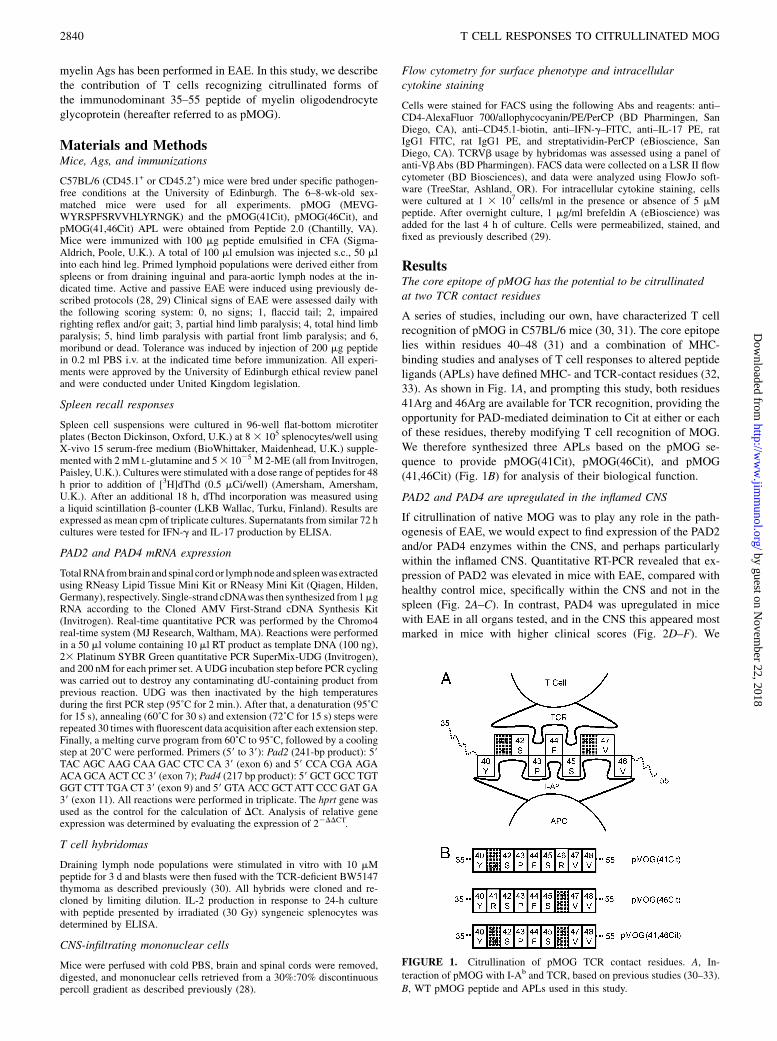
A series of studies, including our own, have characterized T cellrecognition of pMOG in C57BL/6 mice (30, 31). The core epitopelies within residues 40–48 (31) and a combination of MHC-binding studies and analyses of T cell responses to altered peptideligands (APLs) have defined MHC- and TCR-contact residues (32,33). As shown in Fig. 1A, and prompting this study, both residues41Arg and 46Arg are available for TCR recognition, providing theopportunity for PAD-mediated deimination to Cit at either or eachof these residues, thereby modifying T cell recognition of MOG.We therefore synthesized three APLs based on the pMOG se-quence to provide pMOG(41Cit), pMOG(46Cit), and pMOG(41,46Cit) (Fig. 1B) for analysis of their biological function.
PAD2 and PAD4 are upregulated in the inflamed CNS
If citrullination of native MOG was to play any role in the path-ogenesis of EAE, we would expect to find expression of the PAD2and/or PAD4 enzymes within the CNS, and perhaps particularlywithin the inflamed CNS. Quantitative RT-PCR revealed that ex-pression of PAD2 was elevated in mice with EAE, compared withhealthy control mice, specifically within the CNS and not in thespleen (Fig. 2A–C). In contrast, PAD4 was upregulated in micewith EAE in all organs tested, and in the CNS this appeared mostmarked in mice with higher clinical scores (Fig. 2D–F). We
FIGURE 1. Citrullination of pMOG TCR contact residues. A, In-
teraction of pMOG with I-Ab and TCR, based on previous studies (30–33).
B, WT pMOG peptide and APLs used in this study.
2840 T CELL RESPONSES TO CITRULLINATED MOG
by guest on Novem
ber 22, 2018http://w
ww
.jimm
unol.org/D
ownloaded from
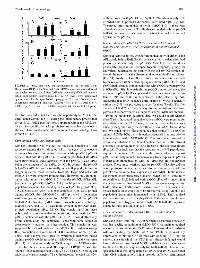
therefore concluded that there was the opportunity for MOG to becitrullinated within the CNS during the inflammatory process thatdrives EAE. PAD2 may be most important within the CNS, be-cause mice specifically lacking this isoform have been previouslyshown to have greatly reduced expression of citrullinated proteinsin the CNS (34).
Citrullinated APLs are immunogenic
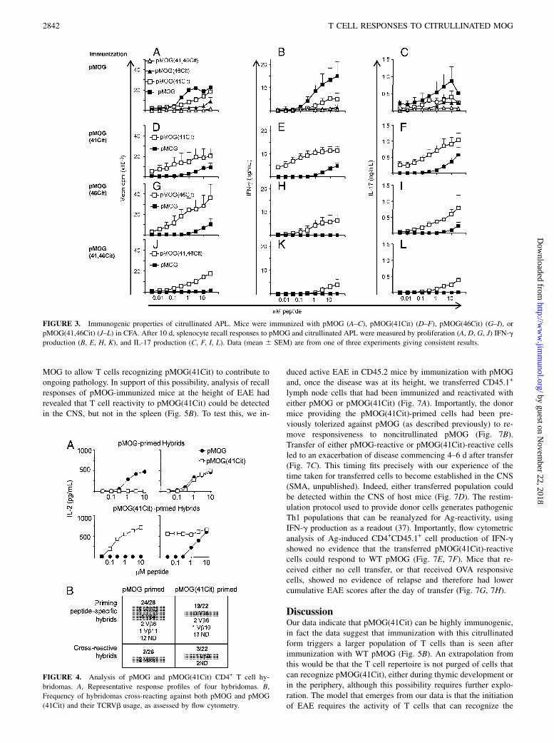
Our next question was whether the mice could mount a T cellresponse against the citrullinated APLs. Analysis of splenocyteresponses from mice immunized against wild-type (WT) pMOGrevealed that both the pMOG(41Cit) and the pMOG(46Cit) APLswere functional as weak agonists, with the pMOG(41Cit) APLsbeing the strongest of these (Fig. 3A–C). In contrast, the pMOG(41,46Cit) APL behaved essentially as a null peptide, failing totrigger any clear recall response from pMOG-primed cells. Allthree APLs were effective immunogens. However, only immuni-zation with either the pMOG(41Cit), or the pMOG(46Cit) APL[and not the pMOG(41,46Cit) APL] could prime an immunepopulation capable of responding to the WT pMOG peptide (Fig.3D–L). Consistent with its higher antigenicity on cells primedagainst pMOG, the pMOG(41Cit) APL provided a populationwith stronger responsiveness for pMOG than did the pMOG(46Cit) APL. Notably, pMOG-driven production of effector cy-tokines (IFNg and IL-17) was more evident in pMOG(41Cit)-primed populations (Fig. 3E–F). The conclusion from thesepolyclonal analyses was that immunization either with the WTpMOG peptide, or with the pMOG(41Cit) APL would effectivelyprime a population that contained T cells with some degree ofcross-reactivity against the other peptide. This conclusion wassupported by a clonal analysis of CD4+ T cell hybridomas (usingIL-2 production as a measure of TCR stimulation of the hybrid-omas). This showed that ∼10% of hybridomas that were raisedagainst pMOG would respond to pMOG(41Cit), and vice versa(Fig. 4). A previous study of TCR usage by pMOG-reactiveT cells has shown that around 50% express TCRVb8.1/2, with the“public” TCR rearrangement using Vb8.2-Jb2.1 (35). Serologicalanalysis of our two panels of T cell hybridomas revealed that 42%
of those primed with pMOG used Vb8 (11/26), whereas only 18%of pMOG(41Cit)-primed hybridomas (4/22) used Vb8 (Fig. 4B).Therefore, after immunization with pMOG(41Cit), there wasa dominant population of T cells that responded only to pMOG(41Cit), but there was also a small fraction that could cross-reactagainst native pMOG.
Immunization with pMOG(41Cit) can initiate EAE, but thisrequires cross-reactive T cell recognition of noncitrullinatedpMOG
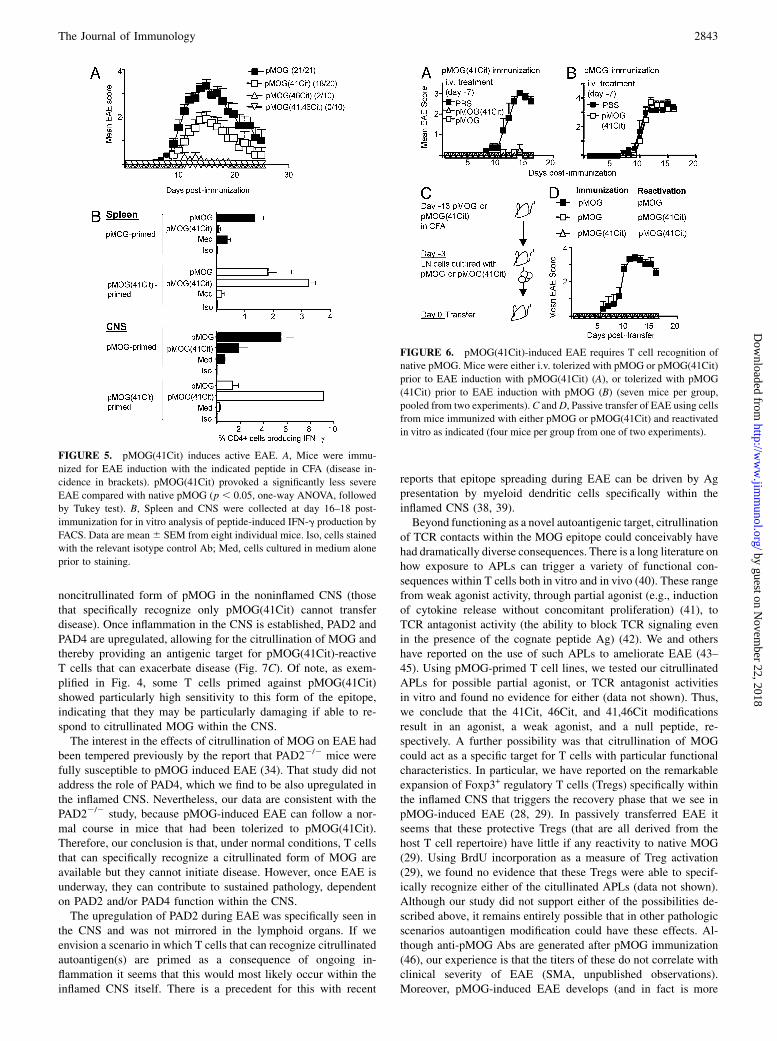
Our next step was to test whether immunization with either of theAPLs could induce EAE. Totally consistent with the data describedpreviously, it was only the pMOG(41Cit) APL that could re-producibly provoke an encephalitogenic response, giving anequivalent incidence to that seen with the WT pMOG peptide, al-though the severity of the disease attained was significantly lower(Fig. 5A). Analysis of recall responses from the CNS revealed ef-fector responses (IFN-g staining) against both pMOG(41Cit) andpMOG in those mice immunized either with pMOG, or with pMOG(41Cit) (Fig. 5B). Interestingly, in pMOG-immunized mice, theresponse to pMOG(41Cit) appeared to be concentrated in the in-flamed CNS and could not be detected in the spleen (Fig. 5B),suggesting that PAD-mediated citrullination of MOG specificallywithin the CNS was providing a target for these T cells. The fre-quencies of IL-17+ cells were always lower, but followed the samepatterns of responsiveness as found with IFN-g (data not shown).From the previously described data, we could not tell whether
those T cells that could recognize native pMOG were required forthe initiation of the EAE lesion, or whether those cells that spe-cifically recognized only the citrullinated form of MOG could dothis. We tested this by tolerizing mice either against WT pMOG oragainst pMOG(41Cit) by i.v. injection of peptide in saline, prior toimmunization with pMOG(41Cit). This tolerance protocol isdemonstrated to lead to deletion of peptide-reactive T cells (36) andprevented the development of EAE in each of the tolerized groups(Fig. 6A). This indicated that the response to the WT peptide wasrequired to initiate EAE; namely, the group tolerized againstpMOG could only mount a (noncross-reactive) response to pMOG(41Cit) after immunization with the APLs but did not developdisease. Those mice tolerized against pMOG(41Cit) also did notdevelop disease, indicating the need for activation by the APL toprovoke the cross-reactive response against pMOG. In the reverseexperiment, mice pretolerized against pMOG(41Cit) were fullysusceptible to EAE induced with pMOG (Fig. 6B), underliningthat a response to citrullinated MOG in vivo was not required forEAE induction. Furthermore, passive transfer experiments re-vealed that disease could only be transferred using lymph nodepopulations from mice immunized with pMOG. This requiredtheir reactivation in vitro with pMOG. If the same lymph nodepopulations were triggered in vitro with pMOG(41Cit), they wereunable to transfer disease (Fig. 6C, 6D).
T cells recognizing citrullinated pMOG can contribute toongoing disease
Our conclusion from the EAE experiments described previouslywas that specific recognition of pMOG(41Cit) was neither requirednor sufficient to initiate the EAE lesion. This would be consistentwith our finding that both PAD2 and PAD4 were markedlyupregulated within the CNS of mice with ongoing EAE (Fig. 2);namely, mice in which the CNS is not already inflamed wouldhave little or no citrullinated MOG available to act as a substratefor those T cells that respond only to pMOG(41Cit). However, wespeculated that this upregulation of PAD2 and PAD4, associatedwith CNS inflammation, might provide sufficient citrullinated
FIGURE 2. Pad2 and Pad4 are upregulated in the inflamed CNS.
Quantitative RT-PCR for Pad2 and Pad4 mRNA expression was performed
on samples taken on day 12 after EAE induction with pMOG, and on those
taken from healthy control mice (0). mRNA levels were normalized
against those for the hprt housekeeping gene. Data are from triplicate
experiments performed. Statistics (Student t test): a, p , 0.001; b, p ,0.005; c, p , 0.01; and d, p , 0.05 compared with the control (0) group.
The Journal of Immunology 2841
by guest on Novem
ber 22, 2018http://w
ww
.jimm
unol.org/D
ownloaded from
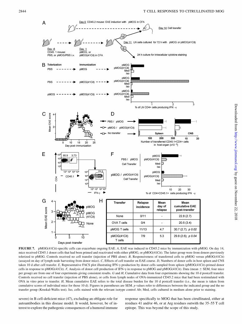
MOG to allow T cells recognizing pMOG(41Cit) to contribute toongoing pathology. In support of this possibility, analysis of recallresponses of pMOG-immunized mice at the height of EAE hadrevealed that T cell reactivity to pMOG(41Cit) could be detectedin the CNS, but not in the spleen (Fig. 5B). To test this, we in-
duced active EAE in CD45.2 mice by immunization with pMOGand, once the disease was at its height, we transferred CD45.1+
lymph node cells that had been immunized and reactivated witheither pMOG or pMOG(41Cit) (Fig. 7A). Importantly, the donormice providing the pMOG(41Cit)-primed cells had been pre-viously tolerized against pMOG (as described previously) to re-move responsiveness to noncitrullinated pMOG (Fig. 7B).Transfer of either pMOG-reactive or pMOG(41Cit)-reactive cellsled to an exacerbation of disease commencing 4–6 d after transfer(Fig. 7C). This timing fits precisely with our experience of thetime taken for transferred cells to become established in the CNS(SMA, unpublished). Indeed, either transferred population couldbe detected within the CNS of host mice (Fig. 7D). The restim-ulation protocol used to provide donor cells generates pathogenicTh1 populations that can be reanalyzed for Ag-reactivity, usingIFN-g production as a readout (37). Importantly, flow cytometricanalysis of Ag-induced CD4+CD45.1+ cell production of IFN-gshowed no evidence that the transferred pMOG(41Cit)-reactivecells could respond to WT pMOG (Fig. 7E, 7F). Mice that re-ceived either no cell transfer, or that received OVA responsivecells, showed no evidence of relapse and therefore had lowercumulative EAE scores after the day of transfer (Fig. 7G, 7H).
DiscussionOur data indicate that pMOG(41Cit) can be highly immunogenic,in fact the data suggest that immunization with this citrullinatedform triggers a larger population of T cells than is seen afterimmunization with WT pMOG (Fig. 5B). An extrapolation fromthis would be that the T cell repertoire is not purged of cells thatcan recognize pMOG(41Cit), either during thymic development orin the periphery, although this possibility requires further explo-ration. The model that emerges from our data is that the initiationof EAE requires the activity of T cells that can recognize the
FIGURE 3. Immunogenic properties of citrullinated APL. Mice were immunized with pMOG (A–C), pMOG(41Cit) (D–F), pMOG(46Cit) (G–I), or
pMOG(41,46Cit) (J–L) in CFA. After 10 d, splenocyte recall responses to pMOG and citrullinated APL were measured by proliferation (A, D, G, J) IFN-g
production (B, E, H, K), and IL-17 production (C, F, I, L). Data (mean 6 SEM) are from one of three experiments giving consistent results.
FIGURE 4. Analysis of pMOG and pMOG(41Cit) CD4+ T cell hy-
bridomas. A, Representative response profiles of four hybridomas. B,
Frequency of hybridomas cross-reacting against both pMOG and pMOG
(41Cit) and their TCRVb usage, as assessed by flow cytometry.
2842 T CELL RESPONSES TO CITRULLINATED MOG
by guest on Novem
ber 22, 2018http://w
ww
.jimm
unol.org/D
ownloaded from
noncitrullinated form of pMOG in the noninflamed CNS (thosethat specifically recognize only pMOG(41Cit) cannot transferdisease). Once inflammation in the CNS is established, PAD2 andPAD4 are upregulated, allowing for the citrullination of MOG andthereby providing an antigenic target for pMOG(41Cit)-reactiveT cells that can exacerbate disease (Fig. 7C). Of note, as exem-plified in Fig. 4, some T cells primed against pMOG(41Cit)showed particularly high sensitivity to this form of the epitope,indicating that they may be particularly damaging if able to re-spond to citrullinated MOG within the CNS.The interest in the effects of citrullination of MOG on EAE had
been tempered previously by the report that PAD22/2 mice werefully susceptible to pMOG induced EAE (34). That study did notaddress the role of PAD4, which we find to be also upregulated inthe inflamed CNS. Nevertheless, our data are consistent with thePAD22/2 study, because pMOG-induced EAE can follow a nor-mal course in mice that had been tolerized to pMOG(41Cit).Therefore, our conclusion is that, under normal conditions, T cellsthat can specifically recognize a citrullinated form of MOG areavailable but they cannot initiate disease. However, once EAE isunderway, they can contribute to sustained pathology, dependenton PAD2 and/or PAD4 function within the CNS.The upregulation of PAD2 during EAE was specifically seen in
the CNS and was not mirrored in the lymphoid organs. If weenvision a scenario in which T cells that can recognize citrullinatedautoantigen(s) are primed as a consequence of ongoing in-flammation it seems that this would most likely occur within theinflamed CNS itself. There is a precedent for this with recent
reports that epitope spreading during EAE can be driven by Agpresentation by myeloid dendritic cells specifically within theinflamed CNS (38, 39).Beyond functioning as a novel autoantigenic target, citrullination
of TCR contacts within the MOG epitope could conceivably havehad dramatically diverse consequences. There is a long literature onhow exposure to APLs can trigger a variety of functional con-sequences within T cells both in vitro and in vivo (40). These rangefrom weak agonist activity, through partial agonist (e.g., inductionof cytokine release without concomitant proliferation) (41), toTCR antagonist activity (the ability to block TCR signaling evenin the presence of the cognate peptide Ag) (42). We and othershave reported on the use of such APLs to ameliorate EAE (43–45). Using pMOG-primed T cell lines, we tested our citrullinatedAPLs for possible partial agonist, or TCR antagonist activitiesin vitro and found no evidence for either (data not shown). Thus,we conclude that the 41Cit, 46Cit, and 41,46Cit modificationsresult in an agonist, a weak agonist, and a null peptide, re-spectively. A further possibility was that citrullination of MOGcould act as a specific target for T cells with particular functionalcharacteristics. In particular, we have reported on the remarkableexpansion of Foxp3+ regulatory T cells (Tregs) specifically withinthe inflamed CNS that triggers the recovery phase that we see inpMOG-induced EAE (28, 29). In passively transferred EAE itseems that these protective Tregs (that are all derived from thehost T cell repertoire) have little if any reactivity to native MOG(29). Using BrdU incorporation as a measure of Treg activation(29), we found no evidence that these Tregs were able to specif-ically recognize either of the citullinated APLs (data not shown).Although our study did not support either of the possibilities de-scribed above, it remains entirely possible that in other pathologicscenarios autoantigen modification could have these effects. Al-though anti-pMOG Abs are generated after pMOG immunization(46), our experience is that the titers of these do not correlate withclinical severity of EAE (SMA, unpublished observations).Moreover, pMOG-induced EAE develops (and in fact is more
FIGURE 6. pMOG(41Cit)-induced EAE requires T cell recognition of
native pMOG. Mice were either i.v. tolerized with pMOG or pMOG(41Cit)
prior to EAE induction with pMOG(41Cit) (A), or tolerized with pMOG
(41Cit) prior to EAE induction with pMOG (B) (seven mice per group,
pooled from two experiments).C andD, Passive transfer of EAE using cells
from mice immunized with either pMOG or pMOG(41Cit) and reactivated
in vitro as indicated (four mice per group from one of two experiments).
FIGURE 5. pMOG(41Cit) induces active EAE. A, Mice were immu-
nized for EAE induction with the indicated peptide in CFA (disease in-
cidence in brackets). pMOG(41Cit) provoked a significantly less severe
EAE compared with native pMOG (p , 0.05, one-way ANOVA, followed
by Tukey test). B, Spleen and CNS were collected at day 16–18 post-
immunization for in vitro analysis of peptide-induced IFN-g production by
FACS. Data are mean6 SEM from eight individual mice. Iso, cells stained
with the relevant isotype control Ab; Med, cells cultured in medium alone
prior to staining.
The Journal of Immunology 2843
by guest on Novem
ber 22, 2018http://w
ww
.jimm
unol.org/D
ownloaded from
severe) in B cell-deficient mice (47), excluding an obligate role forautoantibodies in this disease model. It would, however, be of in-terest to explore the pathogenic consequences of a humoral immune
response specifically to MOG that has been citrullinated, either atresidues 41 and/or 46, or at Arg residues outwith the 35–55 T cellepitope. This was beyond the scope of this study.
FIGURE 7. pMOG(41Cit)-specific cells can exacerbate ongoing EAE. A, EAE was induced in CD45.2 mice by immunization with pMOG. On day 14,
mice received CD45.1 donor cells that had been primed and reactivated with either pMOG, or pMOG(41Cit). The latter group were from donors previously
tolerized to pMOG. Controls received no cell transfer (injection of PBS alone). B, Responsiveness of transferred cells to pMOG versus pMOG(41Cit)
(assayed on day of lymph node harvesting from donor mice). C, Effects of cell transfer on EAE course. D, Numbers of donor cells in host spleen and CNS
taken 10 d after cell transfer. E, Representative FACS plot illustrating IFN-g production by donor cells sampled from spleen (pMOG(41Cit)-primed donor
cells in response to pMOG(41Cit). F, Analysis of donor cell production of IFN-g in response to pMOG and pMOG(41Cit). Data (mean 6 SEM, four mice
per group) are from one of four experiments giving consistent results. G and H, Cumulative data from four experiments showing the 10 d postcell transfer.
Controls received no cell transfer (injection of PBS alone), or cells from lymph nodes of OVA-immunized CD45.2 mice that had been restimulated with
OVA in vitro prior to transfer. H, Mean cumulative EAE refers to the total disease burden for the 10 d postcell transfer (i.e., the mean is taken from
cumulative scores of individual mice for those 10 d). Figures in parentheses are SEM. p values refer to differences between the indicated group and the no
transfer group (Kruskal-Wallis test). Iso, cells stained with the relevant isotype control Ab; Med, cells cultured in medium alone prior to staining.
2844 T CELL RESPONSES TO CITRULLINATED MOG
by guest on Novem
ber 22, 2018http://w
ww
.jimm
unol.org/D
ownloaded from
The evidence for a pathogenic role for citrullinaton of auto-antigens in RA seems beyond doubt (48). Most work in this regardhas focused on humoral immune responses, which are morestraightforward to study. Most compelling is the recent demon-stration of the ability to initiate arthritis with autoantibodies thatspecifically target a citrullinated form of CII (49). Some T cellstudies have been undertaken using citrullinated Ags, however.Reminiscent of earlier observations in celiac disease showing thatwheat gliadin peptides with Gln→Glu substitutions (mimickingthe natural activity of tissue transglutaminase) had enhancedbinding affinities for celiac disease-associated MHC molecules(50, 51), citrullinated vimentin peptides showed enhanced bindingto the RA-associated DRB1p0401 molecule (16). However, ele-vated T cell responses to such peptides have not been reported inautoimmune arthritis (49). A recent report has described the in-duction of arthritis (characterized by synovial hyperplasia andankylosis, but no gross inflammatory infiltrate) in DRB1p0401transgenic mice after immunization with a citrullinated form ofhuman fibrinogen, an autoantigen to which anti-Cit Abs arecommonly found in RA patients (17). Of note, no arthritis wasevident in response to either the noncitrullinated form of humanfibrinogen, or citrullinated mouse fibrinogen. The disease wasassociated with the production of anti-Cit Abs and also showedT cell reactivity to a79–91(84Arg→Cit) peptide of human fibrino-gen, but not to the native form of this peptide. Residue 84 wasmodeled to be a TCR contact residue, but that study did notprovide unequivocal evidence that the T cell response to the cit-rullinated peptide was required for disease; immunization with thea79–91(84Arg→Cit) peptide was not arthritogenic (17). The ef-fects of PTM on T cell reactivity to the autoantigen have beenstudied most extensively in CII-induced arthritis, where glyco-sylation is important (52). In mice expressing either Aq, ortransgenic DR4, the key target for T cells is the 260–270 region.Galactosylation of residue 264 clearly leads to enhanced T cellreactivity and this seems to be also the case in RA (53–55). Usingthe oxidative environment of inflammation as a starting point,a previous study examined malondialdehyde-modification of re-combinant rat MOG and found a more severe form of EAE whenthis form of the autoantigen was used in DBA/1 mice (56). Theauthors concluded that the most likely basis for this was enhanceduptake of the modified MOG by APC via scavenger receptors,rather than any direct effect on TCR recognition.Others have reasoned that recognition of citrullinated MBP by
T cells fromMS patients might reflect an influence on pathogenesis(20). Intriguing evidence that peripheral blood T cell responsesfrom MS patients are more robust when challenged with a cit-rullinated form of an immunodominant MBP peptide than thenoncitrullinated form have lent some support to this view (57).Lewis rats have been reported to develop EAE after active im-munization with a citrullinated form of MBP (21). The im-munodominant epitope of MBP in this setting (72–85) includes anArg at position 76 that acts as a TCR contact residue. Investigationof the effects of an Arg→Cit substitution at this residue concludedthat this was sufficient to abrogate TCR signaling; a MBP (72–85)-reactive T cell clone and lymph node cells from MBP (72–85)-primed rats failed to respond to a 76Cit APL, and vice versa(58). Importantly, although that previous MBP study suggested thecapacity to prime T cells that would respond only to the citrulli-nated form of the epitope, the ability of such cells to contribute topathology was not determined. In this study, we have used precisemodification of a TCR contact residue within the immunodo-minant epitope of MOG to provide proof of principle for this, butonly as far as exacerbating established EAE. Nevertheless, it isconceivable that a pre-established T cell population capable of
recognizing a citrullinated myelin autoantigen could initiate anautoimmune reaction, if PAD2 and/or PAD4 were upregulated inthe CNS by another inflammatory stimulus. In accord with thisthesis, a concise report has shown how citrullination is a generalphenomenon in the inflamed tissues in several diseases of diversecause (3). There are also a wide range of additional PTMs thatmight be either triggered or increased under inflammatory con-ditions (5). The most far-reaching implication of the data pre-sented in this study is that current strategies aimed at identifyingcritical autoantigenic epitopes using the 20 naturally occurringamino acids may be missing a sizeable “hidden” repertoire focusedon modified forms of the autoantigen. Identification of these rep-resents a major challenge.
DisclosuresThe authors have no financial conflicts of interest.
References1. Uy, R., and F. Wold. 1977. Posttranslational covalent modification of proteins.
Science 198: 890–896.2. Doyle, H. A., R. J. Gee, and M. J. Mamula. 2007. Altered immunogenicity of
isoaspartate containing proteins. Autoimmunity 40: 131–137.3. Makrygiannakis, D., E. af Klint, I. E. Lundberg, R. Lofberg, A. K. Ulfgren,
L. Klareskog, and A. I. Catrina. 2006. Citrullination is an inflammation-dependent process. Ann. Rheum. Dis. 65: 1219–1222.
4. Chou, J., J. J. Chen, M. Gross, and B. Roizman. 1995. Association of a M(r)90,000 phosphoprotein with protein kinase PKR in cells exhibiting enhancedphosphorylation of translation initiation factor eIF-2 alpha and premature shutoffof protein synthesis after infection with gamma 134.5- mutants of herpes simplexvirus 1. Proc. Natl. Acad. Sci. USA 92: 10516–10520.
5. Anderton, S. M. 2004. Post-translational modifications of self antigens: im-plications for autoimmunity. Curr. Opin. Immunol. 16: 753–758.
6. Kim, J. K., F. G. Mastronardi, D. D. Wood, D. M. Lubman, R. Zand, andM. A. Moscarello. 2003. Multiple sclerosis: an important role for post-translational modifications of myelin basic protein in pathogenesis. Mol. Cell.Proteomics 2: 453–462.
7. Szekanecz, Z., L. Soos, Z. Szabo, A. Fekete, A. Kapitany, A. Vegvari, S. Sipka,G. Szucs, S. Szanto, and G. Lakos. 2008. Anti-citrullinated protein antibodies inrheumatoid arthritis: as good as it gets? Clin. Rev. Allergy Immunol. 34: 26–31.
8. Harauz, G., and A. A. Musse. 2007. A tale of two citrullines—structural andfunctional aspects of myelin basic protein deimination in health and disease.Neurochem. Res. 32: 137–158.
9. Inagaki, M., H. Takahara, Y. Nishi, K. Sugawara, and C. Sato. 1989. Ca2+-dependent deimination-induced disassembly of intermediate filaments involvesspecific modification of the amino-terminal head domain. J. Biol. Chem. 264:18119–18127.
10. Chavanas, S., M. C. Mechin, R. Nachat, V. Adoue, F. Coudane, G. Serre, andM. Simon. 2006. Peptidylarginine deiminases and deimination in biology andpathology: relevance to skin homeostasis. J. Dermatol. Sci. 44: 63–72.
11. Vossenaar, E. R., T. R. Radstake, A. van der Heijden, M. A. van Mansum,C. Dieteren, D. J. de Rooij, P. Barrera, A. J. Zendman, and W. J. van Venrooij.2004. Expression and activity of citrullinating peptidylarginine deiminase en-zymes in monocytes and macrophages. Ann. Rheum. Dis. 63: 373–381.
12. Asaga, H., K. Akiyama, T. Ohsawa, and A. Ishigami. 2002. Increased and typeII-specific expression of peptidylarginine deiminase in activated microglia butnot hyperplastic astrocytes following kainic acid-evoked neurodegeneration inthe rat brain. Neurosci. Lett. 326: 129–132.
13. Foulquier, C., M. Sebbag, C. Clavel, S. Chapuy-Regaud, R. Al Badine,M. C. Mechin, C. Vincent, R. Nachat, M. Yamada, H. Takahara, et al. 2007.Peptidyl arginine deiminase type 2 (PAD-2) and PAD-4 but not PAD-1, PAD-3,and PAD-6 are expressed in rheumatoid arthritis synovium in close associationwith tissue inflammation. Arthritis Rheum. 56: 3541–3553.
14. Masson-Bessiere, C., M. Sebbag, E. Girbal-Neuhauser, L. Nogueira, C. Vincent,T. Senshu, and G. Serre. 2001. The major synovial targets of the rheumatoidarthritis-specific antifilaggrin autoantibodies are deiminated forms of the alpha-and beta-chains of fibrin. J. Immunol. 166: 4177–4184.
15. Lundberg, K., S. Nijenhuis, E. R. Vossenaar, K. Palmblad, W. J. van Venrooij,L. Klareskog, A. J. Zendman, and H. E. Harris. 2005. Citrullinated proteins haveincreased immunogenicity and arthritogenicity and their presence in arthriticjoints correlates with disease severity. Arthritis Res. Ther. 7: R458–R467.
16. Hill, J. A., S. Southwood, A. Sette, A. M. Jevnikar, D. A. Bell, and E. Cairns.2003. Cutting edge: the conversion of arginine to citrulline allows for a high-affinity peptide interaction with the rheumatoid arthritis-associated HLA-DRB1p0401 MHC class II molecule. J. Immunol. 171: 538–541.
17. Hill, J. A., D. A. Bell, W. Brintnell, D. Yue, B. Wehrli, A. M. Jevnikar,D. M. Lee, W. Hueber, W. H. Robinson, and E. Cairns. 2008. Arthritis inducedby posttranslationally modified (citrullinated) fibrinogen in DR4-IE transgenicmice. J. Exp. Med. 205: 967–979.
18. Moscarello, M. A., D. D. Wood, C. Ackerley, and C. Boulias. 1994. Myelin inmultiple sclerosis is developmentally immature. J. Clin. Invest. 94: 146–154.
The Journal of Immunology 2845
by guest on Novem
ber 22, 2018http://w
ww
.jimm
unol.org/D
ownloaded from

19. Wood, D. D., J. M. Bilbao, P. O’Connors, and M. A. Moscarello. 1996. Acutemultiple sclerosis (Marburg type) is associated with developmentally immaturemyelin basic protein. Ann. Neurol. 40: 18–24.
20. Tranquill, L. R., L. Cao, N. C. Ling, H. Kalbacher, R. M. Martin, andJ. N. Whitaker. 2000. Enhanced T cell responsiveness to citrulline-containingmyelin basic protein in multiple sclerosis patients. Mult. Scler. 6: 220–225.
21. Cao, L., D. Sun, and J. N. Whitaker. 1998. Citrullinated myelin basic proteininduces experimental autoimmune encephalomyelitis in Lewis rats througha diverse T cell repertoire. J. Neuroimmunol. 88: 21–29.
22. Zhou, S. R., M. A. Moscarello, and J. N. Whitaker. 1995. The effects of cit-rullination or variable amino-terminus acylation on the encephalitogenicity ofhuman myelin basic protein in the PL/J mouse. J. Neuroimmunol. 62: 147–152.
23. Akiyama, K., Y. Sakurai, H. Asou, and T. Senshu. 1999. Localization of pepti-dylarginine deiminase type II in a stage-specific immature oligodendrocyte fromrat cerebral hemisphere. Neurosci. Lett. 274: 53–55.
24. Sambandam, T., M. Belousova, M. A. Accaviti-Loper, C. Blanquicett,V. Guercello, R. Raijmakers, and A. P. Nicholas. 2004. Increased peptidylargi-nine deiminase type II in hypoxic astrocytes. Biochem. Biophys. Res. Commun.325: 1324–1329.
25. Nicholas, A. P., T. Sambandam, J. D. Echols, and S. R. Barnum. 2005. Ex-pression of citrullinated proteins in murine experimental autoimmune encepha-lomyelitis. J. Comp. Neurol. 486: 254–266.
26. Raijmakers, R., J. Vogelzangs, J. L. Croxford, P. Wesseling, W. J. van Venrooij,and G. J. Pruijn. 2005. Citrullination of central nervous system proteins duringthe development of experimental autoimmune encephalomyelitis. J. Comp.Neurol. 486: 243–253.
27. Kidd, B. A., P. P. Ho, O. Sharpe, X. Zhao, B. H. Tomooka, J. L. Kanter,L. Steinman, and W. H. Robinson. 2008. Epitope spreading to citrullinated an-tigens in mouse models of autoimmune arthritis and demyelination. ArthritisRes. Ther. 10: R119.
28. McGeachy, M. J., L. A. Stephens, and S. M. Anderton. 2005. Natural recovery andprotection from autoimmune encephalomyelitis: contribution of CD4+CD25+regulatory cells within the central nervous system. J. Immunol. 175: 3025–3032.
29. O’Connor, R. A., K. H. Malpass, and S. M. Anderton. 2007. The inflamed centralnervous system drives the activation and rapid proliferation of Foxp3+ regulatoryT cells. J. Immunol. 179: 958–966.
30. Sweenie, C. H., K. J. Mackenzie, A. Rone-Orugboh, M. Liu, andS. M. Anderton. 2007. Distinct T cell recognition of naturally processed andcryptic epitopes within the immunodominant 35-55 region of myelin oligoden-drocyte glycoprotein. J. Neuroimmunol. 183: 7–16.
31. Mendel, I., N. Kerlero de Rosbo, and A. Ben-Nun. 1995. A myelin oligoden-drocyte glycoprotein peptide induces typical chronic experimental autoimmuneencephalomyelitis in H-2b mice: fine specificity and T cell receptor V betaexpression of encephalitogenic T cells. Eur. J. Immunol. 25: 1951–1959.
32. Ben-Nun, A., N. Kerlero de Rosbo, N. Kaushansky, M. Eisenstein, L. Cohen,J. F. Kaye, and I. Mendel. 2006. Anatomy of T cell autoimmunity to myelinoligodendrocyte glycoprotein (MOG): prime role of MOG44F in selection andcontrol of MOG-reactive T cells in H-2b mice. Eur. J. Immunol. 36: 478–493.
33. Petersen, T. R., E. Bettelli, J. Sidney, A. Sette, V. Kuchroo, and B. T. Backstrom.2004. Characterization of MHC- and TCR-binding residues of the myelin oli-godendrocyte glycoprotein 38-51 peptide. Eur. J. Immunol. 34: 165–173.
34. Raijmakers, R., J. Vogelzangs, J. Raats, M. Panzenbeck, M. Corby, H. Jiang,M. Thibodeau, N. Haynes, W. J. van Venrooij, G. J. Pruijn, and B. Werneburg.2006. Experimental autoimmune encephalomyelitis induction in peptidylargi-nine deiminase 2 knockout mice. J. Comp. Neurol. 498: 217–226.
35. Fazilleau, N., C. Delarasse, C. H. Sweenie, S. M. Anderton, S. Fillatreau,F. A. Lemonnier, D. Pham-Dinh, and J. M. Kanellopoulos. 2006. Persistence ofautoreactive myelin oligodendrocyte glycoprotein (MOG)-specific T cell reper-toires in MOG-expressing mice. Eur. J. Immunol. 36: 533–543.
36. Hochweller, K., and S. M. Anderton. 2005. Kinetics of costimulatory moleculeexpression by T cells and dendritic cells during the induction of tolerance versusimmunity in vivo. Eur. J. Immunol. 35: 1086–1096.
37. O’Connor, R. A., C. T. Prendergast, C. A. Sabatos, C. W. Lau, M. D. Leech,D. C. Wraith, and S. M. Anderton. 2008. Cutting edge: Th1 cells facilitate theentry of Th17 cells to the central nervous system during experimental autoim-mune encephalomyelitis. J. Immunol. 181: 3750–3754.
38. McMahon, E. J., S. L. Bailey, C. V. Castenada, H. Waldner, and S. D. Miller.2005. Epitope spreading initiates in the CNS in two mouse models of multiplesclerosis. Nat. Med. 11: 335–339.
39. Bailey, S. L., B. Schreiner, E. J. McMahon, and S. D. Miller. 2007. CNS myeloidDCs presenting endogenous myelin peptides ‘preferentially’ polarize CD4+ T(H)-17 cells in relapsing EAE. Nat. Immunol. 8: 172–180.
40. Evavold, B. D., J. Sloan-Lancaster, and P. M. Allen. 1993. Tickling the TCR:selective T-cell functions stimulated by altered peptide ligands. Immunol. Today14: 602–609.
41. Evavold, B. D., and P. M. Allen. 1991. Separation of IL-4 production from Thcell proliferation by an altered T cell receptor ligand. Science 252: 1308–1310.
42. Snoke, K., J. Alexander, A. Franco, L. Smith, J. V. Brawley, P. Concannon,H. M. Grey, A. Sette, and P. Wentworth. 1993. The inhibition of different T celllines specific for the same antigen with TCR antagonist peptides. J. Immunol.151: 6815–6821.
43. Anderton, S. M., S. Kissler, A. G. Lamont, and D. C. Wraith. 1999. Therapeuticpotential of TCR antagonists is determined by their ability to modulate a diverserepertoire of autoreactive T cells. Eur. J. Immunol. 29: 1850–1857.
44. Kuchroo, V. K., J. M. Greer, D. Kaul, G. Ishioka, A. Franco, A. Sette,R. A. Sobel, and M. B. Lees. 1994. A single TCR antagonist peptide inhibitsexperimental allergic encephalomyelitis mediated by a diverse T cell repertoire.J. Immunol. 153: 3326–3336.
45. Margot, C. D., M. L. Ford, and B. D. Evavold. 2005. Amelioration of establishedexperimental autoimmune encephalomyelitis by an MHC anchor-substitutedvariant of proteolipid protein 139-151. J. Immunol. 174: 3352–3358.
46. Leech, M. D., C. Y. Chung, A. Culshaw, and S. M. Anderton. 2007. Peptide-based immunotherapy of experimental autoimmune encephalomyelitis withoutanaphylaxis. Eur. J. Immunol. 37: 3576–3581.
47. Fillatreau, S., C. H. Sweenie, M. J. McGeachy, D. Gray, and S. M. Anderton.2002. B cells regulate autoimmunity by provision of IL-10. Nat. Immunol. 3:944–950.
48. van Gaalen, F., A. Ioan-Facsinay, T. W. Huizinga, and R. E. Toes. 2005. Thedevil in the details: the emerging role of anticitrulline autoimmunity in rheu-matoid arthritis. J. Immunol. 175: 5575–5580.
49. Uysal, H., R. Bockermann, K. S. Nandakumar, B. Sehnert, E. Bajtner,A. Engstrom, G. Serre, H. Burkhardt, M. M. Thunnissen, and R. Holmdahl.2009. Structure and pathogenicity of antibodies specific for citrullinated collagentype II in experimental arthritis. J. Exp. Med. 206: 449–462.
50. Molberg, O., S. N. Mcadam, R. Korner, H. Quarsten, C. Kristiansen, L. Madsen,L. Fugger, H. Scott, O. Noren, P. Roepstorff, et al. 1998. Tissue transglutaminaseselectively modifies gliadin peptides that are recognized by gut-derived T cells inceliac disease. Nat. Med. 4: 713–717.
51. Arentz-Hansen, H., R. Korner, O. Molberg, H. Quarsten, W. Vader, Y. M. Kooy,K. E. Lundin, F. Koning, P. Roepstorff, L. M. Sollid, and S. N. McAdam. 2000.The intestinal T cell response to alpha-gliadin in adult celiac disease is focusedon a single deamidated glutamine targeted by tissue transglutaminase. J. Exp.Med. 191: 603–612.
52. Corthay, A., J. Backlund, J. Broddefalk, E. Michaelsson, T. J. Goldschmidt,J. Kihlberg, and R. Holmdahl. 1998. Epitope glycosylation plays a critical rolefor T cell recognition of type II collagen in collagen-induced arthritis. Eur. J.Immunol. 28: 2580–2590.
53. Kjellen, P., U. Brunsberg, J. Broddefalk, B. Hansen, M. Vestberg, I. Ivarsson,A. Engstrom, A. Svejgaard, J. Kihlberg, L. Fugger, and R. Holmdahl. 1998. Thestructural basis of MHC control of collagen-induced arthritis; binding of theimmunodominant type II collagen 256-270 glycopeptide to H-2Aq and H-2Ap
molecules. Eur. J. Immunol. 28: 755–767.54. Backlund, J., A. Treschow, R. Bockermann, B. Holm, L. Holm, S. Issazadeh-
Navikas, J. Kihlberg, and R. Holmdahl. 2002. Glycosylation of type II collagenis of major importance for T cell tolerance and pathology in collagen-inducedarthritis. Eur. J. Immunol. 32: 3776–3784.
55. Backlund, J., S. Carlsen, T. Hoger, B. Holm, L. Fugger, J. Kihlberg,H. Burkhardt, and R. Holmdahl. 2002. Predominant selection of T cells specificfor the glycosylated collagen type II epitope (263-270) in humanized transgenicmice and in rheumatoid arthritis. [see comment] Proc. Natl. Acad. Sci. USA 99:9960–9965.
56. Wallberg, M., J. Bergquist, A. Achour, E. Breij, and R. A. Harris. 2007. Ma-londialdehyde modification of myelin oligodendrocyte glycoprotein leads to in-creased immunogenicity and encephalitogenicity.Eur. J. Immunol. 37: 1986–1995.
57. Deraos, G., K. Chatzantoni, M. T. Matsoukas, T. Tselios, S. Deraos, M. Katsara,P. Papathanasopoulos, D. Vynios, V. Apostolopoulos, A. Mouzaki, andJ. Matsoukas. 2008. Citrullination of linear and cyclic altered peptide ligandsfrom myelin basic protein (MBP(87-99)) epitope elicits a Th1 polarized responseby T cells isolated from multiple sclerosis patients: implications in triggeringdisease. J. Med. Chem. 51: 7834–7842.
58. de Haan, E. C., J. P. Wagenaar-Hilbers, R. M. Liskamp, E. E. Moret, andM. H. Wauben. 2005. Limited plasticity in T cell recognition of modified T cellreceptor contact residues in MHC class II bound peptides. Mol. Immunol. 42:355–364.
2846 T CELL RESPONSES TO CITRULLINATED MOG
by guest on Novem
ber 22, 2018http://w
ww
.jimm
unol.org/D
ownloaded from