
Investigating the role of boundary genes in plant vascular ... · Transgenics were created to...
94
Investigating the role of boundary genes in plant vascular cambiums By Gamalat Allam A thesis submitted to the Faculty of Graduate and Postdoctoral Affairs in partial fulfillment of the requirements for the degree of Master of Science in Biology Carleton University Ottawa, Ontario, Canada © 2018 Gamalat Allam
Transcript of Investigating the role of boundary genes in plant vascular ... · Transgenics were created to...
Investigating the role of boundary genes in plant vascular cambiums
By
Gamalat Allam
A thesis submitted to the Faculty of Graduate and Postdoctoral Affairs in partial
fulfillment of the requirements for the degree of
Master of Science
in
Biology
Carleton University
Ottawa, Ontario, Canada
© 2018 Gamalat Allam
2
ABSTRACT
Meristems play an essential role in plant growth and development. Shoot and root apical
meristems are responsible for primary elongation growth in shoots and roots, respectively.
Secondary or radial growth that follows is dependent on the vascular cambium, a circular meristem
that produces secondary xylem (wood) and secondary phloem (inner bark). Little is known about
the vascular cambium, despite its importance to wood formation in trees. Class I KNOX
homeodomain transcription factors are important regulators of meristem maintenance in plants.
Members of this class, including BREVIPEDICELLUS (BP) maintain the shoot apical meristem
in part by preserving boundaries that keep stem cells separate from differentiating organs. The role
of boundary genes in the vascular cambium is mostly unknown. Here, I provide evidence that
spatial regulation of boundary genes by Class I KNOX genes is important for different reasons in
the vascular cambiums of stem and root-hypocotyl in Arabidopsis thaliana, a model plant species.
In particular, BP repression of boundary genes including BLADE-ON-PETIOLE1 and 2 is
important for the differentiation of reproductive-phase secondary xylem in Arabidopsis root-
hypocotyl but not stem. Populus trichocarpa (Poplar) is a model tree that contains two BOP-like
genes, designed as PtrBPL1 and PtrBPL2. Transgenics were created to examine the expression
and function of these genes in poplar. My data are consistent with a role for boundary genes in
vascular cambiums and shed light on mechanisms controlling secondary growth in trees.

3
ACKNOWLEDGEMENTS
In the name of God, the Most Gracious, the Most Merciful
I first would like to express my deep sincere gratitude to my supervisor Dr. Shelley
Hepworth for her incredible and continuous support of my Master’s degree, her insightful advice,
unbelievable patience, encouragement, and great knowledge. I am also thankful for her
constructive guidance with my research and her help with making figures and editing this thesis.
Providing me with this excellent opportunity in her lab made a real change in my life, which is
greatly appreciated!
I would also like to thank my committee members Dr. Douglas Johnson and Dr. John
Vierula for their guidance as my committee advisors and for providing me with their constructive
feedback, insightful recommendation, and encouragement to help me achieve my precise research
goal.
My deepest thanks to Dr. Jhadeswar Murmu for his patience in teaching experimental
techniques when I first started in the lab and helping me with cloning the RNAi construct. I would
also like to thank Ying Wang for his awesome help and answering my questions. Special thanks
to Adina, who has been a great team player, friend, and family! I would like to acknowledge Chris,
Laura, Kevin for their assistance in the lab and being a remarkable team. Many thanks to all the
fellow lab mates from the Rowland lab for their help with my experiments, productive discussions,
and generating an energetic and enjoyable working environment.
I finally would like to thank my family for their unconditional love, support and
encouragement that has helped me succeed. I owe my deepest gratitude to my husband, Raz
Kareem, for his endless love, incredible support and understanding. I am so blessed to have these
amazing people in my life without whom, I could not have succeeded in accomplishing this goal.
4
PREFACE
This thesis explores mechanisms governing regulation of secondary growth in Arabidopsis
and poplar.
Select figures and text in this thesis appear in the publication: Repression of BLADE-ON-
PETIOLE genes by KNOX homeodomain protein BREVIPEDICELLUS is essential for
differentiation of secondary xylem in Arabidopsis root (2017) Natalie Woerlen, Gamalat
Allam*, Adina Popescu*, Laura Corrigan, Véronique Pautot, and Shelley R. Hepworth. Planta
245, 1079-1090. *These two authors contributed equally to the work.
I carried out the majority of work in this thesis but acknowledge that some of the data were
obtained in collaboration with the individuals listed below:
Natalie Woerlen, Eryang Li, Veronique Pautot, and Shelley Hepworth designed the
research. Natalie Woerlen initiated the work on Arabidopsis root-hypocotyl secondary growth and
wrote the first draft of the published manuscript. Adina Popescu and I contributed equally to
addressing reviewer comments. Laura Corrigan assisted me in quantitative analysis of root
secondary growth. Dr. Eryang Li and Bhaswati Devi initiated the work on poplar and partially
made constructs for functional analysis of PtrBPL1 and PtrBPL2 in Arabidopsis and poplar.
Jhadeswar Murmu finished construction of the RNAi construct for knockdown of PtrBPL1/2
expression in poplar. I would like to thank my supervisor Dr. Shelley Hepworth for making figures
and editing this thesis.

5
TABLE OF CONTENTS
ABSTRACT ................................................................................................................................... 2
ACKNOWLEDGEMENTS ......................................................................................................... 3
PREFACE ...................................................................................................................................... 4
TABLE OF CONTENTS ............................................................................................................. 5
GLOSSARY OF GENETIC TERMS ......................................................................................... 8
GENETIC NOMENCLATURE IN ARABIDOPSIS THALIANA ............................................ 9
LIST OF ABBREVIATIONS .................................................................................................... 10
LIST OF TABLES ...................................................................................................................... 13
LIST OF FIGURES .................................................................................................................... 14
CHAPTER 1: INTRODUCTION .............................................................................................. 15
1.1 Thesis overview .................................................................................................................. 15
1.2 Arabidopsis thaliana as a model plant species ................................................................. 15
1.3 Arabidopsis thaliana life cycle ........................................................................................... 16
1.4 Meristems ........................................................................................................................... 17
1.4.1 Primary growth........................................................................................................... 17
1.4.2 Secondary growth ....................................................................................................... 18
1.4.2.1 Inflorescence stem ............................................................................................... 19
1.4.2.2 Root-hypocotyl .................................................................................................... 19
1.5 Conserved patterns in plant development ...................................................................... 20
1.6 Organization of the SAM .................................................................................................. 20
1.7 Meristem-organ boundaries ............................................................................................. 21
1.8 SAM maintenance ............................................................................................................. 21
1.8.1 WUSCHEL-CLAVATA feedback loop .................................................................... 22
1.8.2 KNOX-BELL homeodomain proteins ...................................................................... 22
1.8.3 Organ boundary genes ............................................................................................... 24
1.9 Vascular cambium............................................................................................................. 25
1.9.1 Stem.............................................................................................................................. 25
1.9.2 Hypocotyl ..................................................................................................................... 27
1.9.3 Poplar tree ................................................................................................................... 27
1.9.3.1. Class I KNOX genes........................................................................................... 28
1.9.3.2 Boundary genes in trees...................................................................................... 29
6
1.10 Thesis rationale and research questions........................................................................ 29
Chapter 2: MATERIALS AND METHODS ............................................................................ 42
2.1 Arabidopsis plant material and growth conditions ........................................................ 42
2.2 Analysis of secondary growth .......................................................................................... 43
2.3 Embedding and sectioning ............................................................................................... 43
2.4 Histochemical analyses ..................................................................................................... 44
2.5 -Glucuronidase (GUS) staining assay ............................................................................ 45
2.6 Poplar plant materials and growth conditions ............................................................... 46
2.7 Constructs for transformation of poplar ........................................................................ 46
2.8 Agrobacterium-mediated transformation of Populus trichocarpa ................................. 47
2.9 Genomic DNA extraction from poplar ............................................................................ 50
2.10 Poplar genotyping .......................................................................................................... 50
2.11 Poplar GUS staining ....................................................................................................... 51
Chapter 3: RESULTS ................................................................................................................. 53
3.1 A gradient of secondary growth in Arabidopsis taproots ............................................. 53
3.2 BOP1/2 loss and gain of function mutants alter xylem fibre differentiation ............... 53
3.3 BP interacts antagonistically with boundary genes to promote xylem fiber
differentiation in roots ............................................................................................................ 54
3.4 BP spatial regulation of boundary genes is important for differentiation of
reproductive-stage xylem ........................................................................................................ 55
3.5 Analysis of PtrBPL1 or PtrBPL2 gain-of-function in Arabidopsis plants .................... 56
3.5.1 Inflorescence stem ....................................................................................................... 56
3.5.2 Root-hypocotyl ............................................................................................................ 57
3.6 Transgenic poplars expressing BOP1:GUS and BOP2:GUS reporter genes ............... 57
3.7 Transgenic poplars expressing a construct for RNAi-mediated PtrBPL1/2 gene
silencing .................................................................................................................................... 58
3.8 Transgenic poplars expressing D35S:BPL1 and D35S:BPL2 transgenes .................... 58
Chapter 4: DISCUSSION........................................................................................................... 73
4.1 Spatial regulation of boundary genes is important for xylem differentiation in roots 73
4.2 Alignment of root and hypocotyl models ........................................................................ 74
4.3 Other SAM-boundary regulators .................................................................................... 74
4.4 Differential impact on xylem II ........................................................................................ 75
4.5 Lignin biosynthesis ............................................................................................................ 76

7
4.6 Comparison to trees .......................................................................................................... 76
4.6.1 Class I KNOX genes ................................................................................................... 76
4.6.2 BLADE-ON-PETIOLE genes ..................................................................................... 77
4.6.3 Transgenic poplar ....................................................................................................... 77
4.7 Final conclusion and future directions ............................................................................ 79
REFERENCES ............................................................................................................................ 80
8
GLOSSARY OF GENETIC TERMS
Loss-of-function: loss or reduction of activity
Gain-of-function: ectopic or increased activity
Phenotypic suppression: shift toward wild type phenotype
Phenotypic enhancement: worsening a mutant phenotype
Redundancy: when there is genetic compensation in the event of a gene loss-of-function
Homolog: genes sharing a common ancestor in evolution
Ortholog: genes in different organisms that descend from a common ancestor (often with the
same function)
Paralog: genes that are related by a duplication event within the genome of an organism

9
GENETIC NOMENCLATURE IN ARABIDOPSIS THALIANA
Wild type gene: BOP1
Wild type protein: BOP1
Loss-of-function mutant (homozygous): bop1
Loss-of-function mutant (hemizygous): bop1/+
Gain-of-function mutant (dominant): bop1-6D
Double mutant: bop1 bop2
Promoter fusion to a gene coding region: 35S:BOP1
Protein fusion: BOP1-GR
10
LIST OF ABBREVIATIONS
AS1 ASYMMETRIC LEAVES1
At Arabidopsis thaliana
ATH1 ARABIDOPSIS THALIANA HOMEOBOX GENE1
ARK ARBORKNOX
BAP 6-benzylaminopurine
BELL BEL1-like
BOP BLADE-ON-PETIOLE
BP BREVIPEDICELLUS
BPL BOP-like
BTB/POZ Bric-a-Brac, Tram Track, Broad Complex/POX virus and Zinc finger
CIM Callus Induction Medium
CLE CLAVATA/ESR related
CLV CLAVATA
Col Columbia (wild-type ecotype of Arabidopsis thaliana)
CUC CUP-SHAPED COTYLEDON
CTAB cetyl trimethylammonium bromide
CZ central zone
ddH2O distilled deionized water
D35S CaMV double 35S Cauliflower Mosaic Virus promoter
DNA deoxyribose nucleic acid
EDTA ethylenediaminetetraacetic acid
FAA formaldehyde-acetic-acid-alcohol
11
GA gibberellin
GUS β-Glucuronidase
h hour
HCl hydrochloric acid
IBA indole-3-butyric acid
KNAT KNOTTED1-LIKE FROM ARABIDOPSIS THALIANA
KNOX KNOTTED1-LIKE HOMEOBOX
LB Lysogeny Broth
LOB Lateral Organ Boundary
MES 2-(N-morpholino) ethanesulfonic acid
miR microRNA
MS Murashige and Skoog
NAA 1-naphthaleneacetic acid
NAC NAM, ATAF, and CUC
NPTII neomycin phosphotransferase II
OC Organizing center
PCR Polymerase chain reaction
PNF POUND-FOOLISH
PNY PENNYWISE
Ptr Populus trichocarpa
PZ peripheral zone
qRT-PCR quantitative reverse transcriptase polymerase chain reaction
RAM Root apical meristem
12
RIM Root induction medium
RNAi RNA interference
RZ rib zone
SAM Shoot apical meristem
SEM Shoot elongation medium
SDS sodium dodecyl sulphate
STM SHOOT MERISTEMLESS
TALE THREE-AMINO ACID-LOOP-EXTENSION
T-DNA transfer DNA
TDZ thiadiazuron
UBQ UBIQUITIN
VC vascular cambium
WT wild-type
w/v weight / volume
WOX WUSCHEL-RELATED HOMEOBOX
WUS WUSCHEL
X-gluc 5-bromo-4-chloro-3-indoxyl--D-glucuronide
2ip 6-(γ, γ-dimethylallylamino)purine
13
LIST OF TABLES
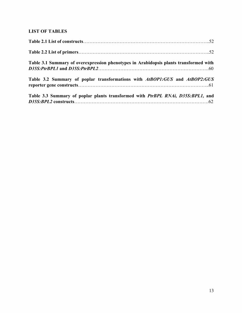
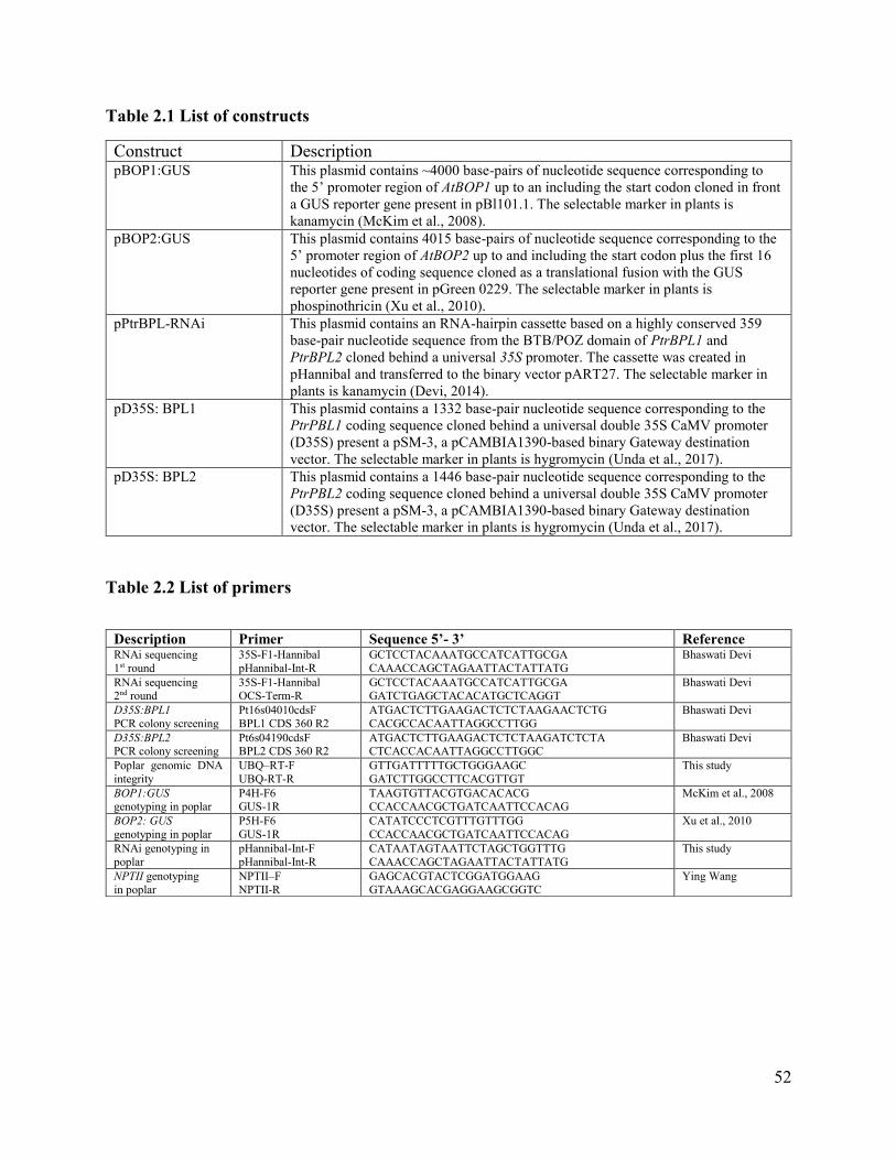
Table 2.1 List of constructs……………………………………………………………………..52
Table 2.2 List of primers………………………………………………………………………..52
Table 3.1 Summary of overexpression phenotypes in Arabidopsis plants transformed with
D35S:PtrBPL1 and D35S:PtrBPL2…………………………………………………………….60
Table 3.2 Summary of poplar transformations with AtBOP1:GUS and AtBOP2:GUS
reporter gene constructs………………………………………………………………………..61
Table 3.3 Summary of poplar plants transformed with PtrBPL RNAi, D35S:BPL1, and
D35S:BPL2 constructs………………………………………………………………………….62

14
LIST OF FIGURES
Figure 1.1 Arabidopsis thaliana life cycle and plant architecture ……………………………30
Figure 1.2 Meristems and vascular organization in Arabidopsis thaliana…………………...32
Figure 1.3 Cross-section of the Arabidopsis hypocotyl viewed by confocal microscopy…...34
Figure 1.4 Organization of the SAM…………………………………………………………...35
Figure 1.5 BLADE-ON-PETIOLE loss and gain-of-function phenotypes…………………...36
Figure 1.6 Stem vascular patterning in Arabidopsis and poplar tree………………………..38
Figure 1.7 STM and BP expression pattern in stem primary vasculature…………………..40
Figure 1.8 Models and research questions…………………………………………………….41
Figure 3.1 Anatomy and developmental gradient of lignin deposition in taproot of wild-type
plants…………………………………………………………………………………………….64
Figure 3.2 Lignin deposition in wild-type and mutant secondarily thickened roots………..66
Figure 3.3 Depiction of bop1 bop2 bp-2 mutants with a weak rescue phenotype and effect of
external GA on bop1-6D xylem formation…………………………………………………….68
Figure 3.4 Boundary gene expression in wild-type and bp-2 secondarily thickened roots...69
Figure 3.5 Lignin deposition in inflorescence stem of Arabidopsis plants expressing
D35S:PtrBPL1 and D35S:PtrBPL2 transgenes………………………………………………..71
Figure 3.6 Lignin deposition in hypocotyl of Arabidopsis plants expressing D35S:PtrBPL1
and D35S:PtrBPL2 transgenes…………………………………………………………………72

15
CHAPTER 1: INTRODUCTION
1.1 Thesis overview
Plant growth and development relies on meristems. Primary meristems at the shoot and
root tips are responsible for vertical growth of shoots and roots, respectively. Vascular meristems
called procambia derived from these meristems provide primary tissues of the vascular system:
xylem and phloem. Secondary growth that follows relies on the vascular cambium, a ring-like
meristem derived from procambium. The vascular cambium allows plant organs to thicken in
circumference through the production of secondary xylem (wood) and secondary phloem (bark).
Knowledge of the vascular cambium is incomplete despite its importance to wood formation in
trees (Nieminen et al., 2015).
Class I KNOX homeodomain transcription factors are important regulators of meristem
activity in land plants (Tsuda and Hake, 2016). Members of this class maintain the SAM in part
by establishing low-growth regions called boundaries that separate forming organs from the
meristem. Disruption of boundaries terminates the stem cell population and impairs organ
formation and patterning (Hepworth and Pautot, 2015). The vascular cambium is a source of stem
cells much like the apical meristem of shoots. How do mechanisms responsible for maintenance
of the SAM apply to the vascular cambium? My thesis investigates the role of boundary genes in
plant secondary growth using Arabidopsis thaliana (thale cress) and Populus trichocarpa (poplar
tree) as study species.
1.2 Arabidopsis thaliana as a model plant species
Arabidopsis thaliana (Arabidopsis) was introduced as genetic model for plant biology
studies in the late 1970’s (Somerville and Koorneef, 2002). This plant is a member of the
16
Brassicaceae family, which includes economically important species like broccoli, cabbage,
horseradish, mustard, and oilseed rape (Somerville and Koorneef, 2002; Koornneef and Meinke,
2010). Arabidopsis was chosen as a model species based on characteristics that make it easy to
work with in a lab. Adult plants are relatively small (30-40 cm in height) with simple growth
requirements and a short life cycle (6-8 weeks) (Meyerowitz, 1989; Somerville and Koorneef,
2002). The plant is a self-fertilizing diploid with prolific seed production. Loss and gain-of-
function mutants are easy to make using chemical mutagens, transposons, or Agrobacterium-
mediated transformation, which can also be used to create transgenic plants. The Arabidopsis
genome is simple and compact, containing five chromosomes whose fully-annotated sequences
can be accessed through The Arabidopsis Information Resource (TAIR; www.arabidopsis.org).
Stock centers in North America, Europe, and Asia are set up to distribute seeds, cDNA clones,
indexed libraries of T-DNA insertion mutants and other resources for a small cost (Koornneef and
Meinke, 2010). Arabidopsis has proved to be one of the most important discovery tools for plant
biology in the last twenty years. Although lacking in economic value, genome composition and
developmental programs are highly conserved with crop species. Information gained from
Arabidopsis has been successfully applied to the improvement of agronomic traits in numerous
crop species (Ferrier et al., 2011; Lavagi et al., 2012).
1.3 Arabidopsis thaliana life cycle
The life cycle of a flowering plant has embryonic, vegetative, and reproductive phases
(Figure 1.1). Embryonic development begins at fertilization. Division of the fertilized egg
produces an embryo within a seed (Wolpert et al., 2015). The mature embryo contains the first
basic set of plant structures: one or two leaves (cotyledons), a stem (hypocotyl), and a primary root
17
(radicle). Post-embryonic development begins at germination and is divided into two phases:
vegetative and reproductive. In Arabidopsis, the vegetative phase is dominated by the production
of leaves. By contrast, the reproductive phase is dominated by the production of lateral branches
and flowers. Stem elongation during the reproductive phase distributes these structures to produce
an inflorescence (Figure 1.1). Each flower is composed of four whorls of floral organs: sepals,
petals, stamens, and carpels. Two fused carpels at the center of the flower form the gynoecium
(female reproductive organ). The gynoecium contains three tissues from top to bottom: the stigma,
style, and ovary which gives rise to ovules internally. Pollen landing on the stigma fertilize the
ovules, which mature into seeds, thus completing the life cycle (Wolpert et al., 2015).
1.4 Meristems
Post-embryonic growth of a plant relies on meristems. Meristems are specialized niches
for stem cells. Different types of meristems can be found in different parts of the plant. Within the
meristem, continuous division of stem cells replenishes the meristem and provides cells for organ
production. Daughter cells exiting the meristem differentiate into all of the specialized cell types
that make up the plant body (Aichinger et al., 2012).
1.4.1 Primary growth
The vertical growth of plants depends on two meristems that originate in the embryo: the
shoot apical meristem (SAM) and the root apical meristem (RAM) (Figure 1.2). The SAM
produces all of the above-ground structures in a plant. By contrast, the RAM produces the root
system (Barton, 2010; Wolpert et al., 2015). Vascular procambial meristems provide the primary
vascular tissue of roots and shoots: xylem and phloem.
18
The vascular anatomy of shoots and roots differs (Figure 1.2). In the stem, individual
vascular bundles are arranged in ring. Each bundle has a sandwich-like structure. Procambial cells
at the center of each “sandwich” divide to make daughter cells that differentiate as xylem on the
inside and phloem on the outside (Sanchez et al., 2012). Vascular tissues in the root and hypocotyl
instead form a central bundle with procambium interspersed between xylem and phloem
(Miyashima et al., 2013; Nieminen et al., 2015). Regardless of origin, xylem provides mechanical
support and transports water and solutes from the roots to the leaves. Phloem transports sugars
from photosynthesis, signaling molecules, and metabolites from leaves to the rest of the plant
(Miyashima et al., 2013; Nieminen et al., 2015).
1.4.2 Secondary growth
Primary growth is followed by secondary growth in which roots and shoots grow in
circumference. This thickening relies on the vascular cambium, a ring-like lateral meristem
derived from procambium (Nieminen et al., 2015). Vascular cambium cells divide to produce
secondary xylem (wood) on the inside and secondary phloem (inner bark) on the outside.
Secondary xylem consists of non-lignified xylem parenchyma interspersed with lignified xylem
vessels and fibres and secondary phloem consists of sieve-elements and their companion cells,
phloem fibres, and phloem parenchyma (Miyashima et al., 2013; Nieminen et al., 2015).
Arabidopsis is a popular model for studying wood development based on extensive secondary
growth in the inflorescence stem, hypocotyl, and roots (Esau, 1965; Chaffey et al., 2002; Beck,
2010; Nieminen et al., 2015). All three tissues establish prominent vasculature similar to
angiosperm trees (Ragni and Hardtke, 2014; Nieminen et al., 2015; Barra-Jiménez and Ragni,
2017).
19
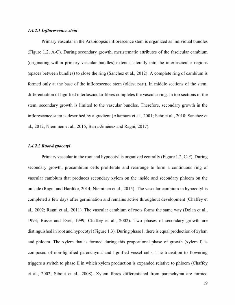
1.4.2.1 Inflorescence stem
Primary vascular in the Arabidopsis inflorescence stem is organized as individual bundles
(Figure 1.2, A-C). During secondary growth, meristematic attributes of the fascicular cambium
(originating within primary vascular bundles) extends laterally into the interfascicular regions
(spaces between bundles) to close the ring (Sanchez et al., 2012). A complete ring of cambium is
formed only at the base of the inflorescence stem (oldest part). In middle sections of the stem,
differentiation of lignified interfascicular fibres completes the vascular ring. In top sections of the
stem, secondary growth is limited to the vascular bundles. Therefore, secondary growth in the
inflorescence stem is described by a gradient (Altamura et al., 2001; Sehr et al., 2010; Sanchez et
al., 2012; Nieminen et al., 2015; Barra-Jiménez and Ragni, 2017).
1.4.2.2 Root-hypocotyl
Primary vascular in the root and hypocotyl is organized centrally (Figure 1.2, C-F). During
secondary growth, procambium cells proliferate and rearrange to form a continuous ring of
vascular cambium that produces secondary xylem on the inside and secondary phloem on the
outside (Ragni and Hardtke, 2014; Nieminen et al., 2015). The vascular cambium in hypocotyl is
completed a few days after germination and remains active throughout development (Chaffey et
al., 2002; Ragni et al., 2011). The vascular cambium of roots forms the same way (Dolan et al.,
1993; Busse and Evet, 1999; Chaffey et al., 2002). Two phases of secondary growth are
distinguished in root and hypocotyl (Figure 1.3). During phase I, there is equal production of xylem
and phloem. The xylem that is formed during this proportional phase of growth (xylem I) is
composed of non-lignified parenchyma and lignified vessel cells. The transition to flowering
triggers a switch to phase II in which xylem production is expanded relative to phloem (Chaffey
et al., 2002; Sibout et al., 2008). Xylem fibres differentiated from parenchyma are formed
20
exclusively during this expansion phase of growth (xylem II), which is seen as a dense ring of
lignified fibers interspersed with vessels (Chaffey et al., 2002; Sibout et al., 2008; Ragni and
Hardtke, 2014; Nieminen et al., 2015).
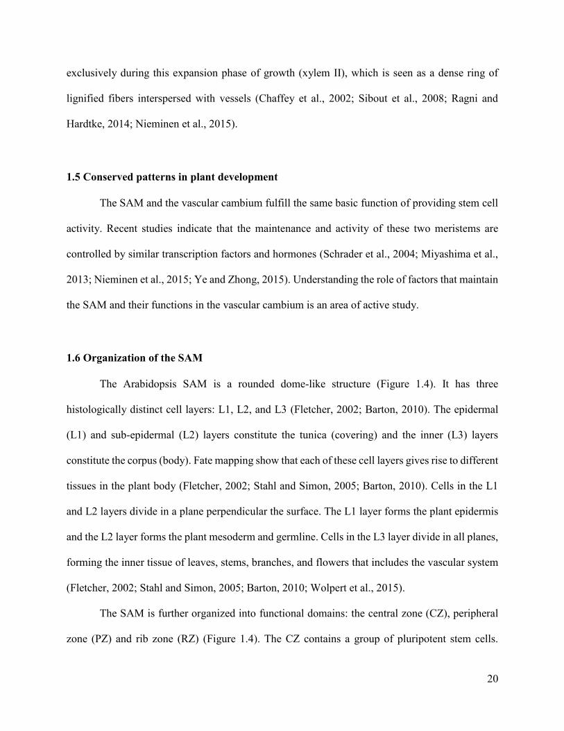
1.5 Conserved patterns in plant development
The SAM and the vascular cambium fulfill the same basic function of providing stem cell
activity. Recent studies indicate that the maintenance and activity of these two meristems are
controlled by similar transcription factors and hormones (Schrader et al., 2004; Miyashima et al.,
2013; Nieminen et al., 2015; Ye and Zhong, 2015). Understanding the role of factors that maintain
the SAM and their functions in the vascular cambium is an area of active study.
1.6 Organization of the SAM
The Arabidopsis SAM is a rounded dome-like structure (Figure 1.4). It has three
histologically distinct cell layers: L1, L2, and L3 (Fletcher, 2002; Barton, 2010). The epidermal
(L1) and sub-epidermal (L2) layers constitute the tunica (covering) and the inner (L3) layers
constitute the corpus (body). Fate mapping show that each of these cell layers gives rise to different
tissues in the plant body (Fletcher, 2002; Stahl and Simon, 2005; Barton, 2010). Cells in the L1
and L2 layers divide in a plane perpendicular the surface. The L1 layer forms the plant epidermis
and the L2 layer forms the plant mesoderm and germline. Cells in the L3 layer divide in all planes,
forming the inner tissue of leaves, stems, branches, and flowers that includes the vascular system
(Fletcher, 2002; Stahl and Simon, 2005; Barton, 2010; Wolpert et al., 2015).
The SAM is further organized into functional domains: the central zone (CZ), peripheral
zone (PZ) and rib zone (RZ) (Figure 1.4). The CZ contains a group of pluripotent stem cells.
21
Daughter cells from division of CZ cells are displaced laterally into the PZ where organogenesis
takes place. The RZ provides cells for upward growth of the stem (Fletcher, 2002; Barton, 2010;
Wolpert et al., 2015). A subdomain of the RZ called the organizing center (OC) controls the size
of the CZ (Barton, 2010; Aichinger et al., 2012; Wolpert et al., 2015). Differential regulation of
activities in the CZ, PZ and RZ are responsible for changing production of leaves, stems, branches,
and flowers throughout the life cycle (Fletcher, 2002; Sanchez et al., 2012; Bencivenga et al.,
2016).
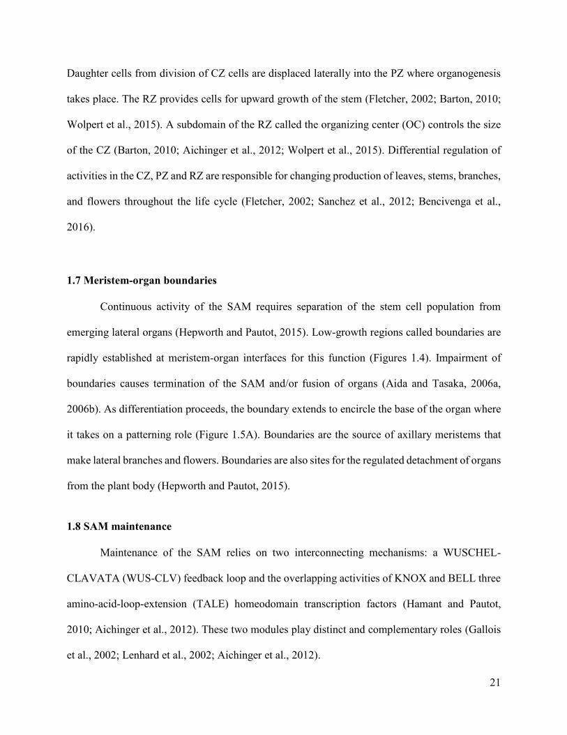
1.7 Meristem-organ boundaries
Continuous activity of the SAM requires separation of the stem cell population from
emerging lateral organs (Hepworth and Pautot, 2015). Low-growth regions called boundaries are
rapidly established at meristem-organ interfaces for this function (Figures 1.4). Impairment of
boundaries causes termination of the SAM and/or fusion of organs (Aida and Tasaka, 2006a,
2006b). As differentiation proceeds, the boundary extends to encircle the base of the organ where
it takes on a patterning role (Figure 1.5A). Boundaries are the source of axillary meristems that
make lateral branches and flowers. Boundaries are also sites for the regulated detachment of organs
from the plant body (Hepworth and Pautot, 2015).
1.8 SAM maintenance
Maintenance of the SAM relies on two interconnecting mechanisms: a WUSCHEL-
CLAVATA (WUS-CLV) feedback loop and the overlapping activities of KNOX and BELL three
amino-acid-loop-extension (TALE) homeodomain transcription factors (Hamant and Pautot,
2010; Aichinger et al., 2012). These two modules play distinct and complementary roles (Gallois
et al., 2002; Lenhard et al., 2002; Aichinger et al., 2012).
22
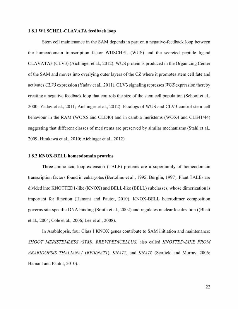
1.8.1 WUSCHEL-CLAVATA feedback loop
Stem cell maintenance in the SAM depends in part on a negative-feedback loop between
the homeodomain transcription factor WUSCHEL (WUS) and the secreted peptide ligand
CLAVATA3 (CLV3) (Aichinger et al., 2012). WUS protein is produced in the Organizing Center
of the SAM and moves into overlying outer layers of the CZ where it promotes stem cell fate and
activates CLV3 expression (Yadav et al., 2011). CLV3 signaling represses WUS expression thereby
creating a negative feedback loop that controls the size of the stem cell population (Schoof et al.,
2000; Yadav et al., 2011; Aichinger et al., 2012). Paralogs of WUS and CLV3 control stem cell
behaviour in the RAM (WOX5 and CLE40) and in cambia meristems (WOX4 and CLE41/44)
suggesting that different classes of meristems are preserved by similar mechanisms (Stahl et al.,
2009; Hirakawa et al., 2010; Aichinger et al., 2012).
1.8.2 KNOX-BELL homeodomain proteins
Three-amino-acid-loop-extension (TALE) proteins are a superfamily of homeodomain
transcription factors found in eukaryotes (Bertolino et al., 1995; Bürglin, 1997). Plant TALEs are
divided into KNOTTED1-like (KNOX) and BELL-like (BELL) subclasses, whose dimerization is
important for function (Hamant and Pautot, 2010). KNOX-BELL heterodimer composition
governs site-specific DNA binding (Smith et al., 2002) and regulates nuclear localization ((Bhatt
et al., 2004; Cole et al., 2006; Lee et al., 2008).
In Arabidopsis, four Class I KNOX genes contribute to SAM initiation and maintenance:
SHOOT MERISTEMLESS (STM), BREVIPEDICELLUS, also called KNOTTED-LIKE FROM
ARABIDOPSIS THALIANA1 (BP/KNAT1), KNAT2, and KNAT6 (Scofield and Murray, 2006;
Hamant and Pautot, 2010).
23
STM is expressed in all subdomains of the SAM except at sites of organ initiation
consistent with essential roles in meristem initiation, meristem maintenance, and the initiation of
boundaries (Endrizzi et al., 1996; Long et al., 1996; Landrein et al., 2015; Balkunde et al., 2017).
Strong stm mutants lack a SAM or terminate growth after forming a few leaves (Endrizzi et al.,
1996; Long et al., 1996). STM directly and indirectly contributes to the expression of other Class
I KNOX genes in subdomains of the SAM (Belles-Boix et al., 2006; Scofield et al., 2013; Scofield
et al., 2014). BP is expressed in the PZ and RZ (Lincoln et al., 1994), KNAT2 is expressed in the
RZ and organ boundaries, and KNAT6 is expressed in organ boundaries (Belles-Boix et al., 2006).
Mutations in BP enhance the phenotype of weak stm mutants and genetic experiments show that
BP can compensate for missing STM activity in the CZ (Byrne et al., 2002; Scofield et al., 2013).
Mutations in KNAT6 also enhance the phenotype of weak stm mutants in organ separation and
SAM maintenance indicating their involvement (Belles-Boix et al., 2006). Genetic experiments
have yet to identify a role for KNAT2 in the SAM (Byrne et al., 2002).
At least three BELL members: PENNYWISE (PNY), POUND-FOOLISH (PNF), and
ARABIDOPSIS THALIANA HOMEOBOX GENE1 (ATH1) interact with STM and allow it to
properly function (Bellaoui et al., 2001; Byrne et al., 2003; Smith and Hake, 2003; Bhatt et al.,
2004; Cole et al., 2006; Kanrar et al., 2006; Rutjens et al., 2009; Li et al., 2012). Interactions with
PNY and other BELLs are required for nuclear import of STM (Cole et al., 2006). Triple mutants
ath1-1 pny pnf have a phenotype similar to weak stm mutants, possibly caused by depletion of
nuclear-localized STM (Rutjens et al., 2009). STM-BELL complexes maintains the SAM by
repressing differentiation of the stem cell population and also by initiating and maintaining organ
boundaries (Hepworth and Pautot, 2015). ATH1, which contributes to boundary establishment, is
expressed in the SAM during vegetative development and downregulated at the transition to

24
flowering (Proveniers et al., 2007; Gómez-Mena and Sablowski, 2008). A specific role for ATH1
in the establishment of boundaries is shown by fused organs in the ath1-3 mutant (Gómez-Mena
and Sablowski, 2008). PNY and PNF are expressed in an overlapping domain with STM (Long et
al., 1996; Smith et al., 2004). The SAM in pny pnf mutants is narrower and prematurely terminates
in a small majority of seedlings (Rutjens et al., 2009; Ung and Smith, 2011). Double mutants fail
to elongate a stem or produce flowers (Smith et al., 2004). These defects are caused by
misexpression of boundary genes including ATH1 and KNAT6 in the SAM. The activity of
boundary genes in the SAM prevents the organized division of cells required for stem elongation
and flower initiation (Andrés et al., 2015; Khan et al., 2015; Bencivenga et al., 2016). Deletion of
boundary genes restores meristem function in pny pnf mutants (Khan et al., 2015). STM activity
in pny pnf mutants must be maintained by additional BELL factors in the SAM whose identities
are unknown.
1.8.3 Organ boundary genes
Class I KNOX proteins STM and BP move from cell to cell (Kim et al., 2003; Kim et al.,
2005). This allows low levels of protein to accumulate in boundaries, contributing to their
initiation, possibly by activating genes that confer boundary identity (Landrein et al., 2015;
Balkunde et al., 2017). CUP-SHAPED COTYLEDON1/2/3 genes belonging to a group of a NAC
(NAM, ATAF, and CUC) domain-containing transcription factors confer boundary identity in
plants (Maugarny et al., 2016). BLADE-ON-PETIOLE1 and 2 (BOP1/2) are a second group of
boundary genes that function in parallel with CUC1/2/3 to pattern developmental boundaries
(Khan et al., 2014).
25
BOP1/2 are closely-related transcriptional coactivator proteins characterized by BTB/POZ
and ankyrin domains (Hepworth et al., 2005; Jun et al., 2010; Khan et al., 2015). BOP1/2 directly
and indirectly promote the expression of boundary genes including ATH1 and KNAT6 to exert their
functions (Khan et al., 2012a; Khan et al., 2012b; Khan et al., 2015). Loss-of-function bop1 bop2
mutations disrupt the patterning of shoot organ boundaries resulting in pleiotropic defects (Figure
1.5B). Mutant characteristics include leafy petioles, defects in the initiation and patterning of floral
meristems, and loss of floral-organ abscission (Hepworth et al., 2005; Norberg et al., 2005; McKim
et al., 2008; Xu et al., 2010; Khan et al., 2014). Conversely, overexpression of BOP1/2 impairs
internode elongation, disrupts vascular patterning and promotes abundant lignification in the stem
(Figure 1.5C) (Norberg et al., 2005; Khan et al., 2012b; Khan et al., 2015). These gain-of-function
phenotypes of BOP1/2 rely on the downstream activities of interacting homeodomain transcription
factors, ATH1 and KNAT6 (Rutjens et al., 2009; Li et al., 2012; Khan et al., 2012a; Khan et al.,
2012b).
1.9 Vascular cambium
1.9.1 Stem
The Arabidopsis inflorescence stem is an established model for wood development (Barra-
Jiménez and Ragni, 2017). During secondary growth, fascicular cambia extend and join into a
continuous ring of meristematic tissue as it happens in trees. In Arabidopsis, this process is only
completed at the base of the stem (Figure 1.6) (Nieminen et al., 2015; Barra-Jiménez and Ragni,
2017).
Class I KNOX homeoproteins STM and BP are important for internode elongation and
vascular patterning during the reproductive phase (Long et al., 1996; Douglas et al., 2002; Venglat

26
et al., 2002; Smith and Hake, 2003; Kanrar et al., 2006; Sanchez et al., 2012). Their combined
activities are proposed to control the timing of stem secondary growth and maintain the vascular
cambium (Mele et al., 2003; Sanchez et al., 2012).
STM is expressed in the RZ of the SAM that gives rise to the stem and in vascular bundles,
including procambium, xylem, and phloem (Figure 1.7) (Long et al., 1996; Sanchez et al., 2012).
BP is also expressed in the RZ and in select stem tissues: phloem and cortex (Figure 1.7) (Lincoln
et al., 1994; Douglas et al., 2002; Venglat et al., 2002; Smith and Hake, 2003). Surprisingly, BP
does not express in the vascular (pro)cambium. An expression pattern outside of the cambial zone
suggests roles for STM and BP beyond maintenance of stem cells in the procambium.
The role of STM in stem vascular patterning is difficult to assess, because knockouts
terminate the SAM (Long et al., 1996). In bp mutants, cortex and epidermal patterning is disturbed
and the organization and spacing of vascular bundles is irregular. Some bundles are
underdeveloped, with xylem elements reduced or lacking in lignin (Douglas et al., 2002; Venglat
et al., 2002; Mele et al., 2003; Smith and Hake, 2003). Cross-sections of the stem show gaps in the
vascular ring and spatial defects in lignin deposition. Further, lignification of interfascicular fibers
and xylem is accelerated compared to wild-type (Mele et al., 2003). Interestingly, all of these bp
defects are the result of misexpression of boundary genes in the stem. Inactivation of BOP1/2
(Khan et al., 2012b) or BOP1/2 downstream effectors: KNAT6 and ATH1 or KNAT2 in
combination with KNAT6 or ATH1 (Ragni et al., 2008; Li et al., 2012; Khan et al., 2012a) greatly
restores internode elongation and normal vascular patterning in bp mutants (Ragni et al., 2008;
Khan et al., 2012a; Khan et al., 2012b). Overexpression BOP1/2 mimics bp defects in stem
patterning showing that spatial regulation of boundary genes in the stem is important for normal
vascular development. Biochemical experiments showed that BP directly represses KNAT2 and

27
KNAT6 (Zhao et al., 2015). PNY, which interacts with BP and partly mediates its activity, directly
represses BOP1/2, ATH1, and KNAT6 (Andrés et al., 2015; Bencivenga et al., 2016).
1.9.2 Hypocotyl
The vascular cambium in the hypocotyl is completed a few days after germination (Sibout
et al. 2008). In contrast to the stem, both STM and BP are highly expressed in the cambial zone. In
addition, these genes are expressed in secondary phloem and developing and mature secondary
xylem, again suggesting a role outside their traditional role in the SAM (Liebsch et al., 2014).
Loss-of-function bp and stm bp mutations had little effect on the size of the cambial zone. Rather,
lignification of xylem fibers and vessels was reduced, resulting in a narrower hypocotyl (Liebsch
et al., 2014). These defects were partially rescued by inactivation of BOP1/2, which are
misexpressed in the cambial zone and developing xylem of bp and stm bp hypocotyls. These data
point to contrasting roles for KNOX spatial regulation of boundary genes in vascular cambiums
of the stem versus the hypocotyl.
1.9.3 Poplar tree
Populus trichocarpa (western balsam poplar) is as an excellent model system for the study
of wood development. This diploid tree has a fully sequenced genome and is a dicot like
Arabidopsis (Jansson and Douglas, 2007). Poplar was chosen for this role in part because it grows
quickly and is easier to transform and propagate in vitro than most other trees (Ma et al., 2004).
Secondary growth in poplar proceeds similarly to the stem of Arabidopsis plants (Figure
1.5). The vascular tissue of primary growth is derived from procambium and arranged in bundles.
Formation of the vascular cambium begins by internode 3 and is complete by about the internode

28
6. Substantial secondary phloem and secondary xylem with well-lignified vessels and fibers are
present by internode 9 (Dharmaawardhana et al., 2010). Further, the arrangement and composition
secondary vasculature in poplar tree compares well to cell types and morphologies found in the
Arabidopsis inflorescence stem and root-hypocotyl (Barra-Jiménez and Ragni, 2017).
1.9.3.1. Class I KNOX genes
Populus contains two Class I KNOX genes, ARBORKNOX1 (PtrARK1) and
ARBORKNOX2 (PtrARK2), which are the functional equivalents of Arabidopsis STM and BP,
respectively (Groover et al., 2006; Du et al., 2009). PtrARK1 expression in the stem is restricted
to the vascular cambium. PtrARK1 overexpression in poplar produces defects in internode
elongation and stem thickness consistent with impairment of secondary growth. Stem cross-
sections show delayed differentiation of secondary vascular cell relative to wild-type plants: the
boundary between the cambium and the secondary xylem is uneven and disrupted, and there are
fewer phloem fibers (Groover et al., 2006). PtrARK2 expression is less restricted to the cambium,
at least initially. Expression is also observed in developing secondary xylem and phloem fibers
and in actively lignifying cell types. In more mature parts of the stem, PtrARK2 expression
becomes mainly localized to the cambium (Du et al., 2009). Transgenic plants overexpressing
PtrARK2 have a thicker cambium and extra secondary phloem is produced at the expense of
phloem fibres and secondary xylem. Overall, there was a decrease in lignified cell types. In plants
expressing an artificial miRNA that lowered the abundance of PtrARK2 mRNA, differentiation of
secondary xylem and phloem fibers was premature similar to the Arabidopsis bp mutant in
inflorescence stem (Du et al., 2009).

29
1.9.3.2 Boundary genes in trees
How boundary genes function in trees in unknown. The poplar genome contains a pair of
BOP-like genes designated as PtrBPL1 (POPTR-0016s04010) and PtrBPL2 (POPTR-006s04190).
These proteins are 93% identical to each other at the amino acid level and have 81.8% amino acid
sequence similarity to AtBOP1 and 83% amino acid sequence similarity to AtBOP2 (Devi, 2014).
Preliminary experiments found that transcripts accumulate in stem vascular tissues consistent with
a vascular cambium role. In addition, PtrBPL1 and PtrBPL2 when expressed in bop1 bop2 plants
under the control of a BOP1 native promoter fully correct mutant phenotypic defects, suggesting
they are functional equivalents of AtBOP1/2 (Devi, 2014). To complete this study, my role was to
investigate the effect of PtrBPL1 and PtrBPL2 gain-of-function in Arabidopsis plants. My
additional role was to generate transgenic poplar plants to investigate the role of boundary genes
in tree secondary growth.
1.10 Thesis rationale and research questions
The Arabidopsis stem and root-hypocotyl are considered to be useful models for wood
development in trees. These tissues are thought to have identical vascular cambia maintained by
similar mechanisms as in the SAM (Figure 1.8). This assumption may be premature. Existing
studies point to contrasting roles for boundary genes in the vascular cambium of Arabidopsis stem
and hypocotyl. In the stem, where BOP1/2 are not normally expressed, misexpression causes gaps
in the vascular ring and accelerates the formation of lignified secondary cell walls. In the
hypocotyl, where BOP1/2 are expressed in secondary phloem, misexpression blocks the formation
of xylem fibers. My thesis partially addresses two questions:
1. Which of these contrasting models applies to the vascular cambium of root?
2. Which model best describes the vascular cambium of trees?
30
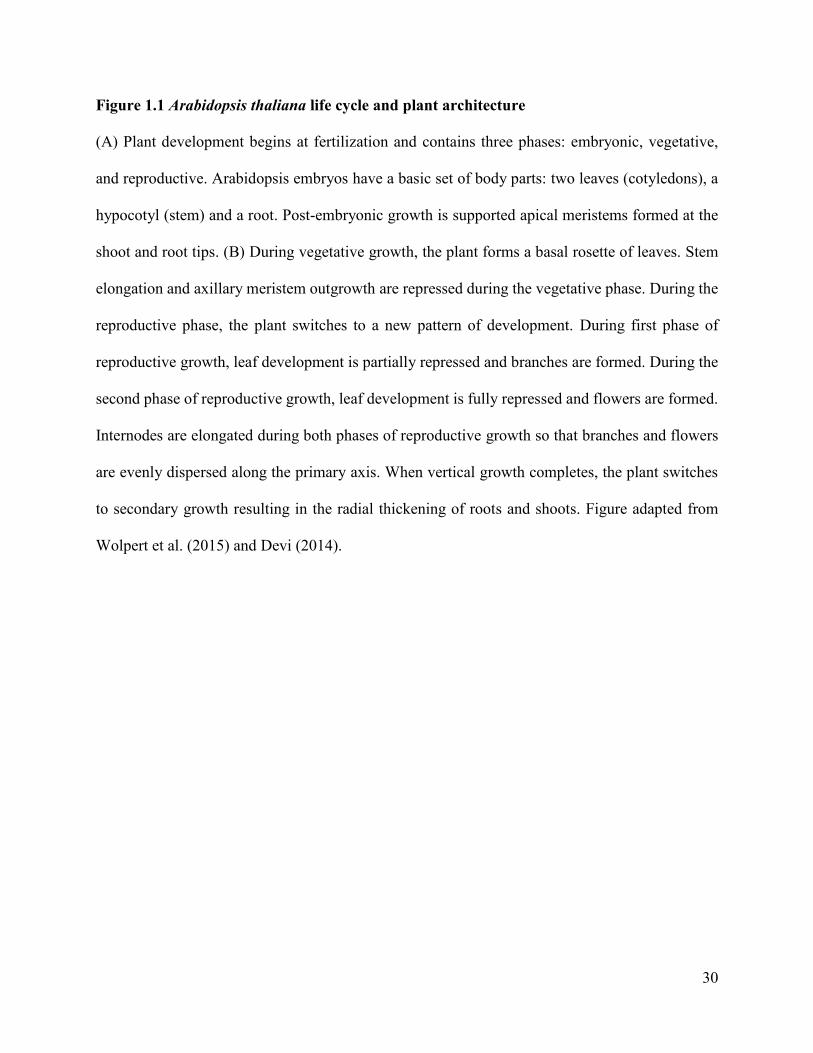
Figure 1.1 Arabidopsis thaliana life cycle and plant architecture
(A) Plant development begins at fertilization and contains three phases: embryonic, vegetative,
and reproductive. Arabidopsis embryos have a basic set of body parts: two leaves (cotyledons), a
hypocotyl (stem) and a root. Post-embryonic growth is supported apical meristems formed at the
shoot and root tips. (B) During vegetative growth, the plant forms a basal rosette of leaves. Stem
elongation and axillary meristem outgrowth are repressed during the vegetative phase. During the
reproductive phase, the plant switches to a new pattern of development. During first phase of
reproductive growth, leaf development is partially repressed and branches are formed. During the
second phase of reproductive growth, leaf development is fully repressed and flowers are formed.
Internodes are elongated during both phases of reproductive growth so that branches and flowers
are evenly dispersed along the primary axis. When vertical growth completes, the plant switches
to secondary growth resulting in the radial thickening of roots and shoots. Figure adapted from
Wolpert et al. (2015) and Devi (2014).

31
32
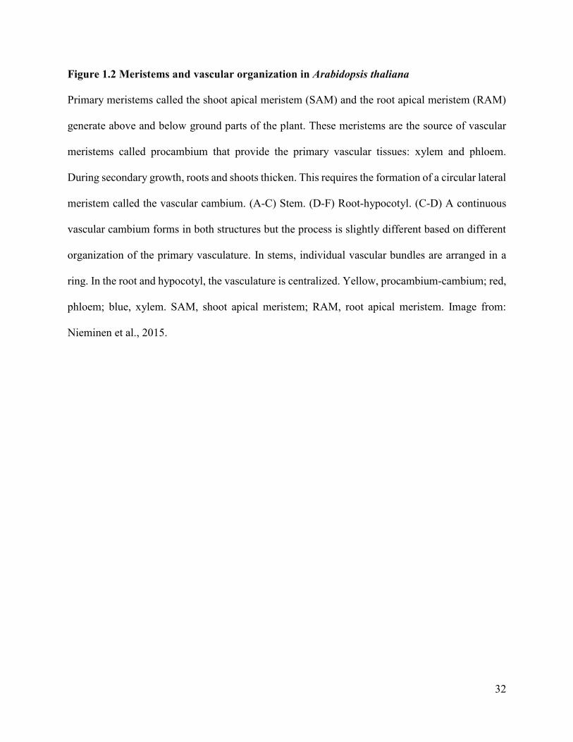
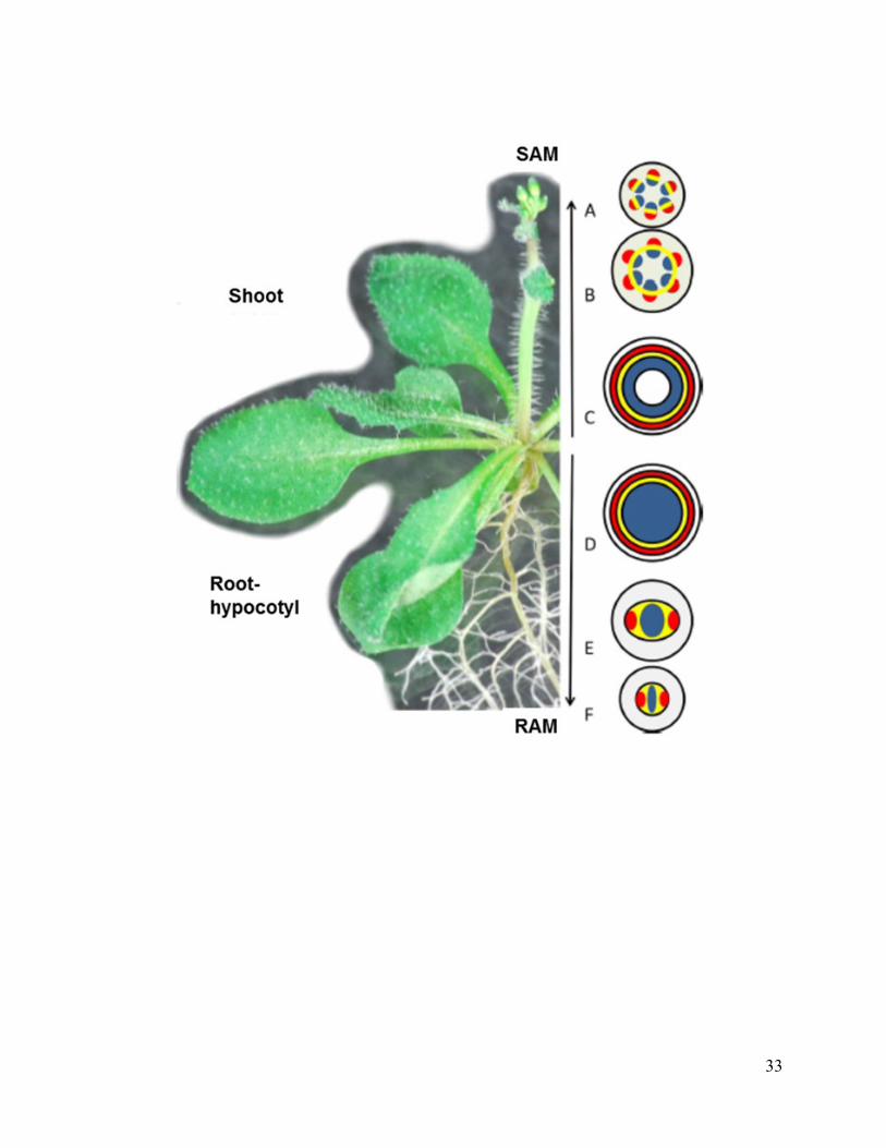
Figure 1.2 Meristems and vascular organization in Arabidopsis thaliana
Primary meristems called the shoot apical meristem (SAM) and the root apical meristem (RAM)
generate above and below ground parts of the plant. These meristems are the source of vascular
meristems called procambium that provide the primary vascular tissues: xylem and phloem.
During secondary growth, roots and shoots thicken. This requires the formation of a circular lateral
meristem called the vascular cambium. (A-C) Stem. (D-F) Root-hypocotyl. (C-D) A continuous
vascular cambium forms in both structures but the process is slightly different based on different
organization of the primary vasculature. In stems, individual vascular bundles are arranged in a
ring. In the root and hypocotyl, the vasculature is centralized. Yellow, procambium-cambium; red,
phloem; blue, xylem. SAM, shoot apical meristem; RAM, root apical meristem. Image from:
Nieminen et al., 2015.
33
34
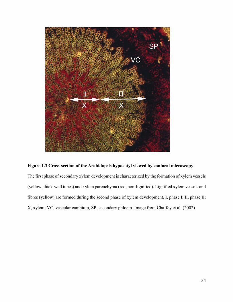
Figure 1.3 Cross-section of the Arabidopsis hypocotyl viewed by confocal microscopy
The first phase of secondary xylem development is characterized by the formation of xylem vessels
(yellow, thick-wall tubes) and xylem parenchyma (red, non-lignified). Lignified xylem vessels and
fibres (yellow) are formed during the second phase of xylem development. I, phase I; II, phase II;
X, xylem; VC, vascular cambium, SP, secondary phloem. Image from Chaffey et al. (2002).

35
Figure 1.4 Organization of the SAM
The SAM in Arabidopsis thaliana contains three histologically distinct cell layers. Cells in the
epidermal (L1) and subepidermal (L2) layers divide anticlinally (←→). By contrast, cells in the
interior (L3) layer divide in all planes. Superimposed on these layers are three functional domains:
the central zone (red), peripheral zone (light blue), and rib zone (yellow). The central zone (CZ)
contains undifferentiated stem cells. Organogenesis takes place in the peripheral zone (PZ). Low-
growth regions called lateral organ boundaries (dark blue) separate emerging organs from
meristem. The rib zone (RZ) provides cells for upward growth of stem. The RZ also contains an
organizing center (OC) that maintains the CZ. Figure modified from Wolpert et al. (2015).
36
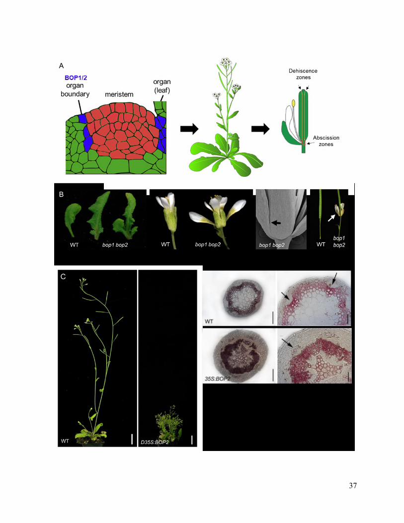
Figure 1.5 BLADE-ON-PETIOLE loss and gain-of-function phenotypes
(A) BOP1/2 are expressed in organ boundaries originating in the shoot apex are pleiotropic
regulators of plant architecture. Boundaries are low-growth domains that separate the meristem
from emerging lateral organs. Subsequently, boundaries extend to encircle the base of shoot organs
serving as an attachment point to the plant body. Boundaries are the source of axillary meristems
that provide lateral branches and flowers. Boundaries are also specialized zones for dehiscence
(opening of anthers or seed pods) or abscission (regulated detachment of plant organs). Images
from Hepworth and Pautot (2015) and Wolpert et al. (2015). (B) Loss-of-function bop1 bop2
mutant phenotypes. From left to right: leafy petioles, defects in the initiation and patterning of
flower meristems, organ fusions, and loss of floral organ abscission. Images from Hepworth et al.,
2005; Khan et al., 2014.
(C) Gain-of-function BOP1/2 phenotypes. Overexpression of BOP1 or BOP2 from a constitutive
promoter inhibits vertical growth and accelerates stem secondary growth. Lignin (pink-stained by
phloroglucinol) deposition is accelerated relative to wild-type stems with occasional gaps in the
vascular ring. The vascular ring is thicker/denser and phloem fibres are lignified (arrows)
compared to wild-type. Images from Khan et al., 2012b and Devi (2014).
37
38
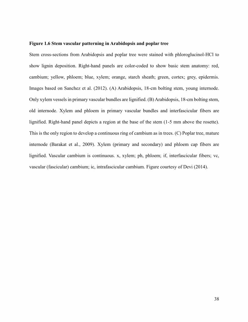
Figure 1.6 Stem vascular patterning in Arabidopsis and poplar tree
Stem cross-sections from Arabidopsis and poplar tree were stained with phloroglucinol-HCl to
show lignin deposition. Right-hand panels are color-coded to show basic stem anatomy: red,
cambium; yellow, phloem; blue, xylem; orange, starch sheath; green, cortex; grey, epidermis.
Images based on Sanchez et al. (2012). (A) Arabidopsis, 18-cm bolting stem, young internode.
Only xylem vessels in primary vascular bundles are lignified. (B) Arabidopsis, 18-cm bolting stem,
old internode. Xylem and phloem in primary vascular bundles and interfascicular fibers are
lignified. Right-hand panel depicts a region at the base of the stem (1-5 mm above the rosette).
This is the only region to develop a continuous ring of cambium as in trees. (C) Poplar tree, mature
internode (Barakat et al., 2009). Xylem (primary and secondary) and phloem cap fibers are
lignified. Vascular cambium is continuous. x, xylem; ph, phloem; if, interfascicular fibers; vc,
vascular (fascicular) cambium; ic, intrafascicular cambium. Figure courtesy of Devi (2014).
39
40
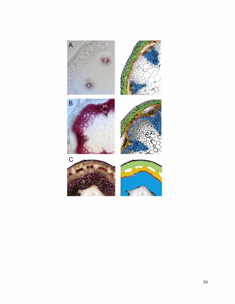
Figure 1.7 STM and BP expression pattern in stem primary vasculature
(A) STM:GUS reporter gene, showing expression of STM in the fascicular cambium, xylem, and
phloem. From: Sanchez et al. (2012). (B) BP:GUS reporter gene, showing expression in the stem
cortex and phloem. Scale bars, 100 µm.

41
Figure 1.8 Models and research questions
The Arabidopsis stem, hypocotyl and root undergo secondary growth and are considered as good
models for understanding wood development in trees. These tissues are thought to have identical
vascular cambia maintained by similar mechanisms as in the SAM (Figure 1.8). This assumption
may be premature. Existing studies point to contrasting roles for boundary genes in the vascular
cambium of Arabidopsis stem and hypocotyl. In the stem, KNOX genes spatially regulate boundary
genes to inhibit lignification of the vascular ring. In the hypocotyl, KNOX genes spatially regulate
boundary genes to promote xylem fiber development. Which of these contrasting models applies
to the vascular cambium of root and which model best describes the vascular cambium of trees?
STM, SHOOT MERISTEMLESS; BP, BREVIPEDICELLUS; VC, vascular cambium; SX,
secondary xylem; SP, secondary phloem.

42
Chapter 2: MATERIALS AND METHODS
2.1 Arabidopsis plant material and growth conditions
The Arabidopsis thaliana accession Columbia (Col-0) was used as wild-type. Plants were
grown in vitro or on soil in growth chambers at 21°C under continuous light (24 h light, intensity
100 µmol m-2 s-1) under long day (16h light/8h dark) photoperiods. Mutants bop1-3 bop2-1
(Hepworth et al., 2005), bp-2 backcrossed into a Col-0 background (Venglat et al., 2002), knat6-2
(Belles-Boix et al., 2006) and ath1-3 (Gómez-Mena and Sablowski, 2008) have been previously
described. The strong activation-tagged overexpression line bop1-6D used in this study has also
been described (Norberg et al., 2005). Reporter lines BOP1:GUS (McKim et al., 2008),
BOP2:GUS (Xu et al., 2010), BP:GUS (Venglat et al., 2002), KNAT6:GUS (Belles-Boix et al.,
2006) and ATH1:GUS (Proveniers et al., 2007) have also been previously described. Double or
triple mutant combinations used in this study were constructed by crossing as described (Woerlen
et al., 2017).
Seeds were surface-sterilized using anhydrous ethanol followed a solution of 0.5% (w/v)
sodium dodecyl sulfate (SDS) and 5% sodium hypochlorite bleach. Sterilized seeds were
subsequently rinsed four times with sterile distilled deionized water (ddH2O) and sown on
bacteriology plates containing Arabidopsis thaliana minimal medium (Haughn and Somerville,
1986). The plated seeds were then incubated for 2-3 days at 4C in the dark to break dormancy,
and then transferred to a growth chamber. When seedlings were 7-10 days old they were
transplanted to steam-sterilized soil (ProMix BX, Premier Horticulture, Rivière-du-Loup, QC)
supplemented with a 1 gram per liter solution of 20-20-20 plant fertilizer (Plant-Prod Inc.,
Brampton, ON) contained in 72-well trays or 3.5-inch square pots as appropriate. Plants were
43
grown to maturity in growth chambers at 21C under continuous light (~100 µmol m-2 sec-1) and
supplemented with water and fertilizer as required.
2.2 Analysis of secondary growth
Inflorescence stem material was harvested from five-week-old plants grown on soil under
long days. Sections were hand-cut from the base of the primary inflorescence stem using a razor
blade (Khan et al., 2012b). Alternatively, the lower 1 cm of stem at the base of the primary
inflorescence was fixed and embedded in wax for sectioning using a microtome.
Root and hypocotyl samples were harvested from seven-week-old plants grown on soil
under long days. Roots were rinsed in distilled water to remove soil. Under a dissecting
microscope, adventitious and lateral roots were removed using a razor blade. The apical 3 mm
length piece of stem directly below the rosette leaves containing the hypocotyl (plus the first mm
of secondary root) was harvested for analysis of secondary growth in the hypocotyl (Liebsch et
al., 2014). An approximately one cm length piece of root directly below was harvested for analysis
of secondary growth in the taproot (Woerlen et al., 2017). The tissue was fixed and embedded in
wax for sectioning using a microtome.
2.3 Embedding and sectioning
Tissues were fixed by submerging in a formaldehyde-acetic-acid-alcohol (FAA: 50% pure
ethanol, 5 % glacial acetic acid, 3.7% formaldehyde 41.3 % H2O) solution for 3-4.5 hours. Samples
were placed under vacuum for 15 minutes and released to remove air bubbles and to promote
penetration of the fixative into the tissue. Fixation was followed by dehydration using an ethanol
series. Tissue was incubated twice in 50% ethanol for 30 minutes, 60% ethanol for 30 minutes,
and 70 % ethanol for 30 minutes followed by storage at 4C until further use. To continue tissue

44
processing, samples were transferred to 85% ethanol for 30 minutes, followed by incubation in
95% ethanol overnight with a few drops of 1% Eosin added to stain the tissue red. The next day,
the tissues were cleared by incubating in 100% ethanol for 60 minutes, followed by two changes
of 100% ethanol for 30 minutes each. This was followed by a xylene series: 25% xylene:75%
ethanol for 30 minutes, 50% xylene:50% ethanol for 30 minutes, 75% xylene:25% ethanol for 30
minutes, then two times 100% xylene for 30 minutes. To slowly permeate the tissue with paraffin
wax (Paraplast Plus, Sigma-Aldrich, St. Louis, Missouri, USA) samples were incubated in a
mixture of 50% xylene: 50% melted wax overnight at 60°C. To melt the wax, paraffin chips were
placed in a large glass bottle and incubated overnight at 60°C. Six wax changes were performed
over three days, spaced at least 6 hours apart. At the end, samples in liquid wax were poured into
petri plates. Once solidified, the molds were stored at 4C until sectioning.
Blocks of embedded tissue were mounted on wooden stubs. A manual rotary microtome
(HM325, Microm International, Waldorff, Germany) was used cut tissue sections (25 µm thick).
Sections were bound to Superfrost Plus microscope slides (Fisher Scientific, Toronto, Ontario,
Canada) by incubating overnight at 42C on a slide warmer (Model 77, Fisher Scientific, Toronto,
Ontario, Canada).
2.4 Histochemical analyses
For the analysis of lignin deposition in stems, wild-type and mutant plants were grown on
soil in continuous light. Hand-sections were cut from the base of five-week-old plants with a razor
blade and placed in 2% phloroglucinol (in 95% ethanol) solution for 5 min. Subsequently, five
drops of concentrated hydrochloric acid were added. Two minutes were allowed for color
development. Samples were transferred to a glass slide and a cover slip was added. Images were
45
immediately collected using a stereomicroscope (SteRIO Discovery V20 equipped with an
Axiocam digital camera (Carl Zeiss, North York, Ontario, Canada).
For the analysis of lignin deposition in fixed tissues, sections mounted on glass slides were
dewaxed by incubating 100% xylenes for 20 minutes at room temperature. Samples were
subsequently incubated in 100% ethanol and 95% ethanol for 30 minutes each. Once ready to take
pictures, 95% ethanol was drained off the slides and replaced with a 2% phloroglucinol solution
for ten minutes at room temperature. Drops of concentrated hydrochloric acid were added over the
entire surface of the slide until pink colour development was visible. A cover slip was added and
images were immediately collected using a stereomicroscope for low-resolution images (as above)
or a compound microscope (Axioimager M2, Carl Zeiss, North York, Ontario, Canada) for high-
resolution images.
Toluidine blue staining was carried out as previously described (O'Brien et al., 1964).
Tissue sections mounted on glass slides were dewaxed as above. After that, tissue was rehydrated
by dipping in 100%, 95%, 85%, 70%, 50% and 30% ethanol solutions and finally pure water for
3 min each. At the end, slides were incubated in staining solution (0.05% toluidine blue O (w/v)
in benzoate buffer, pH 4.4) for approximately 45 seconds. Slides were transferred to distilled water
to remove the excess stain. A cover slip was placed on the top and pictures were immediately
acquired using a stereomicroscope (for low resolution images) or a compound microscope (for
high resolution images).
2.5 -Glucuronidase (GUS) staining assay
Staining for -glucuronidase activity in Arabidopsis plants was carried out as previously
described (Khan et al., 2012b) with small changes. The tissue was placed in chilled 90% acetone
and kept on ice during sample collection. Samples were then placed at room temperature for 15
46
minutes to complete the fixation. The acetone was removed and replaced with a GUS staining
solution that contained 5 mM K3Fe(CN)6, 5 mM K4Fe(CN)6 and 2 mM of 5-bromo-4-chloro-3-
indoxyl--D-glucuronide (X-Gluc). Samples were incubated at 37C for 3 to 24 hours until a
localized blue precipitate was visible.
The staining solution was removed, and samples were processed for sectioning as described
above with minor changes. Samples were dehydrated in 30% and 50% ethanol, fixed in FAA for
2-3 hours, and then transferred to 70% ethanol. tert-butanol was used instead of xylenes during
processing to avoid dissolving the blue precipitate. For dewaxing, slides were incubated in tert-
butanol for 45 minutes at 60°C with occasional shaking. Slides were dipped in 100% xylene for 1-
3 minutes at the end to remove residual traces of wax. After that, the tissue was rehydrated by
dipping slides in 100%, 95%, 85%, 70%, 50% and 30% ethanol solutions and then distilled water
for 3 min each. At the end, sections were mounted in 50% glycerol and a cover slip was added.
Images were collected using a compound microscope.
2.6 Poplar plant materials and growth conditions
Hybrid poplar clone INRA 717-IB4 (Populus tremula x Populus alba) was used as wild-
type. This female poplar clone was kindly provided by Dr. Sharon Regan. For transformation and
micro propagation, plants were grown in vitro and then on soil in growth chambers at 21°C under
continuous light (intensity 100 µmol m-2 s-1). After six months, poplars were transferred to six-
inch pots containing soil and grown in the greenhouse under ambient temperature and natural light.
2.7 Constructs for transformation of poplar
Constructs for transformation of poplar were as previously described (Table 2.1). The
reporter gene plasmid pBOP1:GUS uses kanamycin as a selectable marker in plants (McKim et
al., 2008). The reporter gene plasmid pBOP2:GUS uses phosphinothricin as a selectable marker
47
in plants (Xu et al., 2010). Constructs for overexpression D35S:PtrBPL1 and D35S:PtrBPL2 use
hygromycin as a selectable marker in plants (Devi, 2014).
Our construct for RNAi-mediated knock down of PtrBPL1/2 expression in transgenic
poplar has also been previously described (Devi, 2014). An RNA-hairpin cassette was designed to
co-silence PtrBPL1 and PtrBPL2. This construct is based on a highly conserved 359-base pair
sequence in PtrBPL1 and PtrBPL2 that encodes part of the BTB/POZ domain. The hairpin cassette
was created in pHannibal (Wesley et al., 2001) and composed of sense and antisense BPL1
sequences under the control of a 35S promoter (Devi, 2014). The resulting cassette was verified
by sequencing. Digestion of the plasmid with NotI was used to release a 3.6-kb fragment that was
the cloned into the corresponding site of binary vector pART27 (Gleave, 1992) to create the final
construct. Primers for sequencing are listed in Table 2.2.
2.8 Agrobacterium-mediated transformation of Populus trichocarpa
Transformation of poplar was carried out essentially as described (Meilan and Ma, 2006)
with minor adjustments. Agrobacterium tumefaciens strain C58C1 GV3101 pMP90 was used for
transformations (Koncz and Schell, 1986). Young leaves from growth chamber plants were cut
into small pieces ( 0.5 -1.0 cm x 0.5 -1.0 cm) and then surface-sterilized with 70% (v/v) ethanol
for 1 minute, rinsed in sterile purified water, treated with 10% (v/v) sodium hypochlorite bleach
solution for 3 minutes, followed by three rinses in sterile purified water for 3 minutes each. For
each construct, approximately 10 leaf discs were used per plate x 30 plates for a total of 300.
Immediately after, the explants were pre cultured on Murashige and Skoog (MS) modified basal
medium with Gamborg vitamins supplemented with 30 g/L (w/v) sucrose, 0.20 g/L (w/v) L-
glutamine, 0.25 g/L (w/v) 2-(N-morpholino) ethanesulfonic acid (MES), 10 µM 1-
naphthaleneacetic acid (NAA), and 5 µM 6-(γ, γ-dimethylallylamino) purine (2ip) and 3 g/L (w/v)

48
Gellan gum as the solidifying agent at pH 5.8. The explants were subsequently incubated at 22C
in the dark for 2 days. Agrobacterium tumefaciens strains harboring constructs were streaked onto
Lysogeny Broth (LB) medium plates containing 15 g/L of agar as the solidifying agent and
supplemented with 100 mg/L rifampicin, 40 mg/L gentamycin, and the appropriate antibiotics for
each construct: 50 mg/L kanamycin for D35S:BPL1, D35SS:BPL2, and BOP1:GUS, 25 mg/L
kanamycin and 5 mg/L tetracycline for BOP2:GUS, and 80 mg/L spectinomycin for
35S:RNAiPtrBPL1/2 were used. Plates were then inverted and incubated at 28C for 2-3 days. A
single isolated colony was picked and inoculated into 5mL LB liquid medium (pH 7.0)
supplemented with the same appropriate antibiotics as above. The cultures were grown at 30C
with continuous shaking (250 rpm) for 2 days. The Agrobacterium cultures were then sub cultured
by making a dilution of 1:100 into 5 mL of LB pH 5.4 supplemented with the same antibiotics as
before except eliminating the rifampicin and adding 100 M of acetosyringone. The new culture
was incubated overnight at 28C with shaking to grow the Agrobacterium until an ideal OD 600
of 0.7 was reached (Movahedi et al., 2014). The pre-cultured explants were submerged in the
Agrobacterium–infective suspension and incubated for 1 hr. Excess Agrobacteria were
immediately removed. Petri plates containing the infected leaves were sealed with surgical tape,
covered in silver foil, and co-cultivated for 3 days in the dark at 22C. After this interval, the
infected leaf discs were carefully transferred to 50 ml Falcon tubes containing 40 mL sterile
purified water. The explants were rinsed 5 times with sterile purified water to remove the excess
Agrobacteria. The explants were lastly washed in 35 mL of sterile distilled water with addition of
300 mg/L of cefotaxime and 200 mg/L of timentin for 1 hr with shaking to kill any excess
Agrobacteria. The wounded leaf discs were blotted dry on sterile filter papers to remove the
surface water. The putative transformed explants were transferred to MS callus induction medium
49
(CIM) supplemented with 300 mg/L cefotaxime, 200 mg/L timentin, and suitable selection agent:
10 mg/L kanamycin, 5 mg/L hygromycin, or 1 mg/L phosphinothricin. To rescue leaf disks from
severe Agrobacterium attack, they were observed every day. When bacterial re-growth
contamination occurred, the leaf disks were immediately sub cultured to fresh medium. Every two
weeks, the explants were sub cultured to a fresh CIM medium. Under ideal conditions, callus
formed after 14-21 days. After the selected calluses were formed, they were sub cultured to MS
shoot induction medium (SIM) supplemented with 30g/L (w/v) sucrose, 0.20 g/L (w/v) L-
glutamine, 0.25 g/L (w/v) MES, 0.05 mg/L thiadiazuron (TDZ), 300 mg/L cefotaxime and 200
mg/L timentin at pH 5.8 and an increased concentration of the appropriate selection agent: 25 mg/L
for kanamycin. However, this was not possible for experiments using phosphinothricin or
hygromycin as the selecting agent since one or no transformed calli were produced, respectively.
To induce shoot regeneration, the calli were subsequently incubated in growth chambers at 21°C
under continuous light (intensity 100 µmol m-2 s-1). Subculture was carried out every 2-3 weeks to
a fresh medium. After the selected shoots reached approximately 1 cm with visible leaflets, they
were transferred onto shoot elongation media (SEM). MS shoot elongation medium was
supplemented with 30 g/L (w/v) sucrose, 0.20 g/L (w/v) L-glutamine, 0.25 g/L (w/v) MES, 0.05
mg/L 6-benzylaminopurine (BAP), 300 mg/L cefotaxime, 200 mg/L timentin, and a selecting
agent (25 mg/L) kanamycin adjusted to pH 5.8. When elongated shoots were about 2 cm, they
were individually separated and placed onto ½ MS root induction medium (RIM). The root
induction medium was supplemented with 20 g/L (w/v) sucrose, 0.20 g/L (w/v) L-glutamine, 0.25
MES g/L (w/v), indole-3-butyric acid (IBA) 0.1 mg/L, 100 mg/L timentin and selecting agent
(25mg/L kanamycin) at pH 5.8. Shoots were sub cultured every 2 weeks to a fresh medium. When
the roots were well-developed, plants were transferred to soil and grown in growth chambers.
50
When plants were about six months old, they were moved to the greenhouse for further analysis.
Transformation efficiencies were calculated using the following equation: (number of calli per
total number of the explants) X 100.
2.9 Genomic DNA extraction from poplar
Genomic DNA for PCR-based genotyping was extracted from young poplar leaves using
a CTAB (cetyl trimethylammonium bromide) method as previously described (Fan et al., 2015).
Briefly, ~0.1 g of fresh tissue was ground in liquid nitrogen using a sterilized mortar and pestle.
The resulting powder was transferred to a 1.5 ml microcentrifuge tube and 400 l of pre-heated
CTAB buffer [2% CTAB (w/v), 1.4 M NaCl, 20 mM ethylenediaminetetraacetic acid (EDTA),
and 100 mM Tris-Cl (pH 8.0)] was added to each sample. Samples were gently mixed by inversion.
After incubating for 30 minutes at 65C, 200 μl of chloroform was added. Samples were mixed by
gentle inversion and incubated at room temperature for 10 minutes. After centrifugation at 16,000g
for 5 minutes, the supernatant was transferred into a new tube, mixed with 300 l of isopropanol,
and incubated for 30 minutes at 4C. Finally, the sample was centrifuged at 16,000 g for 5 minutes
to collect the precipitated genomic DNA. The supernatant was removed and the pellet at the bottom
of the tube was washed in 70% ethanol and dissolved in 100 μl of ddH2O. DNA samples were
stored at -20C until further use.
2.10 Poplar genotyping
Genomic DNA from poplar was tested for integrity by PCR-amplifying a 100-bp
fragment corresponding to housekeeping gene UBIQUITIN (Brunner et al., 2004) using primers
UBQ-RT-F- 5´and UBQ-RT-R with an annealing temperature of 58ºC. The AtBOP1:GUS
transgene was detected by PCR-amplifying a 750-bp fragment of DNA spanning the junction

51
between the AtBOP1 promoter and the GUS reporter gene using primers P4H-F6 and GUS-1R
and an annealing temperature of 56ºC. The BOP2:GUS transgene was detected by PCR-amplifying
a 780-bp fragment of DNA spanning the junction between the AtBOP2 promoter and the GUS
reporter gene using primers P5H-F6 and GUS-1R and an annealing temperature of 56°C. The
RNAi transgene was detected by PCR-amplifying a 923-bp fragment of DNA corresponding to
part of the RNA hairpin using primers 35S-F1-Hannibal and Hannibal-Int-R and an annealing
temperature of 55°C. The BOP1:GUS and RNAi constructs were also detected by PCR-
amplifying a 300-bp fragment of DNA corresponding to part of the coding region of the NPTII
gene which confers kanamycin resistance using primers NPTII-F and NPTII-R and an annealing
temperature of 58ºC. See Table 2.2 for primer sequences.
2.11 Poplar GUS staining
Poplar tissue was stained for GUS according to two methods (Khan et al., 2012b; Chen et
al., 2013). A third method for GUS tissue staining was also used (Jefferson et al., 1987) with some
minor changes. Briefly, the samples were collected at room temperature and fixed in 0.3%
formaldehyde in 10 mM MES pH 5.6 and 0.3 M mannitol for 45 minutes. The MES solution was
removed and tissues were subsequently washed several times in 50 mM Na2PO4, pH 7.0. All fixed
tissues were transferred to GUS solution that contained 1 mM of X-Gluc in 50 mM Na2PO4 (pH
7.0) and incubated for 20 minutes to several hours until observing localized blue precipitate. After
staining, tissues were rinsed in 70% ethanol several times to remove chlorophyll. In each case,
Arabidopsis control lines showed blue staining and poplar transgenics did not.
52
Table 2.1 List of constructs
Construct Description pBOP1:GUS This plasmid contains ~4000 base-pairs of nucleotide sequence corresponding to
the 5’ promoter region of AtBOP1 up to an including the start codon cloned in front
a GUS reporter gene present in pBl101.1. The selectable marker in plants is
kanamycin (McKim et al., 2008).
pBOP2:GUS This plasmid contains 4015 base-pairs of nucleotide sequence corresponding to the
5’ promoter region of AtBOP2 up to and including the start codon plus the first 16
nucleotides of coding sequence cloned as a translational fusion with the GUS
reporter gene present in pGreen 0229. The selectable marker in plants is
phospinothricin (Xu et al., 2010).
pPtrBPL-RNAi This plasmid contains an RNA-hairpin cassette based on a highly conserved 359
base-pair nucleotide sequence from the BTB/POZ domain of PtrBPL1 and
PtrBPL2 cloned behind a universal 35S promoter. The cassette was created in
pHannibal and transferred to the binary vector pART27. The selectable marker in
plants is kanamycin (Devi, 2014).
pD35S: BPL1 This plasmid contains a 1332 base-pair nucleotide sequence corresponding to the
PtrPBL1 coding sequence cloned behind a universal double 35S CaMV promoter
(D35S) present a pSM-3, a pCAMBIA1390-based binary Gateway destination
vector. The selectable marker in plants is hygromycin (Unda et al., 2017).
pD35S: BPL2 This plasmid contains a 1446 base-pair nucleotide sequence corresponding to the
PtrPBL2 coding sequence cloned behind a universal double 35S CaMV promoter
(D35S) present a pSM-3, a pCAMBIA1390-based binary Gateway destination
vector. The selectable marker in plants is hygromycin (Unda et al., 2017).
Table 2.2 List of primers
Description Primer Sequence 5’- 3’ Reference RNAi sequencing
1st round
35S-F1-Hannibal
pHannibal-Int-R
GCTCCTACAAATGCCATCATTGCGA
CAAACCAGCTAGAATTACTATTATG
Bhaswati Devi
RNAi sequencing 2nd round
35S-F1-Hannibal OCS-Term-R
GCTCCTACAAATGCCATCATTGCGA GATCTGAGCTACACATGCTCAGGT
Bhaswati Devi
D35S:BPL1
PCR colony screening
Pt16s04010cdsF
BPL1 CDS 360 R2
ATGACTCTTGAAGACTCTCTAAGAACTCTG
CACGCCACAATTAGGCCTTGG
Bhaswati Devi
D35S:BPL2 PCR colony screening
Pt6s04190cdsF BPL2 CDS 360 R2
ATGACTCTTGAAGACTCTCTAAGATCTCTA CTCACCACAATTAGGCCTTGGC
Bhaswati Devi
Poplar genomic DNA
integrity
UBQ–RT-F
UBQ-RT-R
GTTGATTTTTGCTGGGAAGC
GATCTTGGCCTTCACGTTGT
This study
BOP1:GUS genotyping in poplar
P4H-F6 GUS-1R
TAAGTGTTACGTGACACACG CCACCAACGCTGATCAATTCCACAG
McKim et al., 2008
BOP2: GUS
genotyping in poplar
P5H-F6
GUS-1R
CATATCCCTCGTTTGTTTGG
CCACCAACGCTGATCAATTCCACAG
Xu et al., 2010
RNAi genotyping in
poplar
pHannibal-Int-F
pHannibal-Int-R
CATAATAGTAATTCTAGCTGGTTTG
CAAACCAGCTAGAATTACTATTATG
This study
NPTII genotyping
in poplar
NPTII–F
NPTII-R
GAGCACGTACTCGGATGGAAG
GTAAAGCACGAGGAAGCGGTC
Ying Wang

53
Chapter 3: RESULTS
3.1 A gradient of secondary growth in Arabidopsis taproots
The role of boundary genes in root secondary growth has not been investigated. Abundant
secondary growth occurs the Arabidopsis tap root, which encompasses the upper first centimeter
of the primary root. Thickening of roots is presumed to follow the same principles as in hypocotyl
because the primary anatomy of vasculature is similar (Dolan et al., 1993; Buss and Evet 1999;
Chaffey et al., 2002). Cellular organization during secondary growth of the root is depicted in
Figure 3.1A. Two phases of secondary growth are distinguished. During phase I, production of
xylem and phloem is proportional (equal). The xylem produced in this phase is xylem I, which
consist of xylem parenchyma interspaced with lignified vessels. The transition to flowering
triggers a switch to phase II in which xylem production expands relative to phloem (Chaffey et al.
2002; Sibout et al. 2008). During phase II, xylem parenchyma cells differentiate to form lignified
fibres (Chaffey et al., 2002; Sibout et al., 2008). Xylem II is seen as a dense ring of lignified fibers
interspersed with vessels (Chaffey et al., 2002; Ragni and Hardtke, 2014; Nieminen et al., 2015).
To monitor the developmental gradient of secondary growth in roots, transverse sections
were examined from the top, middle, and lower middle of taproots from seven-week-old plants
using phloroglucinol to stain for lignin (Figure 3.1B-E). Clearly defined regions of xylem I and II
were formed at all positions but root diameter was greatest, and morphology was least variable at
the top. Thereafter, sections were taken from the top of the taproot (1-2 mm below the root-
hypocotyl junction) to ensure consistency.
3.2 BOP1/2 loss and gain of function mutants alter xylem fibre differentiation
Functional analysis of BOP1/2 in root secondary growth was assessed by examining the
phenotype of loss and gain-of-function mutants. Transverse sections of taproot from wild-type,

54
bop1 bop2, and bop1-6D (a gain-of-function line) were stained with phloroglucinol (pink) and
toluidine blue (purple) to visualize lignified secondary walls. In bop1 bop2 double mutants, the
overall organization of tissues was preserved, with a slight thickening of xylem II ring relative to
wild-type (Figure 3.2ABJ). The most obvious difference was a significant enlargement of xylem
II relative to wild-type control plants (Figure 3.2AB). By contrast, differentiation of xylem II
including lignified fibers and vessels was missing in bop1-6D gain–of-function mutants (Figure
3.2C). In hemizygous bop1-6D plants, lignified vessels were sometimes formed (Figure 3.3). Thus,
BOP1/2 repress xylem II differentiation.
3.3 BP interacts antagonistically with boundary genes to promote xylem fiber
differentiation in roots
In the stem and hypocotyl, BP represses BOP1/2 in specific subdomains to preserve normal
patterns of secondary growth (Khan et al., 2012b; Liebsch et al., 2014). To test this principle in
roots, secondary growth was analyzed in the taproot of bp and bp bop1 bop2 mutants. Sectioning
showed that xylem II differentiation in bp-2 roots was reduced or absent (Figure 3.2DJ) similar to
bop1-6D mutants (Figure 3.2CJ). Inactivation of BOP1/2 significantly rescued bp-2 defects.
Taproots in bop1 bop2 bp-2 triple mutants contained a lignified xylem II ring similar in thickness
to wild-type control plants (Figure 3.2EJ). Sometimes, lignification of xylem II fibres was
incomplete resulting in a ring of a lower density compared to wild type (Figure 3.3). The diameter
of bp-2 taproots was significantly narrower than wild-type (bp-2, 0.85 ± 0.12 mm versus wild-
type, 1.55 ± 0.13 mm, t test p<0.0001), but was partially restored in bop1 bop2 bp-2 triple mutants
(Figure 3.2J). These data indicate opposing roles for BP and BOP1/2 in controlling root diameter
and cell differentiation during the xylem expansion phase.
55
BOP1/2 regulate aerial development in part through promotion of homeobox genes ATH1
and KNAT6 which are co-expressed in boundaries (Belles-Boix et al., 2006; Gómez-Mena and
Sablowski, 2008; Ragni et al., 2008; Khan et al., 2012b). These genes are also expressed in root
vasculature (Dean et al., 2004; Belles-Boix et al., 2006; Woerlen et al., 2017). Inactivation of
ATH1 and/or KNAT6 partially rescued bp-2 defects in root secondary growth similar to bop1 bop2.
Differentiation of xylem II resumed in all three mutant combination: ath1 bp-2, knat6 bp-2 and
ath1 knat6 bp-2 (Figure 3.2FGHJ). Inactivation of these genes also partially corrected bp-2 defects
in root diameter (Figure 3.2I). These data suggest that BP is a general repressor of boundary genes
in the root.
3.4 BP spatial regulation of boundary genes is important for differentiation of
reproductive-stage xylem
Experiments carried out mainly by Natalie and Adina monitored the expression pattern of
BOP1/2, ATH1, and KNAT6 boundary genes in wild-type and bp-2 roots during secondary growth
(Figure 3.4). In wild-type plants, BOP1:GUS and BOP2:GUS reporter genes were strongly
expressed in secondary phloem and absent in the vascular cambium and secondary xylem
(Woerlen et al., 2017). As predicted, BOP2 expression was strongly upregulated in the xylem
parenchyma cells of bp-2 mutant roots and in xylem II cambial derivatives. Misexpression of
BOP1 was observed in xylem vessel cells but changes in expression of this gene were mild
compared to BOP2 (Woerlen et al., 2017). ATH1:GUS and KNAT6:GUS reporters were also highly
expressed in secondary phloem of wild-type plants. In bp-2 mutants, these genes were strongly
upregulated in xylem parenchyma cells and the cambial zone similar to BOP1/2 (Woerlen et al.,
2017).
56
Collectively, these data reveal that correct spatial regulation of boundary genes is important
for secondary growth in the root-hypocotyl and stem. In roots, BP repression promotes the
production of reproductive-stage xylem. However, BP repression plays a contrasting role in stem
where it delays the formation of lignified interfascicular fibers that complete the vascular ring
(Mele et al., 2003; Khan et al., 2012a; Khan et al., 2012b).
3.5 Analysis of PtrBPL1 or PtrBPL2 gain-of-function in Arabidopsis plants
Poplar tree (Populus trichocarpa) expresses two BOP orthologs in vascular tissues of the
woody stem (Khan et al., 2012b; Devi, 2014). Presumably, spatial regulation of these genes by
KNOX transcription factors preserves normal patterns of wood development. To partially analyze
the function of these genes, I tested their gain-of-function phenotypes in Arabidopsis plants.
3.5.1 Inflorescence stem
Arabidopsis plants that overexpress AtBOP1 or AtBOP2 are short and bushy with abundant
lignin deposition in the stem (Khan et al., 2012b). To test if 35S:PtrBPL1 and 35S:PtrBPL2 have
a similar activity, overexpression lines in Arabidopsis were created (Devi, 2014). A number of
transformed lines had severe defects in stem elongation in the T1 generation. Due to a small
number of plant lines (n=1) with a stable overexpression phenotype transmitting to the T2
generation, I screened for additional lines (Table 3.1). The stems of 5-6 representative T1 plants
were hand-sectioned and stained with phloroglucinol to look for changes in lignin deposition.
Within this population, 35S:PtrBPL1 and 35S:PtrBPL2 transgenic lines often showed a denser
vascular ring and lignified phloem fibers similar to lines overexpressing AtBOP1/2 (Figure 3.5).

57
3.5.2 Root-hypocotyl
Arabidopsis plants that overexpress AtBOP1 or AtBOP2 lack xylem II features in the root
(this study) and the hypocotyl (Liebsch et al., 2014). Analysis of secondary growth in the hypocotyl
of 35S:PtrBPL1 and 35S:PtrBPL2 lines showed at least one 35S:PtrBPL1 transformed line (n=3)
missing xylem II fibers and vessels similar to bop1-6D control plants (Figure 3.2). The xylem II
ring in all 35S:PtrBPL2 transformed lines (n=3) was reduced in thickness compared to wild-type
control plants, similar to bp-2 mutants (Liebsch et al., 2014; Woerlen et al., 2017). These data
suggest that poplar BPL-like genes can functionally substitute in Arabidopsis plants.
3.6 Transgenic poplars expressing BOP1:GUS and BOP2:GUS reporter genes
In poplar, PtrBPL1 and PtrBPL2 are expressed in young leaves, roots, seedlings, and
inflorescences according to microarray transcriptome data compiled by the Botany Array Resource
(www.utoronto.ca/bar). These data closely matched in-house transcript analysis by qRT-PCR
carried out on dissected poplar tissues. Unlike Arabidopsis, PtrBPL1 and PtrBPL2 transcripts in
poplar stem were detected in both xylem and phloem (Devi, 2014). Since these methods do not
provide high-resolution spatial information, my role was to generate transgenic poplar expressing
AtBOP1:GUS and AtBOP2:GUS reporter genes. Cis-regulatory sequences in poplar and
Arabidopsis tend to be conserved (Ding et al., 2012) and so these reporters were expected to give
useful spatial information about gene expression in poplar. Agrobacterium-mediated
transformation of leaf disks (n=300) was used to generate 15 putative AtBOP1:GUS transgenic
lines from callus (Table 3.2). Genotyping confirmed that 11 of 15 lines contained a transgene.
These plants were transferred to soil and assayed for GUS activity using three different methods
(Jefferson et al., 1987; Khan et al., 2012b; Chen et al., 2013). However, zero were positive for
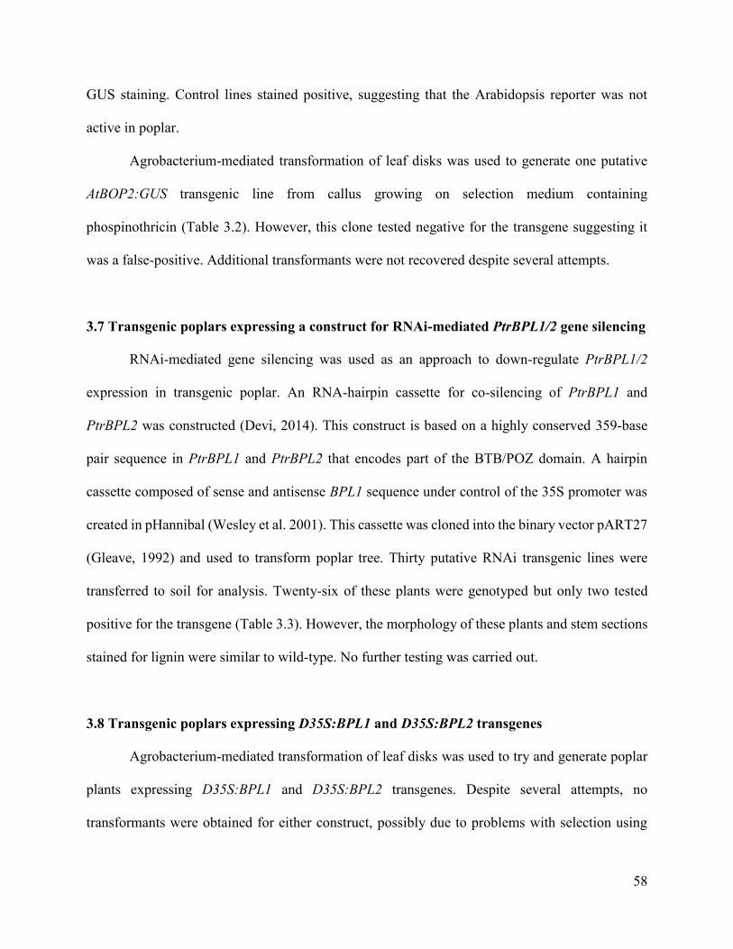
58
GUS staining. Control lines stained positive, suggesting that the Arabidopsis reporter was not
active in poplar.
Agrobacterium-mediated transformation of leaf disks was used to generate one putative
AtBOP2:GUS transgenic line from callus growing on selection medium containing
phospinothricin (Table 3.2). However, this clone tested negative for the transgene suggesting it
was a false-positive. Additional transformants were not recovered despite several attempts.
3.7 Transgenic poplars expressing a construct for RNAi-mediated PtrBPL1/2 gene silencing
RNAi-mediated gene silencing was used as an approach to down-regulate PtrBPL1/2
expression in transgenic poplar. An RNA-hairpin cassette for co-silencing of PtrBPL1 and
PtrBPL2 was constructed (Devi, 2014). This construct is based on a highly conserved 359-base
pair sequence in PtrBPL1 and PtrBPL2 that encodes part of the BTB/POZ domain. A hairpin
cassette composed of sense and antisense BPL1 sequence under control of the 35S promoter was
created in pHannibal (Wesley et al. 2001). This cassette was cloned into the binary vector pART27
(Gleave, 1992) and used to transform poplar tree. Thirty putative RNAi transgenic lines were
transferred to soil for analysis. Twenty-six of these plants were genotyped but only two tested
positive for the transgene (Table 3.3). However, the morphology of these plants and stem sections
stained for lignin were similar to wild-type. No further testing was carried out.
3.8 Transgenic poplars expressing D35S:BPL1 and D35S:BPL2 transgenes
Agrobacterium-mediated transformation of leaf disks was used to try and generate poplar
plants expressing D35S:BPL1 and D35S:BPL2 transgenes. Despite several attempts, no
transformants were obtained for either construct, possibly due to problems with selection using

59
hygromycin. We cannot rule out the possibility that overexpression of PtrBPL1 or PtrBPL2
interferes with callus induction and shoot regeneration.
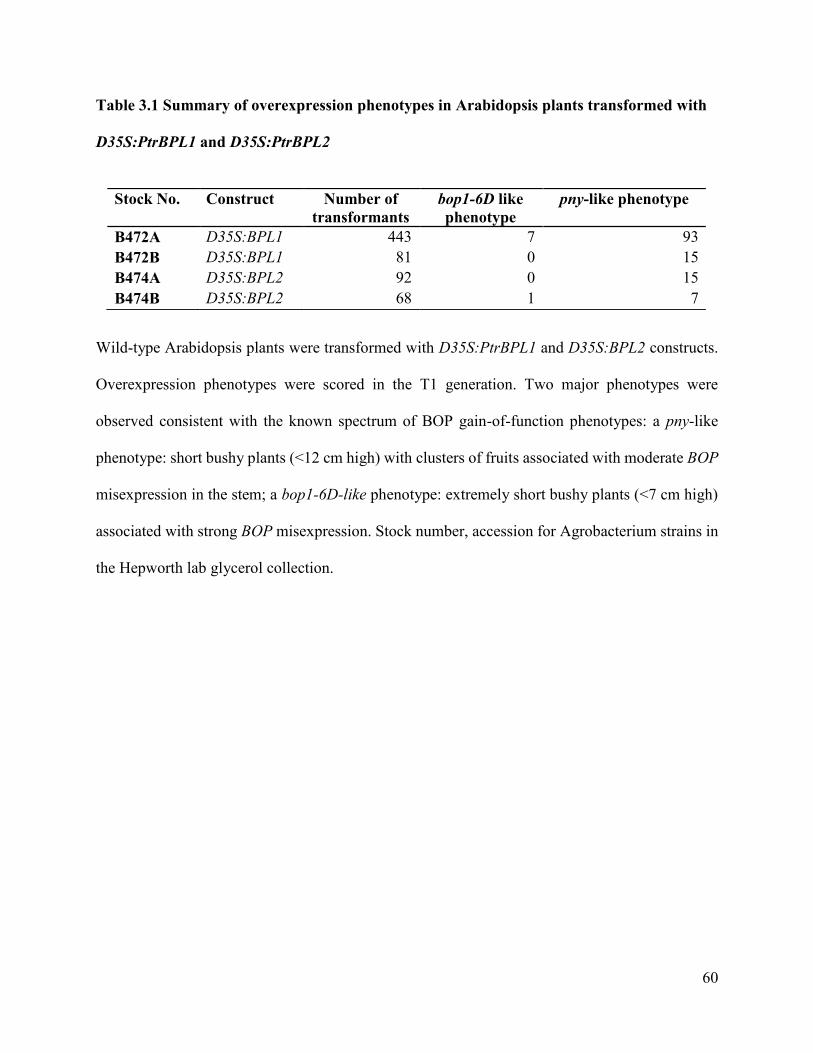
60
Table 3.1 Summary of overexpression phenotypes in Arabidopsis plants transformed with
D35S:PtrBPL1 and D35S:PtrBPL2
Wild-type Arabidopsis plants were transformed with D35S:PtrBPL1 and D35S:BPL2 constructs.
Overexpression phenotypes were scored in the T1 generation. Two major phenotypes were
observed consistent with the known spectrum of BOP gain-of-function phenotypes: a pny-like
phenotype: short bushy plants (<12 cm high) with clusters of fruits associated with moderate BOP
misexpression in the stem; a bop1-6D-like phenotype: extremely short bushy plants (<7 cm high)
associated with strong BOP misexpression. Stock number, accession for Agrobacterium strains in
the Hepworth lab glycerol collection.
Stock No. Construct Number of
transformants
bop1-6D like
phenotype
pny-like phenotype
B472A D35S:BPL1 443 7 93
B472B D35S:BPL1 81 0 15
B474A D35S:BPL2 92 0 15
B474B D35S:BPL2 68 1 7

61
Table 3.2 Summary of poplar transformations with AtBOP1:GUS and AtBOP2:GUS
reporter gene constructs
Reporter construct Genotyping result GUS staining result
AtBOP1:GUS
1A + -
2A + -
3B + -
4A + -
5A + -
6B + -
7A + -
8A + -
9B + -
10A + -
11 not determined not determined
12 not determined. not determined
13A + -
14 not determined. not determined
15 not determined not determined
AtBOP2:GUS
1A - -
1B - -
Leaf disks from poplar wild-type plants (n=300 per construct) were infected with Agrobacterium
cultures from B630 (AtBOP1:GUS) or B639 (AtBOP2:GUS) in the Hepworth lab strain collection.
Fifteen putative AtBOP1:GUS transformed calli grew on selection media containing kanamycin.
Eleven plantlets regenerated from independent calli tested positive for the transgene. All lines
tested negative for GUS staining. One putative AtBOP2:GUS transformed callus grew on selection
media containing phosphinothricin. Two plantlets regenerated from this callus tested negative for
the transgene.

62
Table 3.3 Summary of poplar plants transformed with PtrBPL RNAi, D35S:BPL1, and
D35S:BPL2 constructs
Construct Genotype Result Phenotype result
PtrBPL-RNAi
1AB -
2AC -
3C -
4AB -
5C -
6ABC -
7BC -
8ABC -
9A -
10AB -
11C + WT-like
12ABC -
13B -
14ABC -
15C -
16 not determined
17 not determined
18C -
20B -
21AB -
22C -
23B -
24B + WT-like
25A -
26C -
28BC -
29AB -
30AB -
D35S:BPL1
No transformants
D35S:BPL2
No transformants

63
Leaf disks from poplar wild-type plants (n=300 per construct) were infected with Agrobacterium
cultures from accessions B652 (PtrBPL-RNAi), B472/473/B667 (D35S:BPL1), and
B474/B475/B668 (D35S:BPL2) in the Hepworth lab strain collection. Thirty putative PtrRNAi
transformed calli grew on selection media containing kanamycin. Three plantlets per calli were
moved to soil for further testing. Of these, 2 of 26 genotyped lines tested positive for the transgene
(bold). Phenotypes of positive plants were scored 5-6 weeks after transfer to soil. Morphology of
leaves and shoots were similar to control plants. Lignin deposition in stem sections was also similar
to control plants (data not shown). No further testing was carried out. Hygromycin-resistant calli
were not obtained for leaf disks infected with Agrobacterium containing D35S:BPL1 or
D35S:BPL2 transgenes.

64
Figure 3.1 Anatomy and developmental gradient of lignin deposition in taproot of wild-type
plants
(A) Organization of cell types during root secondary growth. Proliferation of procambium cells
in the central vascular cylinder of the primary root gives rise to the vascular cambium (VC) (Dolan
et al., 1993; Dolan and Roberts, 1995). Cambium cells divide to form secondary phloem (SP)
(outwardly) and secondary xylem (XI and XII) (inwardly). x, primary xylem; XI, proportional
growth phase; XII, xylem expansion phase. A separate meristem called the cork cambium (CC)
derived from the pericycle layer of the central vascular cylinder in the primary root produces the
outer periderm (bark) of secondary thickened roots. The original non-dividing outer layers of the
root (endoderm, cortex, and epidermis) are shed (Dolan and Roberts, 1995). Modified from
(Vishwanath et al., 2015). (B) Taproot of representative seven-week-old wild-type plant.
Horizontal bar indicates the root-hypocotyl junction. Tissues below this junction were harvested
for analysis (arrow) (C–E) Transverse sections were stained with phloroglucinol-HCl to detect
lignin (pink colour). Approximate distances (mm) below the root-hypocotyl junction were as
indicated. Scale bars, 2 mm (B), 0.25 mm (C-E). Figure from: Woerlen et al., 2017.

65
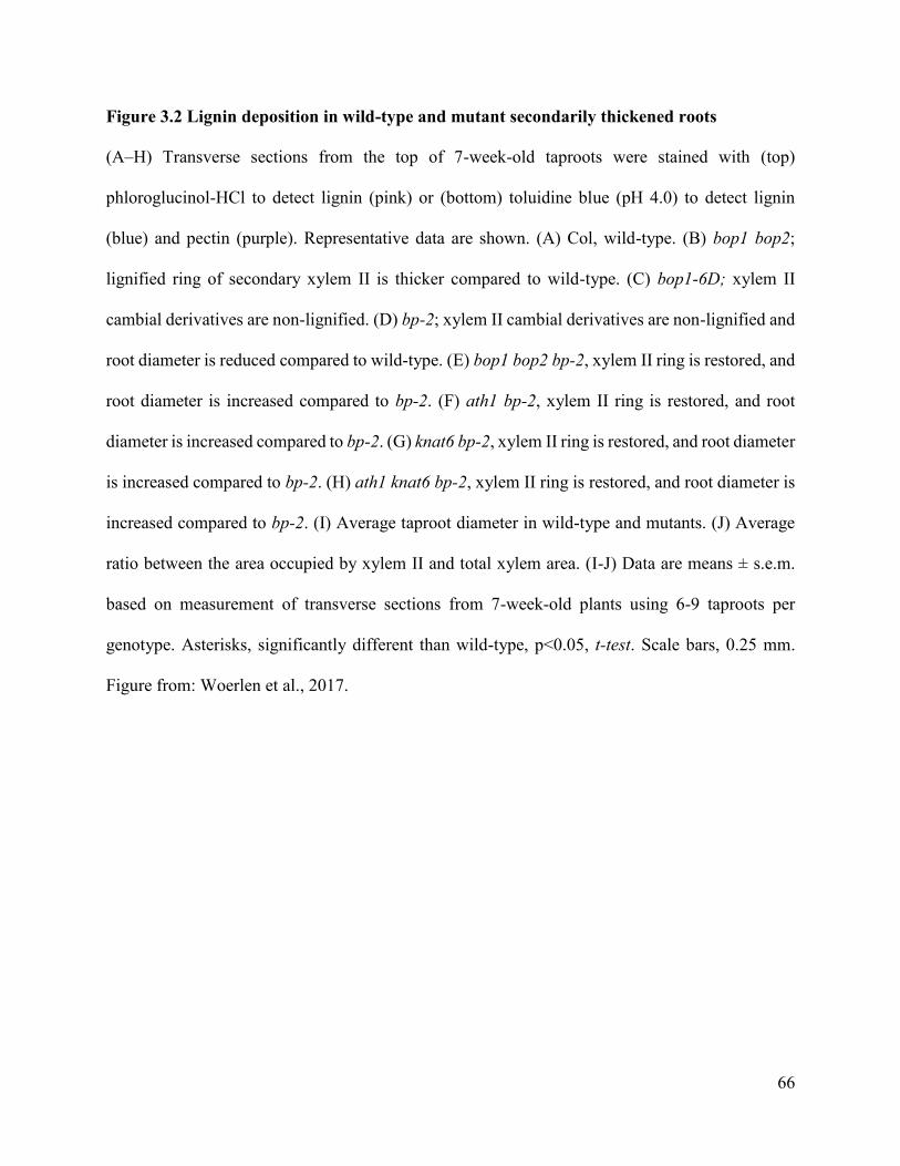
66
Figure 3.2 Lignin deposition in wild-type and mutant secondarily thickened roots
(A–H) Transverse sections from the top of 7-week-old taproots were stained with (top)
phloroglucinol-HCl to detect lignin (pink) or (bottom) toluidine blue (pH 4.0) to detect lignin
(blue) and pectin (purple). Representative data are shown. (A) Col, wild-type. (B) bop1 bop2;
lignified ring of secondary xylem II is thicker compared to wild-type. (C) bop1-6D; xylem II
cambial derivatives are non-lignified. (D) bp-2; xylem II cambial derivatives are non-lignified and
root diameter is reduced compared to wild-type. (E) bop1 bop2 bp-2, xylem II ring is restored, and
root diameter is increased compared to bp-2. (F) ath1 bp-2, xylem II ring is restored, and root
diameter is increased compared to bp-2. (G) knat6 bp-2, xylem II ring is restored, and root diameter
is increased compared to bp-2. (H) ath1 knat6 bp-2, xylem II ring is restored, and root diameter is
increased compared to bp-2. (I) Average taproot diameter in wild-type and mutants. (J) Average
ratio between the area occupied by xylem II and total xylem area. (I-J) Data are means ± s.e.m.
based on measurement of transverse sections from 7-week-old plants using 6-9 taproots per
genotype. Asterisks, significantly different than wild-type, p<0.05, t-test. Scale bars, 0.25 mm.
Figure from: Woerlen et al., 2017.
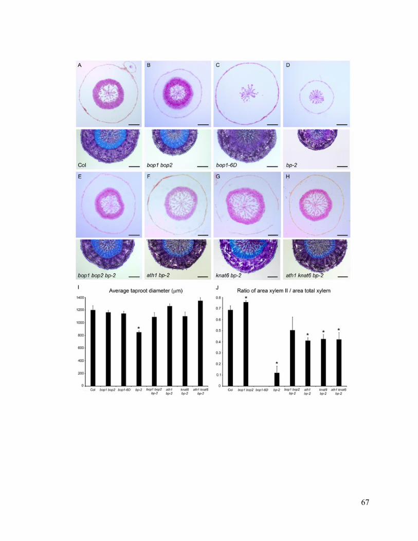
67
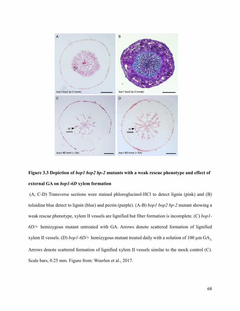
68
Figure 3.3 Depiction of bop1 bop2 bp-2 mutants with a weak rescue phenotype and effect of
external GA on bop1-6D xylem formation
(A, C-D) Transverse sections were stained phloroglucinol-HCl to detect lignin (pink) and (B)
toluidine blue detect to lignin (blue) and pectin (purple). (A-B) bop1 bop2 bp-2 mutant showing a
weak rescue phenotype, xylem II vessels are lignified but fiber formation is incomplete. (C) bop1-
6D/+ hemizygous mutant untreated with GA. Arrows denote scattered formation of lignified
xylem II vessels. (D) bop1-6D/+ hemizygous mutant treated daily with a solution of 100 µm GA3.
Arrows denote scattered formation of lignified xylem II vessels similar to the mock control (C).
Scale bars, 0.25 mm. Figure from: Woerlen et al., 2017.
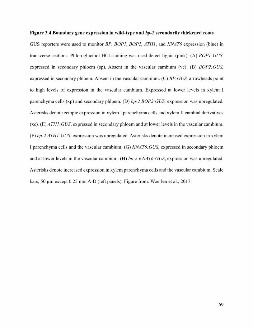
69
Figure 3.4 Boundary gene expression in wild-type and bp-2 secondarily thickened roots
GUS reporters were used to monitor BP, BOP1, BOP2, ATH1, and KNAT6 expression (blue) in
transverse sections. Phloroglucinol-HCl staining was used detect lignin (pink). (A) BOP1:GUS,
expressed in secondary phloem (sp). Absent in the vascular cambium (vc). (B) BOP2:GUS,
expressed in secondary phloem. Absent in the vascular cambium. (C) BP:GUS, arrowheads point
to high levels of expression in the vascular cambium. Expressed at lower levels in xylem I
parenchyma cells (xp) and secondary phloem. (D) bp-2 BOP2:GUS, expression was upregulated.
Asterisks denote ectopic expression in xylem I parenchyma cells and xylem II cambial derivatives
(xc). (E) ATH1:GUS, expressed in secondary phloem and at lower levels in the vascular cambium.
(F) bp-2 ATH1:GUS, expression was upregulated. Asterisks denote increased expression in xylem
I parenchyma cells and the vascular cambium. (G) KNAT6:GUS, expressed in secondary phloem
and at lower levels in the vascular cambium. (H) bp-2 KNAT6:GUS, expression was upregulated.
Asterisks denote increased expression in xylem parenchyma cells and the vascular cambium. Scale
bars, 50 µm except 0.25 mm A-D (left panels). Figure from: Woerlen et al., 2017.
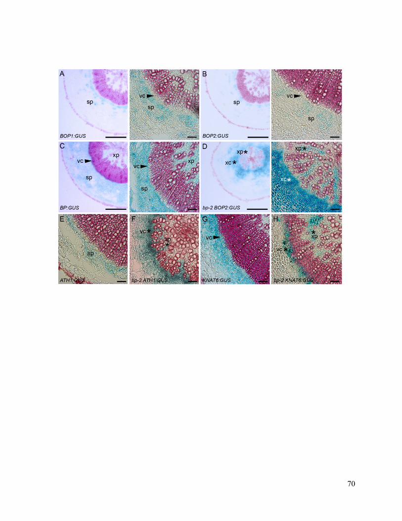
70
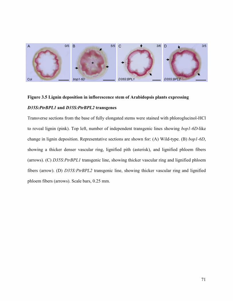
71
Figure 3.5 Lignin deposition in inflorescence stem of Arabidopsis plants expressing
D35S:PtrBPL1 and D35S:PtrBPL2 transgenes
Transverse sections from the base of fully elongated stems were stained with phloroglucinol-HCl
to reveal lignin (pink). Top left, number of independent transgenic lines showing bop1-6D-like
change in lignin deposition. Representative sections are shown for: (A) Wild-type. (B) bop1-6D,
showing a thicker denser vascular ring, lignified pith (asterisk), and lignified phloem fibers
(arrows). (C) D35S:PtrBPL1 transgenic line, showing thicker vascular ring and lignified phloem
fibers (arrow). (D) D35S:PtrBPL2 transgenic line, showing thicker vascular ring and lignified
phloem fibers (arrows). Scale bars, 0.25 mm.

72
Figure 3.6 Lignin deposition in hypocotyl of Arabidopsis plants expressing D35S:PtrBPL1
and D35S:PtrBPL2 transgenes
Transverse sections from the middle of the hypocotyl (1.5 mm below the rosette leaves) were
stained with phloroglucinol-HCl to reveal lignin (pink). Top left, number of independent
transgenic lines with phenotype. Representative sections are shown for: (A) Wild-type, showing a
thick ring of xylem II. (B) bop1-6D, xylem II fibers and vessels are missing. (C) D35S:PtrBPL1
transgenic line, similar to bop1-6D. (D) D35S:PtrBPL2 transgenic line, thickness of xylem II ring
is reduced compared to wild-type. Scale bars, 0.5 mm
73
Chapter 4: DISCUSSION
In Arabidopsis plants, a continuous vascular cambium forms at the inflorescence stem base,
hypocotyl, and root. All three tissues are considered as useful models for wood development in
trees, based on secondary growth characterized by the abundant deposition of lignin (Chaffey et
al., 2002; Strabala and MacMillan, 2013; Nieminen et al., 2015; Barra-Jiménez and Ragni, 2017).
Much like the SAM, the vascular cambium provides stem cell activity, but is organized
differently since cambium cells are not physically separated from differentiating xylem and
phloem. The SAM is maintained by Class I KNOX genes that spatially regulate boundary genes
to allow continuous production of lateral organs. In previous work, we showed that Class I KNOX
genes in inflorescence stem negatively regulate boundary genes and this delays the differentiation
of lignified cell walls. My thesis tested the role of boundary genes in the vascular cambium of
roots, since their roles may be unique in different tissues undergoing secondary growth compared
to the SAM.
4.1 Spatial regulation of boundary genes is important for xylem differentiation in roots
Collectively, our work shows that correct spatial regulation of boundary genes is essential
for normal root secondary growth. In wild-type roots, BOP1/2 are expressed in secondary phloem
but not in the meristematic cambial zone or xylem. Homeobox boundary genes ATH1 and KNAT6
follow a similar pattern. This pattern is maintained by BP, which represses boundary genes in the
cambial zone and in developing xylem. Genetic interactions show that this repression is essential
for the differentiation of lignified fibers and vessels during the expansion phase of secondary
xylem development since inactivation of boundary genes substantially rescues bp patterning
defects in the root. Transgenic plants that overexpress BOP1 have nearly the same phenotype as a
bp mutant indicating that BOP1/2 function downstream of BP in blocking xylem II differentiation.

74
Root development in bp bop1 bop2 and bp ath1 knat6 triple mutants is almost normal suggesting
that other redundant factors control secondary growth. This might ensure robustness and
flexibility.
4.2 Alignment of root and hypocotyl models
My work confirms that regulatory interactions governing secondary growth in the root (this
study) are similar to the hypocotyl (Liebsch et al., 2014). That is, BP in the root-hypocotyl
promotes the differentiation of lignified fibers via repression of boundary genes (this study;
Liebsch et al., 2014). This makes sense since vascular tissues in these organs are centrally arranged
and have a common origin during embryogenesis (Scheres et al., 1994; Scheres et al., 2002; De
Rybel et al., 2016). Mutations that affect radial organization of the root display identical
phenotypes in the hypocotyl (Scheres et al., 1995).
By contrast, vascular tissues in the inflorescence stem are derived from the SAM (De Rybel
et al., 2016). The role of BP in the inflorescence stem more closely resembles its role in the SAM
in that repression of boundary genes delays the differentiation of lignified fibers thereby
maintaining indeterminacy until internode elongation is complete (Mele et al., 2003; Khan et al.,
2012b)
4.3 Other SAM-boundary regulators
In aerial tissues, BOP1/2 are upstream regulators of KNOX-BELL boundary genes ATH1
and KNAT6 (Khan et al., 2012a; Khan et al., 2012b; Khan et al., 2015). KNAT6-ATH1 form a
complex (Rutjens et al., 2009; Li et al., 2012) and function downstream of BOP1/2 in modulating
secondary growth in the inflorescence stem (Khan et al., 2012a; Khan et al., 2012b; Khan et al.,
2015). BP directly represses KNAT6 through recruitment of the chromatin remodeling ATPase
BRAHMA to sites in the promoter (Zhao et al., 2015). Regulatory interactions in the root are
75
probably the same.
BP exerts part of its function as a complex with PNY in the inflorescence stem (Byrne et
al., 2003; Smith and Hake, 2003; Bhatt et al., 2004). Genetic studies show that these two genes
have similar but distinct effects on vascular patterning in the stem (Smith and Hake, 2003). The
size and spacing of vascular bundles in bp stems is irregular with some containing small xylem
elements. These changes precede spatial and temporal defects in lignin deposition (Mele et al.,
2003). Vascular bundles in the pny mutant are crowded with some lacking large xylem vessels
(Smith and Hake, 2003). Overexpression of BOP1/2 recapitulates both bp and pny patterning
defects (Khan et al., 2012b). PNY functions as a direct repressor of BOP1/2, ATH1, and KNAT6
in meristems (Andrés et al., 2015; Bencivenga et al., 2016). The significance of PNY activity in
roots is unknown but may directly repress organ boundary genes in secondary xylem.
4.4 Differential impact on xylem II
Interestingly, BOP1/2 misexpression in the root-hypocotyl have a selective impact on
xylem II formation (see also Liebsch et al., 2014). The switch to xylem II happens at the floral
transition in response to gibberellin (GA) influx from aerial tissues (Ragni et al., 2011).
Misexpression of BOP1/2 in developing xylem may interfere with this signal. BOP1/2
misexpression in the SAM of pny pnf mutants has a similar selective impact on reproductive
development by blocking internode elongation and flower initiation. This deficiency correlates
with reduced responsiveness to GA and changes in the miR156-SQUAMOSA PROMOTER
BINDING PROTEIN-LIKE-miRNA172 module and FLOWERING LOCUS T-FD flowering-time
pathways (Lal et al., 2011; Andrés et al., 2015; Khan et al., 2015). External application of GA to
BOP1 overexpressing plants fails to restore xylem II formation (Figure 3.3) or stem elongation
suggesting that BOP1 does not simply inhibit GA biosynthesis (Khan et al., 2015).
76
4.5 Lignin biosynthesis
Arabidopsis studies focusing on aerial tissues have identified BOP1/2 and BP as opposing
regulators of lignin biosynthesis genes. BP binds directly to the promoter of lignin biosynthesis
genes acting as a repressor (Mele et al., 2003). Also, maize and tobacco plants overexpressing
KNOTTED1 show a reduction in stem lignin content that is associated with down regulation of
key genes in the lignin biosynthesis pathway (Townsley et al., 2013). BOP1/2 has an opposite
effect on genes of the lignin biosynthesis pathway. At least four genes in this pathway are
upregulated in bop1-6D stems [cinnamyl alcohol dehydrogenase 5, cinnamate 4-hydroxylase 1, p-
coumarate 3-hydroxylase 1, and PRXR9GE, which encodes a class III peroxidase] (Khan et al.,
2012b). Transcripts for these genes are also upregulated in bp-2 stems and restored to normal levels
by inactivation of BOP1/2 (Khan et al., 2012b).
4.6 Comparison to trees
4.6.1 Class I KNOX genes
Phenotypic analysis of loss and gain of KNOX effects on secondary growth in poplar show
closest alignment with the Arabidopsis inflorescence stem as a model. ARK1 and ARK2 are the
poplar orthologs of Arabidopsis STM and BP, respectively (Groover et al., 2006; Du et al., 2009;
Du and Groover, 2010). ARK1 expression is concentrated in the cambial zone. Poplar plants
overexpressing ARK1 have short internodes and thin stems in which differentiation of lignified
secondary xylem and phloem is severely inhibited (Groover et al., 2006). ARK2 expression
becomes concentrated in the cambial zone but is initially expressed in developing secondary xylem
and phloem fibers similar to BP in the Arabidopsis hypocotyl and root (Du et al., 2009; Liebsch et
al., 2014; Woerlen et al., 2017). ARK2 overexpression causes short internodes and a thicker
cambium characterized by an overall reduction in production of lignified phloem fibers and

77
secondary xylem. Plants expressing an artificial miRNA that lowers ARK2 transcript abundance
display premature differentiation of secondary xylem and phloem fibers, similar to the bp mutant
in Arabidopsis inflorescence stem (Mele et al., 2003; Du et al., 2009).
4.6.2 BLADE-ON-PETIOLE genes
The poplar genome contains two BOP-like genes designed PtrBPL1 (POPTR-0016s04010)
and PtrBPL2 (POPTR-006s04190) (Devi, 2014). Expression of these orthologs may be spatially
regulated by KNOX transcription factors in trees to preserve normal patterns of wood
development. Arabidopsis plants overexpressing PtrBPL1 and PtrBPL2 display all the same
characteristic defects in stem elongation and lignin deposition associated with overexpression of
AtBOP1 or AtBOP2 (Khan et al., 2012b). Transverse sectioning analysis of 35S:PtrBPL1/2
transgenic lines often showed a denser vascular ring and lignified phloem fibers in the stem, similar
to AtBOP1/2 lines. Secondary growth in the root-hypocotyl was especially sensitive to PtrBPL1/2
overexpression. Formation of xylem II was reduced or blocked in virtually all transgenic lines
regardless of defects in plant height. Taken together, these data show strong functional
conservation between Arabidopsis and poplar BOPs.
4.6.3 Transgenic poplar
PtrBPL1 and PtrBPL2 transcripts are found in xylem and phloem of poplar stem similar to
the root-hypocotyl of Arabidopsis plants (Devi, 2014). We expect their expression to be dominant
in secondary phloem but this remains to be seen. Agrobacterium-mediated transformation of leaf
disks (n=300) produced eleven AtBOP1:GUS genotyped clones equal to a transformation
efficiency of 3.7%. This aligns with 5-37% transformation efficiency reported for poplar,
depending on the Agrobacterium strain and the construct (Meilan and Ma, 2006). Unfortunately,
transgenic poplar expressing the AtBOP1:GUS reporter showed no -glucuronidase activity. This

78
was a surprise, since regulatory elements between Arabidopsis and poplar are highly conserved
and GUS is a common reporter gene in poplar (Ding et al., 2012; Chen et al., 2013). Further, there
are numerous examples in the literature of cross-use of promoters between different plant species
(Lee et al., 1995; Chen et al., 2013; Couzigou et al., 2016). No transformants were obtained for
AtBOP2: GUS using phosphinothricin as a selection agent. This herbicide is used as a selection
agent in poplar but is toxic at low concentrations (De Blocke, 1990; Confaloneiri et al., 2000;
Kutsokon et al., 2013). In the meantime, PtrBPL1 and PtrBPL2 promoters cloned from poplar and
will be fused to GUS for expression in Arabidopsis plants (Estornell et al., 2015). In situ
hybridization is another approach that can be used to directly analyze PtrBPL1/2 transcripts in the
stem of wild-type and ARK2 knockdown poplar plants (Groover et al., 2006; Du et al., 2009).
Attempts to create PtrBPL1/2 loss and gain-of-function mutants in poplar were also
unsuccessful. 35S:PtrBPL RNAi lines were selected in transgenic poplar but only 2 of 26 clones
contained the transgene, revealing a high number of false positives. Positive clones grown to
maturity resembled wild-type and were not analyzed further. RNAi silencing in plants usually
results in a range of phenotypes from weak to strong so it is important to examine a large number
of transformants. Transcript levels were not tested in my lines so it is unclear if silencing was
effective. Dominant negative phenotypes mimicking loss-of-function can also be generated by
overexpressing transcription factors fused to an SRDX repression motif (Hiratsu et al., 2003). This
approach is likely to work in poplar given that transgenic Arabidopsis plants expressing
35S:BOP1-SRDX produce strong loss-of-function phenotypes (Kevin Xiong in the lab). No
transformants were recovered for D35S:PtrBPL1 or D35S:PtrBPL2 in poplar using hygromycin
as a selection agent. This compound is also toxic at low concentrations (Howe, 1991; Tzfira et al.,
1997; Jeon et al., 2016). Remaking these constructs into an expression vector that uses kanamycin
79
as the selectable marker may be the best option going forward.
4.7 Final conclusion and future directions
In conclusion, the vascular cambiums formed in the Arabidopsis stem and the root-
hypocotyl may not be identical. They appear to differ in patterns of expression for Class I KNOX
and boundary genes. Although BP represses boundary genes in both contexts, the developmental
outcome appears to differ. In the root and hypocotyl, BP excludes boundary genes from the
meristematic cambial zone and developing xylem and this is essential for the differentiation of
fibers and vessels during the expansion phase of secondary xylem development (Liebsch et al.,
2014; Woerlen et al., 2017). In the inflorescence stem, BP repression of boundary genes in the
phloem and cortex plays a contrasting role, required in delaying the formation of lignified
interfascicular fibers that complete the vascular ring during secondary growth (Mele et al., 2003;
Khan et al., 2012b). Testing the loss and gain-of-function of BOPs in poplar and examining their
expression patterns in wild-type and ARK1 or ARK2 silenced lines will provide a more complete
picture of how KNOX and boundary genes interact to control wood development in trees.
80
REFERENCES
Aichinger E, Kornet N, Friedrich T, Laux T (2012) Plant stem cell niches. Annu Rev Plant Biol
63: 615-636
Aida M, Tasaka M (2006a) Genetic control of shoot organ boundaries. Curr Opin Plant Biol 9:
72-77
Aida M, Tasaka M (2006b) Morphogenesis and patterning at the organ boundaries in the higher
plant shoot apex. Plant Mol Biol 60: 915-928
Altamura MM, Possenti M, Matteucci A, Baima S, Ruberti I, Morelli G (2001) Development
of the vascular system in the inflorescence stem of Arabidopsis. New Phytol 151: 381-389
Andrés F, Branchat-Romera M, Martínez-Gallegos R, Patel V, Schneeberger K, Jang S,
Altmüller J, Nürnberg P, Coupland G (2015) Floral induction in Arabidopsis by
FLOWERING LOCUS T requires direct repression of BLADE-ON-PETIOLE genes by the
homeodomain protein PENNYWISE. Plant Physiol 169: 2187-2199
Andrés F, Romera-Branchat M, Martínez-Gallegos R, Patel V, Schneeberger K, Jang S,
Altmüller J, Nürnberg P, Coupland G (2015) Floral induction in Arabidopsis by
FLOWERING LOCUS T requires direct repression of BLADE-ON-PETIOLE genes by the
homeodomain protein PENNYWISE. Plant Physiol 169: 2187-2199
Balkunde R, Kitagawa M, Xu XM, Wang J, Jackson D (2017) SHOOT MERISTEMLESS
trafficking controls axillary meristem formation, meristem size and organ boundaries in
Arabidopsis. Plant J 90: 436-446
Barakat A, Bagniewska-Zadworna A, Choi A, Plakkat U, DiLoreto DS, Yallanki P, Carlson
JE (2009) The cinnamyl alcohol dehydrogenase gene family in Populus: phylogeny,
organization, and expression. BMC Plant Biol 9: 26

81
Barra-Jiménez A, Ragni L (2017) Secondary development in the stem: when Arabidopsis and
trees are closer than it seems. Curr Opin Plant Biol 35: 145-151
Barton K (2010) Twenty years on: the inner workings of a shoot apical meristem, a developmental
dynamo. Dev Biol 341: 95-113
Beck CB (2010) An introduction to plant structure and development, Ed 2. Cambridge University
Press, New York
Bellaoui M, Pidkowich MS, Samach A, Kushalappa K, Kohalmi SE, Modrusan Z, Crosby
WL, Haughn WG (2001) The Arabidopsis BELL1 and KNOX TALE homeodomain
proteins interact through a domain conserved between plants and animals. Plant Cell 13:
2455-2470
Belles-Boix E, Hamant O, Witiak SM, Morin H, Traas J, Pautot V (2006) KNAT6: an
Arabidopsis homeobox gene involved in meristem activity and organ separation. Plant Cell
18: 1900-1907
Bencivenga S, Serrano-Mislata A, Bush M, Fox X, Sablowski R (2016) Control of oriented
tissue growth through repression of organ boundary genes promotes stem morphogenesis.
Dev Cell 39: 198-208
Bertolino E, Reimund B, Wildt-Perinic D, Clerc RG (1995) A novel homeobox protein which
recognizes a TGT core and functionally interferes with a retinoid-responsive motif. J Biol
Chem 270: 31178-31188
Bhatt AM, Etchells JP, Canales C, Lagodienko A, Dickinson H (2004) VAAMANA--a BEL1-
like homeodomain protein, interacts with KNOX proteins BP and STM and regulates
inflorescence stem growth in Arabidopsis. Gene 328: 103-111
82
Brunner AM, Yakovlev IA, Strauss SH (2004) Validating internal controls for quantitative plant
gene expression studies. BMC Plant Biol 4: 14
Bürglin TR (1997) Analysis of the TALE superclass homeobox genes (MEIS, PBC, KNOX,
Iroquois, TGIF) reveals a novel domain conserved bewteen plants and animals. Nucleic
Acids Res 25: 4173-4180
Busse JS, Evet RF (1999) Vascular differentiation and transition in seedling of Arabidopsis
thaliana (Brassicacaeae). Int J Plant Sci 160: 241-251
Byrne ME, Groover AT, Fontana JR, Martienssen RA (2003) Phyllotactic pattern and stem
cell fate are determined by the Arabidopsis homeobox gene BELLRINGER. Development
130: 3941-3950
Byrne ME, Simorowski J, Martienssen RA (2002) ASYMMETRIC LEAVES1 reveals KNOX
gene redundancy in Arabidopsis. Development 129: 1957-1965
Chaffey N, Cholewa E, Regan S, Sundberg B (2002) Secondary xylem development in
Arabidopsis: a model for wood formation. Physiol Plant 114: 594-600
Chen Y, Yordanov YS, Ma C, Strauss SH, Busov VB (2013) DR5 as a reporter system to study
auxin response in Populus. Plant Cell Rep 32: 453-463
Chen Z, Wang J, Ye M-X, Li H, Ji L-X, Li Y, Cui D-Q, Liu J-M, An X-M (2013) A novel
moderate constitutive promoter derived from poplar (Populus tomentosa Carrière). Int J
Mol Sci 14: 6187-6204
Cole M, Nolte C, Werr W (2006) Nuclear import of the transcription factor SHOOT
MERISTEMLESS depends on heterodimerization with BLH proteins expressed in discrete
sub-domains of the shoot apical meristem. Nucleic Acid Res 34: 1281-1292
83
Confaloneiri M, Belenghi B, Balestrazzi A, Negri S, Facciotto G, Schenone G, Delledonne M
(2000) Transformation of elite white poplar (Populus alba L.) cv 'Villafranca' and
evaluation of herbicide resistance. Plant Cell Rep 19: 978-982
Couzigou JM, Magne K, Mondy S, Cosson V, Clements J, Ratet P (2016) The legume NOOT-
BOP-COCH-LIKE genes are conserved regulators of abscission, a major agronomical
traits in cultivated crops. New Phytol 209: 228-240
De Blocke M (1990) Factors influencing the tissue culture and the Agrobacterium tumefaciens-
mediated transformation of hybrid aspen and poplar clones. Plant Physiol 93: 1110-1116
De Rybel B, Mähönen AP, Helariutta Y, Weijers D (2016) Plant vascular development: from
early specification to differentiation. Nat Rev Mol Cell Biol 17: 30-40
Dean G, Casson S, Lindsey K (2004) KNAT6 gene of Arabidopsis is expressed in roots and is
required for correct lateral root formation. Plant Mol Biol 54: 71-84
Devi B (2014) Investigating a conserved role for BLADE-ON-PETIOLE and class I TGA bZIP
transcription factors in regulation of inflorescence architecture and lignin biosynthesis in
Arabidopsis thaliana and Populus trichocarpa. Carleton University, Ottawa
Dharmaawardhana P, Brunner AM, Strauss SH (2010) Genome-wide transcriptome analysis
of the transition from primary to secondary stem development in Populus trichocarpa.
BMC Genomics 11: 150
Ding J, Hu H, Li X (2012) Thousands of cis-regulatory sequence combinations are shared by
Arabidopsis and poplar. Plant Physiol 158: 145-155
Dolan L, Janmaat K, Willemsen V, Linstead P, Poethig S, Roberts K, Scheres B (1993)
Cellular organization of the Arabidopsis thaliana root. Development 119: 71-84
84
Dolan L, Roberts K (1995) Secondary thickening in roots of Arabidopsis thaliana: antatomy and
cell surface changes. New Phytol 131: 121-128
Douglas SJ, Chuck G, Dengle RE, Pelecanda L, Riggs CD (2002) KNAT1 and ERECTA regulate
inflorescence architecture in Arabidopsis. Plant Cell 14: 547-558
Du J, Groover A (2010) Transcriptional regulation of secondary growth and wood formation. J
Int Plant Biol 52: 17-27
Du J, Mansfield SD, Groover AT (2009) The Populus homeobox gene ARBORKNOX2 regulates
cell differentiation during secondary growth. Plant J 60: 1000-1014
Endrizzi K, Moussian B, Haecker A, Levin JZ, Laux T (1996) The SHOOT MERISTEMLESS
gene is required for maintenance of undifferentiated cells in the Arabidopsis shoot and
floral meristems and acts at a different regulatory level than the meristem genes WUSCHEL
and ZWILLE. Plant J 10: 967-979
Esau K (1965) Vascular differentiation in plants. Holt, Rinehart and Winston, New York
Estornell LH, Wildhagen M, Pérez-Amador MA, Talón M, Tadeo FR, Butenko MA (2015)
The IDA peptide controls abscission in Arabidopsis and Citrus. Front Plant Sci 6: 1003
Fan D, Liu T, Li C, Jiao B, Li S, Hou Y, Luo K (2015) Efficient CRISPR/Cas9-mediated
targeted mutagenesis in Populus in the first generation. Sci Rep 5: 12217
Ferrier T, Matus JT, Jin J, Reichmann JL (2011) Arabidopsis paves the way: genomic and
network analyses in crops. Curr Opin Biotech 22: 260-270
Fletcher J (2002) Shoot and floral meristem maintenance in Arabidopsis. Annu Rev Plant Biol
53: 45-66
85
Gallois J-L, Woodward C, Reddy GV, Sablowski R (2002) Combined SHOOT
MERISTEMLESS and WUSCHEL triger ectopic organogenesis in Arabidopsis.
Development 129: 3207-3217
Gleave AP (1992) A versatile binary vector system with a T-DNA organisational structure
conducive to efficient integration of cloned DNA into the plant genome. Plant Mol Biol
20: 1203-1207
Gómez-Mena C, Sablowski R (2008) ARABIDOPSIS THALIANA HOMEOBOX GENE1
establishes the basal boundaries of shoot organs and controls stem growth. Plant Cell 20:
2059-2072
Groover AT, Mansfield SD, DiFazio SP, Dupper G, Fontana JR, Millar R, Wang Y (2006)
The Populus homeobox gene ARBORKNOX1 reveals overlapping mechanisms regulating
shoot apical meristem and the vascular cambium. Plant Mol Biol 61: 917-932
Hamant O, Pautot V (2010) Plant development: a TALE story. CR Biol 333: 371-381
Haughn GW, Somerville C (1986) Sulfonylurea-resistant mutants of Arabidopsis thaliana. Mol
Gen Genet 204: 430-434
Hepworth SR, Pautot V (2015) Beyond the divide: boundaries for patterning and stem cell
regulation in plants. Front Plant Sci 6: 1052
Hepworth SR, Zhang Y, McKim S, Li X, Haughn GW (2005) BLADE-ON-PETIOLE-
dependent signaling controls leaf and floral patterning in Arabidopsis. Plant Cell 17: 1434-
1448
Hirakawa Y, Kondo T, Fukuda H (2010) TDIF peptide signaling regulates vascular stem cell
proliferation via the WOX4 homeobox gene in Arabidopsis. Plant Cell 22: 2618-2262

86
Hiratsu K, Matsui K, Koyama T, Ohme-Takagi M (2003) Dominant repression of target genes
by chimeric repressors that include the EAR motif, a repression domain, in Arabidopsis.
Plant J 34: 733-739
Howe GT (1991) Agrobacterium-mediated transformation of hybrid poplar (Populus alba X P.
grandidentata)and analysis of foreign gene expression. Oregon State University
Jansson S, Douglas CJ (2007) Populus: a model system for plant biology. Annu Rev Plant Biol
58: 435-458
Jefferson RA, Kavanagh TA, Bevan MW (1987) GUS fusions: ß-glucuronidase as a sensitive
and versatile gene fusion marker in higher plants. EMBO J 6: 3901-3907
Jeon H-W, Cho J-S, Park E-J, Han K-H, Choi Y-I, Ko J-H (2016) Developing xylem-
preferential expression of PdGA20ox1, a gibberellin 20-oxidase 1 from Pinus densiflora,
improves woody biomass production in a hybrid poplar. Plant Biotech J 14: 1161-1170
Jun JH, Ha CM, Fletcher JC (2010) BLADE-ON-PETIOLE1 coordinates organ determinacy
and axial polarity in arabidopsis by directly activating ASYMMETRIC LEAVES2. Plant Cell
22: 62-76
Kanrar S, Onguka O, Smith HM (2006) Arabidopsis inflorescence architecture requires the
activities of KNOX-BELL homeodomain heterodimers. Planta 224: 1163-1173
Khan M, Ragni L, Tabb P, Salasini BC, Chatfield S, Datla R, Lock J, Kuai X, Després C,
Proveniers M, Yongguo C, Xiang D, Morin H, Citerne S, Hepworth SR, Pautot V
(2015) Repression of lateral organ boundary genes by PENNYWISE and POUND-
FOOLISH is essential for meristem maintenance and flowering in Arabidopsis thaliana.
Plant Physiol 169: 2166-2186

87
Khan M, Tabb P, Hepworth SR (2012a) BLADE-ON-PETIOLE1 and 2 regulate Arabidopsis
inflorescence architecture in conjunction with homeobox genes KNAT6 and ATH1. Plant
Signal Behav 7: 788-792
Khan M, Xu H, Hepworth SR (2014) BLADE-ON-PETIOLE genes: setting boundaries in
development and defense. Plant Sci 215-216: 157-171
Khan M, Xu M, Murmu J, Tabb P, Liu Y, Storey K, McKim SM, Douglas CJ, Hepworth SR
(2012b) Antagonistic interaction of BLADE-ON-PETIOLE1 and 2 with
BREVIPEDICELLUS and PENNYWISE regulates Arabidopsis inflorescence
architecture. Plant Physiol 158: 946-960
Kim J-Y, Yuan Z, D. J (2003) Developmental regulation and significance of KNOX protein
trafficking in Arabidopsis. Development 130: 4351-4362
Kim JY, Rim Y, Wang J, Jackson D (2005) A novel cell-to-cell trafficking assay indicates that
the KNOX homeodomain is necessary and sufficient for intercellular protein and mRNA
trafficking. Genes Dev 19: 788-793
Koncz C, Schell J (1986) The promoter of TL-DNA gene 5 controls the tissue-specific expression
of chimeric genes carried by a novel type of Agrobacterium binary vector. Mol Gen Genet
204: 383-396
Koornneef M, Meinke D (2010) The development of Arabidopsis as a model plant. Plant J 61:
909-921
Kutsokon N, Libantova J, Rudas V, Rashydov N, Grodzinsky D, Ďurechová D (2013)
Advancing protocols for poplars in vitro propagation, regeneration and selection of
transformants. J Microbiol Biotech Food Sci 2: 1447-1454
88
Lal S, Pacis LB, Smith HMS (2011) Regulation of the SQUAMOSA PROMOTER-BINDING
PROTEIN-LIKE genes/microRNA156 module by the homeodomain proteins
PENNYWISE and POUND-FOOLISH in Arabidopsis. Mol Plant 6: 1123-1132
Landrein B, Kiss A, Sassi M, Chauvet A, Das P, Cortizo M, Laufs P, Takeda S, Aida M,
Traas J, Vernoux T, Boudaoud A, Hamant O (2015) Mechanical stress contributes to
the expression of the STM homeobox gene in Arabidopsis shoot meristems. eLife 4:
e07811
Lavagi I, Estelle M, Weckwerth W, Beynon J, Bastow R (2012) From bench to bountiful
harvest: A road map for the next decade of Arabidopsis research. Plant Cell 24: 2240-2247
Lee D, Ellard M, Wanner LA, Davis KR, Douglas CJ (1995) The Arabidopsis thaliana 4-
coumarate:CoA ligase (4CL) gene; stress and developmentally regulated expression and
nucleotide sequence of its DNA. Plant Mol Biol 28: 871-884
Lee J-H, Lin H, Joo S, Goodenough U (2008) Early sexual origins of homeoprotein
heterdimerization and evolution of the plant KNOX/BELL family. Cell 133: 829-840
Lenhard M, Jürgens G, Laux T (2002) The WUSCHEL and SHOOTMERISTEMLESS genes
fulfil complementary roles in Arabidopsis shoot meristem regulation. Development 129:
3195-3206
Li Y, Pi L, Huang H, Xu L (2012) ATH1 and KNAT2 proteins act together in regulation of plant
inflorescence architecture. J Exp Bot 63: 1423-1433
Liebsch D, Sunaryo W, Holmlund M, Norberg M, Zhang J, Hall HC, Helizon H, Jin X,
Helaruitta Y, Nilsson O, Polle A, Fischer U (2014) Class I KNOX transcription factors
promote differentation of cambial derivatives into xylem fibers in the Arabidopsis
hypocotyl. Development 141: 4311-4319
89
Lincoln C, Long J, Yamaguchi J, Serikawa K, Hake S (1994) A knotted1-like homeobox gene
in Arabidopsis is expressed in the vegetative meristem and dramatically alters leaf
morphology when overexpressed in transgenic plants. Plant Cell 6: 1859-1876
Long JA, Moan EI, Medford JI, Barton MK (1996) A member of the KNOTTED class of
homeodomain proteins encoded by the STM gene of Arabidopsis. Nature 379: 66-69
Ma C, Strauss SH, Meilan R (2004) Agrobacterium-mediated transformation of the genome-
sequenced poplar clone, Nisqually-1 (Populus trichocarpa). Plant Mol Biol Report 22: 1-
9
Maugarny A, Goncalves B, Arnaud N, Laufs P (2016) CUC transcription factors: to the
meristem and beyond. In DH Gonzalez, ed, Plant transcription factors: evolutionary,
structural and functional aspects. Academic Press, New York, pp 229-247
McKim SM, Stenvik GE, Butenko MA, Kristiansen W, Cho SK, Hepworth SR, Aalen RB,
Haughn GW (2008) The BLADE-ON-PETIOLE genes are essential for abscission zone
formation in Arabidopsis. Development 135: 1537-1546
Meilan R, Ma C (2006) Poplar (Populus spp.), Vol 344. Humana Press
Mele G, Ori N, Sato Y, Hake S (2003) The knotted1-like homeobox gene BREVIPEDICELLUS
regulates cell differentiation by modulating metabolic pathways. Genes Dev 17: 2088-
2093
Meyerowitz EM (1989) Arabidopsis, a useful weed. Cell 56: 263-269
Miyashima S, Sebastian J, Lee J-Y, Helariutta Y (2013) Stem cell function during plant
vascular development. EMBO J 32: 178-193
Movahedi A, Zhang J, Amirian R, Zhuge Q (2014) An efficient Agrobacterium-mediated
transformation system for poplar. Int J Mol Sci 15: 10789-10793

90
Nieminen K, Blomster T, Helariutta Y, Mahonen AP (2015) Vascular cambium development.
The Arabidopsis Book 13: e0177
Norberg M, Holmlund M, Nilsson O (2005) The BLADE ON PETIOLE genes act redundantly
to control the growth and development of lateral organs. Development 132: 2203-2213
O'Brien TP, Feder N, McCully ME (1964) Polychromatic staining of plant cell walls by
toluidine blue O. Protoplasma 59: 368-373
Proveniers M, Rutjens B, Brand M, Smeekens S (2007) The Arabidopsis TALE homeobox gene
ATH1 controls floral competency through positive regulation of FLC. Plant J 52: 899-913
Ragni L, Belles-Boix E, Gunl M, Pautot V (2008) Interaction of KNAT6 and KNAT2 with
BREVIPEDICELLUS and PENNYWISE in Arabidopsis inflorescences. Plant Cell 20:
888-900
Ragni L, Hardtke CS (2014) Small but thick enough - the Arabidopsis hypocotyl as a model to
study secondary growth. Physiol Plant 151: 164-171
Ragni L, Nieminen K, Pacheco-Villalobos D, Sibout R, Schwechheime C, Hardtke CS (2011)
Mobile gibberellin directly stimulates Arabidopsis hypocotyl xylem expansion. Plant Cell
23: 1322-1336
Rutjens B, Bao D, van Eck-Stouten E, Brand M, Smeekens S, Proveniers M (2009) Shoot
apical meristem function in Arabidopsis requires the combined activities of three BEL1-
like homeodomain proteins. Plant J 58: 641-654
Sanchez P, Nehlin L, Greb T (2012) From thin to thick: major transitions during stem
development. Trends Plant Sci 17: 113-121
Scheres B, Benfey P, Dolan L (2002) Root development. The Arabidopsis Book 1: e0101

91
Scheres B, Di Laurenzio L, Willemsen V, Hauser M-T, Janmaat K, Weisbeek P, Benfey PN
(1995) Mutations affect the radial organisation of the Arabidopsis root display specific
defects throughout the embryonic axis. Development 121: 52-62
Scheres B, Wolkenfelt H, Willemsen V, Terlouw M, Lawson E, Dean C, Weisbeek P (1994)
Embryonic origin of the Arabidopsis primary root and root meristem initials. Development
120: 2475-2487
Schoof H, Lenhard M, Haecker A, Mayer KFX, Jurgens G, Laux T (2000) The stem cell
population of Arabidopsis shoot meristems is maintained by a regulatory loop between the
CLAVATA and WUSCHEL genes. Cell 100: 635-644
Schrader J, Nilsson J, Mellerowicz E, Berglund A, Nilsson P, Hertzberg M, Sandberg G
(2004) A high-resolution transcript profile across the wood-forming meristem of poplar
identifies potential regulators of cambial stem cell identity. Plant Cell 16: 2278-2292
Scofield S, Dewitte W, Murray JAH (2014) STM sustains stem cell function in the Arabidopsis
shoot apical meristem and controls KNOX gene expression independently of the
transcriptional repressor AS1. Plant Signal Behav 9: e28934
Scofield S, Dewitte W, Nieuwland J, Murray JAH (2013) The Arabidopsis homeobox gene
SHOOT MERISTEMLESS has cellular and meristem-organisational roles with differential
requirements for cytokinin and CYCD3 activity. Plant J 75: 53-66
Scofield S, Murray JAH (2006) KNOX gene function in plant stem cell niches. Plant Mol Biol
60: 929-946
Sehr EM, Agusti J, Lehner R, Farmer EE, Schwarz M, Greb T (2010) Analysis of secondary
growth in the Arabidopsis shoot reveals a positive role of jasmonate signaling in cambium
formation. Plant J 63: 811-822
92
Sibout R, Plantegenet S, Hardtke CS (2008) Flowering as a condition for xylem expansion in
Arabidopsis hypocotyl and root. Curr Biol 18: 458-463
Smith HM, Campbell BC, Hake S (2004) Competence to respond to floral inductive signals
requires the homeobox genes PENNYWISE and POUND-FOOLISH. Curr Biol 14: 812-
817
Smith HM, Hake S (2003) The interaction of two homeobox genes, BREVIPEDICELLUS and
PENNYWISE, regulates internode patterning in the Arabidopsis inflorescence. Plant Cell
15: 1717-1727
Smith HMS, Boschke I, Hake S (2002) Selective interaction of plant homeodomain proteins
mediates high DNA-binding affinity. Proc Natl Acad Sci USA 99: 9579-9584
Somerville C, Koorneef M (2002) A fortunate choice: the history of Arabidopsis as a model plant.
Nat Rev Genet 3: 883-889
Stahl Y, Simon R (2005) Plant stem cell niches. Int J Dev Biol 49: 479-489
Stahl Y, Wink RH, Ingram GC, Simon R (2009) A signaling module controlling the stem cell
niche in Arabidopsis root meristems. Curr Biol 19: 909-914
Strabala TJ, MacMillan CP (2013) The Arabidopsis wood model - the case for the inflorescence
stem. Plant Sci 210: 193-205
Townsley BT, Sinha NR, Kang J (2013) KNOX1 genes regulate lignin deposition and
composition in monocots and dicots. Front Plant Sci 4: 121
Tsuda K, Hake S (2016) Homeobox transcription factors and the regulation of meristem
development and maintenance. In DH Gonzalez, ed, Plant transcription factors:
evolutionary, structural and functional aspects. Elsevier Inc., New York, pp 215-288
93
Tzfira T, Jensen CS, Wang W, Zuker A, Vinocur B, Altman A, Vainstein A (1997) Transgenic
Populus tremula: a step-by-step protocol for its Agrobacterium-mediated transformation.
Plant Mol Biol Report 15: 219-235
Unda F, Kim H, Hefer C, Ralph J, Mansfield SD (2017) Altering carbon allocation in hybrid
poplar (Populus alba x grandidentata) impacts cell wall growth and development. Plant
Biotech J 15: 865-878
Ung N, Smith HMS (2011) Regulation of shoot meristem integrity during Arabidopsis vegetative
development. Plant Signal Behav 6: 1250-1252
Venglat SP, Dumonceaux T, Rozwadowski K, Parnell L, Babic V, Keller W, Martienssen R,
Selvaraj G, Datla R (2002) The homeobox gene BREVIPEDICELLUS is a key regulator
of inflorescence architecture in Arabidopsis. Proc Natl Acad Sci USA 99: 4730-4735
Vishwanath SJ, Delude C, Domergue F, Rowland O (2015) Suberin: biosynthesis, regulation,
and polyme assembly of a protective extracellular barrier. Plant Cell Rep 34: 573-586
Wesley SV, Hellwell CA, Smith NA, Wang M, Rouse DT, Liu Q, Gooding PS, Singh SP,
Abbott D, Stoutjesdijk PA, Robinson SP, Gleave AP, Green AG, Waterhouse PM
(2001) Construct design for efficient, effective and high-throughput gene silencing in
plants. Plant J 2001: 581-590
Woerlen N, Allam G, Popescu A, Corrigan L, Pautot V, Hepworth SR (2017) Repression of
BLADE-ON-PETIOLE genes by KNOX homeodomain protein BREVIPEDICELLUS is
essential for differentiation of secondary xylem in Arabidopsis root. Planta 245: 1079-1090
Wolpert L, Tickle C, Martinez Arias A, Lawrence P, Lumsden A, Robertson E, Meyerowitz
E, Smith J (2015) Principles of Development, Ed 5th. Oxford University Press, New York
94
Xu M, Hu T, McKim SM, Murmu J, Haughn GW, Hepworth SR (2010) Arabidopsis BLADE-
ON-PETIOLE1 and 2 promote floral meristem fate and determinacy in a previously
undefined pathway targeting APETALA1 and AGAMOUS-LIKE24. Plant J 63: 974-989
Yadav RK, Perales M, Gruel J, Girke T, Jonsson H, Reddy GV (2011) WUSCHEL protein
movement mediates stem cell homeostasis in the Arabidopsis shoot apex. Genes Dev 25:
2025-2030
Ye Z-H, Zhong R (2015) Molecular control of wood formation in trees. J Exp Bot 66: 4119-4131
Zhao M, Yang S, Chen C-Y, Li C, Shan W, Lu W, Cui Y, Liu X, K W (2015) Arabidopsis
BREVIPEDICELLUS interacts with the SWI2/SNF2 chromatin remodeling ATPase
BRAHMA to regulate KNAT2 and KNAT6 expression in control of inflorescence
architecture. PLoS Genet 11: e1005125


