INTRODUCTION TO THE ASCOMYCETES IB 371 - GENERAL MYCOLOGY LECTURE 16 Tuessday, October 21, 2003.
Upload
kimaiga-hoCategory
view
225download
7
INTRODUCTION TO MYCOLOGY
KIMAIGA H.O
MBChB (University of Nairobi)

Introduction
• Mycology is the study of fungi• Fungi are a group of eukaryotic organisms with
a diversity of morphological appearances depending on species.
• Natural habitat- environment• Fungi are important human pathogens• Used in industries: production of cheese, bread,
wine and beer• Implicated in the spoilage of fruits, grains,
vegetables and jam• Fungi differ from bacteria in several aspects

FEATURE FUNGI BACTERIACell type/Nucleus Eukaryotic ProkaryoticCytoplasm Mitochondria, ER
presentBoth absent
Cell membrane sterols Not there except mycoplasma
Cell wall structurecomponents
Chitin, cellulose, hemicellulose
Peptidoglycan
Spores For reproduction For survivalThermal dimorphism
Yes(some) No

Physiology
• Fungi are better able to withstand certain extreme environmental conditions than most other microorganisms.
• For example, yeasts and moulds can grow in substrates containing concentrations of sugars that inhibit most bacteria, e.g. jams and jellies are spoiled by moulds but not bacteria.
• Yeasts and moulds can generally tolerate more acidic conditions than most other microbes.
• Moulds and most yeasts are aerobic. Some yeasts are facultative.

• Fungi grow over a wide range of temperature, with optimum for most saprophytic species ranging from 22 to 30 oC; pathogenic species have higher temperature optimum, generally 30-37 oC.Some fungi grow at or near 0 oC and thus can cause spoilage of foods (meat, vegetables etc.) in cold storage.
• Fungi are heterotrophic, using a wide variety of materials for nutrition. Some species can use inorganic compounds such as N2 and ammonium salts as source of energy. All fungi can use organic N2, hence culture media for fungi contain peptone.

Comparative physiology of fungi and bacteria.
Characteristic Fungi Bacteria
Optimum pH 3.8 - 5.6 6.5 - 7.5
Optimum temperatures 22 – 30oC (saprohytes)
30 – 37oC (parasites)
20 - 37 oC (mesophiles)
Oxygen requirement Strictly aerobic (moulds)
Facultative (some yeasts)
Aerobic to anaerobic
Light requirement None Some photosynthetic
bacteria occur
Sugar concentration in
laboratory media
4 - 5% 0.5- 1%
Carbon requirement/
Metabolism
Require organic Carbon Require Inorganic and/ or
organic carbon
Antibiotic susceptibility Resistant to penicillin,
tetracycline,
chloromphenicol; sensitive
to griseofulvin
Resistant to griseofulvin;
sensitive to penicillin,
tetracycline,
chloramphenicol.
Cultivation of fungi• Most fungi grow more slowly than bacteria so that
media which support both bacteria and fungi may be over grown by bacterial contaminants in mixed inoculums.
• Where fungi are to be isolated, it is good practice to use a medium that favours their growth, but is not optimal for the growth of bacteria.
• Acidic media (pH 5.6) that incorporate a relatively high sugar concentration are tolerated by moulds but inhibitory to many bacteria.
• Antibiotics may also be used as inhibitory agents.
Classification of fungi
Morphological classification• Fungi are grouped into 4 morphologic
classes:
1. Moulds
2. Yeasts
3. Dimorphic fungi
4. Yeast-like fungi(dimorphic)
Systematic classification
• Based on sexual/asexual reproduction andcharacteristics of sexual spores:
• But differences in opinion are still numerous.
• Fungi classified into kingdom, divisions, subdivisions, classes, subclasses, orders, families, genera and species.
Nomenclature Fungi • The Nomenclature and Taxonomy of fungi is
based on the recommendations of the committee on international rules of Botanical Nomenclature.
1. Division names end in: -mycota2. Sub-division names end in: -mycotina3. Class names end in: -mycetes4. Sub- class names end in: -mycetidae5. Order names end in: -ales6. Family names end in: -aceae7. Genus and species names do not follow any
order.

MOULDS (filamentous fungi)
• Multicellular and composed of branching filaments (hyphae) usually branch and intertwine to form a network called mycelia.
• Have thali (singular, thallus) consisting of two parts:• Spores are resistant resting or dormant cells. A
spore which on germination puts out germ tubes. The germ tubes elongate and branch to form hyphae, via pseudohyphae or pseudomycelia.
• Mycelium (plural, mycelia) - Interwoven mass complex (network) of hyphae (singular, hypha)
• Mycelia can be vegetative or reproductive.

MOLD

• The hyphae of vegetative mycelia penetrate into the medium in order to obtain nutrients; soluble nutrients are absorbed through the walls; insoluble ones are first digested externally by secreted exoenzymesbefore absorption. Hyphae occur in three forms:
•Non-septate- such hyphae have no septa (regular cross walls)
• Septate with uninucleate cells,
• Septate with multinucleate cells- each cell has more than one nucleus in each compartment.

Morphology - Septate hyphae Morphology - Aseptate hyphae


Reproduction
• Reproduction by spores produced by asexual cell division or sexual reproduction
Asexual reproduction/somatic or vegetative reproduction
• Does not involve the union of nuclei, sex cells or sex organs.
• It may be accomplished by:A. Fission of somatic cells yielding two similar daughter
cellsB. Budding of somatic cells or spores, each bud growing
into a new individualC. Fragmentation or disjoining of hyphal cells, each
fragment becoming a new organism, or D. Spore formation.

• Asexual spores, whose function is to disseminate the species, are produced in large numbers. There are many kinds of asexual spores:
a) Sporangiospores- single celled spores formed in sacs called sporangia.
b) Conidiospores (conidia) - are of two types; microconidia or macroconidia. Microconidia are small and single celled. Macroconidia are large and multicelled. Shape colour & arrangement of conidia aid in identification of fungi
c) Arthrospores (oidia) - are single celled spores formed by disjoining of hyphal cells.
d) Chlamydospores - thick-walled, single-celled spores highly resistant to unfavourable conditions; formed from cells of vegetative hyphae.
e) Blastospores- spores formed by budding.

Asexual spores. A: Blastoconidia and pseudohyphae(Candida). B: Chlamydospores(Candida). C: Arthrospores(Coccidioides). D: Sporangia and sporangiospores(Mucor). E: Microconidia(Aspergillus). F: Microconidia and macroconidia(Microsporum).

Asexual spore

Sexual reproduction• Is carried out by fusion of compatible nuclei
of two parent cells. The process begins by joining of two cells and fusion of their protoplasts (plasmogamy). This enables the two haploid nuclei of the mating cells to fuse together (karyogamy) to form a diploid nucleus. This is followed by meiosis, which again reduces the number of chromosomes to the haploid number.
• Sexual spores which are produced by fusion of two nuclei occur less frequently, later, and in smaller numbers than do asexual spores.

• The various types of sexual spores include:a) Ascospores - single-celled spores produced in
a sac called ascus; there are usually 8 ascospores in each ascus.
b) Basidiospores - single-celled spores borne on a club-shaped structure called basidium.
c) Zygospores - large, thick-walled spores formed when the tips of sexually compatible hyphae of certain fungi fuse together.
d) Oospores - spores formed within a special female structure, the oogonium. Fertilization of the eggs or oospheres by the male gametes formed in an antheridium gives rise to oospores.

Sexual spore

YEASTS
• Predominate unicellular• They are commonly oval, but some are elongated and some
spherical.• Yeasts have no flagella or other locomotory organelles seen in
bacteria.• Reproduce asexually by budding. Some species produce buds
that characteristically fail to detach and become elongated; continuation of the budding process then produces a chain of elongated yeast cells called pseudomycelium or true mycelium/ pseudohyphae.
• Yeast colonies are usually soft, opaque, 1–3 mm in size, and cream-colored.
• Primarily identified by ability to ferment sugar and assimilation of carbon and nitrogen compounds• Examples include Cryptococcus, Saccharomyces etc.• Candida, Pityrosporum

• Because the colonies and microscopic morphology of many yeasts are quite similar, yeast species are identified on the basis of physiologic tests and a few key morphologic differences
Company Logo
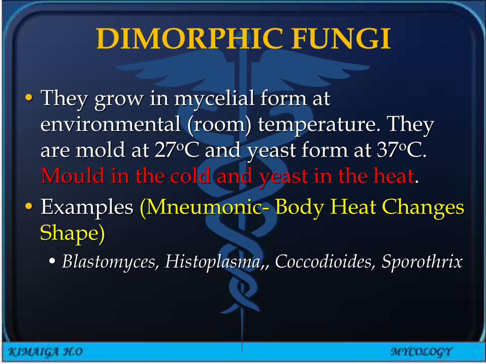
DIMORPHIC FUNGI
• They grow in mycelial form at environmental (room) temperature. They are mold at 27oC and yeast form at 37oC. Mould in the cold and yeast in the heat.
• Examples (Mneumonic- Body Heat Changes Shape)
• Blastomyces, Histoplasma,, Coccodioides, Sporothrix

YEAST-LIKE FUNGI(DIMORPHIC)
• These are fungi that are partly yeasts and partly mould(pseudohyphae/pseudomycelia).
• An example - Candida albicans does grow as yeast form at room temperature and myciliate form at 37oC.

IMPORTANT CELL STRUCTURES
Chitin
• In the fungal cell wall – polysaccharides
• Fungi are insensitive to antibiotics e.g. penicillin that inhibit peptidoglycan
Ergosterol
• In the fungal cell membrane
• Human cell membrane has cholesterol
• Selective action of antifungals based on difference in sterols

Epidemiology & Pathogenesis
• Worldwide distribution
• Causal Agents are saprophytes restricted in their distribution by soil and climatic conditions
• Fungi can cause diseases only if they are provided by their hosts with conditions and nutrients essential for their growth and reproduction.
• Factors that affect the incidence of mycoses degree of exposure causative agent e.g. living conditions, occupation, leisure activities
• Some yeast are commensals of man and cause endogenous infections when there is an imbalance in the host
Disorders/diseases caused by fungi.
1. Mycotoxicoses• Ingestion of foods contaminated by fungal
toxins (rice, maize, groundnuts)e.g.•Aspergillus falvus toxins (aflatoxins)- cause liver
damage, carcinoma• Penicillium toxins• Fusarium toxins
•Hepatotoxins
•Vascular and neurologic toxins- the mouldclaviceps purpura.

2. Fungal poisoning.• Ingestion of poisonous fungi
(Basidiomycetes), e.g. toxic mushrooms;•Amanita phalloides•Amanita muscaria
3.Fungal Allergies• Allergic reactions caused by contact with
fungal spores floating in the air e.g. Aspergillus, Mucor, Penicillium, Candida spp.
• Asthmatic reaction - rapid bronchconstriction

4. MYCOSES
1. Superficial mycoses (dermatomycoses and dermatophytoses)
• Fungal infection
• Those caused by species of Trichophyton, Microsporum, Epidermophyton and Keratomyces (dermatophytes) are called dermatophytoses. Dermatophytoses are commonly called ringworm or tinea (or known as mashilingi locally).
• Those caused by non-dermatophyte fungi e.g. Candida spp are called dermatomycoses.

2. Subcutaneous mycoses: • Mycetoma, Phycomycosis,
Chromoblastomycosis, Sporotrichosis, Lobomycosis, Rhinosporidiosis
3. Systemic mycoses:• Affect internal organs; i.e. All mycoses affecting
tissues deeper than the skin (subcutaneous, deep, opportunistic, actinomycosis
4. Opportunistic mycoses
• Diseases due to infection with normally non-pathogenic fungi as a result of weakened immune system
Lab Diagnosis
• Useful information including residence, previous travel, animal contact, occupation
• Lab investigation involves
• Recognition of pathogen in tissue by microscopy
• Isolation of pathogen in tissue by microscopy
• Isolation of causal fungus in culture
• Serological tests
•Detection of fungal DNA by PCR

Fungal specimens• Biopsies
• Immediately to laboratory in plains simple sterile container (No formalin)
• In formal- Saline for histology• 30% glycerol for transporting • 30%glycerine solution for culture
• Superficial fungi e.g Ringworm- skin scales, nail clippings, hair stubs-Simple envelope, matchbox, petri dish
• Swabs from mucous membranes• Subcutaneous infection- crusts, aspirated pus,
biopsies• Systemic infections- specimen from appropriate
site

• Direct microscopy
•Wet mount with KOH 10%
•Calcoflour white- Fluorescent microscopy
•Gram films- yeast infections
•Giemsa- detect histoplasma capsulatum
•Histology- periodic acid-Schiff (PAS) & Grocott-Gomori methamine silver (GMS) in tissue
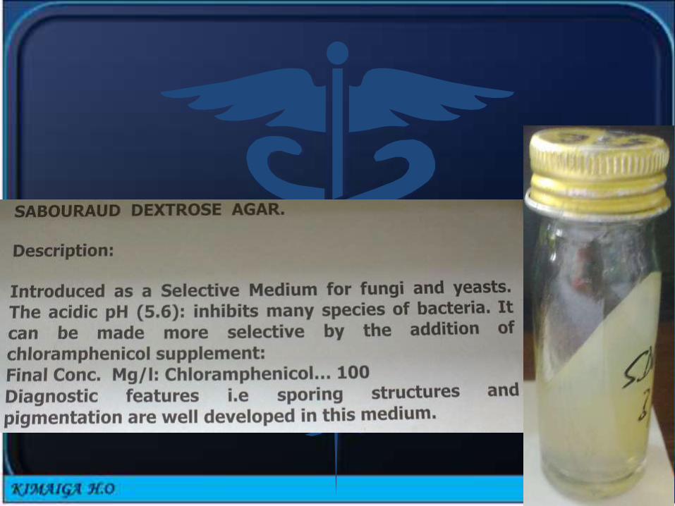
• Culture
•Media SDA (Saboraud dextrose agar, 4 % malt extract agar)
•Chloramphenicol, to reduce bacterial contamination
•Cycloheximide reduce contamination by saprophytic fungi
• Incubated at 25-30 and 37oC
• Yeast grows in 1-5 days
•Moulds identified by macro and microscopic morphology
• Yeasts identified by sugar fermentation, ability to assimilate carbon and nitrogen sources

• Serology
• Immunodiffusion
•Counter-current immune electrophoresis
•Whole cell agglutination
•Complement fixation
• ELISA
• PCR- detection of DNA in blood, serum, broncheoalveolar lavage, sputum e.t.c