Interstitial lung diseases 2012_pdf
17
Interstitial Lung Diseases Dr. Rahul Magazine M.D.(Medicine), D.T.C.D. Department of Pulmonary Medicine
-
Upload
drmanish-kumar -
Category
Health & Medicine
-
view
35 -
download
1
Transcript of Interstitial lung diseases 2012_pdf

Interstitial Lung
Diseases
Dr. Rahul Magazine
M.D.(Medicine), D.T.C.D.
Department of Pulmonary Medicine

Definition
The interstitial lung diseases (ILDs) represent
a large number of heterogeneous disorders
that involve the parenchyma of the lung -- the
alveoli, the alveolar epithelium, the capillary
endothelium, and the spaces between these
structures, as well as the perivascular and
lymphatic tissues.

Classification 1. DPLD of Known Cause (drugs or
association such as collagen vascular disease)
2. Idiopathic interstitial pneumonias
• Idiopathic Pulmonary Fibrosis (UIP pattern)
• IIP other than IPF (e.g. NSIP,DIP,AIP, RB-ILD, COP, LIP)
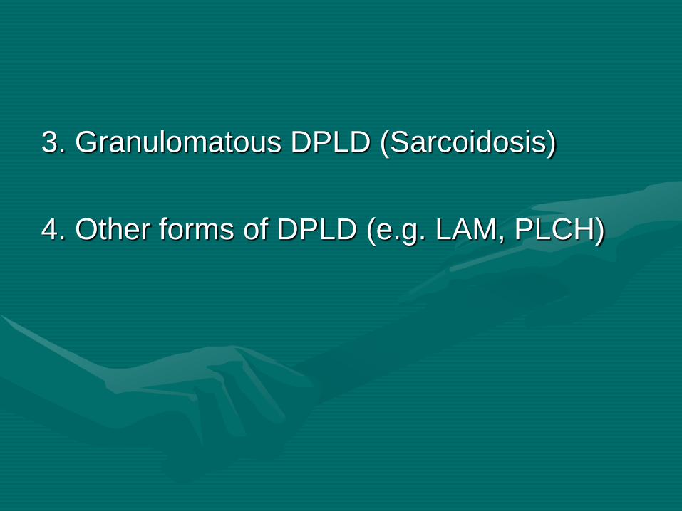
3. Granulomatous DPLD (Sarcoidosis)
4. Other forms of DPLD (e.g. LAM, PLCH)
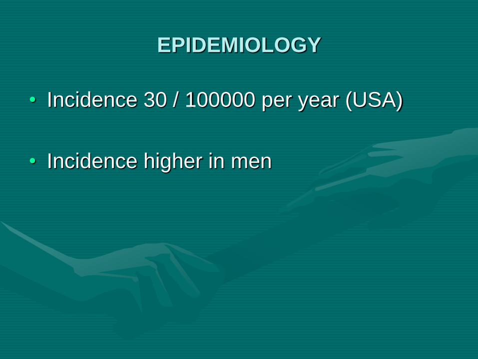
EPIDEMIOLOGY
• Incidence 30 / 100000 per year (USA)
• Incidence higher in men
PATHOGENESIS
ILDs are the result of the superimposed
processes of inflammation and tissue injury and
attempted repair
Two major histopathologic patterns:
GRANULOMATOUS LUNG DISEASE
This process is characterized by an
accumulation of T lymphocytes, macrophages,
and epithelioid cells organized into discrete
structures (granulomas) in the lung
parenchyma. The granulomatous lesions can
progress to fibrosis.

INFLAMMATION AND FIBROSIS
The initial insult is an injury to the epithelial
surface causing inflammation in the air
spaces and alveolar walls. If the disease
becomes chronic, inflammation spreads to
adjacent portions of the interstitium and
vasculature and eventually causes interstitial
fibrosis.

CLINICAL PRESENTATION
HISTORY
Breathlessness (most common): Initially, dyspnea on exertion→ later at rest
Nonproductive cough
Fatigue
Pleuritic chest pain
Hemoptysis-- infrequent
A family history of ILDs should be sought.

D/D BASED ON ONSET: ACUTE ONSET: DAYS TO WEEKS
Acute interstitial pneumonia
Acute pneumonitis from collagen vascular
disease(especially SLE)
Diffuse alveolar hemorrhage
Hypersensitivity pneumonitis
CHRONIC: MONTHS TO YEARS
Idiopathic pulmonary fibrosis
Chronic hypersensitivity pneumonitis
Collagen vascular disease–associated ILD

PHYSICAL EXAMINATION
PULMONARY SIGNS
With advanced disease, patients may have tachypnea and tachycardia, even at rest.
Bilateral, basilar, Velcro-like rales
Signs of pulmonary hypertension

EXTRAPULMONARY SIGNS:
Clubbing (e.g. IPF)
Skin abnormalities, peripheral
lymphadenopathy, hepatosplenomegaly
(SARCOIDOSIS)
Subcutaneous nodules (RHEUMATOID
ARTHRITIS)
Muscle tenderness and proximal weakness
(POLYMYOSITIS)

INVESTIGATIONS
CHEST RADIOGRAPHY
Nodules, linear (reticular) infiltrates, or a
combination of the two (reticulonodular
infiltrates)
Diffuse ground glass pattern– EARLY
Cystic areas (honeycomb pattern)-LATE
HIGH-RESOLUTION CT scan
PULMONARY FUNCTION TESTS
Reduced lung volumes, reduced diffusing
capacity (DLCO ),a normal or supernormal
ratio of FEV1 to FVC, Static lung
compliance is decreased (decreased lung
volume for any given transpulmonary
pressure), and maximal transpulmonary
pressure is increased (a very high
negative pressure must be generated to
open the fibrotic alveoli)
ARTERIAL BLOOD GAS ANALYSIS
BRONCHOSCOPIC STUDIES
Bronchoalveolar lavage
LUNG BIOPSY
Transbronchial biopsy
Open-lung or thoracoscopic biopsy
TREATMENT
Principal aims:
(1) to remove exposure to injurious
agents,
(2) to suppress inflammation to prevent
further destruction of the pulmonary
parenchyma, and
(3) to palliate the manifestations of these
diseases.

CORTICOSTEROIDS: mainstay of therapy
Prednisone, 1 mg/kg for 1 month, followed by 40 mg/day given for 2 months
Gradually tapered (5 mg/week) over several months to a maintenance dose of 15 to 20 mg/day
Corticosteroids are continued until pulmonary function is stable for 1 year
Relapses require returning to high-dose steroids
Cytotoxic agents (Cyclophosphamide)or immunosuppressive agents (Azathioprine) may be used in patients who do not improve on steroid therapy or who cannot tolerate corticosteroids

Thank You