Injectable and porous PLGA microspheres that form · PDF fileInjectable and porous PLGA...
10
Injectable porous microspheres form highly porous scaffolds after injection Qutachi, O.; Vetsch, J.R.; Gill, D.; Cox, H.; Scurr, D.J.; Hofmann, S.; Müller, R.; Quirk, R.A.; Shakesheff, K.M.; Rahman, C.V. Published in: Acta Biomaterialia DOI: 10.1016/j.actbio.2014.08.015 Published: 01/01/2014 Document Version Publisher’s PDF, also known as Version of Record (includes final page, issue and volume numbers) Please check the document version of this publication: • A submitted manuscript is the author's version of the article upon submission and before peer-review. There can be important differences between the submitted version and the official published version of record. People interested in the research are advised to contact the author for the final version of the publication, or visit the DOI to the publisher's website. • The final author version and the galley proof are versions of the publication after peer review. • The final published version features the final layout of the paper including the volume, issue and page numbers. Link to publication Citation for published version (APA): Qutachi, O., Vetsch, J. R., Gill, D., Cox, H., Scurr, D. J., Hofmann, S., ... Rahman, C. V. (2014). Injectable porous microspheres form highly porous scaffolds after injection. Acta Biomaterialia, 10(12), 5090-5098. DOI: 10.1016/j.actbio.2014.08.015 General rights Copyright and moral rights for the publications made accessible in the public portal are retained by the authors and/or other copyright owners and it is a condition of accessing publications that users recognise and abide by the legal requirements associated with these rights. • Users may download and print one copy of any publication from the public portal for the purpose of private study or research. • You may not further distribute the material or use it for any profit-making activity or commercial gain • You may freely distribute the URL identifying the publication in the public portal ? Take down policy If you believe that this document breaches copyright please contact us providing details, and we will remove access to the work immediately and investigate your claim. Download date: 09. May. 2018
Transcript of Injectable and porous PLGA microspheres that form · PDF fileInjectable and porous PLGA...

Injectable porous microspheres form highly porousscaffolds after injectionQutachi, O.; Vetsch, J.R.; Gill, D.; Cox, H.; Scurr, D.J.; Hofmann, S.; Müller, R.; Quirk, R.A.;Shakesheff, K.M.; Rahman, C.V.Published in:Acta Biomaterialia
DOI:10.1016/j.actbio.2014.08.015
Published: 01/01/2014
Document VersionPublisher’s PDF, also known as Version of Record (includes final page, issue and volume numbers)
Please check the document version of this publication:
• A submitted manuscript is the author's version of the article upon submission and before peer-review. There can be important differencesbetween the submitted version and the official published version of record. People interested in the research are advised to contact theauthor for the final version of the publication, or visit the DOI to the publisher's website.• The final author version and the galley proof are versions of the publication after peer review.• The final published version features the final layout of the paper including the volume, issue and page numbers.
Link to publication
Citation for published version (APA):Qutachi, O., Vetsch, J. R., Gill, D., Cox, H., Scurr, D. J., Hofmann, S., ... Rahman, C. V. (2014). Injectableporous microspheres form highly porous scaffolds after injection. Acta Biomaterialia, 10(12), 5090-5098. DOI:10.1016/j.actbio.2014.08.015
General rightsCopyright and moral rights for the publications made accessible in the public portal are retained by the authors and/or other copyright ownersand it is a condition of accessing publications that users recognise and abide by the legal requirements associated with these rights.
• Users may download and print one copy of any publication from the public portal for the purpose of private study or research. • You may not further distribute the material or use it for any profit-making activity or commercial gain • You may freely distribute the URL identifying the publication in the public portal ?
Take down policyIf you believe that this document breaches copyright please contact us providing details, and we will remove access to the work immediatelyand investigate your claim.
Download date: 09. May. 2018
Acta Biomaterialia 10 (2014) 5090–5098
Contents lists available at ScienceDirect
Acta Biomaterialia
journal homepage: www.elsevier .com/locate /ac tabiomat
Injectable and porous PLGA microspheres that form highly porousscaffolds at body temperature
http://dx.doi.org/10.1016/j.actbio.2014.08.0151742-7061/� 2014 Acta Materialia Inc. Published by Elsevier Ltd.This is an open access article under the CC BY-NC-ND license (http://creativecommons.org/licenses/by-nc-nd/3.0/).
⇑ Corresponding author.E-mail address: [email protected] (C.V. Rahman).
Omar Qutachi a, Jolanda R. Vetsch b, Daniel Gill c, Helen Cox c, David J. Scurr a, Sandra Hofmann b,Ralph Müller b, Robin A. Quirk c, Kevin M. Shakesheff a, Cheryl V. Rahman a,⇑a School of Pharmacy, University of Nottingham, University Park, Nottingham NG7 2RD, UKb Institute for Biomechanics, ETH Zurich, Vladimir-Prelog-Weg 3, 8093 Zurich, Switzerlandc RegenTec Ltd, Biocity Nottingham, Pennyfoot Street, Nottingham NG1 1GF, UK
a r t i c l e i n f o
Article history:Received 21 February 2014Received in revised form 19 July 2014Accepted 15 August 2014Available online 23 August 2014
Keywords:PLGAMicrosphereScaffoldPorosityCell delivery
a b s t r a c t
Injectable scaffolds are of interest in the field of regenerative medicine because of their minimally inva-sive mode of delivery. For tissue repair applications, it is essential that such scaffolds have the mechanicalproperties, porosity and pore diameter to support the formation of new tissue. In the current study,porous poly(DL-lactic acid-co-glycolic acid) (PLGA) microspheres were fabricated with an average sizeof 84 ± 24 lm for use as injectable cell carriers. Treatment with ethanolic sodium hydroxide for 2 minwas observed to increase surface porosity without causing the microsphere structure to disintegrate. Thissurface treatment also enabled the microspheres to fuse together at 37 �C to form scaffold structures. Theaverage compressive strength of the scaffolds after 24 h at 37 �C was 0.9 ± 0.1 MPa, and the averageYoung’s modulus was 9.4 ± 1.2 MPa. Scaffold porosity levels were 81.6% on average, with a mean porediameter of 54 ± 38 lm. This study demonstrates a method for fabricating porous PLGA microspheresthat form solid porous scaffolds at body temperature, creating an injectable system capable of supportingNIH-3T3 cell attachment and proliferation in vitro.� 2014 Acta Materialia Inc. Published by Elsevier Ltd. This is an open access article under the CC BY-NC-
ND license (http://creativecommons.org/licenses/by-nc-nd/3.0/).
1. Introduction
The use of polymer scaffolds to deliver cells for regenerativemedicine applications can potentially overcome poor cellengraftment and improve cell survival [1]. In particular, injectablescaffolds show promise for this application, as cells can be mixedhomogeneously with the scaffold formulation prior to injection.The ability to deliver injectable scaffolds in a minimally invasivemanner to a cavity of any size or shape renders them especiallyattractive for clinical use in tissue repair. A number of syntheticpolymers have been investigated for this application to date, includ-ing the biodegradable polymer poly(DL-lactic acid-co-glycolic acid)(PLGA). PLGA is frequently used in regenerative medicine applica-tions, as the degradation rate of the polymer can be controlledand it has FDA approval for certain clinical applications [2,3].PLGA-based scaffold systems have been reported extensively tosupport cell attachment and proliferation, deliver growth factorsin a controlled manner, and support bone regeneration in vivo [4–8].
In order to maintain, induce and restore biological functions,scaffolds for tissue repair require suitable physical properties. Ide-ally, the scaffold should be strong enough to retain its structurewithout exhibiting stiffness that may affect the surrounding tissue[2]. The microstructural properties of the scaffold also play a vitalrole in successful tissue repair, as porosity levels and pore diameterinfluence cell attachment, proliferation and migration in additionto nutrient delivery and waste removal [9,10]. Studies have indi-cated that scaffolds should possess multi-scale porosity involvingboth micro-porosity and macro-porosity, with pore diametersranging from <20 lm to >100 lm [9]. The versatility of chemicallysynthesized polymers such as PLGA is an advantage in this respect,as it enables the fabrication of scaffolds with different porositiesand mechanical properties. However, a delicate balance is requiredin terms of these properties, as increasing the porosity of a scaffoldcauses a subsequent decrease in mechanical properties such ascompressive strength. Attaining suitable porosity and strength inan injectable formulation is therefore a considerable challenge.
One type of injectable scaffold for tissue engineeringapplications involves the use of discreet polymer microspheres.Microspheres can be fabricated using a variety of differentbiodegradable polymers such as chitosan, gelatin and PLGA, and
O. Qutachi et al. / Acta Biomaterialia 10 (2014) 5090–5098 5091
their use for delivery of cells and growth factors for repair oftissues such as bone, skin and brain has been reported [11–15].Building on this strategy, microspheres with surface porosity havestarted to attract interest in the regenerative medicine field. Cellviability, proliferation and differentiation have been shown to beenhanced with porous microsphere systems in comparison withnon-porous microspheres [16,17]. This is most likely due to thepores facilitating improved diffusion of nutrients and oxygen. Anumber of studies have been reported on the fabrication of porousmicrospheres using various natural and synthetic polymers, inaddition to inorganic materials such as tricalcium phosphate[16,18–20]. Techniques employed to induce porosity duringmicrosphere fabrication include salt-leaching or using a porogensuch as gelatin [19,20]. Although such porous microspheres areinjectable and provide micro-porosity, they are unable to fusetogether at body temperature and therefore remain as discretemicrospheres in vivo. This is a drawback for their use in regener-ation of certain tissues such as bone, as they lack the requiredporosity profile and mechanical properties to support tissuerepair.
The current study describes the development of an injectable,porous PLGA scaffold that solidifies in situ at 37 �C. A procedurefor fabricating hollow, porous PLGA microspheres is described,where the microspheres were treated with ethanolic sodiumhydroxide (EtOH–NaOH) to increase the surface porosity. TheEtOH–NaOH treatment enables the microspheres to fuse togetherat 37 �C into solid scaffolds. The scaffold mechanical properties,porosity and ability to support cell attachment and proliferationwere characterized in vitro in order to determine their suitabilityfor use in tissue repair applications.
2. Materials and methods
2.1. Fabrication of porous microspheres
Porous PLGA microspheres were produced by the doubleemulsion method from 20% (w/v) PDLLGA 50:50 (52 kDa, EvonikIndustries, USA) in dichloromethane (DCM, Fisher, UK). One gramof PLGA was dissolved in 5 ml DCM for each batch of microspheres.For the primary emulsion, 250 ll of the porogen (phosphate buf-fered saline (PBS); Gibco, UK) was added to the PLGA/DCM, andthe PLGA/DCM/PBS was homogenized at 9000 rpm for 2 min usinga Silverson L5M homogenizer. The primary water in oil (w/o)emulsion was then homogenized in 0.3% polyvinyl alcohol (PVA;Sigma-Aldrich, UK) at 3000 rpm and the resultant water-in-oil-in-water (w/o/w) double emulsion was left stirringovernight to allow DCM evaporation. The hardened microsphereswere then harvested via distilled water (DW) washing andcentrifugation.
2.2. NaOH treatment of porous microspheres
Porous PLGA microspheres were treated with ethanolic sodiumhydroxide with 30% 0.25 M NaOH (Sigma-Aldrich):70% absoluteethanol (EtOH; Fisher, UK) [12]. This method was chosen becauseethanol is a water-miscible solvent that partially dissolves PLGAand NaOH improves polyester surface wettability by promotingcarboxylic acid exposure on the polymer surface, making it morehydrophilic [21,22]. One gram of microspheres was suspended in10 ml of EtOH–NaOH solution with agitation on a plate rocker for2 min, 3.5 min or 5 min, then poured onto a 50-lm sieve andrinsed with DW before transferring to a 50-ml centrifuge tubeand centrifuged at 1400 rpm for 1 min. This wash and centrifuga-tion step with DW was repeated prior to drying in a freeze-drier(Modulyo, Thermo Electron Corporation, UK) for 24 h.
2.3. Scanning electron microscopy
Microspheres were mounted on aluminium stubs (Agar Scien-tific, UK) and gold-coated using a Balzers SCD030 gold sputtercoater (Balzers Union Ltd., Lichtenstein) with an argon rate of30 mA for 3 min. The structural morphology of the microsphereswas examined by scanning electron microscopy (SEM), using aJEOL 6060L imaging system (JEOL Ltd., Hertfordshire, UK) at 10 kV.
2.4. Microsphere size analysis
The mean microsphere diameter and size distribution wereinvestigated using a Coulter LS230 particle size analyser (Beckman,UK). The microsphere size distribution was then determined as afunction of the microsphere diffraction and plotted as a functionof volume percentage.
2.5. Microsphere pore diameter analysis
The microsphere pore diameter was measured using Image Jsoftware. One hundred pore diameters were measured per imagefrom three separate SEM images from three different batches ofmicrospheres before and after treatment with EtOH–NaOH for2 min.
2.6. Scaffold formation
Triplicate scaffolds were prepared in PTFE moulds producingcylindrical scaffolds 6 mm in diameter and 12 mm high. Micro-spheres were mixed manually with Dulbecco’s Modified Eagle’sMedium (DMEM; Gibco UK) using a spatula at a ratio of 1:1 (micro-spheres to DMEM). The resulting paste was packed into the holesof the mould, and the mould was transferred to a sealed humidifiedchamber containing DMEM at 37 �C. For mechanical testingexperiments, moulds were transferred to a sealed humidifiedchamber containing DMEM at 37 �C, 40 �C and 50 �C for 24 h. After24 h, all scaffolds were removed from the moulds. Scaffolds formicro-computed tomography (lCT) and SEM assessment werefreeze-dried for 24 h and refrigerated until use.
2.7. Mechanical testing
The compressive strength of the scaffolds fabricated using sin-tered microspheres was tested using a TA.HD+ texture analyser(Stable Microsystems, UK) following the ASTM standard D-1621.Scaffolds were tested with the Peltier unit heated to 37 �C and ascaffold contact area of 28.3 mm2. A 50-kg load cell was used forcompression testing. The test speed was set at 0.04 mm s�1. Themechanical testing was performed on triplicate scaffolds sinteredat 37 �C for 6 h and 24 h at 37 �C, 40 �C and 50 �C. A fracture pointwas not observed during compression of the scaffolds, thereforethe compressive strength was measured at 20% strain to comparethe strength after incubating at different temperatures. Thestress–strain curve generated during compression analysis wasused to calculate the Young’s modulus (elastic modulus) as a mea-sure of the stiffness of the scaffolds. The elastic modulus was calcu-lated by determining the slope of the stress–strain curve along theelastic portion of deformation.
2.8. lCT
lCT was performed on dry scaffolds in air on a lCT 40 (ScancoMedical, Brütisellen, Switzerland). Scans were performed at anenergy level of 45 kVp, an intensity of 177 lA, integration time of200 ms and a frame averaging of 4, with a nominal resolution of6 lm. The reconstructed files were Gaussian filtered applying a
5092 O. Qutachi et al. / Acta Biomaterialia 10 (2014) 5090–5098
filter support of 1 voxel and filter width of sigma = 0.8. A volume ofinterest 400 voxels (2.4 mm) in diameter and 500 voxels (3.0 mm)high was chosen for the evaluation. Resulting greyscale imageswere thresholded at a global threshold of 3.3% of the maximal greyvalue. Objects smaller than 40 voxels were removed using compo-nent labelling and neglected for further evaluation. The resultingthree-dimensional (3-D) volumes were evaluated as describedpreviously for scaffold porosity, pore diameter distribution andaverage pore diameter [23,24].
2.9. Mouse 3T3 fibroblast cell culture
NIH-3T3 fibroblast cells were cultured in DMEM medium(Gibco, UK) supplemented with 10% foetal calf serum, 1% antibi-otic/antimycotic solution, 1% L-glutamine (2 mM) and 1% non-essential amino acids (Sigma-Aldrich, UK). RFP-NIH-3T3 cells werepreviously genetically labelled with red fluorescent protein asdescribed by Dixon and colleagues [25]. RFP-MIH-3T3 cells werecultured in the same media as NIH-3T3. All cells were maintainedin a humidified tissue-culture incubator at 37 �C and with 5% CO2.
2.10. Cell seeding on scaffolds
For scaffolds fabricated in the presence of cells, microsphereswere mixed manually with DMEM (Gibco UK) containing 2 � 105
NIH-3T3 fibroblasts using a spatula at a ratio of 1:1 (microspheresto DMEM). The resulting paste was packed into the holes of themould, and the mould was transferred to a sealed humidifiedchamber containing DMEM at 37 �C. After 24 h at 37 �C, all scaf-folds were removed from the moulds, transferred into 12-well cellculture plates with fresh medium (1 ml per well) and incubated at37 �C.
2.11. Cell viability assay
The PrestoBlue cell viability assay (Invitrogen Life Sciences, UK)was performed on day 3, 6 and 9 post-seeding on four scaffolds pertime point. Each scaffold was submerged in 1 ml of media, and111 ll of Prestoblue was added to each well. The scaffolds werethen incubated at 37 �C for 25 min. Three 100-ll media sampleswere taken from each well and were read on a Tecan plate readerwith the excitation wavelength set to 535 nm and the emissionwavelength set at 615 nm.
2.12. Toluidine blue staining
Toluidine blue (Sigma-Aldrich, UK) staining was performed onday 7 post-seeding on triplicate scaffolds. Each scaffold waswashed twice with DW and fixed in formalin solution (Sigma-Aldrich, UK) for 5 min at room temperature. Each scaffold was thenwashed twice in DW, and 1% toluidine blue was added to each wellfor 5 min at room temperature. Finally, the scaffolds were washedfive times in DW and visualized under a light microscope.
2.13. Statistical analysis
Statistical analysis was performed using GraphPad Prism(Version 3) analysis software. Differences among groups weredetermined by ANOVA Tukey–Kramer Multiple Comparisons Testand were considered to be significantly different if p < 0.05.
3. Results
3.1. Fabrication of porous PLGA microspheres
PLGA microspheres were fabricated using a double emulsionprocess as described in Section 2.1 (Fig. 1A). Microspheres withsurface porosity were generated using PBS in the internal aqueousphase during the fabrication process, as shown in Fig. 1B. Porediameters were enlarged through treatment with EtOH–NaOH, asdescribed in Section 2.2, which degraded the surface layer of thePLGA microspheres (Fig. 1C–E). Different lengths of time of surfacetreatment with EtOH–NaOH were assessed (2 min, 3.5 min and5 min). Increased porosity compared with non-treated micro-spheres was observed in the SEM images. However, treating themicrospheres for 3.5 or 5 min resulted in signs of structuraldamage to the microspheres such as cracks on the surface and dis-integration of the microspheres (Fig. 1D, E). Microsphere diameterwas measured as described in Section 2.4, using triplicate batchesof non-treated microspheres and microspheres treated with EtOH–NaOH for 2, 3.5 and 5 min. The microsphere size distribution wassimilar for each batch of microspheres regardless of treatment time(Fig. 2). The average diameter of non-treated microspheres was81 ± 26 lm, and the average diameter of treated porous micro-spheres was 84 ± 24 lm, 81 ± 24 lm and 82 ± 24 lm for 2, 3.5and 5 min treatment, respectively (Fig. 2). In order to avoid thepossibility of over-treatment with EtOH–NaOH and structuraldamage to the microspheres, a treatment time of 2 min wasselected for porous microsphere fabrication. Microsphere porediameters were measured before and after EtOH–NaOH treatmentfor 2 min, as described in Section 2.5. Pore diameters were6 ± 2 lm on average before treatment, with a maximum porediameter of 9 lm. Following EtOH–NaOH treatment, pore diame-ters were 8 ± 3 lm on average, with a maximum of 15 lm.
3.2. Scaffold formation using porous PLGA microspheres
Porous microspheres were packed into a PTFE mould and incu-bated at 37 �C for 24 h both before and after EtOH–NaOH treat-ment, as described in Section 2.6. Microspheres treated withEtOH–NaOH for 2 min formed solid scaffold structures over 24 hat 37 �C, as shown in Fig. 3A. SEM images show the microspheresfusing together to create the scaffold structure (Fig. 3B, C). Micro-spheres that were not treated with EtOH–NaOH did not fusetogether over 24 h at 37 �C (Fig. 3D). As shown in Fig. 3E and F,treatment with EtOH–NaOH for 3.5 or 5 min allowed the micro-spheres to fuse. However, deformation of microsphere structureand collapse of pores could be visualized in these scaffolds, partic-ularly when microspheres were treated with EtOH–NaOH for5 min prior to scaffold fabrication. Therefore, the treatment timeof 2 min was selected to produce scaffolds for further analysis interms of their mechanical and microstructural properties. Asnon-treated microspheres did not form scaffolds, resulting in loosemicrospheres being removed from the mould after this time, anon-treated microsphere group was not included in the furtherstudies described herein, as these were performed on wholescaffolds.
3.3. Mechanical properties of scaffolds fabricated with porous PLGAmicrospheres
The compressive strength and Young’s modulus of scaffoldsprepared with porous microspheres was assessed using a textureanalyser, as described in Section 2.7. The microspheres were fabri-cated with PLGA that has a glass transition temperature (Tg) of�50 �C. The EtOH–NaOH treated PLGA microspheres were
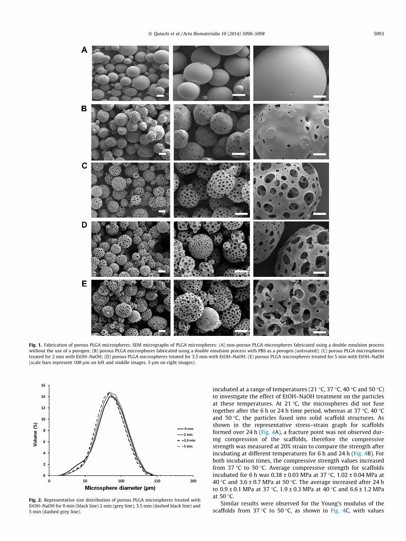
Fig. 1. Fabrication of porous PLGA microspheres. SEM micrographs of PLGA microspheres: (A) non-porous PLGA microspheres fabricated using a double emulsion processwithout the use of a porogen; (B) porous PLGA microspheres fabricated using a double emulsion process with PBS as a porogen (untreated); (C) porous PLGA microspherestreated for 2 min with EtOH–NaOH; (D) porous PLGA microspheres treated for 3.5 min with EtOH–NaOH; (E) porous PLGA microspheres treated for 5 min with EtOH–NaOH(scale bars represent 100 lm on left and middle images, 5 lm on right images).
Fig. 2. Representative size distribution of porous PLGA microspheres treated withEtOH–NaOH for 0 min (black line) 2 min (grey line), 3.5 min (dashed black line) and5 min (dashed grey line).
O. Qutachi et al. / Acta Biomaterialia 10 (2014) 5090–5098 5093
incubated at a range of temperatures (21 �C, 37 �C, 40 �C and 50 �C)to investigate the effect of EtOH–NaOH treatment on the particlesat these temperatures. At 21 �C, the microspheres did not fusetogether after the 6 h or 24 h time period, whereas at 37 �C, 40 �Cand 50 �C, the particles fused into solid scaffold structures. Asshown in the representative stress–strain graph for scaffoldsformed over 24 h (Fig. 4A), a fracture point was not observed dur-ing compression of the scaffolds, therefore the compressivestrength was measured at 20% strain to compare the strength afterincubating at different temperatures for 6 h and 24 h (Fig. 4B). Forboth incubation times, the compressive strength values increasedfrom 37 �C to 50 �C. Average compressive strength for scaffoldsincubated for 6 h was 0.38 ± 0.03 MPa at 37 �C, 1.02 ± 0.04 MPa at40 �C and 3.6 ± 0.7 MPa at 50 �C. The average increased after 24 hto 0.9 ± 0.1 MPa at 37 �C, 1.9 ± 0.3 MPa at 40 �C and 6.6 ± 1.2 MPaat 50 �C.
Similar results were observed for the Young’s modulus of thescaffolds from 37 �C to 50 �C, as shown in Fig. 4C, with values
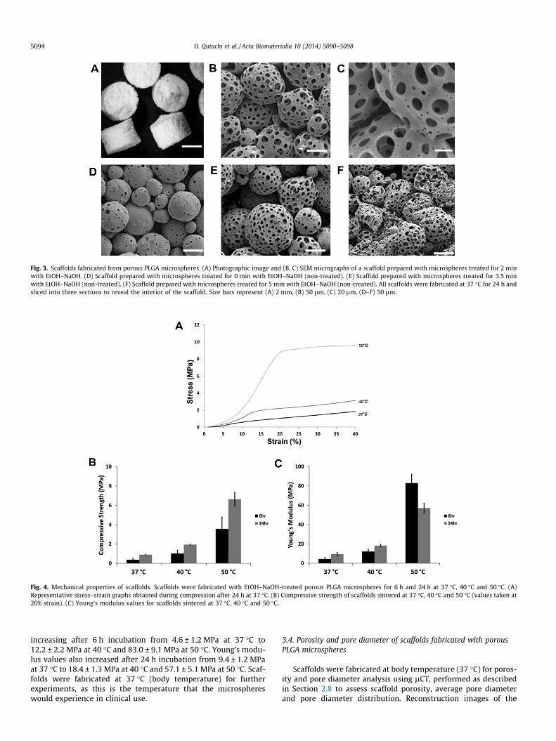
Fig. 3. Scaffolds fabricated from porous PLGA microspheres. (A) Photographic image and (B, C) SEM micrographs of a scaffold prepared with microspheres treated for 2 minwith EtOH–NaOH. (D) Scaffold prepared with microspheres treated for 0 min with EtOH–NaOH (non-treated). (E) Scaffold prepared with microspheres treated for 3.5 minwith EtOH–NaOH (non-treated). (F) Scaffold prepared with microspheres treated for 5 min with EtOH–NaOH (non-treated). All scaffolds were fabricated at 37 �C for 24 h andsliced into three sections to reveal the interior of the scaffold. Size bars represent (A) 2 mm, (B) 50 lm, (C) 20 lm, (D–F) 50 lm.
Fig. 4. Mechanical properties of scaffolds. Scaffolds were fabricated with EtOH–NaOH-treated porous PLGA microspheres for 6 h and 24 h at 37 �C, 40 �C and 50 �C. (A)Representative stress–strain graphs obtained during compression after 24 h at 37 �C. (B) Compressive strength of scaffolds sintered at 37 �C, 40 �C and 50 �C (values taken at20% strain). (C) Young’s modulus values for scaffolds sintered at 37 �C, 40 �C and 50 �C.
5094 O. Qutachi et al. / Acta Biomaterialia 10 (2014) 5090–5098
increasing after 6 h incubation from 4.6 ± 1.2 MPa at 37 �C to12.2 ± 2.2 MPa at 40 �C and 83.0 ± 9.1 MPa at 50 �C. Young’s modu-lus values also increased after 24 h incubation from 9.4 ± 1.2 MPaat 37 �C to 18.4 ± 1.3 MPa at 40 �C and 57.1 ± 5.1 MPa at 50 �C. Scaf-folds were fabricated at 37 �C (body temperature) for furtherexperiments, as this is the temperature that the microsphereswould experience in clinical use.
3.4. Porosity and pore diameter of scaffolds fabricated with porousPLGA microspheres
Scaffolds were fabricated at body temperature (37 �C) for poros-ity and pore diameter analysis using lCT, performed as describedin Section 2.8 to assess scaffold porosity, average pore diameterand pore diameter distribution. Reconstruction images of the
O. Qutachi et al. / Acta Biomaterialia 10 (2014) 5090–5098 5095
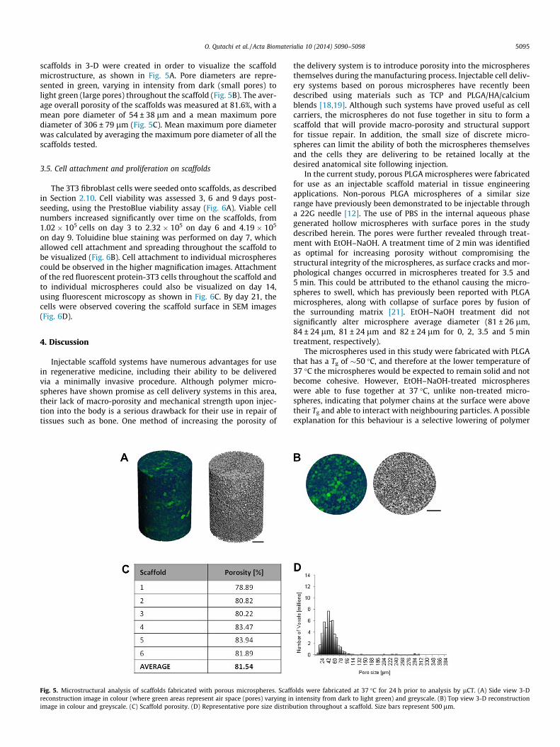
scaffolds in 3-D were created in order to visualize the scaffoldmicrostructure, as shown in Fig. 5A. Pore diameters are repre-sented in green, varying in intensity from dark (small pores) tolight green (large pores) throughout the scaffold (Fig. 5B). The aver-age overall porosity of the scaffolds was measured at 81.6%, with amean pore diameter of 54 ± 38 lm and a mean maximum porediameter of 306 ± 79 lm (Fig. 5C). Mean maximum pore diameterwas calculated by averaging the maximum pore diameter of all thescaffolds tested.
3.5. Cell attachment and proliferation on scaffolds
The 3T3 fibroblast cells were seeded onto scaffolds, as describedin Section 2.10. Cell viability was assessed 3, 6 and 9 days post-seeding, using the PrestoBlue viability assay (Fig. 6A). Viable cellnumbers increased significantly over time on the scaffolds, from1.02 � 105 cells on day 3 to 2.32 � 105 on day 6 and 4.19 � 105
on day 9. Toluidine blue staining was performed on day 7, whichallowed cell attachment and spreading throughout the scaffold tobe visualized (Fig. 6B). Cell attachment to individual microspherescould be observed in the higher magnification images. Attachmentof the red fluorescent protein-3T3 cells throughout the scaffold andto individual microspheres could also be visualized on day 14,using fluorescent microscopy as shown in Fig. 6C. By day 21, thecells were observed covering the scaffold surface in SEM images(Fig. 6D).
4. Discussion
Injectable scaffold systems have numerous advantages for usein regenerative medicine, including their ability to be deliveredvia a minimally invasive procedure. Although polymer micro-spheres have shown promise as cell delivery systems in this area,their lack of macro-porosity and mechanical strength upon injec-tion into the body is a serious drawback for their use in repair oftissues such as bone. One method of increasing the porosity of
Fig. 5. Microstructural analysis of scaffolds fabricated with porous microspheres. Scaffreconstruction image in colour (where green areas represent air space (pores) varying inimage in colour and greyscale. (C) Scaffold porosity. (D) Representative pore size distrib
the delivery system is to introduce porosity into the microspheresthemselves during the manufacturing process. Injectable cell deliv-ery systems based on porous microspheres have recently beendescribed using materials such as TCP and PLGA/HA/calciumblends [18,19]. Although such systems have proved useful as cellcarriers, the microspheres do not fuse together in situ to form ascaffold that will provide macro-porosity and structural supportfor tissue repair. In addition, the small size of discrete micro-spheres can limit the ability of both the microspheres themselvesand the cells they are delivering to be retained locally at thedesired anatomical site following injection.
In the current study, porous PLGA microspheres were fabricatedfor use as an injectable scaffold material in tissue engineeringapplications. Non-porous PLGA microspheres of a similar sizerange have previously been demonstrated to be injectable througha 22G needle [12]. The use of PBS in the internal aqueous phasegenerated hollow microspheres with surface pores in the studydescribed herein. The pores were further revealed through treat-ment with EtOH–NaOH. A treatment time of 2 min was identifiedas optimal for increasing porosity without compromising thestructural integrity of the microspheres, as surface cracks and mor-phological changes occurred in microspheres treated for 3.5 and5 min. This could be attributed to the ethanol causing the micro-spheres to swell, which has previously been reported with PLGAmicrospheres, along with collapse of surface pores by fusion ofthe surrounding matrix [21]. EtOH–NaOH treatment did notsignificantly alter microsphere average diameter (81 ± 26 lm,84 ± 24 lm, 81 ± 24 lm and 82 ± 24 lm for 0, 2, 3.5 and 5 mintreatment, respectively).
The microspheres used in this study were fabricated with PLGAthat has a Tg of �50 �C, and therefore at the lower temperature of37 �C the microspheres would be expected to remain solid and notbecome cohesive. However, EtOH–NaOH-treated microsphereswere able to fuse together at 37 �C, unlike non-treated micro-spheres, indicating that polymer chains at the surface were abovetheir Tg and able to interact with neighbouring particles. A possibleexplanation for this behaviour is a selective lowering of polymer
olds were fabricated at 37 �C for 24 h prior to analysis by lCT. (A) Side view 3-Dintensity from dark to light green) and greyscale. (B) Top view 3-D reconstruction
ution throughout a scaffold. Size bars represent 500 lm.
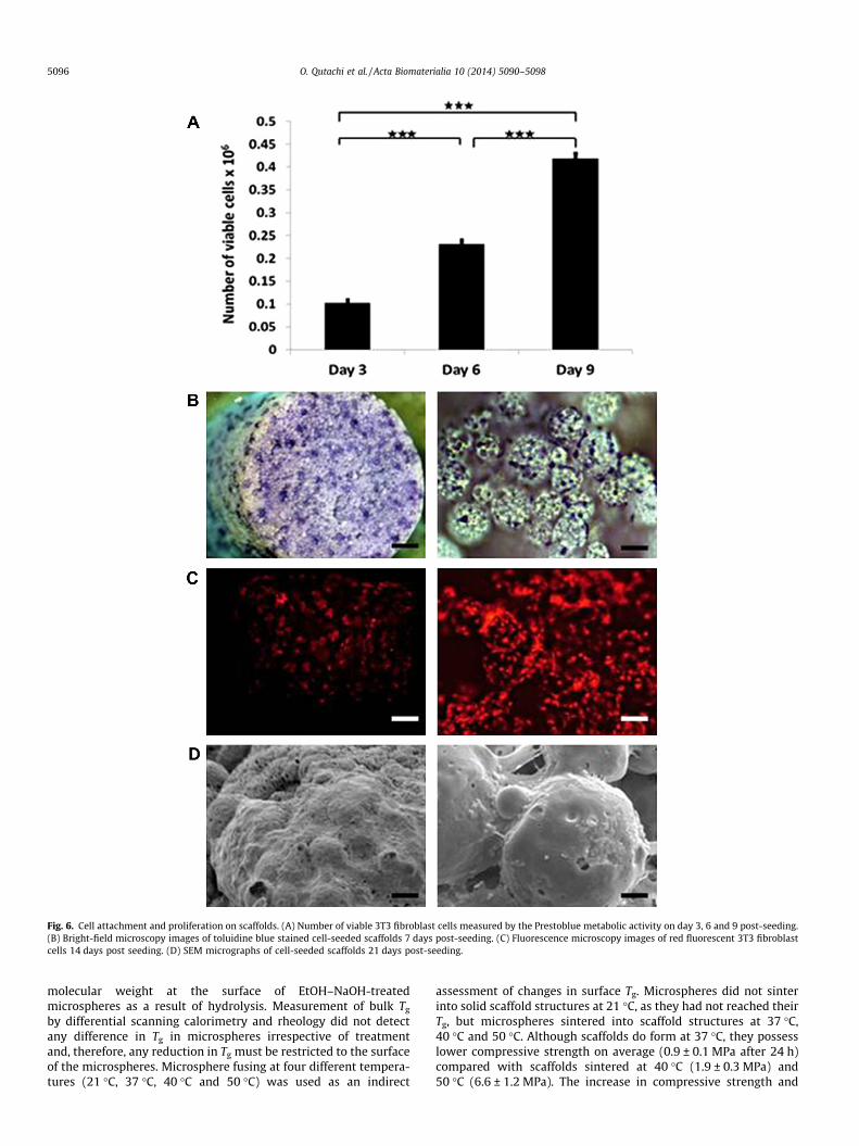
Fig. 6. Cell attachment and proliferation on scaffolds. (A) Number of viable 3T3 fibroblast cells measured by the Prestoblue metabolic activity on day 3, 6 and 9 post-seeding.(B) Bright-field microscopy images of toluidine blue stained cell-seeded scaffolds 7 days post-seeding. (C) Fluorescence microscopy images of red fluorescent 3T3 fibroblastcells 14 days post seeding. (D) SEM micrographs of cell-seeded scaffolds 21 days post-seeding.
5096 O. Qutachi et al. / Acta Biomaterialia 10 (2014) 5090–5098
molecular weight at the surface of EtOH–NaOH-treatedmicrospheres as a result of hydrolysis. Measurement of bulk Tg
by differential scanning calorimetry and rheology did not detectany difference in Tg in microspheres irrespective of treatmentand, therefore, any reduction in Tg must be restricted to the surfaceof the microspheres. Microsphere fusing at four different tempera-tures (21 �C, 37 �C, 40 �C and 50 �C) was used as an indirect
assessment of changes in surface Tg. Microspheres did not sinterinto solid scaffold structures at 21 �C, as they had not reached theirTg, but microspheres sintered into scaffold structures at 37 �C,40 �C and 50 �C. Although scaffolds do form at 37 �C, they possesslower compressive strength on average (0.9 ± 0.1 MPa after 24 h)compared with scaffolds sintered at 40 �C (1.9 ± 0.3 MPa) and50 �C (6.6 ± 1.2 MPa). The increase in compressive strength and
O. Qutachi et al. / Acta Biomaterialia 10 (2014) 5090–5098 5097
stiffness of the scaffolds as the sintering temperature was raisedindicated that the microspheres were able to fuse together moreas the temperature was raised closer to the PLGA glass transitiontemperature (50 �C).
Overall porosity level and pore diameter range are criticalproperties of a biomaterial scaffold. They play a vital role in tissueformation, as they allow migration and proliferation of cells intothe scaffold structure as well as supporting vascularization [26].Higher levels of porosity have been reported to enhance bone in-growth in vivo [27]. In addition, a porous surface improvesmechanical interlocking between the implant biomaterial and thesurrounding tissue, providing greater mechanical stability at thiscritical interface [28]. Scaffolds for tissue repair ideally requireboth micro-porosity (pore diameters of <20 lm) and macro-poros-ity (pore diameters of >100 lm) [9]. The injectable scaffoldsdescribed herein possess a mean pore diameter of 54 ± 38 lm.Many injectable scaffolds are hydrogel-based and therefore oftenlack the micro- and macro-porosity optimal for cell migrationand proliferation. Solid scaffolds can be engineered with varyinglevels of porosity, but these are usually preformed and thereforemust be implanted. For example, implantable porous scaffoldsdeveloped from PLGA microspheres can be fabricated at 49 �C, withporosity levels ranging from 66 to 74% [29]. PLGA has been used inan injectable format when dissolved in a solvent such as tetragly-col, and injectable scaffolds of 72% average porosity have been cre-ated using such a system [17]. However, in addition to the solventleaching out from the formulation in situ, the scaffolds displayed arelatively low average compressive strength of 0.1 MPa [17].
A delicate balance is required in terms of mechanical propertiesand porosity in scaffold systems, as increasing the porosity of ascaffold commonly results in a subsequent decrease in compres-sive strength. For example, porous PLGA scaffolds have been devel-oped with relatively high compressive strength compared with thepresent system, such as scaffolds prepared from PLGA–TiO2 micro-spheres, which displayed average compressive strength values of4.8 MPa [30]. However, such scaffolds displayed comparativelylower porosity ranging from 30 to 40%. The present authors previ-ously reported the fabrication of scaffolds for bone regenerationusing non-porous PLGA/poly(ethylene glycol) (PEG) microparticlesthat can be either implanted or injected and solidify at 37 �C.Although they display higher compressive strength and Young’smodulus values than the system described herein (2 MPa and40 MPa, respectively, after 2 h at 37 �C), the scaffolds producedusing this formulation had lower overall porosity with an averageof �40% [31]. It is clear that attaining a suitable balance betweenporosity and strength in an injectable scaffold formulation is a con-siderable challenge.
In addition to possessing suitable physical properties to supporttissue regeneration, scaffolds must also be capable of facilitatingcell delivery. In this regard, cell adhesion to the biomaterial iscrucial for cell survival, proliferation and eventual differentiationinto a specific cell type. Although PLGA has been extensively inves-tigated in tissue engineering scaffolds, this material does havesome drawbacks with regard to cell adhesion caused by its highhydrophobicity. In order to improve cell attachment, researchershave employed methods of modifying the surface of the PLGA, suchas pre-coating PLGA microspheres with the adhesion molecules ofRGD peptide, or treating microspheres with plasma polymerizedallylamine [12,13,32]. In the current study, PLGA microsphereswere not directly treated to enhance cell attachment, but theywere treated with EtOH–NaOH to increase surface porosity. Asdescribed in Section 2.9, the 3T3 fibroblast cell suspension wasmixed with microspheres using a spatula, and the resulting pastewas packed into a mould, which was transferred to a sealedhumidified chamber containing DMEM at 37 �C. After 24 h,scaffolds were removed from mould, transferred into 12-well cell
culture plates with fresh medium and incubated at 37 �C. The abil-ity of the cells to attach to and proliferate on the microsphereswithin the scaffolds following the fabrication process was assessed.Viable cell number increased over time in culture from1 � 105 cells per scaffold on day 3 to 4 � 105 on day 9, demonstrat-ing proliferation of the 3T3 cells over the 9-day time period. Cellattachment was visualized on the scaffolds using toluidine stainingand fluorescence microscopy, with cells appearing to attach well tothe microspheres within the scaffolds and to spread over the scaf-fold structure. An increase in the number of attached cells wasobserved over time in culture, culminating with a monolayer ofcells covering the particles by day 21. NIH-3T3 fibroblasts havepreviously been shown to attach and proliferate on injectable por-ous PLGA microspheres 535 lm in size, with surface pores up to20 lm, fabricated using ammonium bicarbonate as a porogen,but these microspheres did not form scaffolds in situ [33]. Futurework will investigate the ability of the injectable scaffold systemdescribed in this study to deliver viable cells and regenerate tissuein vivo.
5. Conclusion
In this study, hollow, porous PLGA microspheres were devel-oped, which fuse together at 37 �C to form highly porous scaffoldstructures in situ that support cell attachment and proliferation.To the best of the present authors’ knowledge, this is the first dem-onstration of the use of porous PLGA microspheres that physicallyfuse together without the use of solvents at body temperature asan injectable scaffold system. This technology is a promisingminimally invasive approach formation of porous biodegradablescaffolds in situ for tissue repair applications.
Disclosures
There are no potential conflicts of interest to disclose for thiswork.
Acknowledgments
This work was funded by the European Community’s FP7project Biodesign EUFP7-NMP.20102.3-1 (grant: 262948) and theEuropean Research Council under the European Community’sSeventh FP7 project FP72007-2013 (grant: 227845). The authorswould also like to thank Dr. James Dixon (University of Notting-ham) for kindly providing the red fluorescent labelled NIH-3T3cells.
References
[1] Mooney DJ, Vandenburgh H. Cell delivery mechanisms for tissue repair. CellStem Cell 2008;2:205–13.
[2] Yang S, Leong KF, Du Z, Chua CK. The design of scaffolds for use in tissueengineering. Part I. Traditional factors. Tissue Eng 2001;7:679–89.
[3] Jiang T, Abdel-Fattah WI, Laurencin CT. In vitro evaluation of chitosan/poly(lactic acid-glycolic acid) sintered microsphere scaffolds for bone tissueengineering. Biomaterials 2006;27:4894–903.
[4] White LJ, Kirby GTS, Cox HC, Qodratnama R, Qutachi O, Rose FRAJ, et al.Accelerating protein release from microparticles for regenerative medicineapplications. Mater Sci Eng C Mater Biol Appl 2013;33:2578–83.
[5] Ren T, Ren J, Jia X, Pan K. The bone formation in vitro and mandibular defectrepair using PLGA porous scaffolds. J Biomed Mater Res A 2005;74A:562–9.
[6] Rahman CV, Ben-David D, Dhillon A, Kuhn G, Gould TW, Muller R, et al.Controlled release of BMP-2 from a sintered polymer scaffold enhances bonerepair in a mouse calvarial defect model. J Tissue Eng Regen Med2012;8:59–66.
[7] Curran JM, Fawcett S, Hamilton L, Rhodes NP, Rahman CV, Alexander M, et al.The osteogenic response of mesenchymal stem cells to an injectable PLGAbone regeneration system. Biomaterials 2013;34:9352–64.
[8] Kirby GTS, White LJ, Rahman CV, Cox HC, Qutachi O, Rose FRAJ, et al. PLGA-based microparticles for the sustained release of BMP-2. Polymers2011;3:571–86.

5098 O. Qutachi et al. / Acta Biomaterialia 10 (2014) 5090–5098
[9] Bose S, Roy M, Bandyopadhyay A. Recent advances in bone tissue engineeringscaffolds. Trends Biotechnol 2012;30:546–54.
[10] Murphy CM, O’Brien FJ, Little DG, Schindeler A. Cell-scaffold interactions in thebone tissue engineering triad. Eur Cell Mater 2013;26:120–32.
[11] Bhat A, Dreifke MB, Kandimalla Y, Gomez C, Ebraheim NA, Jayasuriya AC.Evaluation of cross-linked chitosan microparticles for bone regeneration. JTissue Eng Regen Med 2010;4:532–42.
[12] Bible E, Chau DY, Alexander MR, Price J, Shakesheff KM, Modo M. Attachmentof stem cells to scaffold particles for intra-cerebral transplantation. Nat Protoc2009;4:1440–53.
[13] Bible E, Chau DY, Alexander MR, Price J, Shakesheff KM, Modo M. The supportof neural stem cells transplanted into stroke-induced brain cavities by PLGAparticles. Biomaterials 2009;30:2985–94.
[14] Bible E, Qutachi O, Chau DY, Alexander MR, Shakesheff KM, Modo M. Neo-vascularization of the stroke cavity by implantation of human neural stem cellson VEGF-releasing PLGA microparticles. Biomaterials 2012;33:7435–46.
[15] Kawai K, Suzuki S, Tabata Y, Ikada Y, Nishimura Y. Accelerated tissueregeneration through incorporation of basic fibroblast growth factor-impregnated gelatin microspheres into artificial dermis. Biomaterials2000;21:489–99.
[16] Kang S-W, La W-G, Kim B-S. Open macroporous poly(lactic-co-glycolic acid)microspheres as an injectable scaffold for cartilage tissue engineering. JBiomater Sci Polym Ed 2009;20:399–409.
[17] Krebs MD, Sutter KA, Lin ASP, Guldberg RE, Alsberg E. Injectable poly(lactic-co-glycolic) acid scaffolds with in situ pore formation for tissue engineering.Acta Biomater 2009;5:2847–59.
[18] Cheng D, Gao H, Hao L, Cao X, Wang Y. Facile development of a hollowcomposite microsphere with porous surface for cell delivery. Mater Lett2013;111:238–41.
[19] Ryu T-K, Oh M-J, Moon S-K, Paik D-H, Kim S-E, Park J-H, et al. Uniformtricalcium phosphate beads with an open porous structure for tissueengineering. Colloids Surf B: Biointerfaces 2013;112:368–73.
[20] Yang Y, Bajaj N, Xu P, Ohn K, Tsifansky MD, Yeo Y. Development of highlyporous large PLGA microparticles for pulmonary drug delivery. Biomaterials2009;30:1947–53.
[21] Kim HK, Chung HJ, Park TG. Biodegradable polymeric microspheres with‘‘open/closed’’ pores for sustained release of human growth hormone. J ControlRel 2006;112:167–74.
[22] Gao J, Niklason L, Langer R. Surface hydrolysis of poly(glycolic acid) meshesincreases the seeding density of vascular smooth muscle cells. J Biomed MaterRes 1998;42:417–24.
[23] Hildebrand T, Laib A, Müller R, Dequeker J, Rüegsegger P. Direct three-dimensional morphometric analysis of human cancellous bone:microstructural data from spine, femur, iliac crest, and calcaneus. J BoneMiner Res 1999;14:1167–74.
[24] van Lenthe GH, Hagenmüller H, Bohner M, Hollister SJ, Meinel L, Müller R.Nondestructive micro-computed tomography for biological imaging andquantification of scaffold–bone interaction in vivo. Biomaterials2007;28:2479–90.
[25] Dixon JE, Dick E, Rajamohan D, Shakesheff KM, Denning C. Directeddifferentiation of human embryonic stem cells to interrogate the cardiacgene regulatory network. Mol Ther 2011;19:1695–703.
[26] Kuboki Y, Takita H, Kobayashi D, Tsuruga E, Inoue M, Murata M, et al. BMP-induced osteogenesis on the surface of hydroxyapatite with geometricallyfeasible and nonfeasible structures: topology of osteogenesis. J Biomed MaterRes 1998;39:190–9.
[27] Karageorgiou V, Kaplan D. Porosity of 3D biomaterial scaffolds andosteogenesis. Biomaterials 2005;26:5474–91.
[28] Story BJ, Wagner WR, Gaisser DM, Cook SD, Rust-Dawicki AM. In vivoperformance of a modified CSTi dental implant coating. Int J Oral MaxillofacImplants 1998;13:749–57.
[29] Clark A, Milbrandt TA, Hilt JZ, Puleo DA. Mechanical properties and dual drugdelivery application of poly(lactic-co-glycolic acid) scaffolds fabricated with apoly(b-amino ester) porogen. Acta Biomater. 2014;10:2125–32.
[30] Wang Y, Shi X, Ren L, Yao Y, Zhang F, Wang D-A. Poly(lactide-co-glycolide)/titania composite microsphere-sintered scaffolds for bone tissueengineering applications. J Biomed Mater Res B: Appl Biomater 2010;93B:84–92.
[31] Rahman CV, Kuhn G, White LJ, Kirby GTS, Varghese OP, McLaren JS, et al. PLGA/PEG-hydrogel composite scaffolds with controllable mechanical properties. JBiomed Mater Res B: Appl Biomater 2013;101B:648–55.
[32] Park JS, Yang HN, Jeon SY, Woo DG, Na K, Park KH. Osteogenic differentiation ofhuman mesenchymal stem cells using RGD-modified BMP-2 coatedmicrospheres. Biomaterials 2010;31:6239–48.
[33] Kim TK, Yoon JJ, Lee DS, Park TG. Gas foamed open porous biodegradablepolymeric microspheres. Biomaterials 2006;27:152–9.