Three-Dimensional Porous Graphene-Metal Oxide Composite ... · PDF fileThree-Dimensional...
15
Three-Dimensional Porous Graphene-Metal Oxide Composite Microspheres: Preparation and Application in Li-Ion Batteries Seung Ho Choi 1,2 , Jung-Kul Lee 2 (), and Yun Chan Kang 1 () Nano Res., Just Accepted Manuscript • DOI: 10.1007/s12274-014-0646-1 http://www.thenanoresearch.com on November 24 2014 © Tsinghua University Press 2014 Just Accepted This is a “Just Accepted” manuscript, which has been examined by the peer-review process and has been accepted for publication. A “Just Accepted” manuscript is published online shortly after its acceptance, which is prior to technical editing and formatting and author proofing. Tsinghua University Press (TUP) provides “Just Accepted” as an optional and free service which allows authors to make their results available to the research community as soon as possible after acceptance. After a manuscript has been technically edited and formatted, it will be removed from the “Just Accepted” Web site and published as an ASAP article. Please note that technical editing may introduce minor changes to the manuscript text and/or graphics which may affect the content, and all legal disclaimers that apply to the journal pertain. In no event shall TUP be held responsible for errors or consequences arising from the use of any information contained in these “Just Accepted” manuscripts. To cite this manuscript please use its Digital Object Identifier (DOI®), which is identical for all formats of publication. Nano Research DOI 10.1007/s12274-014-0646-1
Transcript of Three-Dimensional Porous Graphene-Metal Oxide Composite ... · PDF fileThree-Dimensional...

Nano Res
1
Three-Dimensional Porous Graphene-Metal Oxide
Composite Microspheres: Preparation and Application
in Li-Ion Batteries
Seung Ho Choi1,2, Jung-Kul Lee2(), and Yun Chan Kang1()
Nano Res., Just Accepted Manuscript • DOI: 10.1007/s12274-014-0646-1
http://www.thenanoresearch.com on November 24 2014
© Tsinghua University Press 2014
Just Accepted
This is a “Just Accepted” manuscript, which has been examined by the peer-review process and has been
accepted for publication. A “Just Accepted” manuscript is published online shortly after its acceptance,
which is prior to technical editing and formatting and author proofing. Tsinghua University Press (TUP)
provides “Just Accepted” as an optional and free service which allows authors to make their results available
to the research community as soon as possible after acceptance. After a manuscript has been technically
edited and formatted, it will be removed from the “Just Accepted” Web site and published as an ASAP
article. Please note that technical editing may introduce minor changes to the manuscript text and/or
graphics which may affect the content, and all legal disclaimers that apply to the journal pertain. In no event
shall TUP be held responsible for errors or consequences arising from the use of any information contained
in these “Just Accepted” manuscripts. To cite this manuscript please use its Digital Object Identifier (DOI®),
which is identical for all formats of publication.
Nano Research
DOI 10.1007/s12274-014-0646-1

TABLE OF CONTENTS (TOC)
Three-Dimensional Porous Graphene-Metal Oxide
Composite Microspheres: Preparation and Application
in Li-Ion Batteries
Seung Ho Choi1,2, Jung-Kul Lee2,*, Yun Chan Kang1,*
[1] Department of Materials Science and Engineering,
Korea University, Republic of Korea.
[2] Department of Chemical Engineering, Konkuk
University, Republic of Korea.
The new three-dimensional (3D) porous graphene-metal oxide
composite microspheres are prepared by a one-pot spray pyrolysis
process. 3D porous SnO2-graphene microspheres selected as the first
target material showed superior electrochemical properties as anode
materials for lithium ion batteries. Discharge capacity of the 3D porous
SnO2-graphene microspheres after 500 cycles at a high current density
of 2 A g-1 is 1009 mA h g-1.

Three-Dimensional Porous Graphene-Metal Oxide
Composite Microspheres: Preparation and Application
in Li-Ion Batteries
Seung Ho Choi1,2, Jung-Kul Lee2(), and Yun Chan Kang1()
Received: day month year
Revised: day month year
Accepted: day month year
(automatically inserted by
the publisher)
© Tsinghua University Press
and Springer-Verlag Berlin
Heidelberg 2014
KEYWORDS
Graphene • Metal oxide •
Nanostructures •
Electrode material•
Batteries
ABSTRACT
The use of new three-dimensional (3D) porous graphene-metal oxide
composite microspheres that has been studied as anode material for lithium
ion batteries (LIBs) is firstly introduced here. 3D graphene microspheres are
aggregates of individual hollow graphene nanospheres composed of sheet
graphene. Metal oxide nanocrystals are uniformly distributed over the
graphene surface of the microspheres. The 3D porous graphene-SnO2
microspheres were selected as the first target material for investigation
because of their superior electrochemical properties. The 3D porous
graphene-SnO2 and graphene microspheres, and bare SnO2 powders,
deliver discharge capacities of 1009, 196, and 52 mA h g-1, respectively, after
500 cycles at a current density of 2 A g-1. The 3D porous graphene-SnO2
microspheres have uniquely low charge transfer resistances and high
lithium ion diffusivities before and after cycling.
1. Introduction
Two-dimensional (2D) graphene-metal oxide
composites with unique combinations of electronic,
chemical, and mechanical properties are considered
promising materials for various applications such
as batteries, supercapacitors, sensors, and catalysts
[1-9]. In continuous and large-scale production of
2D material by liquid solution processes,
graphene-based composites are subject to serious
aggregation between the graphene sheets owing to
van der Waals forces [10-14]. A change to
three-dimensional (3D) graphene from 2D graphene
sheets is not only a good solution for aggregation
phenomenon, but also introduces desirable
characteristics of fast ionic and electronic transport,
high specific area and strong mechanical properties
[10-20].
In energy storage fields, various 3D
graphene-metal oxide structures, such as porous
graphene film, graphene foam, and graphene ball
hybrids, are more significant than the 2D forms. 3D
graphene structures maintain the superior intrinsic
properties of graphene sheets, such as large surface
areas, novel physical properties and excellent
electrochemical properties, and can further exhibit
Nano Research
DOI (automatically inserted by the publisher)
Review Article/Research Article Please choose one
Address correspondence to Yun Chan Kang, [email protected]; Jung-Kul Lee, [email protected].

| www.editorialmanager.com/nare/default.asp
Nano Res.
improved functions through addition of metal
oxides [21-34]. For example, a 3D graphene
structure with porous morphology has many
advantages such as easy electrolyte penetration and
fast Li+ diffusion for lithium ion batteries [26-34].
Also, 3D graphene sheets with their outstanding
electrical conductivity and flexibility act as an
excellent support, and as a buffer layer that
mitigates volume changes during Li-ion
insertion/extraction by absorption of stress, thus
improving the structural stability and cyclability of
electrode materials [26-34]. In particular, 3D
graphene-metal oxide composites have been
reported to possess excellent electrochemical
properties through a range of energy storage
applications [21-34].
Choice of a synthesis method of 3D
graphene-metal oxide architectures for facile,
continuous, and large-scale production is extremely
important. In previous reports, 3D graphene-metal
oxide composite structures were mainly prepared
using a chemical vapor deposition (CVD) process or
a multistep solution process [15-18,35-39]. CVD
synthesized graphene-metal oxide is commonly
grown on a flat metal foil or thin film greatly
limiting its application in energy storage due to low
production yield [16-18]. Compared with the CVD
method, solution processes, including
hydrothermal, self-assembly, and freeze-drying
techniques, are large scale and less costly, and the
resulting 3D graphene-metal oxide architectures
have desirable features of high porosity and pore
structure with meso and macropores [35-39].
However, obstacles to use of the solution process
are the multiple steps, long reaction times, and use
of a toxic reducing agent for graphene oxide
reduction [35-39]. As potential commercial products
for industrial applications, 3D graphene-metal
oxide spherical powders with regular morphology
and non-aggregating properties are attractive.
Therefore, development of a new continuous and
one-step process for 3D graphene-metal oxide
spherical hybrids remains a large and essential
challenge. In this study, we first synthesized the
new structured 3D graphene-metal oxide spherical
powders with macroporous structure for anode
applications of LIBs. 3D porous graphene
microspheres are aggregates of individual hollow
graphene nanospheres formed by graphene sheets.
Metal oxide nanocrystals were uniformly
distributed over the graphene sheets forming the
microspheres. The hollow graphene nanosphere
structure accommodates the volume change of
metal oxides during repeated lithium insertion and
extraction and prevents growth of active material
crystals during cycling.
In this study, 3D graphene-SnO2 microspheres
with porous structure were selected as the first
target material because SnO2 nanostructured
materials with various morphologies have been
widely studied as anode materials for LIBs.
Polystyrene (PS) nanobeads were used as sacrificial
templates in creating a porous structure [14,29]. 3D
porous graphene-SnO2 composite microspheres
showed high reversible capacity and excellent
cycling and fast rate performance. Our approach to
fabricating a 3D porous graphene structure
powders may be valuable in developing this
application for energy storage devices.
2. Experimental
2.1 Synthesis of 3D porous graphene-SnO2
composite microspheres.
Graphene oxide (GO) was synthesized using a
modified Hummer’s method [40]. 3D porous
graphene-SnO2 microspheres were directly
prepared by ultrasonic spray pyrolysis at 800oC; a
schematic of the apparatus is shown in Figure S10.
A quartz reactor of length 1200 mm and diameter 50
mm was used, with a nitrogen flow rate (carrier
gas) of 10 L min-1. The as-obtained GO was
redispersed in distilled water and exfoliated by
ultrasonication to generate GO sheets. 500 mL of the
exfoliated GO solution (1 mg ml-1) was added to 1.4

www.theNanoResearch.com∣www.Springer.com/journal/12274 | Nano Research
Nano Res.
g of Sn oxalate (13 mM) in H2O2 solution. Then, 4.2
g of 100-nm PS nanobeads was added into the
solution of GO sheets and Sn. Subsequently, the
prepared solution with Sn oxalate salt, PS
nanobeads, and GO sheets was dispersed in
distilled water by ultrasonication.
2.2 Characterizations
The crystal structures of the powders were
investigated by X-ray diffractometry (XRD), (X’pert
PRO MPD), using Cu Kradiation (= 1.5418 Å ).
The morphological features were investigated using
field-emission scanning electron microscopy
(FE-SEM, Hitachi S-4800), and high-resolution
transmission electron microscopy (HR-TEM,
JEM-2100F), at a working voltage of 200 kV. The
specific surface areas of the 3D porous
graphene-SnO2 composite microspheres were
calculated from a Brunauer–Emmett–Teller (BET)
analysis of nitrogen adsorption measurements
(TriStar 3000). The powders were also investigated
using X-ray photoelectron spectroscopy (XPS),
(ESCALAB-210) with Al K radiation (1486.6 eV).
Thermal gravimetric analysis (TGA, SDT Q600) was
performed in air at a heating rate of 10°C min-1 to
determine the amount of graphene in the composite
microspheres.
2.3 Electrochemical Measurements
Capacities and cycling properties of the powders
were determined using a 2032-type coin cell format.
The electrode was prepared from a mixture
containing 70 wt% of the active material, 15 wt% of
Super P, and 15 wt% of sodium carboxymethyl
cellulose (CMC) binder. Lithium metal and
microporous polypropylene film were used as
counter electrode and separator, respectively. The
electrolyte was 1 M LiPF6 in a 1:1 mixture by
volume of ethylene carbonate/dimethyl carbonate
(EC/DMC) with 5% fluoroethylene carbonate.
Charge-discharge characteristics of the samples
were determined through cycling in the potential
range 0.001–3.0 V at various fixed current densities.
Scheme 1. Schematic diagram for the formation process of 3D porous graphene-SnO2 composite microsphere.

| www.editorialmanager.com/nare/default.asp
Nano Res.
Cyclic voltammetry (CV) measurements were
carried out at a scan rate of 0.1 mV s-1. Dimensions
of the negative electrode were 1 cm × 1 cm and mass
loading was approximately 1.2 mg cm-2.
3. Results and discussion
3D porous graphene-metal oxide composite
microspheres were prepared by a one-pot spray
pyrolysis process. The SnO2 nanocrystals of active
material for lithium storage were distributed inside
individual hollow graphene nanosphere consisting of
graphene sheets. A schematic diagram for the
formation of 3D porous SnO2-graphene microspheres
by one-pot spray pyrolysis is shown in Scheme 1.
Droplets containing PS nanobeads, fragments of
graphene oxide sheets, and tin oxalate, were formed
by an ultrasonic nebulizer. Drying these droplets
produced the PS-tin oxalate-graphene oxide
composite powder, in which PS nanobeads were
uniformly distributed among the graphene oxide
sheets and tin oxalate. Graphene oxide binds with PS
nanobeads in water through a hydrophobic
interaction [41,42]. In consequence, PS nanobeads
improve the stability of a colloidal solution of
graphene oxide sheets. Elimination of the PS
nanobeads by thermal decomposition into CO2 and
H2O resulted in 3D porous graphene microspheres
with numerous tiny hollow individual nanospheres.
Decomposition of tin oxalate and thermal reduction
of graphene oxide sheets resulted in SnO2-graphene
composite microsphere [40]. Overall, 3D porous
SnO2-graphene microspheres were formed from
single droplets by one-pot spray pyrolysis, where
process conditions included a short residence time of
3 s in a hot wall reactor maintained at 800°C under a
nitrogen atmosphere. Ultrafine SnO2 nanocrystals
were uniformly distributed over the 3D porous
graphene microspheres. The size of nanospheres
making up a 3D porous microsphere could be easily
manipulated by controlling the PS nanobeads’ size,
as shown in Figure S1.
Morphologies of the 3D porous SnO2-graphene
Figure 1. Morphologies of the 3D porous graphene-SnO2
composite microspheres: a,b) SEM images, c-e) TEM images, f)
SAED pattern, and g) elemental mapping images of Sn and C
components.
composite microspheres prepared by the one-pot
spray pyrolysis method described above are shown
in Figure 1. The concentration of tin oxalate was 13
mM. SEM images of the composite microspheres
show an embossed structure, and TEM images show
numerous tiny hollow graphene nanospheres. The
high resolution TEM image shown in Figure 1e
reveals graphene sheets with multiple layers forming
the skin of individual graphene nanospheres.
Ultrafine SnO2 nanocrystals of several nanometers in
size were uniformly distributed over the graphene
sheets, as shown in Figure 1e. The Figure S2 exhibits
clear lattice fringes with d spacings of 0.34 nm and
0.26 nm, which can be attributed to the (110) and (101)
planes of rutile SnO2, respectively [29]. The selected
area electron diffraction (SAED) pattern of the
composite powders as shown in Figure 1f shows the
ring-like mode characteristic of polycrystalline SnO2
[43]. The elemental mapping images in Figure 1g
show that the uniform distributions of the Sn and C
components originated from the SnO2 nanocrystals
and graphene sheets, respectively, uniformly

www.theNanoResearch.com∣www.Springer.com/journal/12274 | Nano Research
Nano Res.
Figure 2. TEM images and elemental mapping images of the
3D porous graphene-SnO2 composite microspheres prepared
from spray solution with (a-d) low and (e-h) high concentration
of Sn oxalate (8 mM and 18 mM): a-c) TEM images of low
SnO2, d) elemental mapping images of low SnO2, e-g) TEM
images of high SnO2, and h) elemental mapping images of high
SnO2.
distributed over the 3D porous SnO2-graphene
composite microspheres. Figure 2 show the
morphologies and elemental mapping images of the
graphene-SnO2 composite microspheres prepared
from the spray solutions with low (8 mM) and high
(18 mM) concentrations of tin oxalate. The
graphene-SnO2 composite microspheres had 3D
porous structure and uniform distribution of
ultrafine SnO2 nanocrystals over the microspheres
regardless of SnO2 contents in the composite
powders. Morphologies of the 3D porous graphene
microspheres, not containing SnO2, prepared by the
one-pot spray pyrolysis method from a spray
solution of PS nanobeads and graphene oxide sheets,
are shown in Figure 3. The graphene microspheres
had similar structure to the
Figure 3. Morphologies and SAED pattern of the 3D porous
graphene microspheres: a,b) SEM images, c,d) TEM images,
and e) SAED pattern.
SnO2-graphene composite microspheres. The high
resolution TEM image in Figure 3d shows graphene
sheets with multiple layers forming the skin of
individual drupelets. The SAED pattern in Figure
3e shows sets of bright spots and faint spots,
indicating that the sample consists of randomly
oriented graphene layers [44,45].
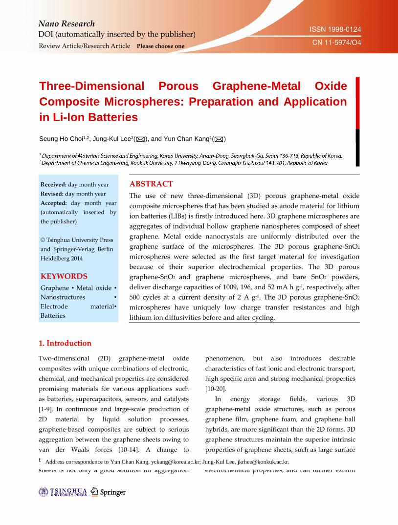
Figure 4a shows the XRD patterns of both the 3D
porous graphene and graphene-SnO2 microspheres
prepared directly by spray pyrolysis at
temperatures of 800 °C. The XRD pattern of the
graphene microspheres shows a broadened
diffraction peak around 23o similar to reduced
graphene oxide, while the SnO2-graphene
microspheres exhibits pure crystal structure of
rutile SnO2 (JCPDS card no. 41-1445) [46]. The mean
crystallite size of SnO2 nanocrystals dispersed on
graphene microspheres calculated from the (110)
peak using Scherer’s formula was an ultrafine 4 nm.
The pore-size distributions of the 3D porous
SnO2-graphene and graphene microspheres were
investigated by nitrogen isothermal adsorption, and
the results are shown in Figure 4b. Both samples
had similar pore size distributions, including both
mesopores (2–50 nm) and macropores. BET surface
areas of the SnO2-graphene and graphene
microspheres were 120 and 200 m2 g-1, respectively.
| www.editorialmanager.com/nare/default.asp
Nano Res.
The graphene content in the 3D porous
SnO2-graphene microspheres was evaluated to be
19.5 wt% by thermogravimetric analysis (Figure S3).
Figures 4c and 4d show the XPS profiles of C1s
acquired from the 3D porous graphene
microspheres (Figure 4c) and 3D porous
graphene-SnO2 microspheres (Figure 4d). The C1s
peak in the XPS profile could be attributed to sp2
bonded carbon (C–C), epoxy and alkoxy groups
(C–O) and carbonyl and carboxylic (C=O) groups,
which corresponded to peaks at 284.6, 286.6, and
288.1 eV, respectively [47]. The XPS profiles showed
sharp peaks at around 284.6 eV, which could be
assigned to graphitic carbon [47]. The relative
carbon content of the sp2 bonded carbon at 286.4 eV
was 83% in the 3D porous SnO2-graphene
microspheres. The carbonyl and carboxylic (C=O)
groups were not observed in the XPS C1s profile of
the graphene-SnO2 composite microspheres due to
the complete reduction of GO sheets into reduced
GO sheets. Analysis by XPS indicated that the
thermal reduction of GO sheets
containing oxygen functional
groups into graphene occurred
when the preparation
temperature was set to 800 °C
[47]. The overall XPS spectrum
of the 3D porous SnO2-graphene
microspheres (Figure S4)
showed clear Sn 3d5/2 (487.3eV)
and Sn 3d3/2 (495.8eV) peaks [48].
The electrochemical properties
of the 3D porous SnO2-graphene
microspheres were evaluated by
assembling a coin-type half-cell.
In addition, we tested
electrochemical properties of 3D
porous graphene microspheres
without SnO2 nanocrystals, and
bare SnO2 microspheres, both
prepared by spray pyrolysis under
the same conditions; the bare SnO2
microspheres had a spherical shape and dense
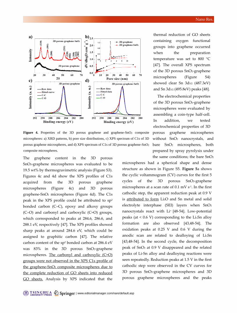
structure as shown in Figure S5. Figure 5a shows
the cyclic voltammogram (CV) curves for the first 5
cycles of the 3D porous SnO2-graphene
microspheres at a scan rate of 0.1 mV s-1. In the first
cathodic step, the apparent reduction peak at 0.9 V
is attributed to form Li2O and Sn metal and solid
electrolyte interphase (SEI) layers when SnO2
nanocrystals react with Li+ [48-54]. Low-potential
peaks (at < 0.6 V) corresponding to the LixSn alloy
formation are also observed [43,48-54]. The
oxidation peaks at 0.25 V and 0.6 V during the
anodic scan are related to dealloying of LixSn
[43,48-54]. In the second cycle, the decomposition
peak of SnO2 at 0.9 V disappeared and the related
peaks of Li-Sn alloy and dealloying reactions were
seen repeatedly. Reduction peaks at 1.5 V in the first
cathodic step were observed in the CV curves for
3D porous SnO2-graphene microspheres and 3D
porous graphene microspheres and the peaks
Figure 4. Properties of the 3D porous graphene and graphene-SnO2 composite
microspheres: a) XRD patterns, b) pore size distributions, c) XPS spectrum of C1s of 3D
porous graphene microspheres, and d) XPS spectrum of C1s of 3D porous graphene-SnO2
composite microspheres.
www.theNanoResearch.com∣www.Springer.com/journal/12274 | Nano Research
Nano Res.
disappeared from the second cycles as shown in
Figures 5a and S6. The reduction peak at 1.5 V in
the first cathodic step is attributable to irreversible
lithium insertion into graphene layers of the 3D
porous structure. Figure 5b shows the initial charge
and discharge curves of the 3D porous
SnO2-graphene and graphene microspheres and
bare SnO2 powders. The operating cut-off voltages
were 0.001 and 3 V at a current density of 2 A g-1.
The initial discharging and charging specific
capacities of the 3D porous graphene microspheres
were 1244 and 389 mA h g-1, respectively.
Coulombic efficiency (CE) of the graphene
microspheres observed in the first cycle was low at
32%, which is an unavoidable phenomenon in
reduced graphene oxide materials [55,56].
Electrochemical properties of reduced graphene
oxide are affected by synthesis methods, and
various lithium storage properties have been
previously reported [6-8]. However, most reduced
graphene oxide has low initial CE due to the high
production of SEI layers resulting from the high
specific surface area and to irreversible lithium ion
storage at highly active
defect sites [49-52]. 3D
porous SnO2-graphene
microspheres delivered
discharge and charge
capacities of 1586 mA h
g-1 and 1010 mA h g-1 in
the first cycle, and the
corresponding initial
CE was 64%. Bare SnO2
delivered discharge
and charge capacities
of 1397 mA h g-1 and
935 mA h g-1 in the first
cycle, with a
corresponding initial CE
of 67%. The irreversible
capacity loss of the bare
SnO2 powders is
attributed to the
formation of Li2O from SnO2 and SEI layers at the
electrode–electrolyte interface [43,48-54].
Cycling performances of the 3D porous
SnO2-graphene and graphene microspheres and
bare SnO2 powders at a constant current density of
2 A g-1 are shown in Figure 5c. In comparison, after
500 cycles the 3D porous SnO2-graphene and
graphene microspheres and bare SnO2 powders
delivered discharge capacities of 1009, 196, and 52
mA h g-1, respectively, and the corresponding
capacity retentions measured after the first cycles
were 96, 47, and 5%. Coulombic efficiency of 3D
porous SnO2-graphene microspheres reached
approximately 99% after the tenth cycle and
remained at this value in subsequent cycles. The
average Coulombic efficiency from 100th to 500th
cycles of these SnO2-graphene microspheres was
99.8%. The increase in the discharge capacities of
the 3D porous SnO2-graphene microspheres after
100 cycles had been attributed to the formation of a
gel-like reversible polymer film on the microsphere
Figure 5. Electrochemical properties of the 3D porous graphene and graphene-SnO2 composite
microspheres and bare SnO2 powders: a) CV curves, b) initial charge/discharge curves at a current
density of 2 A g-1, c) cycling performances at a current density of 2 A g-1, and d) rate performance
and Coulombic efficiencies of 3D porous graphene-SnO2 composite microspheres.

| www.editorialmanager.com/nare/default.asp
Nano Res.
surfaces [57-59]. The 3D porous SnO2-graphene
composite microspheres have excellent
electrochemical properties regardless of SnO2
contents (see Figure S7a). The long-term cycling
performance of 3D porous SnO2-graphene
microspheres at a high value of the current density,
5 A g-1, is shown in Figure S7b. The 2nd and 1000th
discharge capacities of the 3D porous
graphene-SnO2 microspheres were 905 and 730 mA
h g-1, and the Coulombic efficiency was reached at
99.7% after 50 cycles. The discharging and charging
rate performance of the 3D porous graphene-SnO2
microspheres are shown in Figure 5d. As can be
seen, the average reversible discharge capacities
were 1020, 875, 785, 716, and 660 mA h g-1 at current
densities of 1, 3, 5, 7, and 9 A g-1, respectively. After
high-rate charge-discharge cycling, discharge
capacity recovered to a value as high as 941 mA h
g-1 for a lower current density of 1 A g-1.
TEM images as shown in Figure S8 showed that
3D porous graphene-SnO2 microspheres maintained
their overall morphology after cycling, but bare
SnO2 powders lost their original morphologies after
the 200th cycle. The 3D graphene backbone of the
graphene-SnO2 microspheres was maintained, as
shown in the TEM images. The Sn component
remained uniformly dispersed over the
microspheres without aggregation even after
cycling, as shown in the elemental mapping images
in Figure S8b. The 3D porous graphene structure
suppressed the aggregation of SnO2 nanocrystals
during repeated cycling. The graphene increases the
electrical conductivity of the electrode and is able to
accommodate the strain induced by volume change
of SnO2. It also facilitated fast transportation of
electrons and lithium ions, which is responsible for
the good cycling stability and rate capability [59-61].
On the other hand, changes in the bare SnO2
powders caused disconnection between active
material, conducting carbon, and copper foil,
resulting in rapid capacity fading upon cycling. The
good conductivity and morphological uniqueness
of 3D porous graphene-SnO2 microspheres
contribute to their excellent electrochemical
performance, and were confirmed by
electrochemical impedance spectroscopy (EIS)
measurements. Nyquist plots of the electrodes
consisted of a semicircle in the medium frequency
region and an inclined line at low frequencies. The
medium-frequency semicircle was attributed to
charge-transfer resistance between active material
and electrolyte, and the low frequency portion of
the trace corresponded to the lithium diffusion
process within the electrodes [62-64]. The charge
transfer resistance of 3D porous graphene-SnO2
microspheres was much smaller than that of bare
SnO2 powders before cycling, as shown in Figure
S9a. Figure S9b shows the relationship between the
real part of the impedance spectrum Zre and ω-1/2
(where ω = 2πf is angular frequency) in the
low-frequency region, before cycling. The gradual
low-frequency slope (the Warburg impedance
coefficient) of Zre versus ω-1/2 indicates high lithium
ion diffusivity for 3D porous graphene-SnO2
microspheres [63,64]. The unique structure of 3D
porous graphene-SnO2 microspheres resulted in
low charge transfer resistance and high lithium ion
diffusivity. Figures S9c and S9d show the Nyquist
plot and the Zre - ω-1/2 relationship after 200 cycles.
The microspheres continued to have low charge
transfer resistance and high lithium ion diffusivity
even after 200 cycles. On the other hand, the charge
transfer resistance of bare SnO2 powders increased
after cycling. The excellent electrochemical
performance of 3D porous graphene-SnO2
microspheres is due to their structural stability and
graphene’s synergy effects.
4. Concluisons
In this study, electrochemical properties of 3D porous
graphene-SnO2 microspheres were compared with

www.theNanoResearch.com∣www.Springer.com/journal/12274 | Nano Research
Nano Res.
those of 3D porous graphene microspheres (without
SnO2) and bare SnO2 powders, prepared by the same
process. 3D porous graphene-SnO2 microspheres
were formed from single droplets using the one-pot
spray pyrolysis method. Ultrafine SnO2 nanocrystals
were found to be uniformly distributed within the
individual graphene nanospheres, which consisted of
graphene layers. The size of individual graphene
nanospheres forming the 3D porous microspheres,
and the content of SnO2 active material, were easily
controlled through the size of polystyrene (PS)
nanobeads used as sacrificial templates, and the
concentration of tin salt dissolved in the spray
solution, respectively. 3D porous graphene-SnO2
composite microspheres showed high reversible
capacity and good cycling performance at high
current density. The good conductivity and
morphological uniqueness of 3D porous
graphene-SnO2 microspheres contribute to excellent
electrochemical performance. The process developed
here could be applied in the preparation of various
types of 3D porous graphene-metal oxide composite
microspheres for a wide range of applications,
including energy storage devices.
Acknowledgements
This work was supported by the National Research
Foundation of Korea (NRF) grant funded by the
Korea government (MEST) (No.
2012R1A2A2A02046367). This work was supported
by the Energy Efficiency & Resources Core
Technology Program of the Korea Institute of
Energy Technology Evaluation and Planning
(KETEP), granted financial resource from the
Ministry of Trade, Industry & Energy, Republic of
Korea (201320200000420).
Electronic Supplementary Material: Supplementary
material (Schematic diagrams of 3D graphene,
HR-TEM, TG, and XPS data of the 3D porous
graphene-SnO2 composite microsphere, SEM image
of the bare SnO2 powders, CV curves of the 3D
porous graphene, Cycling performances of the 3D
porous graphene-SnO2 composite, TEM images of the
porous graphene-SnO2 composite after cycling, EIS
data of the 3D porous graphene-SnO2 composite and
the bare SnO2, Schematic diagram of spray pyrolysis
process, TEM image of the 3D porous graphene-SnO2
composite microspheres prepared using 40-nm PS
nanobeads, SEM image of the crushed 3D porous
graphene-SnO2 composite microspheres) is available
in the online version of this article at
http://dx.doi.org/10.1007/s12274-***-****-*
(automatically inserted by the publisher).
References [1] Zhu, Y.; Murali, S.; Cai, W.; Li, X.; Suk, J. W.; Potts, J. R.;
Ruoff, R. S. Graphene and Graphene Oxide: Synthesis,
Properties, and Applications. Adv. Mater. 2010, 22, 3906–3924.
[2] Huang, X.; Zeng, Z.; Fan, Z.; Liu, J.; Zhang, H.
Graphene-Based Electrodes. Adv. Mater. 2012, 24, 5979–6004.
[3] Guo, S. J.; Dong, S. J. Graphene Nanosheet: Synthesis,
Molecular Engineering, Thin Film, Hybrids, and Energy and
Analytical Applications. Chem. Soc. Rev. 2011, 40, 2644–2672.
[4] Liu, Y. X.; Dong, X. C.; Chen, P. Biological and Chemical
Sensors based on Graphene Materials. Chem. Soc. Rev. 2012, 41,
2283–2307.
[5] Jiang, J.; Li, Y. Y.; Liu, J. P.; Huang, X. T.; Yuan, C. Z.; Lou,
X. W. Recent Advances in Metal Oxide-based Electrode
Architecture Design for Electrochemical Energy Storage. Adv.
Mater. 2012, 24, 5166–5180.
[6] Wu, Z. S.; Zhou, G. M.; Yin, L. C.; Ren, W. C.; Li, F.; Cheng,
H. M. Graphene/Metal Oxide Composite Electrode Materials for
Energy Storage. Nano Energy 2012, 1, 107–131.
[7] Huang, X.; Tan, C. L.; Yin, Z. Y.; Zhang, H. 25th Anniversary
Article: Hybrid Nanostructures Based on Two-Dimensional
Nanomaterials. Adv. Mater. 2014, 26, 2185–2204.
[8] Armstrong, M. J.; O’Dwyer, C.; Macklin, W. J.; Holmes, J. D.
Evaluating the Performance of Nanostructured Materials as
Lithium-Ion Battery Electrodes. Nano Res. 2014, 7, 1-62.
[9] Leng, K.; Zhang, F.; Zhang, L.; Zhang, T.; Wu, Y.; Lu, Y.;
Huang, Y.; Chen, Y. Graphene-Based Li-Ion Hybrid
Supercapacitors with Ultrahigh Performance. Nano Res. 2013,

| www.editorialmanager.com/nare/default.asp
Nano Res.
6, 581–592.
[10] Huang, X.; Qian, K.; Yang, J.; Zhang, J.; Li, L.; Yu, C.;
Zhao, D. Functional Nanoporous Graphene Foams with
Controlled Pore Sizes. Adv. Mater. 2012, 24, 4419–4423.
[11] Luo, J.; Jang, H. D.; Sun, T.; Xiao, L.; He, Z.; Katsoulidis,
A. P.; Kanatzidis, M. G.; Gibson, J. M.; Huang, J. X.
Compression and Aggregation-Resistant Particles of Crumpled
Soft Sheets. ACS Nano 2011, 5, 8943–8949.
[12] Mao, S.; Wen, Z. H.; Kim, H. J.; Lu, G. H.; Hurley, P.; Chen,
J. H. A General Approach to One-Pot Fabrication of Crumpled
Graphene-Based Nanohybrids for Energy Applications. ACS
Nano 2012, 6, 7505–7513.
[13] Chen, Y.; Guo, F.; Jachak, A.; Kim, S. P;. Datta, D.; Liu, J.;
Kulaots, I.; Vaslet, C.; Jang, H. D.; Huang, J.; Kane, A.; Shenoy,
V. B.; Hurt, R. H. Aerosol Synthesis of Cargo-Filled Graphene
Nanosacks. Nano Lett. 2012, 12, 1996–2002.
[14] Choi, B. G.; Yang, M. H.; Hong, W. H.; Choi, J. W.; Huh, Y.
S. 3D Macroporous Graphene Frameworks for Supercapacitors
with High Energy and Power Densities. ACS Nano 2012, 6,
4020–4028.
[15] Chen, Z. P.; Ren, W. C.; Gao, L. B.; Liu, B. L.; Pei, S. F.;
Cheng, H. M. Three-Dimensional Flexible and Conductive
Interconnected Graphene Networks Grown by Chemical Vapour
Deposition. Nat. Mater. 2011, 10, 424–428.
[16] Li, C.; Shi, G. Q. Three-Dimensional Graphene
Architectures. Nanoscale 2012, 4, 5549–5563.
[17] Cao, X. H.; Shi, Y. M.; Shi, W. H.; Lu, G.; Huang, X.; Yan,
Q. Y.; Zhang, Q. C.; Zhang, H. Preparation of Novel 3D
Graphene Networks for Supercapacitor Applications. Small 2011,
7, 3163–3168.
[18] Yoon, J. C.; Lee, J. S.; Kim, S. I.; Kim, K. H.; Jang, J. H.
Three-Dimensional Graphene Nano-Networks with High Quality
and Mass Production Capability via Precursor-Assisted Chemical
Vapor Deposition. Sci. Rep. 2013, 3, 1788.
[19] Xie, X.; Yu, G.; Liu, N.; Bao, Z.; Criddle, C. S.; Cui, Y.
Graphene–Sponges as High-Performance Low-Cost Anodes for
Microbial Fuel Cells. Energy Environ. Sci. 2012, 5, 6862–6866.
[20] Sohn, K.; Na, Y. J.; Chang, H.; Roh, K. M.; Jang, H. D.;
Huang, J. Oil Absorbing Graphene Capsules by Capillary
Molding. Chem. Commun. 2012, 8, 5968–5970.
[21] He, Y.; Chen, W. J.; Li, X. D.; Zhang, Z. X.; Fu, J. C.; Zhao,
C. H.; Xie, E. Q. Freestanding Three-Dimensional
Graphene/MnO2 Composite Networks As Ultralight and Flexible
Supercapacitor Electrodes. ACS Nano 2013, 7, 174–182.
[22] Cao, X.; Yin, Z.; Zhang, H. Three-Dimensional Graphene
Materials: Preparation, Structures and Application in
Supercapacitors. Energy Environ. Sci. 2014, 7, 1850–1865.
[23] Dong, X. C.; Cao, Y. F.; Wang, J.; Park, M. B. C.; Wang, L.
H.; Huang, W.; Chen, P. Hybrid Structure of Zinc Oxide
Nanorods and Three Dimensional Graphene Foam for
Supercapacitor and Electrochemical Sensor Applications. RSC
Adv. 2012, 2, 4364–4369.
[24] Wang, X. B.; Zhang, Y. J.; Zhi, C. Y.; Wang, X.; Tang, D. M.;
Xu, Y. B.; Weng, Q. H.; Jiang, X. F.; Mitome, M.; Golberg, D.;
Bando, Y. Three-Dimensional Strutted Graphene Grown by
Substrate-Free Sugar Blowing for High-Power-Density
Supercapacitors. Nat. Comm. 2013, 4, 2905.
[25] Xu, Z. W.; Li, Z.; Holt, C. M. B.; Tan, X. H.; Wang, H. L.;
Amirkhiz, B. S.; Stephenson, T.; Mitlin, D. Electrochemical
Supercapacitor Electrodes from Sponge-like Graphene
Nanoarchitectures with Ultrahigh Power Density. J. Phys. Chem.
Lett. 2012, 3, 2928–2933.
[26] Wei, W.; Yang, S. B.; Zhou, H. X.; Lieberwirth, I.; Feng, X.
L.; Müllen, K. 3D Graphene Foams Cross-linked with
Pre-encapsulated Fe3O4 Nanospheres for Enhanced Lithium
Storage. Adv. Mater. 2013, 25, 2909–2914.
[27] Luo, J. S.; Liu, J. L.; Zeng, Z. Y.; Ng, C. F.; Ma, L. J.; Zhang,
H.; Lin, J. Y.; Shen, Z. X.; Fan, H. J. Three-Dimensional
Graphene Foam Supported Fe3O4 Lithium Battery Anodes with
Long Cycle Life and High Rate Capability. Nano Lett. 2013, 13,
6136–6143.
[28] Cao, X. H.; Shi, Y. M.; Shi, W. H.; Rui, X. H.; Yan, Q. Y.;
Kong, J.; Zhang, H. Preparation of MoS2-Coated
Three-Dimensional Graphene Networks for High-Performance
Anode Material in Lithium-Ion Batteries. Small 2013, 9,
3433–3438.
[29] Huang, X.; Yu, H.; Chen, J.; Lu, Z. Y.; Yazami, R.; Hng, H.
H. Ultrahigh Rate Capabilities of Lithium-Ion Batteries from 3D
Ordered Hierarchically Porous Electrodes with Entrapped Active
Nanoparticles Configuration. Adv. Mater. 2014, 26, 1296–1303.
[30] Liu, X.; Cheng, J.; Li, W.; Zhong, X.; Yang, Z.; Gu, L.; Yu,
Y. Superior Lithium Storage in a 3D Macroporous Graphene
Framework/SnO2 Nanocomposite. Nanoscale 2014, 6,
7817–7822.

www.theNanoResearch.com∣www.Springer.com/journal/12274 | Nano Research
Nano Res.
[31] Zhu, J. X.; Yang, D.; Rui, X. H.; Sim, D. H.; Yu, H.; Hng, H.
H.; Hoster, H. E.; Ajayan, P. M.; Yan, Q. Y. Facile Preparation of
Ordered Porous Graphene–Metal Oxide@C Binder-Free
Electrodes with High Li Storage Performance. Small 2013, 9,
3390–3397.
[32] Cao, X. H.; Zheng, B.; Rui, X. H.; Shi, W. H.; Yan, Q. Y.;
Zhang, H. Metal Oxide-Coated Three-Dimensional Graphene
Prepared by the Use of Metal–Organic Frameworks as Precursors.
Angew. Chem. Int. Ed. 2014, 53, 1404–1409.
[33] Ji, J. Y.; Ji, H. X.; Zhang, L. L.; Zhao, X.; Bai, X.; Fan, X. B.;
Zhang, F. B.; Ruoff, R. S. Graphene-Encapsulated Si on
Ultrathin-Graphite Foam as Anode for High Capacity
Lithium-Ion Batteries. Adv. Mater. 2013, 25, 4673–4677.
[34] Li, N.; Chen, Z. P.; Ren, W. C.; Li, F.; Cheng, H. M. Flexible
Graphene-based Lithium Ion Batteries with Ultrafast Charge and
Discharge Rates. PNAS 2012, 109, 17360–17365.
[35] Gong, Y. J.; Yang, S. B.; Liu, Z.; Ma, L. L.; Vajtai, R.;
Ajayan, P. M. Graphene-Network-Backboned Architectures for
High-Performance Lithium Storage. Adv. Mater. 2013, 25,
3979–3984.
[36] Chen, W. F.; Li, S. R.; Chen, C. H.; Yan, L. F. Self-Assembly
and Embedding of Nanoparticles by In Situ Reduced Graphene
for Preparation of a 3D Graphene/Nanoparticle Aerogel. Adv.
Mater. 2011, 23, 5679–5683.
[37] Huang, X. D.; Sun, B.; Chen, S. Q.; Wang, G. X.
Self-Assembling Synthesis of Free-standing Nanoporous
Graphene–Transition-Metal Oxide Flexible Electrodes for
High-Performance Lithium-Ion Batteries and Supercapacitors.
Chem. Asian J. 2014, 9, 206–211.
[38] Xiao, L.; Wu, D. Q.; Han, S.; Huang, Y. S.; Li, S.; He, M. Z.;
Zhang, F.; Feng, X. L. Self-Assembled Fe2O3/Graphene Aerogel
with High Lithium Storage Performance. ACS Appl. Mater.
Interfaces 2013, 5, 3764–3769.
[39] Nardecchia, S.; Carriazo, D.; Ferrer, M. L.; Gutiérrez, M. C.;
Monte, F. Three Dimensional Macroporous Architectures and
Aerogels Built of Carbon Nanotubes and/or Graphene:Synthesis
and Applications. Chem. Soc. Rev. 2013, 42, 794–830.
[40] Choi, S. H.; Kang, Y. C. Crumpled Graphene–Molybdenum
Oxide Composite Powders: Preparation and Application in
Lithium-Ion Batteries. ChemSusChem 2014, 7, 523–529.
[41] Zhang, T. Y.; Li, X. Q.; Kang, S. Z.; Qin, L. X.; Yan, W. F.;
Mua, J. Facile Assembly and Properties of Polystyrene
Microsphere/Reduced Graphene Oxide/Ag Composite. J. Colloid
Interface Sci. 2013, 402, 279–283.
[42] Zhang, W. L.; Liu, Y. D.; Choi, H. J. Graphene Oxide
Coated Core–Shell Structured Polystyrene Microspheres and
Their Electrorheological Characteristics under Applied Electric
Field. J. Mater. Chem. 2011, 21, 6916–6921.
[43] Zhou, X.; Wan, L. J.; Guo, Y. G. Binding SnO2 Nanocrystals
in Nitrogen-Doped Graphene Sheets as Anode Materials for
Lithium-Ion Batteries. Adv. Mater. 2013, 25, 2152–2157.
[44] Stankovich, S.; Dikin, D. A.; Dommett, G. H. B.; Kohlhaas,
K. M.; Zimney, E. J.; Stach, E. A.; Piner, R. D.; Nguyen, S. B. T.;
Ruoff, R. S. Graphene-based Composite Materials. Nature 2006,
442, 282–286.
[45] Kim, T. Y.; Kang, H. C.; Tung, T. T.; Lee, J. D.; Kim, H. K.;
Yang, W. S.; Yoon, H. G.; Suh, K. S. Ionic Liquid-Assisted
Microwave Reduction of Graphite Oxide for Supercapacitors.
RSC Adv. 2012, 2, 8808–8812.
[46] Seema, H.; Kemp, K. C.; Chandra, V.; Kim, K. S.
Graphene–SnO2 Composites for Highly Efficient Photocatalytic
Degradation of Methylene Blue under Sunlight. Nanotechnology
2012, 23, 355705.
[47] Beidaghi, M.; Wang, C. Micro-Supercapacitors Based on
Interdigital Electrodes of Reduced Graphene Oxide and Carbon
Nanotube Composites with Ultrahigh Power Handling
Performance. Adv. Funct. Mater. 2012, 22, 4501–4510.
[48] Li, Y. M.; Lv, X. J.; Lu, J.; Li, J. H. Preparation of
SnO2-Nanocrystal/Graphene-Nanosheets Composites and Their
Lithium Storage Ability. J. Phys. Chem. C 2010, 114,
21770–21774.
[49] Li, L.; Kovalchuk, A.; Tour, J. M. SnO2–Reduced Graphene
Oxide Nanoribbons as Anodes for Lithium Ion Batteries with
Enhanced Cycling Stability. Nano Res. 2014, 7, 1319–1326.
[50] Lin, J.; Peng, Z. W.; Xiang, C. S.; Ruan, G. D.; Yan, Z.;
Natelson, D.; Tour, J. M. Graphene Nanoribbon and
Nanostructured SnO2 Composite Anodes for Lithium Ion
Batteries. ACS Nano 2013, 7, 6001–6006.
[51] Zhang, C. F.; Peng, X.; Guo, Z. P.; Cai, C. B.; Chen, Z. X.;
Wexler, D.; Li, S.; Liu, H. K. Carbon-Coated SnO2/Graphene
Nanosheets as Highly Reversible Anode Materials for Lithium
Ion Batteries. Carbon 2012, 50, 1897–1903.
[52] Huang, Y.; Wu, D.; Wang, J.; Han, S.; Lv, L.; Zhang, F.;
Feng, X. Amphiphilic Polymer Promoted Assembly of
| www.editorialmanager.com/nare/default.asp
Nano Res.
Macroporous Graphene/SnO2 Frameworks with Tunable
Porosity for High-Performance Lithium Storage. Small 2014,
10, 2226–2232.
[53] Lee, C. W.; Seo, S. D.; Kim, D. W.; Park, S.; Jin, K.; Kim,
D. W.; Hong, K. S. Heteroepitaxial Growth of ZnO Nanosheet
Bands on ZnCo2O4 Submicron Rods Toward High-Performance
Li Ion Battery Electrodes. Nano Res. 2013, 6, 348–355.
[54] Lee, C. W.; Seo, S. D.; Kim, D. W.; Park, S.; Jin, K.; Kim,
D. W.; Hong, K. S. Heteroepitaxial Growth of ZnO Nanosheet
Bands on ZnCo2O4 Submicron Rods Toward High-Performance
Li Ion Battery Electrodes. Nano Res. 2013, 6, 348-355.
[55] Xiang, H. F.; Li, Z. D.; Xie, K.; Jiang, J. Z.; Chen, J. J.;
Lian, P. C.; Wu, J. S.; Yud, Y.; Wang, H. H. Graphene Sheets as
Anode Materials for Li-Ion Batteries: Preparation, Structure,
Electrochemical Properties and Mechanism for Lithium Storage.
RSC Adv. 2012, 2, 6792–6799.
[56] Yang, J.; Liao, Q.; Zhou, X.; Liu, X.; Tang, J. Efficient
Synthesis of Graphene-based Powder via in Situ Spray Pyrolysis
and Its Application in Lithium Ion Batteries. RSC Adv. 2013, 3,
16449–16455.
[57] Li, S.; Li, A.; Zhang, R.; He, Y.; Zhai, Y.; Xu, L.
Hierarchical Porous Metal Ferrite Ball-In-Ball Hollow Spheres:
General Synthesis, Formation Mechanism, and High
Performance as Anode Materials for Li-Ion Batteries. Nano Res.
2014, 7, 1116–1127.
[58] Choi, S. H.; Kang, Y. C. Using Simple Spray Pyrolysis to
Prepare Yolk–Shell-Structured ZnO–Mn3O4 Systems with the
Optimum Composition for Superior Electrochemical Properties.
Chem. Eur. J. 2014, 20, 3014–3018.
[59] Wang, D.; Yang, J.; Li, X.; Geng, D.; Li, R.; Cai, M.; Sham,
T. K.; Sun, X. Layer by Layer Assembly of Sandwiched
Graphene/SnO2 Nanorod/Carbon Nanostructures with Ultrahigh
Lithium Ion Storage Properties. Energy Environ. Sci. 2013, 6,
2900–2906.
[60] Wang, D.; Li, X.; Wang, J.; Yang, J.; Geng, D.; Li, R.; Cai,
M.; Sham, T. K.; Sun, X. Defect-Rich Crystalline SnO2
Immobilized on Graphene Nanosheets with Enhanced Cycle
Performance for Li Ion Batteries. J. Phys. Chem. C 2012, 116,
22149–22156.
[61] Zhou, G.; Wang, D. W.; Yin, L. C.; Li, N.; Li, F.; Cheng, H.
M. Oxygen Bridges between NiO Nanosheets and Graphene for
Improvement of Lithium Storage. ACS Nano 2012, 6,
3214–3223.
[62] Choi, S. H.; Kang, Y. C. Yolk–Shell, Hollow, and
Single-Crystalline ZnCo2O4 Powders: Preparation Using a
Simple One-Pot Process and Application in Lithium-Ion Batteries.
ChemSusChem 2013, 6, 2111–2116.
[63] Park, M. S.; Kang, Y. M.; Wang, G. X.; Dou, S. X.; Liu, H.
K. The Effect of Morphological Modification on the
Electrochemical Properties of SnO2 Nanomaterials. Adv. Funct.
Mater. 2008, 18, 455–461.
[64] Ko, Y. N.; Park, S. B.; Jung, K. Y.; Kang, Y. C. One-Pot
Facile Synthesis of Ant-Cave-Structured Metal Oxide−Carbon
Microballs by Continuous Process for Use as Anode Materials in
Li-Ion Batteries. Nano Lett. 2013, 13, 5462–5466.


![Synthesis, Properties and Potential Applications of Porous ...graphene. Shown in Fig. 2 is the band structure for graphene and hydrogenated porous graphene (HPG) [56]. For pure graphene](https://static.fdocuments.us/doc/165x107/5e69b1218c0b164bfe524e90/synthesis-properties-and-potential-applications-of-porous-graphene-shown-in.jpg)