Inhibition of HSV-1 replication and reactivation by the mutation-insensitive transcription inhibitor...
11
Antiviral Research 58 (2003) 35–45 Inhibition of HSV-1 replication and reactivation by the mutation-insensitive transcription inhibitor tetra-O-glycyl-nordihydroguaiaretic acid Richard Park, Paul E. Giza, David E. Mold, Ru Chih C. Huang ∗ Department of Biology, The Johns Hopkins University, Baltimore, MD 21218, USA Received 16 April 2002; accepted 28 August 2002 Abstract Methylated derivatives of nordihydroguaiaretic acid (NDGA)were previously shown to be potent mutation-resistant inhibitors of herpes simplex virus type 1 (HSV-1) which target Sp1 protein binding to critical viral promoters. The hydrophobic nature of these agents, however, renders them relatively water-insoluble and, therefore, limits their applicability. We report here on the anti-HSV-1 properties of a related but water-soluble glycylated derivative of NDGA, tetra-O-glycyl-NDGA (G 4 N). In yield reduction assays, G 4 N inhibited replication of laboratory and clinical strains of wild type HSV-1 and ACV-resistant (HSV-1 R ) strains of HSV-1 in a dose-dependent manner, with average IC 50 values of 4.7 and 3.2 M against wild-type and HSV-1 R strains, respectively. An MTT-based cytotoxicity assay revealed a TC 50 value of 73.2 M for G 4 N on Vero cells, with no reduction in viability detected at concentrations below 30 M. Similar to its methylated counterparts, G 4 N was found to inhibit transcription of the HSV-1 ICP4 gene, a major immediate early viral regulator, and gel mobility shift assays showed it can block Sp1 protein binding to cognate sites on the ICP4 promoter. In anticipation of its potential use as a systemic anti-HSV-1 agent, we tested G 4 N in a murine trigeminal ganglia (TG) explant model system, and found G 4 N was able to prevent HSV-1 reactivation from explanted and cultured latently infected TG. © 2002 Elsevier Science B.V. All rights reserved. Keywords: Herpes simplex virus type 1; Tetra-O-glycyl-nordihydroguaiaretic acid; G 4 NM 4 N; Sp1; Latency 1. Introduction The herpes simplex virus type 1 (HSV-1) is an enveloped DNA virus afflicting greater than 70% of the human popu- lation. In humans, an HSV-1 infection normally results in primary and recurrent mucoepithelial lesions (herpes labi- alis) and in some instances leads to disseminated disease, encephalitis and blindness. Following a primary infection, HSV-1 establishes a permanent latent infection within the sensory ganglia of the host. This latent reservoir of virus may then reactivate following appropriate stimuli and serve as a site for future recurrent infection (Whitley, 1996; Roizman and Sears, 1996). At particular risk of recurrence, due to the increased severity of infection and the generation of drug-resistant strains of HSV-1, are those with com- promised immune systems such as AIDS patients, cancer patients, and patients undergoing organ transplantation (De Logu et al., 2000). ∗ Corresponding author. Tel.: +1-410-516-5181; fax: +1-410-516-5213. E-mail address: [email protected] (R.C.C. Huang). Clinically, the most widely used and successful chemother- apeutic agents in the systemic treatment of HSV-1 infec- tion are nucleoside analogue agents, in particular acyclovir (ACV) which targets viral DNA synthesis via HSV thymi- dine kinase activity (Hirsch et al., 1996). Studies have shown nucleoside analogue agents to also be successful topical treatments for herpes labialis (Straten et al., 2001) and systemic agents for minimizing the establishment and reactivation from latency (Efstathiou et al., 1999). Despite the effectiveness and high selectivity of nucleoside ana- logue agents, however, the development of ACV-resistant and nucleoside analogue-resistant strains of mutant HSV have been reported in immunocompromised individuals (Crumpacker et al., 1982; Crumpacker, 2001). The ability of HSV strains to readily mutate in response to conven- tional chemical agents underscores a need to continually develop novel anti-HSV agents that will substitute for or complement ACV and nucleoside analogues. During an HSV-1 lytic infection, viral genes are expressed in a tightly regulated cascade. In this cascade, several of the HSV-1 genes are transcriptionally regulated by Sp1 protein 0166-3542/02/$ – see front matter © 2002 Elsevier Science B.V. All rights reserved. doi:10.1016/S0166-3542(02)00165-1
-
Upload
richard-park -
Category
Documents
-
view
213 -
download
0
Transcript of Inhibition of HSV-1 replication and reactivation by the mutation-insensitive transcription inhibitor...
Antiviral Research 58 (2003) 35–45
Inhibition of HSV-1 replication and reactivation by themutation-insensitive transcription inhibitortetra-O-glycyl-nordihydroguaiaretic acid
Richard Park, Paul E. Giza, David E. Mold, Ru Chih C. Huang∗Department of Biology, The Johns Hopkins University, Baltimore, MD 21218, USA
Received 16 April 2002; accepted 28 August 2002
Abstract
Methylated derivatives of nordihydroguaiaretic acid (NDGA)were previously shown to be potent mutation-resistant inhibitors of herpessimplex virus type 1 (HSV-1) which target Sp1 protein binding to critical viral promoters. The hydrophobic nature of these agents, however,renders them relatively water-insoluble and, therefore, limits their applicability. We report here on the anti-HSV-1 properties of a relatedbut water-soluble glycylated derivative of NDGA, tetra-O-glycyl-NDGA (G4N). In yield reduction assays, G4N inhibited replication oflaboratory and clinical strains of wild type HSV-1 and ACV-resistant (HSV-1R) strains of HSV-1 in a dose-dependent manner, with averageIC50 values of 4.7 and 3.2�M against wild-type and HSV-1R strains, respectively. An MTT-based cytotoxicity assay revealed a TC50
value of 73.2�M for G4N on Vero cells, with no reduction in viability detected at concentrations below 30�M. Similar to its methylatedcounterparts, G4N was found to inhibit transcription of the HSV-1 ICP4 gene, a major immediate early viral regulator, and gel mobilityshift assays showed it can block Sp1 protein binding to cognate sites on the ICP4 promoter. In anticipation of its potential use as a systemicanti-HSV-1 agent, we tested G4N in a murine trigeminal ganglia (TG) explant model system, and found G4N was able to prevent HSV-1reactivation from explanted and cultured latently infected TG.© 2002 Elsevier Science B.V. All rights reserved.
Keywords: Herpes simplex virus type 1; Tetra-O-glycyl-nordihydroguaiaretic acid; G4N M4N; Sp1; Latency
1. Introduction
The herpes simplex virus type 1 (HSV-1) is an envelopedDNA virus afflicting greater than 70% of the human popu-lation. In humans, an HSV-1 infection normally results inprimary and recurrent mucoepithelial lesions (herpes labi-alis) and in some instances leads to disseminated disease,encephalitis and blindness. Following a primary infection,HSV-1 establishes a permanent latent infection within thesensory ganglia of the host. This latent reservoir of virusmay then reactivate following appropriate stimuli and serveas a site for future recurrent infection (Whitley, 1996;Roizman and Sears, 1996). At particular risk of recurrence,due to the increased severity of infection and the generationof drug-resistant strains of HSV-1, are those with com-promised immune systems such as AIDS patients, cancerpatients, and patients undergoing organ transplantation (DeLogu et al., 2000).
∗ Corresponding author. Tel.:+1-410-516-5181; fax:+1-410-516-5213.E-mail address: [email protected] (R.C.C. Huang).
Clinically, the most widely used and successful chemother-apeutic agents in the systemic treatment of HSV-1 infec-tion are nucleoside analogue agents, in particular acyclovir(ACV) which targets viral DNA synthesis via HSV thymi-dine kinase activity (Hirsch et al., 1996). Studies haveshown nucleoside analogue agents to also be successfultopical treatments for herpes labialis (Straten et al., 2001)and systemic agents for minimizing the establishment andreactivation from latency (Efstathiou et al., 1999). Despitethe effectiveness and high selectivity of nucleoside ana-logue agents, however, the development of ACV-resistantand nucleoside analogue-resistant strains of mutant HSVhave been reported in immunocompromised individuals(Crumpacker et al., 1982; Crumpacker, 2001). The abilityof HSV strains to readily mutate in response to conven-tional chemical agents underscores a need to continuallydevelop novel anti-HSV agents that will substitute for orcomplement ACV and nucleoside analogues.
During an HSV-1 lytic infection, viral genes are expressedin a tightly regulated cascade. In this cascade, several of theHSV-1 genes are transcriptionally regulated by Sp1 protein
0166-3542/02/$ – see front matter © 2002 Elsevier Science B.V. All rights reserved.doi:10.1016/S0166-3542(02)00165-1
36 R. Park et al. / Antiviral Research 58 (2003) 35–45
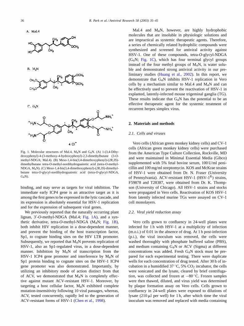
Fig. 1. Molecular structures of Mal.4, M4N and G4N. (A) 1-(3,4-Dihy-droxyphenyl)-4-(3-methoxy-4-hydroxyphenyl)-2,3-dimethylbutane (3-O-methyl-NDGA; Mal.4). (B) Meso-1,4-bis(3,4-dimethoxyphenyl)-(2R,3S)-dimethylbutane tetra-O-methyl-nordihydroguairetic acid (tetra-O-methyl-NDGA, M4N). (C) Meso-1,4-bis(3,4-dimethoxyphenyl)-(2R,3S)-dimethyl-butane tetra-O-glycyl-nordihydroguairetic acid (tetra-O-glycyl-NDGA,G4N).
binding, and may serve as targets for viral inhibition. Theimmediate early ICP4 gene is an attractive target as it isamong the first genes to be expressed in the lytic cascade, andits expression is absolutely essential for HSV-1 replicationand for the expression of subsequent viral genes.
We previously reported that the naturally occurring plantlignan, 3′-O-methyl-NDGA (Mal.4; Fig. 1A), and a syn-thetic derivative, tetra-O-methyl-NDGA (M4N; Fig. 1B),both inhibit HIV replication in a dose-dependent manner,and prevent the binding of the host transcription factor,Sp1, to cognate binding sites on the HIV LTR promoter.Subsequently, we reported that M4N prevents replication ofHSV-1, also an Sp1-regulated virus, in a dose-dependentmanner. Inhibition by M4N of transcription from theHSV-1 ICP4 gene promoter and interference by M4N ofSp1 protein binding to cognate sites on the HSV-1 ICP4gene promoter was also demonstrated. Importantly, byutilizing an inhibitory mode of action distinct from thatof ACV, we demonstrated that M4N is completely effec-tive against mutant ACV-resistant HSV-1. Moreover, bytargeting a host cellular factor, M4N exhibited completemutation-insensitivity following 10 viral passages, whereas,ACV, tested concurrently, rapidly led to the generation ofACV-resistant forms of HSV-1 (Chen et al., 1998).
Mal.4 and M4N, however, are highly hydrophobicmolecules that are insoluble in physiologic solutions andare impractical as systemic therapeutic agents. Therefore,a series of chemically related hydrophilic compounds weresynthesized and screened for antiviral activity againstHSV-1. One of these compounds, tetra-O-glycyl-NDGA(G4N; Fig. 1C), which has four terminal glycyl groupsinstead of the four methyl groups of M4N, is water solu-ble and demonstrated strong antiviral activity in our pre-liminary studies (Huang et al., 2002). In this report, wedemonstrate that G4N inhibits HSV-1 replication in Verocells by a mechanism similar to Mal.4 and M4N and canbe effectively used to prevent the reactivation of HSV-1 inexplanted, latently-infected mouse trigeminal ganglia (TG).These results indicate that G4N has the potential to be aneffective therapeutic agent for the systemic treatment ofrecurrent herpes simplex virus.
2. Materials and methods
2.1. Cells and viruses
Vero cells (African green monkey kidney cells) and CV-1cells (African green monkey kidney cells) were purchasedfrom the American Type Culture Collection, Rockville, MDand were maintained in Minimal Essential Media (Gibco)supplemented with 5% fetal bovine serum, 100 U/ml peni-cillin and 100 mg/ml streptomycin. KOS and McKrae strainsof HSV-1 were obtained from Dr. N. Fraser (Universityof Pennsylvania). ACV-resistant HSV-1 (HSV-1R) strains,F59878 and T28387, were obtained from Dr. K. Thomp-son (University of Chicago). All HSV-1 strains and stockswere propagated in Vero cells. Reactivation of KOS HSV-1from latently infected murine TGs were assayed on CV-1cell monolayers.
2.2. Viral yield reduction assay
Vero cells grown to confluency in 24-well plates wereinfected for 1 h with HSV-1 at a multiplicity of infection(m.o.i.) of 0.01 in the absence of drug. At 1 h post-infection(p.i.), the viral inoculum was removed, the cells werewashed thoroughly with phosphate buffered saline (PBS),and medium containing G4N or ACV (Sigma) at differentconcentrations was added. Fresh G4N stock must be pre-pared for each experimental testing. There were duplicatewells for each concentration of drug tested. After 30 h of in-cubation in a humidified 37◦C, 5% CO2 incubator, the cellswere sonicated and the lysate, cleared by brief centrifuga-tion, was collected and frozen at−80◦C. Frozen sampleswere then thawed, diluted, and virus yield was determinedby plaque formation assay on Vero cells. Cells grown toconfluency in 24-well plates were exposed to dilutions oflysate (210�l per well) for 1 h, after which time the viralinoculum was removed and replaced with media containing

R. Park et al. / Antiviral Research 58 (2003) 35–45 37
0.5% methyl cellulose (1.2 ml per well). After 3 days of in-cubation at 37◦C, the methyl cellulose media was removedand the cells were fixed and stained for 20 min with 0.06%formalin, 1% crystal violet and 0.64% NaCl. The stainedcells were rinsed gently with tap water and air dried. Titersof virus yield were then determined by the enumeration ofplaques. The IC50 was determined as the drug concentrationrequired to reduce plaque formation by 50%.
2.3. MTT cytotoxicity assay
Vero cells grown to confluency or to 50% confluency in24-well plates were exposed to media containing differentconcentrations of G4N in a volume of 1.5 ml per well. TheG4N concentrations were the same ones used in the viralyield reduction experiments and duplicate wells of each con-centration were tested. After a 30 h incubation, the mediawas replaced with 0.5 ml per well MTT solution PBS con-taining 5% fetal bovine serum, penicillin, streptomycin and0.5 mg/ml MTT (Sigma). After a 2-h incubation, the MTTsolution was replaced with 1 ml of DMSO. The cells wererocked gently for 20 min to allow solubilization of the incor-porated formazan crystals by the DMSO. The OD570 valueof each DMSO sample was measured and the relative num-ber of viable cells was determined after correction of the val-ues for absorbance due to turbidity from cell debris (OD690).The TC50 was calculated as the drug concentration whichreduced cell viability by 50%. The viability of murine TGwas assayed in a similar manner.
2.4. RT-PCR
Total RNA was isolated from HSV-1 infected Verocells (MOI of 1) by the method ofChomczynski andSacchi (1987). HSV-1 ICP4 mRNA levels were measuredsemi-quantitatively by RT-PCR analysis using oligonu-cleotide primers specific for a portion of the ICP4 coding re-gion (nucleotides+1113 to+1411 from the start site of tran-scription). The upstream primer was 5′-GCAGTACGCCCT-GATCAC-3′ and the downstream primer was 5′-CAGGCT-GGTCAGCAGGAA-3′. In brief, RNA samples were treatedwith RNAsin (Promega) and RNAse-free DNAse to removeany contaminating genomic DNA. First strand synthesiswas primed with the ICP4-specific downstream primer andperformed using MMLV RT enzyme and PCRX Enhancer(Gibco) at 42◦C for 45 min. Amplification of the first strandsynthesis products was then carried out by PCR using TaqDNA polymerase and PCRX Enhancer under the follow-ing thermocycler conditions: 94◦C for 3 min, 30 cycles of94◦C for 55 s, 60◦C for 55 s, 72◦C for 1 min and a final10 min 72◦C extension. The PCR products were visualizedby electrophoresis on a 1.8% agarose gel and ethidium bro-mide staining. The bands were photographed, scanned andband intensities were quantified usingIMAGEQUANT soft-ware. RT-minus negative controls which received Milli-QH2O in place of MMLV RT enzyme were performed for
all samples and showed there was no DNA contamination(Park, JHU, Ph.D. Thesis, 2002). RNA was isolated at thetime before much viral DNA replication had occurred and,thus, the viral genome copy number was still relativelylow (approximately MOI of 1), therefore, standard DNAsetreatment (2 U per 38�l reaction) was sufficient to avoidcontamination with viral DNA.
2.5. Gel mobility shift assay
The DNA template used for the gel mobility shift assaywas a 36 bp DNA fragment spanning nucleotides−101to −66 from the start site of transcription of the HSV-1ICP4 promoter, which contains the Sp1 protein bindingsites most important for ICP4 gene transcription. The tem-plate was formed by annealing the single stranded comple-mentary oligonucleotides: 5′-GTTCGGGGCGGGCCCGC-CTGGGGGGCGGGGGGCCGG-3′ and 5′-CCGGCCCCC-CGCCCCCCAGG and labeling with32P-dATP using theKlenow fragment of DNA Polymerase I (Promega). The la-beled DNA (5 ng) was incubated for 30 min at room temper-ature with various concentrations of G4N in reaction buffercontaining 10 mM Tris (pH 7.5), 0.7 mM HEPES (pH 7.7),30 mM KCl, 1 mM EDTA, 0.8 mM MgCl2, 0.6�M ZnSO4,10% glycerol, 5 mM DTT and 0.5 mg/ml BSA. Recombi-nant Sp1 protein (Sjottem et al., 1997) was then added andthe reaction mixture was incubated at room temperature foran additional 30 min. Reactions were electrophoresed ona 5% polyacrylamide gel in 0.25× TBE buffer containing2.5% glycerol. The dried gel was then exposed to X-rayfilm for autoradiography and32P emissions were quanti-fied by phosphorimager analysis using a Packard InstantImager.
2.6. Mouse trigeminal ganglia system of HSV latencyand reactivation
Mice were anesthetized by an intraperitoneal injection ofketamine (100 mg/kg) and xylazine (10 mg/kg), and bothcorneas were lightly scarified using a 26 gauge needle. A5�l aliquot of KOS HSV-1 containing 1.0 × 10⁶ pfu wasthen placed on each eye and the eye was gently massaged.After >28 days p.i., mice were euthanized and the TG wererapidly and aseptically explanted into growth media and in-cubated in a humidified 37◦C, 5% CO2 incubator. Vari-ous concentrations of freshly made G4N in PBS (or a PBScontrol) were administered to the TGs every 48 h for a pe-riod of 8 h up to either 5 or 14 days post-explantation.Prior to each 8-h G4N treatment, a sample of culture me-dia was transferred to CV-1 cell monolayers to assay forthe presence of reactivated infectious HSV-1 virions. Atthe end of G4N treatment, an MTT cytotoxicity assay wasperformed to ensure that all TGs remained viable. AfterMTT analysis, PCR using HSV-1 specific primers was per-formed to ensure all TGs had been latently infected withHSV-1.

38 R. Park et al. / Antiviral Research 58 (2003) 35–45
3. Results
3.1. G4N inhibits replication of wild type andACV-resistant HSV-1 in Vero cells
Results from our previous studies showed that M4Ninhibits the growth of HSV-1 (Sm44 strain) in Vero cellcultures in a dose-dependent fashion with an IC50 of11.7�M. This was shown in comparison with ACV whichhad an IC50 of 7.54�M. It was also demonstrated thatbecause of its novel mode of inhibition, M4N exhibitsno viral cross-resistance against ACV-resistant strains ofHSV-1 (HSV-1R; Chen et al., 1998). In this study, G4Nwas evaluated in comparison with ACV for its inhibitoryactivity against four strains of HSV-1: two wild-type strains(KOS, a laboratory strain and McKrae, a virulent clinicalisolate), and two HSV-1R mutant strains. The viral yieldreduction assay results are shown inFig. 2. G4N inhibitedwild-type HSV-1 production in a dose-dependent mannerwith IC50 values of 3.6 and 4.5�M, and IC90 values of15.7 and 34.3�M for KOS and McKrae strains of HSV-1,respectively. ACV inhibited both wild type strains in a sim-ilar dose-dependent manner with IC50 values of 5.4 and3.9�M. Thus, G4N exhibits very similar antiviral potencyagainst wild-type HSV-1 strains as ACV, and it appears tobe slightly more effective than M4N, although this may bedue to a difference in viral strain.
Cross-resistance is observed among different drugs tar-geting like viral mechanisms. Since G4N is hypothesizedto function as M4N and target a mechanism distinct fromACV, it was expected that there would be no cross-resistanceof HSV-1R strains against G4N. Cross-resistance against
Fig. 2. Inhibition of wild-type HSV-1 by G4N and ACV. The effect of G4N and ACV on the replication of two wild-type strains of HSV-1 in Vero cellswas determined by viral yield reduction assay. KOS, a laboratory HSV-1 strain; McKrae, a virulent clinical isolate of HSV-1. Each concentration wasdone in duplicate. The IC50 was determined as the drug concentration required to reduce plaque formation by 50%.
G4N was tested using the two HSV-1R strains, F59878and T28387, under assay conditions and drug concentra-tions identical to those used for the wild-type viruses. Theresults (Fig. 3) show that the IC50 values for ACV were20–40-fold higher against the HSV-1R strains than againstwild-type strains. G4N, however, retained complete effec-tiveness against both HSV-1R strains with IC50 values of 2.1and 4.3�M. Thus, there is no cross-resistance of HSV-1R
strains against G4N.
3.2. Cytotoxicity of G4N towards Vero cells
The cytotoxicity of a prospective antiviral drug must beinvestigated in order to gauge its safety, and to investigatewhether the observed antiviral effect is an indirect result ofa deleterious effect upon the host cell. This is of particularconcern for G4N which targets a host cellular factor.
The cytotoxicity of G4N toward Vero cells was deter-mined by MTT analysis using the same drug concentrationsand incubations times as the viral yield reduction experi-ments. Since G4N may exhibit different toxicities againstdividing and non-dividing cells, its toxicity was tested forboth 100% confluent non-dividing cells and 50% confluentactively dividing cells. After 30 h of incubation, no toxicitywas seen up to 30�M (Fig. 4). At 50�M of G4N, 70.1% ofnon-dividing Vero cells, and 40.8% of actively dividing Verocells remained viable. At 100�M, 27.0% of non-dividingcells, and 7.3% of dividing Vero cells remained viable. Thisgives a TC50 value for G4N towards Vero cells of 73.2�Mfor non-dividing cells, and 46.9�M for dividing cells. TheIC50 value for G4N of 3.6–4.5�M strongly indicates thatthe G4N-induced inhibition of HSV-1 does not result from

R. Park et al. / Antiviral Research 58 (2003) 35–45 39
Fig. 3. Inhibition of ACV-resistant strains of HSV-1 by G4N and ACV. Using the same conditions as used inFig. 2, the reduction of viral yield by G4Nand ACV was tested using the two HSV-1R strains, F59878 and T28387.
host cell injury, since the cells remain completely viable atthis concentration.
3.3. Inhibition of HSV-1 ICP4 gene transcriptionby G4N during lytic infection
Earlier reporter gene experiments showed that M4N in-hibits transcription from the HSV-1 ICP4 gene promoter,presumably by interfering with required host Sp1 transcrip-tion factor binding to the ICP4 promoter region.
Fig. 4. Cytotoxicity of G4N towards Vero cells. Cell viability of actively dividing (50% confluent) and stationary (100% confluent) Vero cells incubatedin G4N-containing media was measured using MTT analysis. Viability was expressed relative to the no drug treatment control (100%).
To examine whether G4N prevents HSV-1 ICP4 genetranscription during a lytic infection, ICP4 mRNA levels inHSV-1 infected, G4N-treated Vero cells were measured byRT-PCR. Confluent Vero cells were infected with HSV-1at an m.o.i. of 1 and treated with varying concentrations ofG4N. At 3 h p.i., when ICP4 gene transcription is at a max-imum, RNA was isolated and analyzed by RT-PCR usingHSV-1 ICP4-specific primers. The PCR product of 298 bpwas resolved on an agarose gel, stained with ethidiumbromide and photographed (Fig. 5A). RT-minus negative

40 R. Park et al. / Antiviral Research 58 (2003) 35–45
Fig. 5. RT-PCR analysis of G4N inhibition of HSV-1 ICP4 gene transcription in vivo. Confluent Vero cells were infected with HSV-1 (MOI of 1) andtreated with varying concentrations of G4N. At 3 h p.i., RNA was isolated and ICP4 mRNA levels was detected by RT-PCR using ICP4 specific primers.DNA bands were resolved by agarose gel electrophoresis, stained with ethidium bromide, and quantified by phosphorimager analysis with backgroundsubtraction. (A) Reduction by G4N of HSV-1 ICP4 mRNA expression, resolved on a 1.8% agarose minigel. (B) HSV-1 ICP4 bands from (A) werequantitated using IMAGEQUANT software and represented graphically. The 0�M band represents 100% expression.
controls showed there was no detectable DNA contami-nation (Park, 2002). Quantitation of the 298 bp band bydensitometry followed by background subtraction (Fig. 5B)showed ICP4 mRNA levels were reduced to 15% of un-treated controls at 5�M G4N. At 10 and 25�M G4N, ICP4mRNA levels were further reduced to 12 and 4%, respec-tively, showing that G4N causes a dose-dependent decreasein HSV-1 ICP4 gene transcription during a lytic infection.
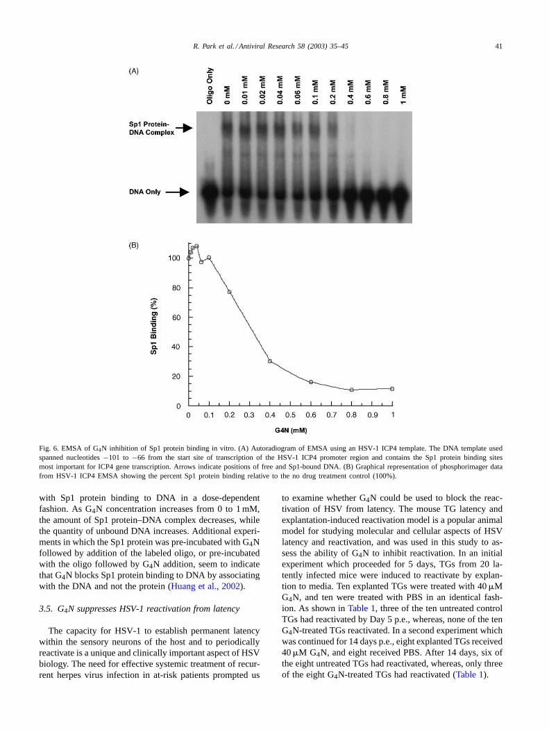
3.4. G4N inhibits binding of Sp1 protein to the HSV-1ICP4 gene promoter in vitro
Previous experiments showed that the mode of viral in-hibition by Mal.4 and M4N was due to interference withthe binding of the Sp1 transcription factor to its cognatebinding sites on critical HIV and HSV viral gene promoters
(Gnabre et al., 1995; Chen et al., 1998). We hypothesizedthat structurally similar G4N inhibits HSV-1 ICP4 genetranscription via the same mechanism. The ability of G4Nto prevent Sp1 protein binding to the HSV-1 ICP4 promoterwas examined here in vitro by electrophoretic mobility shiftanalysis (EMSA). A portion of the HSV-1 ICP4 promoterregion containing the most proximal Sp1 protein bindingsites, reported to be the most important for gene transcrip-tion (Jones and Tjian, 1985), was labeled and pre-incubatedfor 30 min with varying concentrations of G4N. Recom-binant Sp1 protein was then added to the reaction for anadditional 30 min and the mixture was electrophoresed on anon-denaturing polyacrylamide gel (Fig. 6A). The relativeintensities of the bands corresponding to Sp1 protein–DNAcomplexes were quantified by phosphorimager analysis, andare shown inFig. 6B. The data shows that G4N interferes
R. Park et al. / Antiviral Research 58 (2003) 35–45 41
Fig. 6. EMSA of G4N inhibition of Sp1 protein binding in vitro. (A) Autoradiogram of EMSA using an HSV-1 ICP4 template. The DNA template usedspanned nucleotides−101 to −66 from the start site of transcription of the HSV-1 ICP4 promoter region and contains the Sp1 protein binding sitesmost important for ICP4 gene transcription. Arrows indicate positions of free and Sp1-bound DNA. (B) Graphical representation of phosphorimager datafrom HSV-1 ICP4 EMSA showing the percent Sp1 protein binding relative to the no drug treatment control (100%).
with Sp1 protein binding to DNA in a dose-dependentfashion. As G4N concentration increases from 0 to 1 mM,the amount of Sp1 protein–DNA complex decreases, whilethe quantity of unbound DNA increases. Additional experi-ments in which the Sp1 protein was pre-incubated with G4Nfollowed by addition of the labeled oligo, or pre-incubatedwith the oligo followed by G4N addition, seem to indicatethat G4N blocks Sp1 protein binding to DNA by associatingwith the DNA and not the protein (Huang et al., 2002).
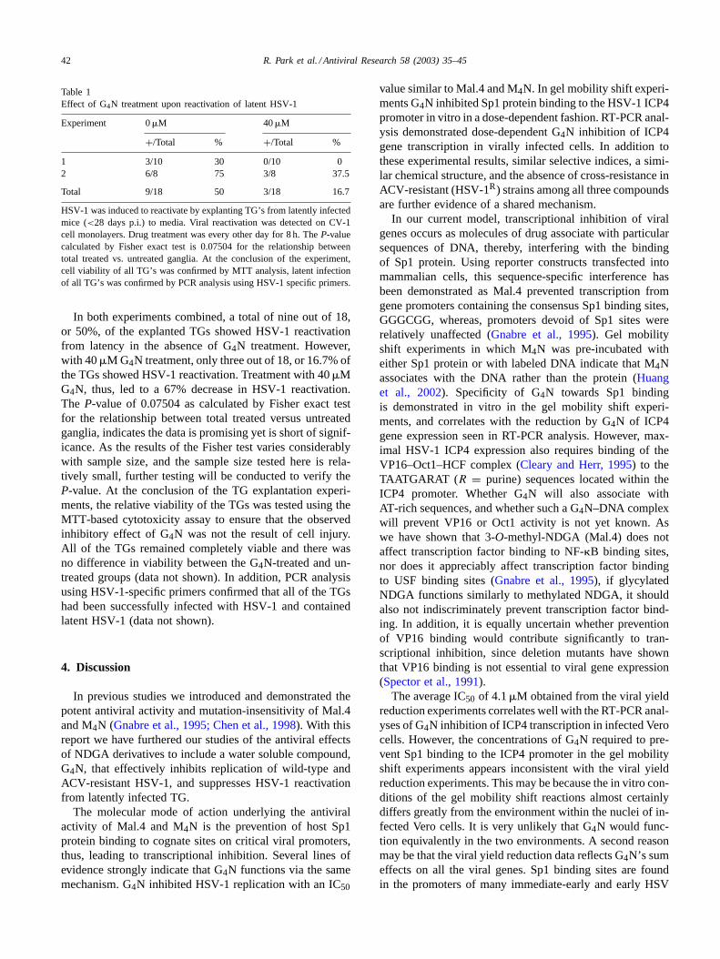
3.5. G4N suppresses HSV-1 reactivation from latency
The capacity for HSV-1 to establish permanent latencywithin the sensory neurons of the host and to periodicallyreactivate is a unique and clinically important aspect of HSVbiology. The need for effective systemic treatment of recur-rent herpes virus infection in at-risk patients prompted us
to examine whether G4N could be used to block the reac-tivation of HSV from latency. The mouse TG latency andexplantation-induced reactivation model is a popular animalmodel for studying molecular and cellular aspects of HSVlatency and reactivation, and was used in this study to as-sess the ability of G4N to inhibit reactivation. In an initialexperiment which proceeded for 5 days, TGs from 20 la-tently infected mice were induced to reactivate by explan-tion to media. Ten explanted TGs were treated with 40�MG4N, and ten were treated with PBS in an identical fash-ion. As shown inTable 1, three of the ten untreated controlTGs had reactivated by Day 5 p.e., whereas, none of the tenG4N-treated TGs reactivated. In a second experiment whichwas continued for 14 days p.e., eight explanted TGs received40�M G4N, and eight received PBS. After 14 days, six ofthe eight untreated TGs had reactivated, whereas, only threeof the eight G4N-treated TGs had reactivated (Table 1).
42 R. Park et al. / Antiviral Research 58 (2003) 35–45
Table 1Effect of G4N treatment upon reactivation of latent HSV-1
Experiment 0�M 40�M
+/Total % +/Total %
1 3/10 30 0/10 02 6/8 75 3/8 37.5
Total 9/18 50 3/18 16.7
HSV-1 was induced to reactivate by explanting TG’s from latently infectedmice (<28 days p.i.) to media. Viral reactivation was detected on CV-1cell monolayers. Drug treatment was every other day for 8 h. TheP-valuecalculated by Fisher exact test is 0.07504 for the relationship betweentotal treated vs. untreated ganglia. At the conclusion of the experiment,cell viability of all TG’s was confirmed by MTT analysis, latent infectionof all TG’s was confirmed by PCR analysis using HSV-1 specific primers.
In both experiments combined, a total of nine out of 18,or 50%, of the explanted TGs showed HSV-1 reactivationfrom latency in the absence of G4N treatment. However,with 40�M G4N treatment, only three out of 18, or 16.7% ofthe TGs showed HSV-1 reactivation. Treatment with 40�MG4N, thus, led to a 67% decrease in HSV-1 reactivation.The P-value of 0.07504 as calculated by Fisher exact testfor the relationship between total treated versus untreatedganglia, indicates the data is promising yet is short of signif-icance. As the results of the Fisher test varies considerablywith sample size, and the sample size tested here is rela-tively small, further testing will be conducted to verify theP-value. At the conclusion of the TG explantation experi-ments, the relative viability of the TGs was tested using theMTT-based cytotoxicity assay to ensure that the observedinhibitory effect of G4N was not the result of cell injury.All of the TGs remained completely viable and there wasno difference in viability between the G4N-treated and un-treated groups (data not shown). In addition, PCR analysisusing HSV-1-specific primers confirmed that all of the TGshad been successfully infected with HSV-1 and containedlatent HSV-1 (data not shown).
4. Discussion
In previous studies we introduced and demonstrated thepotent antiviral activity and mutation-insensitivity of Mal.4and M4N (Gnabre et al., 1995; Chen et al., 1998). With thisreport we have furthered our studies of the antiviral effectsof NDGA derivatives to include a water soluble compound,G4N, that effectively inhibits replication of wild-type andACV-resistant HSV-1, and suppresses HSV-1 reactivationfrom latently infected TG.
The molecular mode of action underlying the antiviralactivity of Mal.4 and M4N is the prevention of host Sp1protein binding to cognate sites on critical viral promoters,thus, leading to transcriptional inhibition. Several lines ofevidence strongly indicate that G4N functions via the samemechanism. G4N inhibited HSV-1 replication with an IC50
value similar to Mal.4 and M4N. In gel mobility shift experi-ments G4N inhibited Sp1 protein binding to the HSV-1 ICP4promoter in vitro in a dose-dependent fashion. RT-PCR anal-ysis demonstrated dose-dependent G4N inhibition of ICP4gene transcription in virally infected cells. In addition tothese experimental results, similar selective indices, a simi-lar chemical structure, and the absence of cross-resistance inACV-resistant (HSV-1R) strains among all three compoundsare further evidence of a shared mechanism.
In our current model, transcriptional inhibition of viralgenes occurs as molecules of drug associate with particularsequences of DNA, thereby, interfering with the bindingof Sp1 protein. Using reporter constructs transfected intomammalian cells, this sequence-specific interference hasbeen demonstrated as Mal.4 prevented transcription fromgene promoters containing the consensus Sp1 binding sites,GGGCGG, whereas, promoters devoid of Sp1 sites wererelatively unaffected (Gnabre et al., 1995). Gel mobilityshift experiments in which M4N was pre-incubated witheither Sp1 protein or with labeled DNA indicate that M4Nassociates with the DNA rather than the protein (Huanget al., 2002). Specificity of G4N towards Sp1 bindingis demonstrated in vitro in the gel mobility shift experi-ments, and correlates with the reduction by G4N of ICP4gene expression seen in RT-PCR analysis. However, max-imal HSV-1 ICP4 expression also requires binding of theVP16–Oct1–HCF complex (Cleary and Herr, 1995) to theTAATGARAT (R = purine) sequences located within theICP4 promoter. Whether G4N will also associate withAT-rich sequences, and whether such a G4N–DNA complexwill prevent VP16 or Oct1 activity is not yet known. Aswe have shown that 3-O-methyl-NDGA (Mal.4) does notaffect transcription factor binding to NF-�B binding sites,nor does it appreciably affect transcription factor bindingto USF binding sites (Gnabre et al., 1995), if glycylatedNDGA functions similarly to methylated NDGA, it shouldalso not indiscriminately prevent transcription factor bind-ing. In addition, it is equally uncertain whether preventionof VP16 binding would contribute significantly to tran-scriptional inhibition, since deletion mutants have shownthat VP16 binding is not essential to viral gene expression(Spector et al., 1991).
The average IC50 of 4.1�M obtained from the viral yieldreduction experiments correlates well with the RT-PCR anal-yses of G4N inhibition of ICP4 transcription in infected Verocells. However, the concentrations of G4N required to pre-vent Sp1 binding to the ICP4 promoter in the gel mobilityshift experiments appears inconsistent with the viral yieldreduction experiments. This may be because the in vitro con-ditions of the gel mobility shift reactions almost certainlydiffers greatly from the environment within the nuclei of in-fected Vero cells. It is very unlikely that G4N would func-tion equivalently in the two environments. A second reasonmay be that the viral yield reduction data reflects G4N’s sumeffects on all the viral genes. Sp1 binding sites are foundin the promoters of many immediate-early and early HSV
R. Park et al. / Antiviral Research 58 (2003) 35–45 43
genes, any or all of which may be affected by G4N to varyingdegrees, and lead to viral inhibition. The gel mobility shiftdata shows G4N’s effects on only two of the eight ICP4 Sp1binding sites. It is, therefore, likely that lower concentra-tions of G4N would suffice to inhibit viral replication, thanwhat is required to inhibit Sp1 binding at particular ICP4Sp1 binding sites.
Since the G4N inhibitory mechanism is completely dis-tinct from the antiviral mechanism of nucleoside analogueagents, there is little possibility of cross-resistance betweenthese two classes of drugs. We have shown in previous stud-ies that mutant HSV-1R strains have no resistance againstM4N or Mal.4, and the studies presented here demonstrateG4N is as equally potent against HSV-1R as it is againstwild-type HSV-1. In addition to being effective as a soletreatment against mutant HSV-1R, G4N’s independent modeof action also raises the possibility of combining it with nu-cleoside analogue treatments in an antiviral cocktail whichtargets multiple points in HSV-1 replication.
Another corollary to the mode of action of Mal.4, M4Nand G4N is mutation-insensitivity. The capacity of harm-ful viral and bacterial strains to mutate in response tochemotherapy presents a rapidly increasing problem in newdrug design and the implementation of therapeutic strate-gies. Viral inhibitors which target viral factors have theadvantages of high specificity and low cytoxicity. However,in the clinical setting, prolonged use of these drugs placesselective pressure upon the viral population to generateharmful resistant strains containing mutant proteins againstwhich the drug is ineffective. Mutant ACV-resistant strainsof HSV-1 have been commonly reported to contain alteredTK and DNA polymerase enzymes (Hirsch et al., 1996),and repeated use of AZT and even the most effective reversetranscriptase (RT) and protease inhibitors leads to resistantHIV strains containing mutations found within HIV RT andprotease enzymes (Richman, 2001). In contrast, host factors,unlike viral factors, are under no selective pressure to mutateand are in general structurally invariable. Thus, inhibitorslike G4N which target host factors have the advantages ofbeing relatively mutation-resistant, are potentially effectiveagainst a spectrum of related or unrelated viruses, and mostsignificantly will be less prone to generate drug-resistantviral strains. This should be qualified, however, by the un-likelihood of any single drug, including G4N, displayingcomplete mutation-resistance over prolonged clinical treat-ment. It is conceivable that resistance to G4N may be con-ferred by mutations to the Sp1 binding site which preventG4N association but retain Sp1 binding, or by the use ofalternative transcription factors for viral gene transcription.
The Sp1 protein is an abundant and ubiquitous transcrip-tion factor that regulates the basal expression of many cellu-lar genes in addition to playing critical roles in cell growthand development. Therefore, inhibition of its binding toDNA by G4N might be expected to have a profound delete-rious effect on the viability of host cells. However, the datapresented here for G4N and our previous studies with Mal.4
and M4N show, quite to the contrary, that these compoundsare relatively non-toxic to cells in culture. Experiments alsoindicate that G4N and M4N are likely to be less cytotoxicto primary, non-dividing cells than to proliferating cells inculture. In fact, we have previously shown that M4N canselectively eliminate C3 cell-induced tumors in mice whilesparing the surrounding normal stationary cells (Heller et al.,2001), and 8 h treatment of explanted mouse TG with G4Nshowed no cytotoxicity up to 120�M. We hypothesize thatstationary cells whose Sp1-regulated cell cycle genes are in-active and require less transcription factor activity would beless sensitive to the inhibitory activities of M4N and G4N,and would, thus, suffer less toxic effects than proliferatingcells. Of particular note with regard to toxicity, are the find-ings that subdermal injection of M4N showed no toxicity inmice (Heller et al., 2001), and topical application of G4N todorsal regions of guinea pigs twice daily at concentrationsup to 150 mM also showed no toxicity (Park, 2002). The lowabsorption of M4N by cells may contribute to the low tox-icity seen in animals including mice, rats, rabbits and dogs(Heller, 2002). Similar experiments have been done for G4Nin guinea pigs, and examinations of animal tissues of fol-lowing local administration of G4N in vivo showed no his-tological differences from non-treated tissues (Park, 2002)and will be reported separately.
The requirement of Sp1 in endogenous gene expressionin eukaryotic cells has not been extensively studied. Sp1 hasbeen associated with the cell cycle, chromatin remodeling,and the maintenance of methylation-free CpG islands. It hasbeen shown that many chromosomal genes previously shownto be Sp1-dependent in gene transfection experiments, in-cluding several cell cycle genes, remain active in the absenceof Sp1. While Sp1 protein is necessary for early embryonicdevelopment, it is not required for cell growth and differen-tiation (Marin et al., 1997). Although a high rate of proviraltranscription is essential for viral replication in differentiatedcells, there may be no such great demand for such large out-put of cellular transcripts in quiescent host cells. In contrast,rapidly dividing cells do require elevated levels of cell cycleprotein in order to carry out cell division in a time-dependentmanner. We have found treatment of C3 cells with 40�MM4N for 72 h caused a marked decrease in CDC2 proteinand mRNA levels, which requires Sp1 binding. The sametreatment, however, did not affect levels of cyclin B protein,whose promoter does not require Sp1 protein binding, nordid it affect GAPDH transcription (Heller, 2002).
Even in proliferating Vero cells, G4N exhibits no cyto-toxicity within the therapeutic range of 0–30�M, with cy-totoxic effects being evident only at concentrations abovethis range. A TC50 value of 73.2�M combined with an IC50value of 4.5�M yields a selective index (TC50/IC50) forG4N in Vero cells of 16.3, which is similar to the selectiveindex of 13.7 reported previously for M4N. Contingent uponactivity seen in infected laboratory animals, it may be spec-ulated that the relatively low selective index for G4N indi-cates that when possible it should be used therapeutically at

44 R. Park et al. / Antiviral Research 58 (2003) 35–45
its lowest effective concentration to avoid toxicity. Due tothis, G4N may be most useful as a routine complement toACV in controlling and precluding the exponential growthof ACV-resistant (HSV-1R) strains of HSV. Since HSV-1R
strains remain sensitive to G4N, the small initial populationof HSV-1R mutants generated during early ACV treatmentmay be kept under control by supplementing with low con-centrations of G4N. If so, supplementing ACV with smallamounts of G4N may result in a continually low IC50 valuefor ACV, thus, reducing the dosage, the toxicity, and the costof treatment.
Whereas, the insolubility of Mal.4 and M4N restricts theirantiviral use to very localized applications such as the topi-cal treatment of herpes labialis, G4N’s high water-solubilityextends the advantages of this class of compounds to the sys-temic treatment of recurrent HSV-1 infection. The precisemolecular mechanisms of HSV reactivation from latency arenot well understood. Perhaps the best inhibitors of HSV re-activation would act upon the signal transduction pathwayleading to HSV reactivation. Given the little which is knownabout signaling mechanisms involved in HSV-1 reactivation,however, it may be difficult at this point to demonstrate thatG4N inhibition of a cellular promoter associated with HSV-1reactivation contributes to the inhibition we observed. How-ever, since G4N can block the initiation of transcription ofeven the earliest viral genes, it may be particularly effectiveat preventing HSV reactivation, since it would preclude theinitiation of the HSV lytic gene expression cascade. In con-trast, nucleoside analogue agents are active only at the pointof viral DNA replication when HSV has already reactivatedand the lytic gene transcription cascade has progressed be-yond immediate early gene and early gene expression.
Indeed, animal studies of the impact of nucleoside ana-logue agents upon HSV latency and reactivation havefocused upon reducing the number of latently infected neu-rons or reducing the viral copy number per cell during theestablishment of latency, with the implication that thesereductions would in turn reduce the frequency and severityof future HSV reactivation (Efstathiou et al., 1999). Suchstudies have focused mainly on valaciclovir and famciclovir(oral pro-drugs of ACV and penciclovir, respectively) andhave shown some success in reducing the establishment ofHSV latency in mice when administered early and at highdosage. Several clinical trials have shown oral ACV to behighly effective in the suppression of HSV recurrence evenduring prolonged treatment over several years (Kaplowitzet al., 1991). ACV effectively interrupts the further progressof viral DNA replication, thereby, suppressing a recurrenceas well as an acute infection; however, it does not act uponthe reactivation mechanism. The mechanisms causing HSVreactivation are yet unclear, however, are likely to directlyimpinge upona gene expression, which G4N inhibits. Theresults above show that G4N directly and significantly re-duces the frequency of HSV reactivation from explantedlatently infected mouse TG. Therefore, as a systemic agentG4N may be useful in more urgent clinical cases, such as
those in which the patients are severely immunocompro-mised, where it is necessary to prevent the initiation of anyHSV reactivation rather than reduce its severity. Experimentsdemonstrating G4N’s ability to prevent in vivo HSV reacti-vation in latently infected animals are now being prepared.
As with the issue of ACV-resistance, G4N has the po-tential to be most useful therapeutically as a complementto nucleoside analogues in the systemic treatment of HSVreactivation. It is a common practice to administer systemicand prophylactic anti-HSV treatment to HSV-seropositivepatients in order to minimize the frequent HSV-1 reacti-vation resulting from tissue damaging procedures such asexcimer laser keratectomy for the correction of corneal re-fractive errors (Gilbert, 2001), cytoreductive chemotherapyin malignant cancer treatment (Khan and Wingard, 2001),or facial restructuring of the face and neck (Asbell, 2000).Guinea pig experiments have been performed which showM4N and G4N prevent HSV-1 induced cutaneous lesionsand viral shedding when applied as a topical agent in vivo(Park, 2002). Supplementing current systemic nucleosideanalogue regimens with G4N may improve their efficacyby providing a two-pronged treatment of HSV reactivation,with G4N reducing the frequency of reactivation, and bothnucleoside analogue reagents and G4N reducing the severityof reactivation.
Acknowledgements
This work was supported by a grant to R.C. Huang fromthe National Institutes of Health (1RO1DE12165). We wouldlike to thank Dr. Kenneth Thompson, University of Chicago,for providing specimens of HSV-1R strains, F59878 andT28387, and Dr. Nigel Fraser, University of Pennsylvania,for providing specimens of KOS and McKrae strains ofHSV-1. We are also grateful to Dr. Prashant Desai, JohnsHopkins University, and Dr. Ying-Hsu Su, University ofPennsylvania, for their guidance in establishing the mouselatency system.
References
Asbell, P.A., 2000. Valacyclovir for the prevention of recurrent herpessimplex virus eye disease after excimer laser photokeratectomy. Trans.Am. Ophthalmol. Soc. 98, 285–303.
Chen, H., Teng, L., Li, J., Park, R., Mold, D.E., Gnabre, J., Hwu,J.R., Tseng, W.N., Huang, R.C., 1998. Antiviral activities of methy-lated noridhydroguaiaretic acids. Part 2. Targeting herpes simplexvirus replication by the mutation insensitive transcription inhibitortetra-O-methyl-NDGA. J. Med. Chem. 41, 3001–3007.
Chomczynski, P., Sacchi, N., 1987. Single-step method of RNA isolationby acid guianidium thiocyanate–phenol–chloroform extraction. Anal.Biochem. 162, 156–159.
Cleary, M.A., Herr, W., 1995. Mechanisms and flexibility in DNA se-quence recognition and VP16-induced complex formation by the Oct-1POU domain. Mol. Cell. Biol. 14, 2090–2100.
Crumpacker, C., 2001. Antiviral agents. In: Knipe, D.M., Howley, P.M.,Griffin, D.E., Lamb, R.A., Martin, M.A., Roizman, B. et al. (Eds.),

R. Park et al. / Antiviral Research 58 (2003) 35–45 45
Fields Virology, 4th edition. Lippincott Williams and Wilkins, Philadel-phia, pp. 393–433.
Crumpacker, C.S., Schnipper, L.E., Marlow, S.I., Kowalsky, P.N., Hershey,B.J., Levin, M.J., 1982. Resistance to antiviral drugs of herpes simplexvirus isolated from a patient treated with acyclovir. N. Engl. J. Med.306, 343–346.
De Logu, A., Loy, G., Pellerano, M.L., Bonsignore, L., Schivo, M.L.,2000. Inactivation of HSV-1 and HSV-2 and prevention of cell-to-cellvirus spread bySantolina insularis essential oil. Antiviral Res. 48,177–185.
Efstathiou, S., Field, H.J., Griffiths, P.D., Kern, E.R., Sacks, S.L., Sawtell,N.M., Stanberry, L.R., 1999. Herpes simplex virus latency and nucle-oside analogues. Antiviral Res. 41, 85–100.
Gilbert, S., 2001. Improving the outcome of facial resurfacing—preventionof herpes simplex virus type 1 reactivation. J. Antimicrob. Chemother.47, 29–34.
Gnabre, J.N., Brady, J.N., Clanton, D.J., Ito, Y., Dittmer, J., Bates, R.B.,Huang, R.C., 1995. Inhibition of human immuno deficiency virustype 1 transcription and replication by DNA sequence-selective plantlignans. Proc. Natl. Acad. Sci. 92, 11239–11243.
Heller, J.D., 2002. Discovery and mechanism of the anti-cancer chemother-apeutic tetra-O-methyl nordihydroguaiaretic acid. Ph.D. thesis, JohnsHopkins University, UMI Dissertations Publishing.
Heller, J.D., Kuo, J., Wu, T.C., Kast, M.W., Huang, R.C., 2001.Tetra-O-methyl nordihydroguiaretic acid induces G2 arrest in mam-malian cells and exhibits tumoricidal activity in vivo. Cancer Res. 61,5499–5504.
Hirsch, M.S., Kaplan, J.C., D’Aquila, R.T., 1996. Antiviral agents. In:Fields, B.N., Knipe, D.M., Howley, P.M., Chanock, R.M., Melnick,J.C., Monath, T.P. et al. (Eds.), Fields Virology, 3rd edition. RavenPress, Philadelphia, pp. 431–466.
Huang, R.C., Li, Y., Giza, P.E., Gnabre, J.N., Abd-Elazem, I.S.,King, K.Y., Hwu, J.R., 2002. Novel anti-viral agent tetraglycylated
nordihydroguaiaretic acid hydrochloride as a host dependent viral in-hibitor. Antiviral Res, in press.
Jones, K.A., Tjian, R., 1985. Sp1 binds to promoter sequences and ac-tivates herpes simplex virus ‘immediate-early’ gene transcription invitro. Nature 317, 179–182.
Kaplowitz, L.G., Baker, D., Gelb, L., Blythe, J., et al., 1991. Prolongedcontinuous acyclovir treatment of normal adults with frequently recur-ring genital herpes simplex virus infection. J. Am. Med. Assoc. 265,747–751.
Khan, S.A., Wingard, J.R., 2001. Infection and mucosal injury in cancertreatment. J. Natl. Cancer Inst. Monogr. 29, 31–36.
Marin, M., Karis, A., Visser, P., Grosveld, F., Philipsen, S., 1997. Tran-scription factor Sp1 is essential for early embryonic development butdispensable for cell growth and differentiation. Cell 89, 619–628.
Park, R., 2002. Inhibition of the Herpes simplex virus type I by threeNDGA derivatives: Mal.4, M4N, and G4N. Ph.D. thesis, Johns HopkinsUniversity, UMI Dissertations Publishing.
Richman, D.D., 2001. HIV chemotherapy. Nature 410, 995–1001.Roizman, B., Sears, A.E., 1996. Herpes simplex viruses and their repli-
cation. In: Fields, B.N., Knipe, D.M., Howley, P.M., Chanock, R.M.,Melnick, J.C., Monath, T.P. et al. (Eds.), Fields Virology, 3rd edition.Raven Press, Philadelphia, pp. 2231–2295.
Sjottem, E., Andersen, C., Johansen, T., 1997. Structural and functionalanalyses of DNA bending induced by Sp1 family transcription factors.J. Mol. Biol. 267, 490–504.
Spector, D., Purves, F., Roizman, B., 1991. Role of alpha-transinducingfactor (VP16) in the induction of alpha genes within the context ofviral genomes. J. Virol. 65, 3504–3513.
Straten, M.V., Carrasco, D., Lee, P., Tyring, S.K., 2001. A review ofantiviral therapy for herpes labialis. Arch. Dermatol. 137, 1232–1235.
Whitley, R.J., 1996. Herpes simplex viruses. In: Fields, B.N., Knipe,D.M., Howley, P.M., Chanock, R.M., Melnick, J.C., Monath, T.P. et al.(Eds.), Fields Virology, 3rd edition. Raven Press, Philadelphia.